Evolution of the Modern Resectoscope
Dedication of the Jefferson Museum of Urology in 2004 during our Centennial Celebration. Shown here with one of the antique ACMI incandescent bulb cystoscopy sets are Dr. Frank D’Elia, Dr. Leonard Gomella, and museum benefactor Dr. Leonard Frank. Also seen are the original museum curators Dr. Dolores Byrne and former Chair of Urology Dr. S. Grant Mulholland.
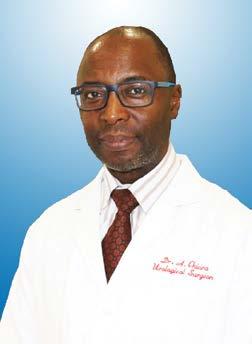
Historical Artifacts from Our Museum of Urology By Thomas Hardacker, MD, MBA The Leonard Frank Museum of Urology was dedicated during our Departmental Centennial celebration in 2004. Located in the Department of Urology Academic Office in the College building, it houses many historical instruments and artifacts. The museum displays several types of resectoscopes that developed over the last 100 years used primarily in the treatment of prostatitic enlargement. In this article Dr. Tom Hardacker, Chief Resident in the Department of Urology, discusses the evolution of the resectoscope and highlights some the instruments displayed in our museum. Over the last two centuries, the field of Urology had made great strides in the treatment of benign prostatic hypertrophy or BPH. In the 1800’s, as modern surgery was progressing, the options for surgical relief of prostatic obstruction were still 16
Year in Review
limited to an open suprapubic prostatectomy or blind transurethral cutting of prostatic tissue. Significant advances in transurethral surgery began in 1830, when George James Guthrie passed a concealed knife per urethra to incise prostatic tissue, though this was not done under direct visualization and was without a mechanism to control bleeding. This technique was improved upon when Enrico Bottini introduced a mechanism to deliver electrocautery in 1873. It was not until the 20th Century that there were significant advances. In 1909, Hugh Hampton Young, the father of modern urology, developed a “cold punch” in which an inner tube sheared off prostatic tissue in a more controlled manner. Edwin Beer, A.R. Stevens, and H.G. Bugbee all contributed the use of unipolar current in resection, resulting in improved hemostasis and would enable subsequent improvements on the procedure. Maximillian Stern developed the first recognized resectoscope in 1926, and T.M.
Davis made it possible to alternate between unipolar and bipolar current—a huge advancement that enabled both cutting and coagulation via the same mechanism. These advancements set the stage for Joseph McCarthy’s development of a more refined resectoscope, and one that allowed for greatly improved visualization of the procedure. He added a lens system that widened the visual field, a non-conducting outer sheath, and with the help of W.T. Bovie, separate channels for cutting and coagulating current. The SternMcCarthy resectoscope became the initial predecessor to the current resectoscope and made TURP a feasible procedure to a wide number of urologists (Figure 1). The first significant modification to this resectoscope was made by Reed Nesbit, who developed a loop that was pushed against a spring and would return to its initial position once released. This was the first development that enabled delivery of energy current by a one-handed technique, and allowed the surgeon to place a finger in the rectum so