FOREWORD
Dear readers,
f or us, 2024 was a year dominated by the evaluation procedure conducted by the Leibniz Association. With a look back at the recent changes in our research focus and the associated challenges, this regular evaluation represented an important milestone for us. The intensive preparations were demanding, but they gave us lots of valuable ideas and new insights.
It was also especially important for us to make our research even more accessible to different audiences. That is why we have explored new avenues in order to involve the interested public even more closely in our work and promote dialogue – whether through the provision of information, new participatory event formats or intensive discussions, such as on the topic of animal experiments. Whenever the latter are unavoidable, we make use of innovative analytical methods combined with artificial intelligence to reduce the number of experimental animals in line with the 3R principle. Transparent communication on this topic is a matter close to our hearts – which is why we support initiatives such as the “Initiative Transparente Tierversuche” (Transparent Animal Experimentation Initiative).
At the end of the year, we suffered a painful loss: following a short, serious illness, our long-standing Chief Financial Officer, Jürgen Bethke, sadly passed away. We are deeply saddened by his sudden death and are grateful for his tireless dedication. With great energy, foresight and personal warmth, he helped shape the institute over three decades – in both calm and challenging times.
On the following pages, we would like to highlight a few examples of what happened at ISAS over the past year, both at a personal and scientific level.
I hope you enjoy reading our Annual Report!

CONT ENTS
DUAL COMMITMENT
At ISAS, many employees take on additional tasks alongside their regular duties, helping to shape the work of the institute in a variety of ways. Their commitment to taking on dual responsibilities enriches the collaborative atmosphere and strengthens the ongoing development of the institute in the long term.
As ombudspersons, PhD spokespersons, programme directors or members of the works council, they bolster scientific integrity, represent the interests of their colleagues and constructively support departmental and structural changes at the institute. They raise issues from various divisions at an early stage, highlight needs in research and administration and thus promote dialogue across research groups and departments.
This voluntary commitment is not only an expression of a sense of responsibility and drive to shape the instituteʼs work – it is also an essential component in ensuring that the research infrastructure at ISAS remains vibrant and adaptable. It plays a role in helping to further develop structures, improve processes and put good ideas into practice quickly. In the following pages we introduce some of the people at ISAS who are engaged in such dual roles and provide an insight into their duties and motivation.
My job as an ombudsperson at ISAS is ...
What are you doing at ISAS, Yvonne?
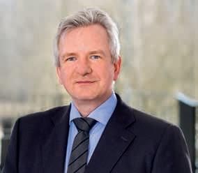
Dr Yvonne Reinders has been working as a scientist at the institute since 2018. Her research focuses on proteomics. In 2020, the biochemist successfully applied for an honorary position as an ombudsperson for good scientific practice. Ever since, she has been one of two independent contacts for all questions regarding the rules for safeguarding good scientific practice. To gain an insight into her work as an ombudsperson, the editorial team asked her to complete the following sentences.
to advise employees on good scientific practice and avoid possible discrepancies. A large part of my job and that of my colleague, Dr Roland Hergenröder, who is also an ombudsperson, is therefore prevention through education. We regularly advise all researchers – from students and doctoral candidates to experienced colleagues – on the principles of good scientific practice. Indications of scientific misconduct can also be reported to us so that we can deal with them in accordance with the ISAS guidelines for safeguarding good scientific practice. We also offer support in the event of conflicts.
It is particularly important to me ...
that I am always available as a point of contact for my colleagues at all career levels. My aim is to create an atmosphere in which everyone can turn to me with confidence. I therefore give regular talks to strengthen knowledge of good scientific practice and to constantly reawaken awareness for this among all researchers at the institute.
Good scientific practice ...
to put it simply, means ensuring that one's own behaviour complies with the relevant guidelines – and thus ensuring scientifically correct conduct. Scientific errors can occur anywhere, including in a lack of knowledge. It is therefore crucial that good scientific practice is considered by all employees, regardless of their position.
GOOD SCIENTIFIC PRACTICE
Good scientific practice comprises ethical and methodological standards that serve as a foundation for scientific work. The principles of good scientific practice include, among other things, researchers applying current methods and continuously checking the accuracy and comprehensibility of their results. The prerequisites for this are responsible, conscientious, and transparent behaviour, proper documentation and respectful treatment of other researchers and their research contributions.
In addition to her role as a research associate, Dr Yvonne Reinders advocates good scientific practice at ISAS.
Duo of PhD Advocates
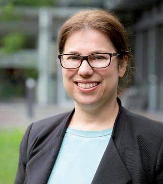
Emanuel Lange and Felix-Levin Hormann have been representing the interests of the 35 doctoral students at ISAS since March 2024. As democratically elected PhD spokes-persons, they act as mediators when it comes to ques-tions, problems and issues concerning the junior researchers. They also organise various events, such as the PhD Breakfast Club and the annual Summer School.
As part of the structured PhD training programme, the two training days organised by the doctoral candidates themselves include various scientific lectures and poster sessions. These are also intended to bolster social interaction and exchange.
“We want to provide a supporting programme and a network to connect the doctoral candidates at the institute – also because we do a lot of interdisciplinary research at ISAS,” says Hormann, doctoral candidate in the Lipidomics research group. The two take on the tasks of PhD spokespersons in addition to their research at ISAS. They
also benefit from this themselves, says Lange, doctoral candidate in the Multidimensional Omics Data Analysis research group: “Through our work as PhD spokespersons, we can not only improve our organisational skills, but also learn to take on responsibility and gain initial experience in leadership and project planning.”
There is no set term of office for PhD spokespersons at ISAS. However, like their predecessors, Lange and Hormann expect to remain in office for about two years. During this time, there are a few more things they would like to achieve. “We would like to organise an excursion for all PhD students, establish more networking events and gather information and experience for our successors in a more accessible form,” says Lange regarding the duoʼs plans.
Felix-levin Hormann (left) and Emanuel lange have been PhD spokespersons at ISAS since March 2024.
Responsibility & Creativity for Safe Procedures in the Laboratory
Storing, using and disposing of chemicals – safely: Luisa Röbisch is the hazardous substances officer at ISAS and point of contact for everything in relation to handling hazardous substances. She thus supports the ISAS safety expert and advises employees on the correct storage conditions, labelling and disposal of chemicals. Her responsibilities also include checking and taking stock of the cadastre of chemicals.
“In addition to my routine tasks, as a hazardous substances officer I have the opportunity to modernise and improve processes. I enjoy finding new ways of making laboratory work more efficient for my colleagues and myself,” says Röbisch.
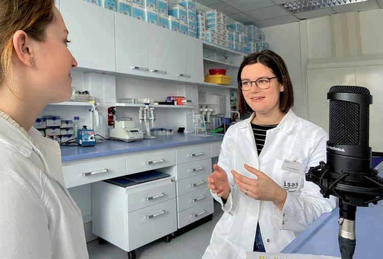
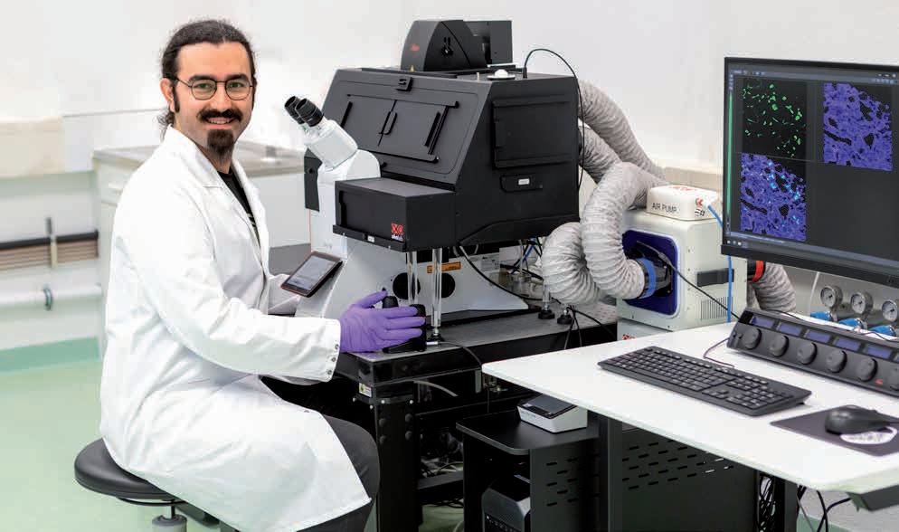
The 32-year-oldʼs “main job” is also in the laboratory: as a technical assistant in the Bioimaging research group, Röbisch has been ensuring the smooth running of research processes since 2022. She provides an insight into her work in episode 9 of the ISAS podcast “NACHGEFORSCHT – DIE LIVESCHALTE INS LABOR”. In it, among other things, she explains why she always needs a touch of creativity in her everyday work in addition to technical know-how.
EPISODE 9 : Hinter den Kulissen der Mikroskopie –die Arbeitswelt einer Technischen Assistentin / German Episode
https://www.isas.de/kompakt/ isas-wissenschaftspodcast-folge-9


live in the lab – luisa Röbisch (right) in conversation with presenter Cheyenne Peters
New Research Programme Complements Existing Structures
Researchers from different specialist disciplines at ISAS are working hand in hand to develop measurement strategies for integrated, cross-scale multi-parameter analysis, including strategies for data interpretation. Biologists, chemists, computer scientists, immunologists, pharmacologists, physicians and physicists, among others, are collaborating on projects that are part of four research programmes. The institute reexamined its research programmes and established a new programme in 2024. In addition, ISAS uses a strategy fund to foster scientific ideas beyond the four established programmes, the expansion of research infrastructures and young researchers. The research programmes are scientifically coordinated by the following individuals, who each head a research group and thus play a dual role.
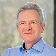
Prof. Dr Albert Sickmann
The Multi-Omics research programme (► p. 50) combines qualitative, quantitative and time-resolved methods for the analysis of lipids, metabolites and proteins. The measurements taken in this way enable us to gain a better understanding of the dynamic regulation of metabolic processes, for example in the context of cardiovascular diseases or cancer.
Chemist, Coordinator of the Multi-Omics research programme, Head of the Proteomics research group & ISAS Chairman of the Board
Proteomics Research Group
Prof. Dr Albert Sickmann
T: +49 (0)231 1392-100
E: albert.sickmann@isas.de
The MS-Based Imaging research programme (► p. 18) has been complementing the other three research programmes since 2024. It combines qualitative and spatially resolved analyses using mass spectrometry imaging. The spatial resolution and tracking of metabolic components such as lipids and metabolites should contribute to a mechanistic understanding of disease processes.
Lipidomics
Junior Research Group
Prof. Dr Sven Heiles
T: +49 (0)231 1392-4202
E: sven.heiles@isas.de
Bioimaging Research Group
Prof. Dr Anika Grüneboom
T: +49 (0)231 1392-239
E: anika.grueneboom@isas.de
The Pathomechanisms research programme
(► p. 76) brings together the methodological developments of the other programmes based on specific questions concerning the genesis of diseases such as cardiovascular diseases. ISAS wants to transfer its analyses into clinical practice so that researchers can use the technologies developed to identify and validate molecular changes that cause cardiovascular diseases and represent possible target molecules for active pharmaceutical ingredients or potential biomarkers. (SR)
The 3D Molecular Pathology research programme (► p. 30) focuses on high-resolution temporal and spatial analyses of physiological and pathological states in whole organs, tissue structures and cells, down to the molecular constituents. For the optical imaging, the researchers combine various microscopy techniques with AI-assisted analysis and visualisation of their imaging data.
Cardiovascular Pharmacology Research Group
Prof. Dr Kristina l orenz
T: +49 (0)231 1392-103
E: kristina.lorenz@isas.de
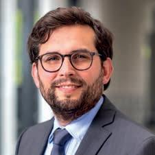
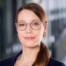
Prof. Dr Kristina Lorenz Pharmacologist, Coordinator of the Pathomechanisms research programme, Head of the Cardiovascular Pharmacology research group
Prof. Dr Sven Heiles
Chemist, Coordinator of the MS-Based Imaging research programme & Head of the Lipidomics junior research group
Prof. Dr Anika Grüneboom Immunologist, 3D Molecular Pathology programme Coordinator, Head of the Bioimaging research group
Between Code & Council
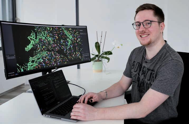
I work at ISAS as a software engineer in the research group AMBIOM – Analysis of Microscopic BIOMedical Images. I primarily write code for plug-ins, extensions that is, for the image analysis platform napari. Researchers use the programme to display and evaluate multidimensional images, such as microscope images.
Since April 2018, I have also been involved in the instituteʼs works council, where we represent the interests of the employees vis-à-vis the employer. For example, we conclude internal agreements and look after the interests of severely disabled employees. There are seven of us in total, each with different roles and responsibilities. In principle, the works council relies on the close cooperation of all members, each of whom contributes their own expertise. I am more versed in, for example, technical feasibility or data protection issues, than other topics.
I joined the works council at ISAS after a colleague informed me about it. To begin with, I wasnʼt sure if I was the right person for the job, but I decided to stand for election. I enjoy advocating for the interests of my colleagues. In my first term of office, I
only took on a few smaller tasks. Even though as a so-called substitute member I wasnʼt part of the core team, I still got acquainted with the internal operations of the works council. Now I am a full member and in my second term.
At 27, I am the youngest member, but I see that more as a strength. Everyone brings their own experience and expertise to the table. With age comes more experience. That means people just have different backgrounds, different views and different areas of expertise. As a result, I may see things from a different angle, so I think this is a valuable mix. It means the interests of all ISAS employees are represented in the best possible way.
We are always happy when someone wants to get involved in the works council. After all, itʼs important to have one. Itʼs there for the employees and gives them security. Getting involved in the works council also means doing something good for yourself and your colleagues.
(Protocol: LK)
lennart Kowitz has been working as a software engineer in the AMBIOM research group since October 2021 and is also studying computer science at Tu Dortmund university. From September 2017 to July 2019, he completed an apprenticeship as an IT specialist at ISAS.
PEOPLE
Mourning the Loss of Jürgen Bethke
ISAS mourns the loss of its former Chief Financial Officer Jürgen Bethke, who passed away on December 12, 2024, after a short, serious illness.
We are shocked and saddened by his sudden passing and will remember Jürgen Bethke as a warm-hearted and humorous colleague who was deeply committed to ISAS.
Jürgen Bethke served the institute for over 35 years, including over 15 years on the Executive Board. With his critical eye and love of debate, with his energy and breadth of vision, he shaped and influenced the development of ISAS over many years of service. He was also a long-standing participant in various committees within the Leibniz Association, including the Administrative Committee and the Finance Committee, and was involved in numerous appointment procedures.
With his passing, we have lost not only our Chief Financial Officer but also a wonderful person who was full of the joys of life. His death has left a void. We feel a sense of profound sadness but, at the same time, also immense gratitude for everything that Jürgen Bethke achieved for the institute.
Jürgen Bethke worked at ISAS for over 35 years, including over 15 years as Chief Financial Officer.
Prof. Dr Albert Sickmann Joins acatech
At acatech, the German National Academy of Science and Engineering, researchers from various disciplines (engineering, the natural sciences, medicine, and the humanities and social sciences) work on strategic engineering and technology policy issues, providing advice to policymakers and the general public. The academy currently has 400 members from Germany and abroad. One such member is bioanalyst and Chair of the ISAS Executive Board
Prof. Dr Albert Sickmann, who joined acatech as an ordinary member in 2024.
Proteomics Research Group
Prof. Dr Albert Sickmann
T: +49 (0)231 1392-100
E: albert.sickmann@isas.de
New Preclinical Metabolomics Research Group Launched under Prof. Dr Dr Alpaslan Tasdogan
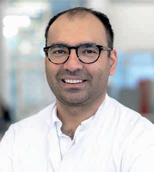
Since May 2024, there has been a new research group at ISAS. The Preclinical Metabolomics group seeks to investigate metabolic heterogeneity in tumours and their metastases using the technologies developed at the institute. Head of the research group is Prof. Dr Dr Alpaslan Tasdogan, a clinician who – in addition to his new position at ISAS – also leads the Institute for Tumour Metabolism at the Department of Dermatology at University Hospital Essen and is Professor of Dermatology and Tumour Metabolism in the Faculty of Medicine at the University of Duisburg-Essen.
Preclinical Metabolomics Research Group
Prof. Dr Dr Alpaslan Tasdogan
T: +49 (0)231 1392-100
E: alpaslan.tasdogan@isas.de
Building on questions from the Department of Dermatology, the group at ISAS works with mouse models, in vitro models such as cell cultures, and samples from patients, which it studies using techniques such as mass spectrometry (MS) and MALDI imaging MS. Among others, the researchers collaborate with the Lipidomics, Proteomics and Spatial Metabolomics research groups at ISAS. A fundamental part of this work involves translating these results into clinical practice according to the “from bench to bedside” principle. In the long term, the scientists hope to use these insights to identify new, targeted therapies that take as their starting point metabolic changes in cancer cells during treatment or metastasis.
At ISAS, Prof. Dr Albert Sickmann heads the department of Bioanalytics and the Proteomics research group.
Prof. Dr Dr Alpaslan Tasdogan has received several awards for his research, including an ERC Starting Grant, an Emmy Noether Independent Junior Research Group and a Peter Hans Hofschneider Professorship of Molecular Medicine.
Spatial Metabolomics Junior Research Group: Dr Karl
Smith Takes over as Head
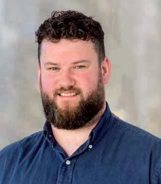
Dr Karl Smith was already very familiar with the work when he took over as Head of the Spatial Metabolomics junior research group on October 1, 2024. The chemist had already been conducting research in the same group as a postdoctoral researcher from June 2022 through September 2024. The 32-year-old was therefore able to continue the interdisciplinary collaboration with other ISAS research groups and external partners without any interruption.
The young father had already gained experience abroad before moving to Germany. A postdoctoral position had previously taken him from his native Ireland to the National High Magnetic Field Laboratory in Florida, USA, where he spent several years.
The junior research group, funded by the Federal Ministry of Education and Research (Bundesministerium für Bildung und Forschung, BMBF), was established at ISAS in 2021. The group aims to develop a multi-method approach that allows metabolic processes to be analysed simultaneously in terms of space and time. To achieve this, Smithʼs team combines complementary technologies such as mass spectrometry-based imaging and nuclear magnetic resonance spectroscopy (NMR).
(SR)
Spatial Metabolomics
Junior Research Group
Dr Karl Smith
T: +49 (0)231 1392-4210
E: karl.smith@isas.de
The Federal Ministry for Education and Research (Bundesministerium für Bildung und Forschung, BMBF) is funding the MSCoreSys-associated junior research group Spatial Metabolomics under the funding number 161 l 0271.
Dr Karl Smith took over as Head of the Spatial Metabolomics junior research group in October 2024.
After
completing
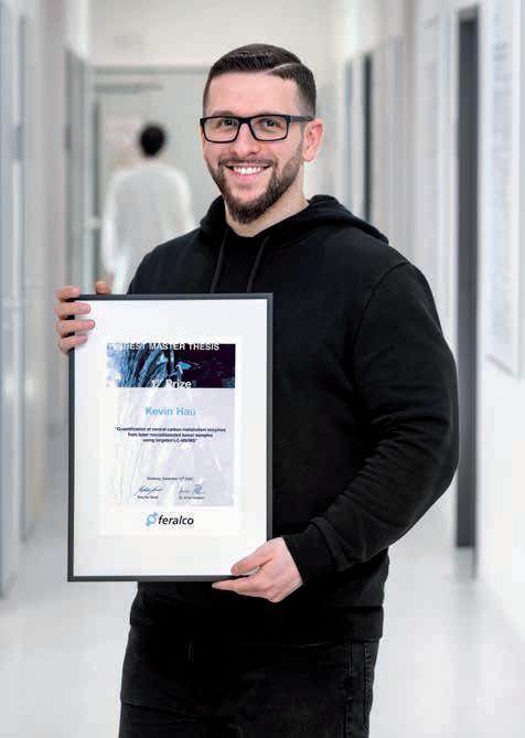
ISAS Congratulates Kevin Hau on the Prize for his Masterʼs Thesis
Kevin Hau received a special award for his thesis at ISAS on changes in the metabolism of tumour cells. The 26-year-old was honoured with the Feralco Water Award in December 2024. The prize is awarded annually by the chemical company Feralco Germany to students with outstanding masterʼs theses in the Water Science programme at the University of Duisburg-Essen. The award is endowed with 1,500 euros. Hau shares the prize with three other graduates who also achieved top marks.
For his masterʼs thesis, Hau conducted research in the Proteomics group where he had already completed his bachelorʼs thesis before. “I like the research at ISAS so much because it is about helping patients in the long term,” says Hau about his work at the institute. The chemist studied metabolic changes in tumour cells of malignant squamous cell carcinoma (a type of skin cancer) in mouse models for his masterʼs thesis. Laser microdissection enabled him to obtain precise, contamination-free samples of metastatic tissue from the lungs and liver. He then used targeted liquid chromatography-tandem mass spectrometry (LC-MS/MS) to quantitatively analyse these samples for proteins involved in central carbon metabolism. The method developed should provide information on the pathogenesis of tumour cells and, in the long term, help to identify new therapeutic approaches.
successfully
his bachelorʼs and masterʼs thesis, Kevin Hau remains with the Proteomics research group at ISAS. For his doctorate, he is researching the consequential effects caused by myocardial infarction in the interdisciplinary DFG Research Training Group TCI repAMI.
After completing his masterʼs degree, Hau remained in the Proteomics research group at ISAS. Since April 2024, he has been doing his doctorate in the DFG Research Training Group »RTG 2989 Targeting Cellular Interfaces in Reperfused Acute Myocardial Infarction (TCI repAMI)« (► info box). For his project, the doctoral student is carrying out multi-omics analyses of left ventricular cardiomyocytes (heart muscle cells) at ISAS. He aims to gain a better understanding of the cellular mechanisms involved in the recovery of the heart after a myocardial infarction.
»RTG 2989 TARGETING
CELLULAR INTERFACES
IN REPERFUSED
ACUTE MYOCARDIAL INFARCTION (TCI REPAMI)«
The Research Training Group TCI repAMI deals with consequential injury following a heart attack. An emergency reperfusion procedure – that is, the rapid reopening of a coronary vessel – can trigger inflammatory processes. These processes are underpinned by an interaction between specific immune, vascular and heart muscle cells.
Proteomics Research Group
Prof. Dr Albert Sickmann
T: +49 (0)231 1392-100
E: albert.sickmann@isas.de
TCI repAMI aims to analyse this interaction with the aim of identifying new treatment options for heart attack patients. It follows the bed-to-benchto-bed principle: once the researchers have determined the clinical problem, they draw up the experimental design in the lab, analyse and evaluate the research data, and bring these results back to the bedside for contextualisation in the clinical setting. The Research Training Group is a collaboration between the University of Duisburg-Essen, including University Hospital Essen, and ISAS. In total, it comprises eleven subprojects, each forming part of the three research areas of immune cells, vascular cells and heart muscle cells. One of the key elements of the group is interdisciplinary training. Accordingly, tandem teams each consisting of two experts from clinical practice and fundamental research supervise a total of 33 doctoral candidates.
Funded by the German Research Foundation (Deutsche Forschungsgemeinschaft, DFG) – project number 449437943.
(AB)
Poster Prize for Darleen Hüser
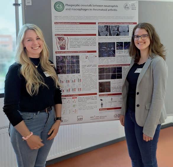
Held in Munich in October 2024, the internal retreat of the Collaborative Research Centre of the German Research Foundation (Deutsche Forschungsgemeinschaft) Transregio 332 (CRC/TRR 332) »Neutrophils: Origin, Fate & Function« (► p. 87) ended very pleasantly for Darleen Hüser. The doctoral candidate and her colleague Eva Gričar, a PhD student at the University of Münster, won first prize for a poster about their tandem project.
Bioimaging Research Group
Prof. Dr Anika Grüneboom
T: +49 (0)231 1392-239
E: anika.grueneboom@isas.de
Funded by the German Research Foundation (Deutsche Forschungsgemeinschaft, DFG) –project number 449437943.
The researchers’ work in Dortmund and Münster deals with the topic of »Phagocytic crosstalk between neutrophils and macrophages« in rheumatoid arthritis. In other words, their project deals with how these immune cells communicate with one another. Specifically, the two scientists study neutrophil transmigration in knee joints of diseased mice. At ISAS, Hüser analyses the samples using confocal laser scanning microscopy (CLSM) and light sheet fluorescence microscopy (LSFM) with immunofluorescence staining. Gričar uses flow cytometry for her analyses in order to define the various neutrophil population clusters in the clinical picture within cell cultures.
Retreat participants selected the prize-winners from the posters produced by all of the doctoral candidates. Hüser is delighted at this academic recognition of her joint work (aside from the award, the prize also included a tablet): “We’re proud that the research data and therefore the considerable effort we invested in the lab have been recognised.”
(LK)
Darleen Hüser (left) and Eva Gričar jointly present their research at the TRR 332 retreat in Munich.
Prof. Dr Norbert Esser: Farewell into Retirement
ISAS could count on Prof. Dr Norbert Esserʼs work for some 21 years – during which the doctor of physics held various different roles at the institute. In October 2004, Esser took up the position of Head of the instituteʼs Berlin site. This was followed in 2006 by a shared appointment as W3 Professor of Interface and Surface Analytics by TU Berlin as well as various posts at ISAS. As Head of the Berlin site, Esser was a member of the Executive Board from 2008 to 2020, during which he was Chair of the Executive Board for three years. To mark his retirement, ISAS organised a scientific colloquium in Berlin on July 19.
“We thank Norbert Esser for his dedication during his many years of service at ISAS. From 2011 onwards, he supported the institute on its journey from the materials sciences to the life sciences,” says Prof. Dr Albert Sickmann, Chair of the ISAS Executive Board. He believes this was a forward-looking change and essential for the institute’s move to develop analytics for health research.
On behalf of the entire Executive Board, Sickmann also expressed his gratitude for Esser’s support of young scientists and highlighted his commitment outside of the institute – including on the Board of Members of the Joint Initiative of Non-University Research Institutes in Adlershof (Initiativgemeinschaft Außeruniversitärer Forschungseinrichtungen in Adlershof, IGAFA). Moreover, Esser was involved in the »Thin Films« association of the German Physical Society (Deutsche Physikalische Gesellschaft, DPG), including in the capacity of spokesperson, for many years.
Esser studied physics at RWTH Aachen University and went on to obtain his doctorate at TU Berlin in 1991 with a dissertation on metal-semiconductor interfaces. He then turned his attention to optical spectroscopy at interfaces with a view to sounding out the extent to which “spectral fingerprints” can be used to derive a quantitative understanding at the atomic/molecular level.
In addition to the vibronic and electronic properties of surfaces, 1D atomic nanostructures and functional interfaces, his work also focused on the optical properties of “new” materials such as wide-bandgap semiconductors – always in close collaboration with various research groups in the field of solid state theory. Interdisciplinary applications and methodological developments of optical analysis methods were an integral part of Esser’s work at ISASʼ Berlin site, often in close collaboration with companies on the Adlershof campus.
Prof. Dr Norbert Esser was Head of ISASʼ Berlin site from 2004 to 2024.
MS-BASED IMAGING
Most illnesses or medical conditions, including cardiovascular diseases or tumours, are associated with localised and heterogeneous changes of biochemical cascades within some cells. The reason is that cells often respond differently when triggered with external stimuli such as low oxygen levels, viruses, bacteria, or when genetic alteration occurs. This leads to a complex spatial assembly of diseased or infected cells surrounded by healthy tissue, which is responsible for the medically relevant phenotype (referring to the appearance, development, and behaviour of an organism). To fully understand these phenotypes on a molecular level, analytical methods need to be capable of mapping spatially confined molecular changes.
Mass spectrometry imaging (MSI) enables the label-free localisation of hundreds of biochemical substances such as metabolites, lipids, peptides, drugs, etc. from single cells to tissue sections. This technical capability has provided new molecular insights into diseases such as cancer, diabetes, neurodegenerative, and metabolic disorders. Although MSI methods have been developed and refined in recent years, multiple key aspects of the analysis pipeline need to be improved to fully benefit biomedical and clinical research.
The aim of the Mass Spectrometry (MS)-Based Imaging research programme is to develop and combine matrix-assisted laser desorption ionisation (MALDI) MSI with microscopy techniques. The overall goal is to enable a spatial tracking of downstream products of proteins that indicate the actions of enzymes within cells, for instance lipids and metabolites. The method development of MS-based imaging methods goes hand in hand with applications in the field of heart in rare genetic disease, mechanistic understanding of metabolite and lipid regulation in cardiovascular disfunctions, tumourous changes, and the influence of small molecules during parasite or virus infection.
Improving the performance of MALDI-MSI sources
One requirement for the visualisation of small molecules in tissue sections is sufficient ion signal throughout the measurement, ideally without influences from matrix background or other analytes. Therefore, the scientists involved in this programme are dedicating a huge part of their work to improving the performance of MALDIMSI sources with regard to the overall ion signal, reduced ion suppression effects, increased coverage of lipids and metabolites in one MSI run. For this, the researchers are combining different ionisation sources with MALDI-MSI, for instance ISAS’s flexible microtube plasma (FμTP). The scientists will then test the optimised ion sources for the analysis of lipids and metabolites in cells and cardiovascular disease-associated tissues (inflamed heart tissue after an infarction and Fabry disease) obtained from mouse models and human samples.
Developing and optimising different sample preparation strategies
Another aspect that can significantly influence the quality of MSI results is sample preparation. Tissue washing steps may lower the total salt content, additives may improve ionisation efficiency, or
Miniaturisation Research Group
PD Dr Joachim Franzke
T: +49 (0)231 1392-174/199
E: joachim.franzke@isas.de
Preclinical Metabolomics
Research Group
Prof. Dr Dr Alpaslan Tasdogan
T: +49 (0)231 1392-100
E: alpaslan.tasdogan@isas.de
AMBIOM – Analysis of Microscopic BIOMedical Images
Junior Reseach Group
Dr Jianxu Chen
T: +49 (0)231 1392-217
E: jianxu.chen@isas.de
Lipidomics
Junior Research Group
Prof. Dr Sven Heiles
T: +49 (0)231 1392-4202
E: sven.heiles@isas.de
Multidimensional Omics
Data Analysis
Junior Research Group
Prof. Dr Robert Heyer
T: +49 (0)231 1392-271
E: robert.heyer@isas.de
Spatial Metabolomics
Junior Research Group
Dr Karl Smith
T: +49 (0)231 1392-4210
E: karl.smith@isas.de
Mass spectrometry imaging enables the examination of tissue slices and the precise localisation of thousands of molecules in one experiment.
3D-PRINTED FUNNEL AND ION MOBILITY SPECTROMETER
Combination is also key when it comes to MSI set-ups. In the MS-Based Imaging research programme, the scientists aim to develop a miniaturised ion funnel and a 3D printed stand-alone drift tube ion mobility spectrometer that is compatible with MSI technologies to improve the ion transfer, its sensitivity, and enable separation of ion populations, respectively. It is expected that these devices can significantly improve the performance of MSI set-ups for molecularly resolved spatial profiling, are customisable in size thanks to 3D printing, and are therefore compatible with multiple MSI set-ups.
chemical derivatisation can enhance the signal of selected compound classes and help elucidate the molecular structure of analytes. For this reason, the development and improvement of sample preparation strategies are part of the work in the MS-based imaging research programme. Specifically, these include:
• optimising protocols for MSI metabolites of the citric acid cycle,
• minimising ion suppression effects by removing salts or suppression analytes,
• on-tissue derivatisation methods to target low abundant or hard to ionise compounds focusing on steroids, oxidised lipids, sphingolipids,
• chemical derivatisation methods to structurally characterise analytes or validate compound annotations, and
• increasing the compatibility of MSI sample preparation with modalities such as fluorescence and Raman microscopy.
Library of quantified lipid and metabolite values
Quantification is difficult with MSI, because ion suppression effects could depend on the tissue type or the histological structures of the tissue. That is why the researchers at ISAS intend to combine their improved ion sources and optimised sample preparation strategies with absolute quantification results from established shotgun and liquid chromatography (LC) MS/MS experiments. One of their goals is to create a library of quantified lipid and metabolite data for a range of tissue samples using MALDI-MSI and to compare the results with established shotgun and LC-MS/MS values. Another aim is to develop a software tool that allows to use these libraries to quantify compounds in preclinical samples (genetically altered mouse models) and clinical samples.
New Ionisation Method: From Open Questions to Closed Plasma
Mass spectrometry is one of the most important analytical methods in medical chemistry. It works only with particles that are electrically charged – or ionised, to be more precise. Ionisation is often achieved using a plasma, an excited gas. Researchers like to use noble gases for this, traditionally in particular helium. More recently, however, the availability of helium has been affected by supply bottlenecks and sharp price increases. Until now it has also been unclear exactly how ionisation works in a plasma. However, ISAS scientists have published a series of studies that not only shed new light on the ionisation mechanism, but have also led to a new variant: their ‘closed microtube plasma’ (CµTP) works without a continuous supply of gas, making it particularly resource-efficient.
For ionisation by plasma, one technique used by researchers is dielectrically impeded discharge, in which two metal electrodes are insulated from one another by a suitable material and connected to an alternating voltage. An electrically conductive plasma then forms in the gas space between the electrodes even at room temperature. One variant of this is the “flexible microtube plasma” (FμTP) developed at ISAS. With this method, the plasma is generated in a fine, flexible glass capillary through which a noble gas continuously flows. At the end of the capillary and therefore outside the plasma, sample molecules can be added and ionised in a particularly gentle or ‘soft’ way. This is particularly important when analysing medically relevant large molecules such as proteins and lipids. With other analytical methods, these would easily disintegrate, thus making their identification by mass spectra difficult or even impossible.
Economical Ion Source
An ion source that works without a continuous gas flow could be relevant not only scientifically but also economically. In some cases the price of helium has doubled in recent years, and some laboratories have even had to shut down equipment at times because the gas was rationed.
Miniaturisation Research Group
PD Dr Joachim Franzke
T: +49 (0)231 1392-174/199
E: joachim.franzke@isas.de
This is the mechanism that is described in textbooks.
Noble gases such as helium are particularly suitable for soft ionisation, as they are chemically inert and therefore do not react readily with other substances. In addition, metastable helium atoms – helium atoms that have an excited state with a very long lifetime – can effectively ionise other molecules. For years, researchers had assumed that soft ionisation took place as a chain of collisions. During “Penning ionization”, metastable atoms or molecules collide with nitrogen molecules from the surrounding air. The resulting positively charged nitrogen ions collide and ionise with water molecules in the air, which in turn ionise the substances under investigation through further collisions. “This is the mechanism that is described in textbooks,” says
Luisa Speicher, doctoral candidate in the Miniaturisation research group. The explanation seemed plausible, since a helium plasma has a high energy level and can effectively transfer this energy to the nitrogen and water molecules with their lower energy. However, experiments have repeatedly shown that noble gases such as krypton and xenon also ionise analytical substances well in a plasma. However, the energy levels of these gases are too low to excite nitrogen as described – so the old theory could not apply to all noble gases.
Like a series of stop-motion shots
In order to find out what really happens, the scientists in the Miniaturisation research group developed an analytical method called plasma optical emission phoresis spectroscopy (POEPS). This enabled them to monitor exactly how the charged and excited particles in the plasma are activated in time and space in an FμTP for the first time. The method works rather like a series of stop-motion shots. Firstly the researchers record the glow emitted by the plasma. This glow contains
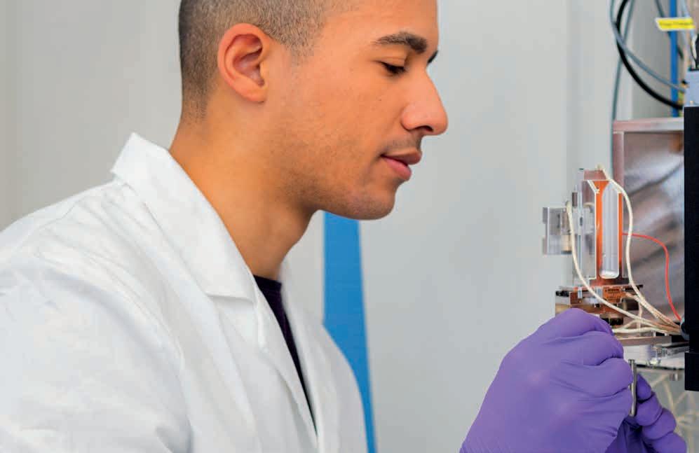
Luisa Speicher, doctoral candidate in the Miniaturisation research group, knows the mass spectrometers and ionisation sources at ISAS inside out.
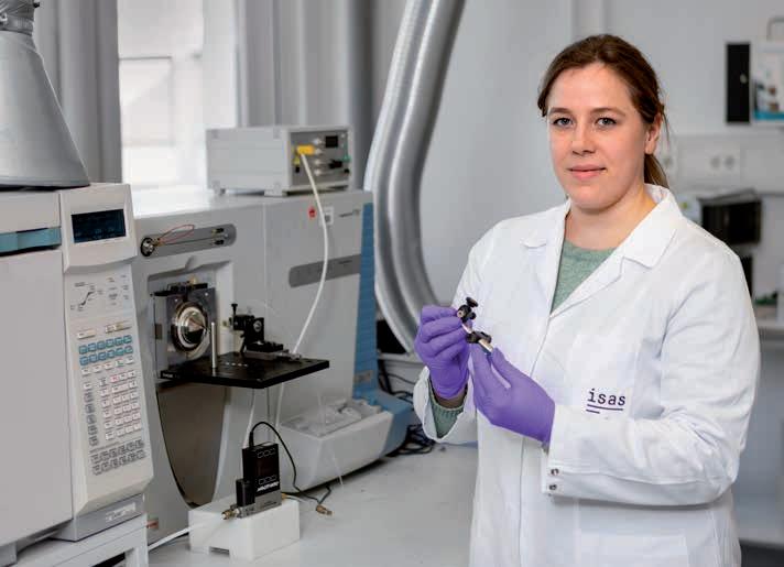
Caiyan Tian, doctoral candidate in the Miniaturisation research group, often works in the dark to record the glow of the plasma.
different colours (wavelengths), as each type of particle in the plasma emits light with characteristic colours when excited – rather like fireworks in which different chemical elements produce different colour effects. The scientists use a special camera to record these coloured light signals and to determine precisely when and where which colour appears and with what intensity.
The researchers analyse the images in two different ways. In the first analysis, they consider the temporal dimension – how the brightness of the different colours changes over time. This tells them in which order and with what dynamics the different particles in the plasma are activated. In the second analysis, they concentrate on the spatial dimension – at which points in the thin glass tube the particles become energised or electrically charged. “This approach makes it easier to observe even small differences which, in the twodimensional colour representations that are otherwise used, are superimposed,” explains Caiyan Tian, doctoral candidate in the Miniaturisation research group. The scientists published their new method in the journal Spectrochimica Acta Part B: Atomic Spectroscopy.
This approach makes it easier to observe even small differences.
The POEPS measurements indicate that the ionisation outside the capillaries is not caused by collisions between gas particles of the plasma and the ambient air, but rather by a short-term localised change in potential. The researchers elaborated on this hypothesis in the journal Analytical and Bioanalytical Chemistry, in which they explained that ions are deposited on the glass wall within the plasma capillary and polarise the glass. The resulting electric field is strong enough to ionise molecules outside the capillary by electron impact.
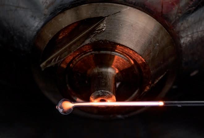
For the test, the scientists used helium instead of ambient air as a “diagnostic gas” outside the plasma capillary. They observed that the helium was excited even through a glass wall – a clear indication that it was not the direct contact between the plasma gas and the environment that caused the ionisation. The glass wall also rules out other possible mechanisms such as photoionisation, says Speicher. “If photons caused the ionisation, we would no longer be able to observe any effects through the glass wall.” The group has also published this experiment in the journal Analytical and Bioanalytical Chemistry. Based on their findings, the ISAS researchers developed an even more efficient plasma source for mass spectrometry. Whereas in the FμTP, gas constantly flows through the capillary and the plasma that builds up partially escapes from the tube, it was now possible to completely enclose the plasma in a closed glass tube. “At some point we came up with the idea that we could weld the tube completely closed – almost like a miniature fluorescent tube,” summarises Speicher.
The new closed µ-tube plasma, CµTP, achieves a similar ionisation efficiency to the previous technology – but without the need to continuously supply gas. “That
The closed µ-tube plasma (CµTP) completely encloses the diagnostic gas, in this case neon. The photo was taken by Dr Daniel Foest, research associate in the group and keen amateur photographer.
makes the ionisation source not only more cost-effective, but also portable,” says Tian. “Itʼs so compact that it can even be combined with other ionisation sources as an additional ionisation module.” The new technology, also presented in the journal Analytical and Bioanalytical Chemistry, has already been patented. The researchers are now working on optimising the new microplasma source for imaging mass spectrometry.
(UE)
Speicher, L., Song, H., Ahlmann, N., Foest, D., Höving, S., Brandt, S., Niu, G., Franzke, J., Tian, C. (2024) Soft ionization mechanisms in flexible µ-tube plasma – from FµTP to closed µ-tube plasma. Analytical and Bioanalytical Chemistry, 416, 4919-4927. https://doi.org/10.1007/s00216-024-05420-8
NUCLEAR: A Training Network at the Heart of Cancer Research
NUCLEAR is an interdisciplinary European training network for doctoral students. Its aim is to train young scientists on the metabolic regulation of genome function and cell identity, an area that is relevant to stem cell biology and cancer research. To this end, the European Union is funding NUCLEAR as a Marie Skłodowska-Curie Doctoral Network to the tune of approximately four million euros through the programme Horizon Europe. The network was launched in November 2024. From 2025 on, twelve partner organisations will be participating in training the network’s 17 PhD students, including universities such as the University of Cambridge, Charité – Universitätsmedizin Berlin, non-university research institutions such as ISAS, companies from industry, and a patients’ association. All of these participants enrich the network by contributing different skills from disciplines including stem cell biology, precision nutrition, mass spectrometry and drug development.
At ISAS, the Lipidomics research group is participating in the network under the leadership of Prof. Dr Sven Heiles. The PhD training focuses on the visualisation of acyl-coenzyme A (acyl-CoA) and associated metabolites (products of metabolism) in subcellular resolution using matrix-assisted laser desorption/ionisation (MALDI).
Specifically, this NUCLEAR project is about developing methods including mass spectrometry imaging and combining them with complementary techniques such as fluorescence microscopy to reveal how metabolites
such as acyl-CoA organise themselves spatially in cells and in the cell nucleus. In the next step, the ISAS researchers want to use the resulting information to clarify the influence of these metabolites on chromatin (a complex of DNA and proteins in the nucleus) and the regulation of cancer-inducing genes. As one of several subprojects, the aim is to use the results from ISAS to help identify metabolic weak points in cancer cells for new therapeutic strategies. (SR)
Funded by the European u nion under grant agreement number 101166838. Views and opinions expressed are however those of the author(s) only and do not necessarily reflect those of the European u nion. Neither the European u nion nor the granting authority can be held responsible for them.
Everything is Relative: New Approaches to Mass Spectrometry Imaging
When researchers examine tissues in order to get to the bottom of disease mechanisms, they often need to choose between two fundamental approaches. On the one hand, they can mark substances specifically and, for example, use microscopic techniques to investigate how many of them are present in the tissue. For this quantitative analysis, however, the researchers must already know what they’re looking for. On the other hand, they can use methods such as mass spectrometry imaging to see what metabolic products, for example, are present in the tissue in the first place. Until now, however, it has been difficult to read off the quantity of the substances from this qualitative data. Now, with the participation of ISAS, a group of researchers has refined a method that allows not only qualitative but also quantitative analysis – and, for the first time, for a whole class of substances.
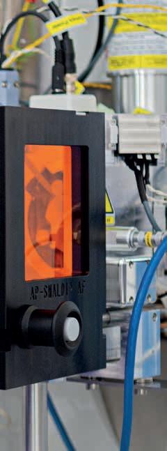
To this end, the team led by Prof. Dr Bernhard Spengler from Justus Liebig University Giessen and Prof. Dr Sven Heiles, leader of the Lipidomics research group at ISAS, have combined two analytical techniques known as “atmospheric pressure scanning microprobe matrixassisted laser desorption/ionisation mass spectrometry imaging” (AP-SMALDI MSI) and “nanoflow hydrophilicinteraction liquid chromatography tandem mass spectrometry” (nano-HILIC MS/MS). Using this combined method, they investigated the molecular processes taking place in schistosomiasis, a neglected tropical disease with over 200 million sufferers worldwide. The researchers published their results in the journal Analytical Chemistry.
Molecular insights into schistosomiasis
Schistosomiasis, also known as bilharzia, is triggered when people come into contact with water contaminated with the larvae of Schistosoma worms. These tiny parasites enter the body by penetrating the skin and lay eggs that are often deposited in the organs – especially the liver. The human immune system responds by forming granulomas (spherical tissue structures) around the eggs. These structures are actually intended to encapsulate the parasites to prevent their spread. However, this defensive response often leads to inflam-
mation, which can cause conditions including fibrosis – that is, chronic scarring – of the liver. Until now, it has been difficult to determine what this process entails on the molecular level.
In their study, the researchers focused on a specific group of fats known as glycosphingolipids (GSLs). These glycolipids, which are a key constituent of the cell membrane, are structured like a lollipop: with a fat-soluble “backbone”, known as a ceramide, which is anchored in the membrane, and a water-soluble sugar headgroup that projects outwards. GSLs play a key role in the communication between cells and in immune responses, acting as “immunomodulators”: antibodies, endogenous cells and immune cells detect GSLs and produce an immune response accordingly. So far, however, there has been no way of determining how many of these molecules are present in a specific tissue using imaging.
Finally, chemical imaging with relative quantities
In their method, the team led by Spengler and Heiles combined the chemical analysis of nano-HILIC MS/MS with the high-resolution imaging of AP-SMALDI MSI in order to examine the liver tissue of healthy hamsters and hamsters infected with Schistosoma. In AP-SMALDI MSI, a fine laser beam scans the tissue point by point
and breaks off molecules, which are identified by the mass spectrometer. This produces a chemical image (► p. 28) of the tissue, resembling the image produced by a thermal imaging camera but showing the spatial distribution of certain molecules rather than temperatures. By optimising the pixel resolution of the APSMALDI MSI method to as little as three micrometres – about a 20th of the thickness of a human hair – the scientists were able to recognise even the finest structures within the granulomas and therefore to distinguish between antigens and endogenous GSLs, for example, in the case of the parasitic infection.
The key advance is that, with their method, the researchers were able not only to see where GSLs were located in the animals’ tissue but also to compare the relative amounts in different tissue regions – an important step that goes beyond just imaging. Specifically, the team identified 60 different GSL species and established that 50 of them occurred in greater quantities in infected tissue. Of these molecules, 44 were directly connected with schistosomiasis-related granuloma formation.
More than just an image – a paradigm shift
This work represents a paradigm shift, moving away from purely qualitative imaging (“what is where?”) to quantitative analysis (“how much is where?”). The developed method not only opens up new insights into
the pathology of schistosomiasis but also has potential applications in various areas of biomedicine where the spatial distribution of lipids and other biomolecules plays a significant role. At ISAS, for example, Heiles and his team are working on analytical techniques for use in a genetic lipid storage disorder known as Fabry disease. In patients, this disorder leads to an accumulation of GSLs that is harmful in the long term. The ability to better understand and analyse this process could potentially pave the way for new therapies.
Luh, D., Heiles, S., Roderfeld, M., Grevelding, C.G., Roeb, E., Spengler, B. (2024) Hepatic Topology of Glycosphingolipids in Schistosoma mansoni-Infected Hamsters. Analytical Chemistry, 96(16), 6311-6320.
https://doi.org/10.1021/acs.analchem.3c05846
When a Picture Is Not Worth a Thousand Words
How and where exactly does inflammation occur? Is it possible to identify the affected cells at an early stage and decrypt the underlying processes? Yes, in some cases, this is indeed possible. These analyses often use bioanalytical imaging techniques, such as mass spectrometry imaging. This technique can visualise the spatial distribution of metabolic products or pharmaceutical substances in cells and tissue sections by assembling intensity information from spatially resolved mass spectrometry data into distribution images with the help of computer programs. This often produces beautiful images. However, in order to be relevant to clinical research, mass spectrometry imaging can and must make the step from pretty, qualitative images to absolutely quantifiable statements. After all, only a limited
Prof. Dr Sven Heiles holds a junior professorship at the university of Duisburg-Essen and leads the lipidomics research group at ISAS. He also coordinates the MS-Based Imaging research programme.
(UE)
amount of information can be derived from the images themselves. This is due to our human perception of colour and intensity, which is often misleading. Lipids (fats) “light up” relative to sugars in mass spectrometry images, for example, due to their chemical properties even though the lipid and sugar concentrations in some tissues are very similar. Although sugar metabolism plays a key role in many disrupted biochemical processes, carbohydrates appear to be less important in the images at first glance. Understanding and correcting this subjective distortion using analytical techniques represents a significant challenge in our area of research. We can only succeed in capturing the information contained in the images objectively if, in addition to the qualitative information – that is, the distribution of metabolic products – we can also provide access to insights into the relative or absolute quantity of substances. It is precisely this quantitative information that the mass spectrometry images don’t readily deliver – because the signal intensity of the metabolic products can only be indirectly linked to the frequency of the biomolecules in the sample.
At ISAS, we take a multistep approach to this challenge. We have already succeeded in the relative quantification of sugar-containing lipids – known as glycosphingolipids (GSLs). We can therefore evaluate the relative change in GSLs between individual tissue structures or different disease states. In bioanalytics, for example, this approach has been used in work on parasitic pathogens such as Schistosoma mansoni and the metabolic disorder known as Fabry disease. We’re only able to do this thanks to the most accurate control of sampling, validated sample preparation, standardisation of the measurement procedure and validation of the results. Now, we want to expand this method to other groups of substances. By applying internal standards with a known concentration to the sample as a reference value, we want to enable absolute quantification of substances in future by comparison of endogenous compounds with internal standards. It is only if this absolute quantification succeeds that mass spectrometry imaging could be used to make decisions on disease stage and therapeutic approaches for patients without comparison with control groups.
I’m convinced that pretty pictures alone are not enough. We will need to establish relative and, above all, absolute quantification in mass spectrometry imaging in order to help provide our clinical partners with valuable, objective, measurable data.
(Guest article by Prof. Dr Sven Heiles)
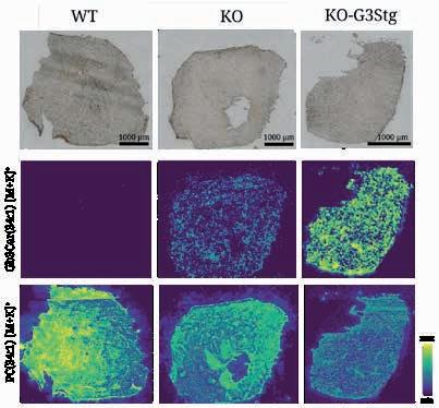
Lipid deposits in the heart in a mouse model for Fabry disease
The image shows heart tissue from mice in three groups: on the left, the tissue is from healthy wild type (WT) animals. In the middle, it is from knock-out (KO) mice in which the gene for the enzyme α-galactosidase A has been specifically switched off. This enzyme normally breaks down glycosphingolipids (GSls) but is diminished in people with Fabry disease. In the right column are the samples from knock-out mice that also produce greater quantities of GSls (K0-G3Stg), exacerbating the disease characteristics. The top row shows the tissue under an optical microscope. The middle and bottom rows each show the same sections using mass spectrometry imaging: in the middle row, the disease-relevant GSl Gb3Cer 34:1 is highlighted in colour, while the bottom row shows the cell membrane lipid PC 34:1, which occurs in the normal state, for comparison. The colour scale of the measurement results from minimum to maximum intensity clearly shows that the stronger the typical Fabry-disease conditions, the greater the quantity of harmful GSls deposited in the heart tissue.
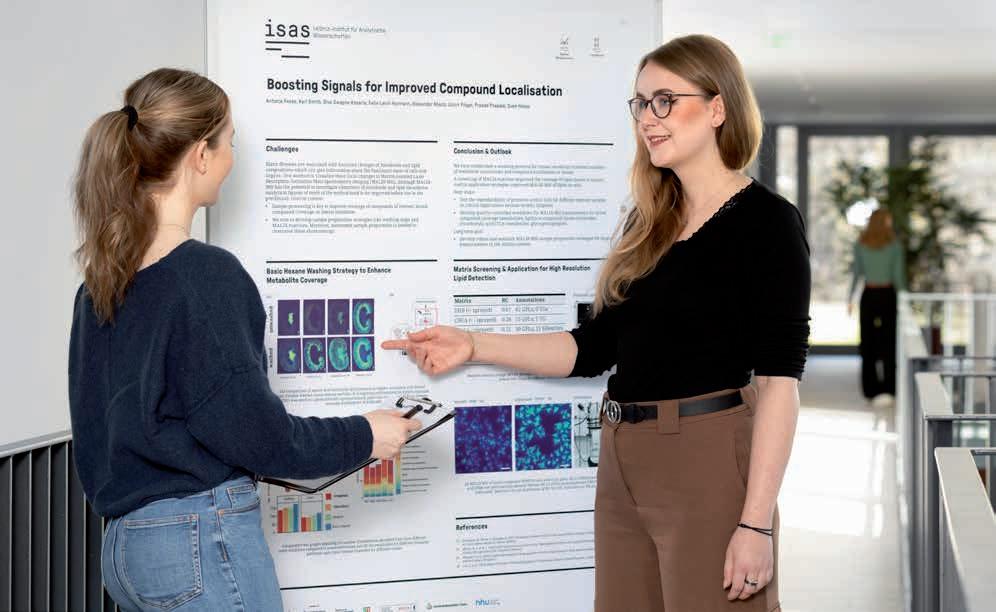
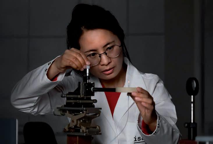
What’s going on here, Antonia Fecke?
Spatial Metabolomics
Junior Research Group
Dr Karl Smith
T: +49 (0)231 1392-4210
E: karl.smith@isas.de
Communications Team
Sara Rebein
T: +49 (0)231 1392-234
E: sara.rebein@isas.de
The photo shows me during my presentation training with our science editor (on the left) for the evaluation by the Leibniz Senate. During my presentation I introduced two optimised strategies for sample preparation for mass spectrometry imaging that we are working on in the Spatial Metabolomics research group together with the Lipidomics group. Here, in the training session a few weeks before the evaluation, Iʼm showing kidney sections, for example, in which we were able to improve the signal intensity of various metabolites through an additional washing step. Unlike poster presentations at conferences, I had to prepare the content differently for the evaluation, as many of the reviewers came from a different specialist field. Everything had to be clearer, more concise and more recognisable in the context of our research programme. To ensure that I felt confident on this important day, we doctoral candidates and post-doctoral researchers trained intensively with the Communications team beforehand. Together we formulated key messages, rehearsed possible questions from the reviewers and discussed strategies to combat nervousness. All of this not only helped me for the evaluation, but also for other presentations in the longer term. In an academic environment, you often get feedback on the content, but rarely on your presentation style. Rehearsing in a relaxed atmosphere was therefore really helpful. In particular, I will remember to always formulate key messages. That will enable me to react flexibly to questions and stay calm even in unfamiliar situations.
Antonia Fecke is a doctoral candidate in the Spatial Metabolomics junior research group.
3D MOLECULAR PATHOLOGY
Modern imaging methods are regarded as a key technology in first-class medical research. At ISAS, the research programme 3D Molecular Pathology focuses on temporally and spatially high resolution visualisations and measurements of physiological and pathological states in whole organs, the tissue structures and cells of which they are composed of, down to the molecular components which are essential for the function of the cells.
Inflammation as a basis of many pathological processes & positive events
Various research groups at ISAS are working on different projects to elucidate the molecular and cellular processes that underlie immuno-vascular interactions under inflammatory conditions. The researchers investigate these cell-cell interactions, both in acute inflammatory processes and in chronic autoimmune disorders. Inflammation is the basis of many pathological processes in the human body. In addition to injuries or infections as triggers, also internal events like a vascular blockage can lead to an inflammatory reaction. Examples for these so-called sterile or aseptic inflammations (meaning no pathogens are involved in their development) are for example heart attacks, strokes, autoimmune diseases like rheumatoid arthritis, or cancer. Sterile inflammation is characterised by a massive infiltration of activated immune cells (“inflammatory cells”) into the inflamed tissue and a systemic flooding (of the whole body) with soluble inflammatory mediators.
However, immune cells that migrate into inflammatory sites can also perform important positive tasks in sterile inflammation, such as the regeneration of tissue damage, the local restriction of inflammatory foci by encapsulation or the fight against tumours. For this reason, it is difficult to clearly classify the role of an immunological infiltrate as “harmful” or “beneficial”. Both, the molecular context in which the immune reaction takes place and its timing in relation to the triggering event are essential when evaluating the impact of an immune response to sterile inflammation, and thus also for the question of how to treat patients most efficiently.
Combination of complementary methods for full-scale analyses
Using light sheet fluorescence microscopy (LSFM), high-resolution confocal laser scanning microscopy (CLSM) and Raman microscopy for example, scientists at ISAS identify and validate biomarkers to accelerate the early detection of adverse conditions such as cardiovascular or autoimmune diseases, and their impact on systemic integrity (for example resulting in an immune dysfunction). To translate this basic research into clinical practice, there is a close cooperation, for example, with the Institute for Experimental Immunology & Imaging at the University Hospital Essen.
Moreover, the researchers develop complementary new microscopy techniques which are designed to massively increase the throughput of samples, and therefore the speed of analyses. In addition, the scientists use artificial intelligence (AI) to analyse entire organs down to the level of individual cells in experimental disease models in mice or in tissue and blood samples from patients. Depending on the microscope used, one individual sample can produce hundreds of images. Without AI, an in-depth rapid quantification and understanding of the biological information contained in these images would not be possible, nor would it be possible to administer it efficiently. Therefore,
Researchers at ISAS are developing and optimising imaging methods to analyse the infiltration of immune cells in knee joints and their interaction in rheumatoid arthritis, for example. using a confocal microscope, the immunologists analyse cryosections of murine (from mice) knee joints in a healthy and a diseased state.
Biofluorescence
Research Group
Prof. Dr Matthias Gunzer
T: +49 (0)231 1392-100
E: matthias.gunzer@isas.de
Bioimaging Research Group
Prof. Dr Anika Grüneboom
T: +49 (0)231 1392-239
E: anika.grueneboom@isas.de
Cardiovascular Pharmacology
Research Group
Prof. Dr Kristina l orenz
T: +49 (0)231 1392-103
E: kristina.lorenz@isas.de
NMR Metabolomics
Research Group
Dr Roland Hergenröder
T: +49 (0)231 1392-178
E: roland.hergenroeder@isas.de
Proteomics Research Group
Prof. Dr Albert Sickmann
T: +49 (0)231 1392-100
E: albert.sickmann@isas.de
AMBIOM – Analysis of Microscopic BIOMedical Images
Junior Research Group
Dr Jianxu Chen
T: +49 (0)231 1392-217
E: jianxu.chen@isas.de
Multidimensional Omics
Data Analysis
Junior Research Group
Prof. Dr Robert Heyer
T: +49 (0)231 1392-271
E: robert.heyer@isas.de
The Federal Ministry of Education and Research (Bundesministerium für Bildung und Forschung, BMBF) is funding the MSCoreSysassociated junior research group AMBIOM –Analysis of Microscopic BIOMedical Images under the funding code 161 l 0272.
microscopy is only one of many areas of application in medical imaging where AI is continuously revolutionising the processing of huge quantities of data.
The combination of LSFM and CLSM allows scientist to carry out a three-dimensional analysis of biological samples from the macroscopic to the subcellular level. However, in order to be able to characterise morphological and functional changes in inflammatory tissues with their fundamental mechanisms in molecular detail and over a period of time, scientists at ISAS combine timeresolved CLSM, LSFM and complementary analytical technologies such as mass spectrometry (MS), mass spectrometry imaging (MSI), and high-dimensional flow cytometry.
Striving towards a multimodal analytics workflow with nondestructive, integrative analyses
Since a disease mechanism is not only decisively influenced by the function of a biomolecule in a system but also its precise occurrence in time and space, combining microscopic methods with general and locally-resolved MS paves the way for entirely new diagnosis options in the future. At present, many of the stated imaging methods still inevitably lead to a destruction of the samples. This means that analyses are restricted to using individual techniques, which may also be mutually exclusive. This is problematic, especially regarding rare samples like human tissue biopsies, because comprehensive analyses are only possible to a limited extent. In the 3D Molecular Pathology programme, ISAS researchers therefore work on harmonising and combining complementary imaging and analytical methods with the aim of obtaining new non-destructive integrative measurement strategies. The purpose of this cross-scale multimethod concept – in the form of 4D analyses – is to enable the location- and time-resolved, quantitative in vivo analysis of biologically relevant components at the cellular to molecular level. Key technical innovations are required to enable a truly comprehensive multimodal and multidimensional analysis, and therefore for an overall understanding of biomedically relevant processes. In the long run, these emerging new analytical technologies are supposed to be integrated into clinical diagnostics which in turn should lead to improved prevention and early disease diagnosis as well as personalised therapies.
A Cause of Immunodeficiency Identified
Each year, 250,000 to 300,000 people in Germany have a stroke or heart attack. This often results in disruption to their immune system and life-threatening infections. Until 2024, little was known about the underlying mechanisms. Then, a team of scientists from University Hospital Essen and ISAS discovered a previously unknown cause, including an approach to treatment. They published their results in Nature Cardiovascular Research.
The study was led by Prof. Dr Matthias Gunzer (Director of the Institute for Experimental Immunology and Imaging, IEII, at the University of Duisburg-Essen and Head of the Biospectroscopy department at ISAS) and Dr Vikramjeet Singh, Head of the Stroke Immunology Unit at IEII. Together with other scientists, they demonstrated a dramatic reduction in the quantity of IgA antibodies (which are essential for defending against infections in the blood) one to three days after a person suffers a stroke or heart attack. Antibodies exist as several subtypes, known as immunoglobulins (Ig), and are produced in specialised cells (plasma cells) in the intestine.
NETs: formation of hundreds of small clots in the blood vessels
To get to grips with the mechanism behind the loss of antibodies and to use these insights to improve treatment for patients, the researchers carried out experiments using mouse models. Mice also exhibit a loss of IgA in their blood and stool following a stroke or heart attack. The scientists discovered that DNA fibres are a previously unknown factor in the loss of immune defence. These DNA fibres, known as neutrophil extracellular traps (NETs), originate from the nucleus of another type of immune cell, known as a neutrophil. Following a stroke or heart attack, NETs are released into the blood in large quantities by highly activated neutrophils and can directly kill the plasma cells in the intestine. Probably an even more important effect of NETs is the formation of hundreds of small clots in
the blood vessels supplying the plasma cells in the intestine. This results in an insufficient blood supply, and the Ig-forming cells die off in large numbers.
Therapy to maintain an intact immune system despite a stroke or heart attack
The immunologists and their teams not only succeeded in proving a causal relationship between stroke, heart attack and immunodeficiency, but also demonstrated a new approach to treatment: if you destroy the NETs using the enzyme DNase, or if you prevent their release using a substance with a novel mode of action, the immune defence remains intact. The researchers were able to demonstrate this both in the mouse model and – in the case of DNase – in subsequent clinical trials. “Until now, no therapeutic approaches could be developed because the cause of the immune deficiency was unclear. A treatment that breaks down the NETs or even prevents them from forming in the first place could be a promising new approach to maintaining the immune defence in patients after a stroke or heart attack. It may be possible to prevent serious secondary infectious diseases or even death,” says Gunzer.
(UDE / ISAS)
Biofluorescence
Research Group
Prof. Dr Matthias Gunzer
T: +49 (0)231 1392-100
E: matthias.gunzer@isas.de
3 Questions for Dr Ali Ata Tuz
Dr Ali Ata Tuz completed his doctorate on the causes of immunodeficiency after strokes at the Institute of Experimental Immunology and Imaging (IEII) at University Hospital Essen. The results of his research, conducted in collaboration with ISAS, were published in the journal Nature Cardiovascular Research (► p. 33). Having studied medicine in Turkey, Tuz has thus now obtained his doctorate in medicine in Germany. Since then, he has been researching various imaging techniques – including confocal and light sheet fluorescence microscopy (LSFM) – in the Bioimaging research group at ISAS in order to investigate the behaviour of immune cells.
1How did your path lead you from medicine into application-oriented basic research?
Tuz: Right from the beginning of my medical studies in Turkey, I was interested in basic research, and I am particularly fascinated by neuroscience. I want to know how our brain works. Thatʼs why during
research visits as part of my studies, for example at Yale University in the USA or at the University of Heidelberg, I studied different types of neurological disease, brain tumours or certain cell types in the brain. There is still a lot that we donʼt know in this field. The combination of basic research methods and clinical questions was par-
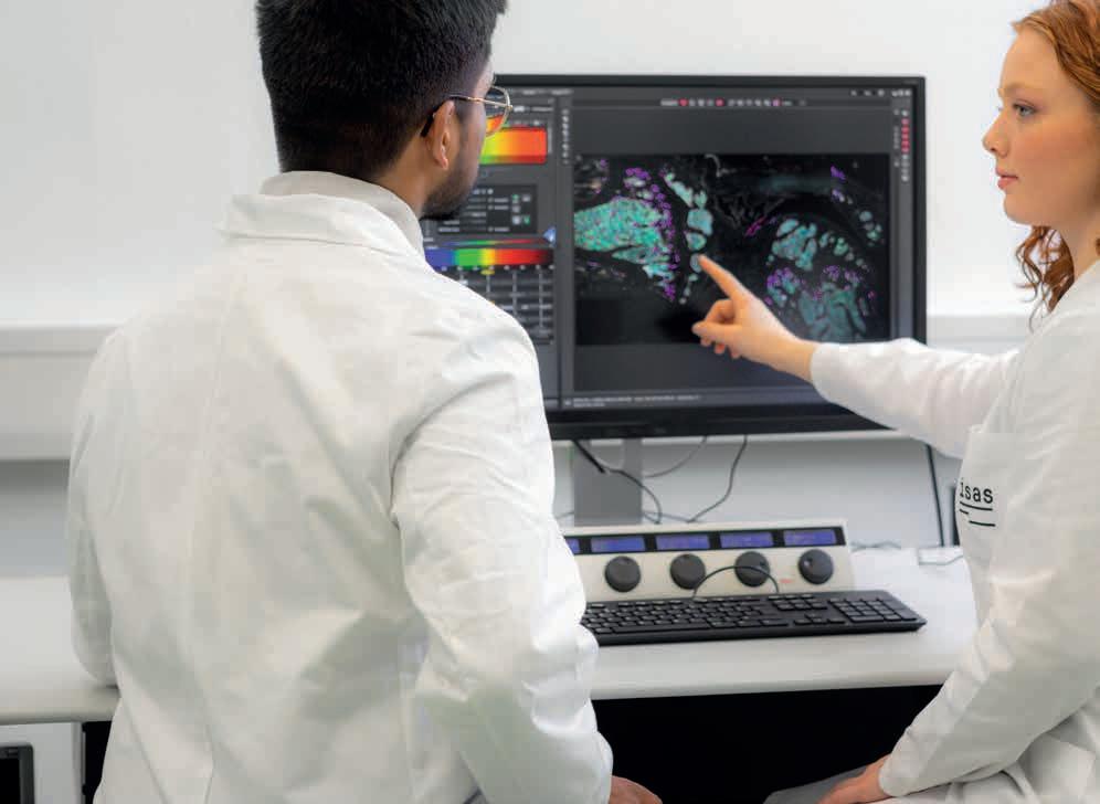
Dr Ali Ata Tuz uses a confocal microscope to prepare images of immune cells in tissue sections. He later analyses the high-resolution pictures on the computer.
ticularly fascinating for me here. My interest in neurological and neuroimmunological research then led me to my doctorate in Germany: I want to plan and carry out experiments myself and get to know the different research methods better instead of working only in the clinical field. At the IEII in Essen, I was able to combine neuroscience and imaging, the two aspects that interest me the most, during my doctorate. Even as a post-doctoral researcher at ISAS, I still work with various imaging methods, such as light sheet fluorescence microscopy.
2How did you proceed during your doctorate to get to the bottom of the causes of immunodeficiency after a stroke or heart attack?
Tuz: The basic idea of my doctorate was always to apply the results later in the clinic. In this field of research, we already knew that patients often have problems with infections after a stroke, something that indicates an immunodeficiency. An important part of the research process is to constantly define new research questions based on initial hypotheses and later also on the results. So we asked ourselves why this immunodeficiency occurs. If we know the cause, we can identify specific points on the signalling pathway. Researchers can then develop drugs specifically for these “targets”. However, there are still a lot of unanswered questions before the findings can really be applied in the clinic. For example, different times of drug administration after a stroke and the dose of the drugs still need to be researched. It is also a normal part of research that we often expect certain results at the beginning and then something completely new emerges.
3In your work you combine various methods, such as different microscopy techniques, mass spectrometry or AI-based 3D image analysis. What role does interdisciplinary collaboration play in your research?
Tuz: The cooperation with the different research groups has helped me a lot in analysing questions from different perspectives, and thus to achieve better results. I carried out my doctorate under the supervision of Prof. Dr Matthias Gunzer, Director of the IEII and Head of the Biospectroscopy department at ISAS. During that time I learned a lot about different microscopy techniques, for example. I was also able to learn a lot from scientists with different backgrounds who supported me, for example, with microscopy, analysing results or planning experiments with mouse models. At ISAS, I worked closely with the Bioimaging, AMBIOM and Proteomics research groups. Prof. Dr Anika Grüneboom, who is now my research group leader here at ISAS, helped us, for example, to carry out experiments with the confocal microscope and the light sheet fluorescence microscope. We also worked a lot with colleagues from the clinic because we also analysed blood and plasma samples from patients for the publication.
(Interview was conducted by AB.)
Bioimaging Research Group
Prof. Dr Anika Grüneboom
T: +49 (0)231 1392-239
E: anika.grueneboom@isas.de
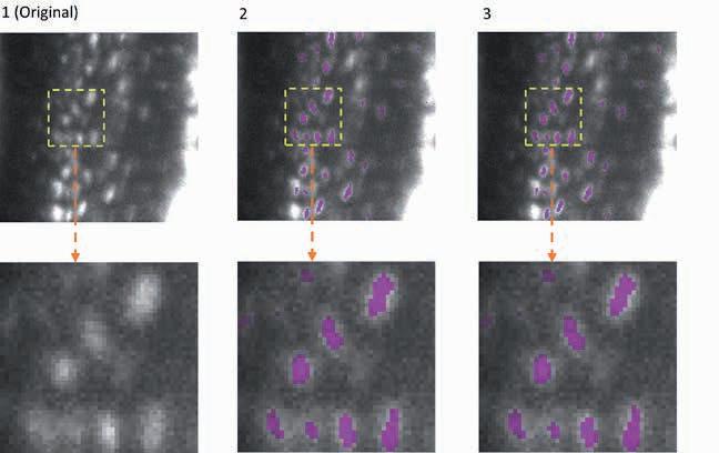
EfficientBioAI: New Open-Source Software Makes AI Models Lighter & Greener
The adaptability of EfficientBioAI was tested using several applications. One example is the 3D semantic segmentation. The AI model identifies specific structures within a three-dimensional cellular environment. The illustration shows the segmentation of osteocytes (mature bone cells) in images of mouse bones. The images were taken using light sheet fluorescence microscopy. The figure shows the original image (column 1), the segmentation by the FNet 3D model (column 2) from the MMV_Im2Im Toolbox for image-toimage transformation, and the segmentation after compression of FNet 3D by EfficientBioAI (column 3). The comparison (columns 2 and 3) shows that compression does not reduce the accuracy of the segmentation at all.
Artificial intelligence (AI) has become an indispensable component in the analysis of microscopic data. However, while AI models are becoming better and more complex, the computing power and associated energy consumption are also increasing. Researchers at ISAS and Peking University have therefore created a free compression software that allows scientists to run existing bioimaging AI models faster and with significantly lower energy consumption. The researchers have now presented their user-friendly toolbox called EfficientBioAI (open source) in Nature Methods.
Modern microscopy techniques produce a large number of high-resolution images, and individual data sets can comprise thousands of them. Scientists often use AI-supported software to reliably analyse these data sets. However, as AI models become more complex, the latency (processing time) for images can significantly increase. “High network latency, for example with particularly large images, leads to higher computing power and ultimately to
Yu Zhou has been a PhD student in the junior research group AMBIOM – Analysis of Microscopic BIOMedical Images at ISAS since September 2022. He previously studied biomedical engineering in Sweden, China and Switzerland.
increased energy consumption,” says Dr Jianxu Chen, Head of the AMBIOM – Analysis of Microscopic BIOMedical Images junior research group at ISAS.
A well-known technique finds new applications
To avoid high latency in image analysis, especially on devices with restricted computing power, researchers use sophisticated algorithms to compress the AI models. This means they reduce the amount of computations in the models while retaining comparable prediction accuracy. “Model compression is a technique that is widely used in the field of digital image processing, known as computer vision, and AI to make models lighter and greener,” explains Chen. Researchers combine various strategies to reduce memory consumption, speed up model inference, the ‘thought process’ of the model – and thus save energy. Pruning, for example, is used to remove excess nodes from the neural network. “These techniques are often still unknown in the bioimaging community. Therefore, we wanted to develop a ready-to-use and simple solution to apply them to common AI tools in bioimaging,” says Yu Zhou, the paper’s first author and PhD student at AMBIOM.
Energy savings of up to 81 per cent
To put their new toolbox to test, the researchers led by Chen tested their software on several real-life applications. With different hardware and various bioimaging analysis tasks, the compression techniques were able to significantly reduce latency and cut energy consumption by between 12.5 and 80.6 per cent. “Our tests show that EfficientBioAI can significantly increase the efficiency of neural networks in bioimaging without limiting the accuracy of the models,” summarises Chen. He illustrates the energy savings using the commonly
used CellPose model as an example: if a thousand users were to use the toolbox to compress the model and apply it to the
Our tests show that EfficientBioAI can significantly increase the efficiency of neural networks in bioimaging without limiting the accuracy of the models.
Jump Target ORF dataset (around one million microscope images of cells) they could save energy equivalent to the emissions of a car journey of around 7,300 miles (approx. 11,750 kilometres).
No special knowledge required
The authors are keen to make EfficientBioAI accessible to as many scientists in biomedical research as possible. Researchers can install the software and seamlessly integrate it into existing PyTorch libraries (open-source programme library for the Python programming language). For some widely used models, such as Cellpose, researchers can therefore use the software without having to make any changes to the code themselves. To support specific change requests, the group also provides several demos and tutorials. With just a few changed lines of code, the toolbox can then also be applied to customised AI models.
About EfficientBioAI
EfficientBioAI is a ready-to-use and opensource compression software for AI models in the field of bioimaging. The plug-andplay toolbox is kept simple for standard use, but offers customisable functions. These include adjustable compression
levels and effortless switching between the central processing unit (CPU) and graphics processing unit (GPU). The researchers are constantly developing the toolbox and are already working on making it available for MacOS in addition to Linux (Ubuntu 20.04, Debian 10) and Windows 10. At present, the focus of the toolbox is on improving the inference efficiency of pre-trained models rather than increasing efficiency during the training phase. However, the researchers are continuously developing and expanding the toolbox.
Biofluorescence
Research Group
Prof. Dr Matthias Gunzer
T: +49 (0)231 1392-100
E: matthias.gunzer@isas.de
Bioimaging Research Group
Prof. Dr Anika Grüneboom
T: +49 (0)231 1392-239
E: anika.grueneboom@isas.de
Proteomics Research Group
Prof. Dr Albert Sickmann
T: +49 (0)231 1392-100
E: albert.sickmann@isas.de
AMBIOM – Analysis of Microscopic BIOMedical Images
Junior Research Group
Dr Jianxu Chen
T: +49 (0)231 1392-217
E: jianxu.chen@isas.de
Zhou, Y., Cao, J., Sonneck, J., Banerjee, S., Dörr, S., Grüneboom, A., Lorenz, A., Zhang, S. & Chen, J. (2024) EfficientBioAI: making bioimaging AI models efficient in energy and latency. Nature Methods, 21(3), 368-369.
https://doi.org/10.1038/s41592-024-02167-z
“It’s very important to keep all the details –and that’s what we’re aiming for”
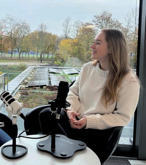
Your groupʼs name AMBIOM is short for Analysis of Microscopic BIOMedical Images. Would you like to give us a quick intro into what you do?
Chen: Since the last maybe 20 years, people have been starting to use microscopy for more than just looking at something. They want to quantify things in a larger scale. So thatʼs what we are doing. We develop computer algorithms, software and tools to help researchers analyse their biomedical images at large scales to answer their biomedical questions. Weʼre doing this in two ways. One is a pure method-developing direction. For example, when we want to detect or analyse a lot of images and make the process five times faster, the more the better. Itʼs a very clearly defined problem. We also have collaborations where we work closely with biologists or biomedical researchers here at ISAS. For example, one of our projects is to virtually tag cells that might be undergoing cell death. Instead of physically using fluorescent markers, we use AI techniques to predict how these fluorescent markers would look like. In that case, you reduce the amount of work from a biomedical experimental perspective.
You frequently address resource awareness in biomedical imaging in your research. Why is that a concern in this field?
Chen: This can be viewed from several different aspects. The first thing is carbon footprint or energy consumption. Running artificial intelligence models uses lots of energy and generates a lot of CO2. Another sustainability issue would be storage. Tons of data are being generated all over the world. Be it from a regular CT, MRI or X-ray scan or from biology labs where we collect our microscopic images. All this data needs to be stored somewhere. Data and knowledge management
In the “NACHGEFORSCHT – DIE lIVESCHAlTE INS lABOR” podcast, Dr Jianxu Chen (left) talks to presenter Cheyenne Peters about his work. With his junior research group AMBIOM – Analysis of Microscopic BIOMedical Images, the computer scientist is researching new tools and algorithms for biomedical image analysis, among other things.
are also important factors when it comes to resource awareness. We need to efficiently utilise human expertise when it comes to developing AI models for research purposes.
How is AMBIOM addressing those challenges?
Chen: The EfficientBioAI toolkit is a very concrete step in which we are trying to reduce the energy part. Scientists can run existing bioimaging models through our wrapper to speed things up and reduce energy by more than 80 percent. The second part is about storage. Because of the fast development of microscopy technologies we are starting to have higher resolution and higher throughput. To store those larger images, we developed two different image compression methods. Both achieved very promising results without losing critical information. Scientists in microscopy spend years and years to acquire all this imagery with stunning details and then after compression, you lose all details? I mean, it doesnʼt make sense, right? So itʼs very important to keep all the details and that is what we’re aiming for.
How do you ensure your tools are accessible and user-friendly for other scientists?
Chen: In order to make this really easily accessible for different scientists, not only for the master developers, we need to understand what they prefer. So, we did a lot of collaborations with different labs and we con-
stantly improve our tools according to their feedback. I think a very important thing is community. We are always open to have people come to us and reach out if they have questions or if they want to collaborate.
(The interview was conducted by CP.)
(Editorʼs note: This interview is an excerpt from the episode 10 – in English – of the ISAS podcast “NACHGEFORSCHT – DIE LIVESCHALTE INS LABOR”. It has been shortened and edited for our Annual Report.)

EPISODE 10: Resource Awareness in Biomedical Image Analysis
https://www.isas.de/en/en-kompakt/podcast-nachgeforscht-die-liveschalteins-labor-folge-10-resource-awarenessin-biomedical-image-analysis
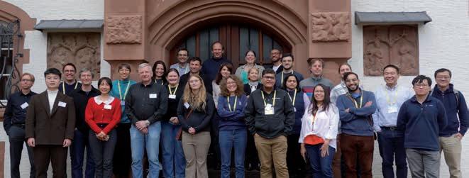
Dagstuhl Seminar on the Emerging Issues in Bioimaging AI Publications & Research
The rapid development of Artificial Intelligence (AI) opens up new questions in bioimaging AI publications and research. How can scientists properly validate the methods used in quantitative biological analyses? What are the ethical considerations in bioimaging AI research and publications, and what are future research directions of bioimaging AI focusing on validation and robustness? The participants of the seminar »The Emerging Issues in Bioimaging AI Publications and Research« at Schloss Dagstuhl – Leibniz Center for Informatics addressed these questions from January 21 to 24, 2024.
Dr Jianxu Chen, Head of the junior research group AMBIOM at ISAS, organised the seminar together with Dr Florian Jug, group leader and Head of the Image Analysis Department at the Human Technopole in Milan (Italy), Dr Susanne Rafelski, Deputy Director of scientific programmes at the Allen Institute for Cell Science in Seattle (US), and Dr Shanghang Zhang, tenure track assistant professor at the School of Computer Science, Peking University (China).
Open-ended dialogue and critical reflection
In structured presentations and interactive discussions, a diverse group of experts and promising early-career researchers from experimental biology, computational biology, bioimage analysis, computer vision, and AI research came together to discuss bioimaging in an interdisciplinary environment. Unlike conventional conferences, Dagstuhl Seminars are specifically designed to encourage open-ended dialogue and critical reflection rather than the presentation of finalised results. This
format allows participants to explore unfinished work and new ideas on emerging challenges in interdisciplinary fields.
The four organisers have documented the topics, discussions and conclusions of the seminar in a report available for further reading.
Chen, J., Jug, F., Rafelski, S., Zhang, S. (2024) The Emerging Issues in Bioimaging AI Publications and Research (Dagstuhl Seminar 24042). Dagstuhl Reports, 14(1), 90-107.
https://doi.org/10.4230/DagRep.14.1.90
More than 25 researchers took part in the Dagstuhl seminar 24042.

P ROGRAMME DEVELOPMENT: A COMPASS
Research that is both socially relevant and future-oriented must undergo constant transformation. For ISAS, this means: in order to be able to pursue its mission – developing analytical methods that can be combined to create new integrative measurement strategies for health research – it firstly requires continuous fine-tuning of its own direction. And secondly, budding ideas need sufficient time, resources and space to take shape. This is what the institute’s own programme development is designed for.
In this way, ISAS promotes promising new scientific ideas that do not (yet) fit into the canon of one of the research programmes. These ideas have the opportunity to grow beyond the established programmes and thus, prospectively, with their potential inclusion, provide approaches for the further development of ISAS research.
(SR)
Bioimaging Research Group
Prof. Dr Anika Grüneboom
T: +49 (0)231 1392-239
E: anika.grueneboom@isas.de
ERC-Sulfaging
Dr habil. Miloš Filipović
T: +49 (0)231 1392-4173
E: milos.filipovic@isas.de
Preclinical Metabolomics
Research Group
Prof. Dr Dr Alpaslan Tasdogan
T: +49 (0)231 1392-100
E: alpaslan.tasdogan@isas.de
“Moving to ISAS changed my whole view on mass spectrometry”
Dr habil. Miloš Filipović joined ISAS in October 2020, after moving his group ERC-Sulfaging from Bordeaux, France to Dortmund. His EU-funded research focusses on the connection between ageing processes and gasotransmitter signalling, hydrogen sulfide (H₂S) in particular. In 2024, the 42-year-old accepted an offer for a full professorship for Molecular Biosciences at the University of Glasgow, Scotland. As he will be leaving ISAS in spring 2025, this poses a fitting occasion to reflect on the past four years and review his time at the institute in a short interview.
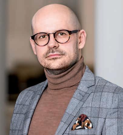
What was you most memorable finding in the past four years?
Filipović: In 2024, we submitted a paper on the molecular mechanism of ergothioneine, a compound found in mushrooms. Our results hint that it elongates the health span of ageing animals through provision of H2S which protects cells from oxidative stress through the process of persulfidation (► p. 45). It’s an interesting story because it started more as a backup plan for a thesis, where a couple of things didn’t work as planned. Out of necessity, we had to come up with something quick and had no high expectations at all. It somehow just perfectly landed in our laps, as the whole mechanism turned out to be right within our area of expertise. We already had the network and the resources to get to work right away. It was really all kind of serendipity in a way and quite unusual.
How would you describe your research at ISAS?
ERC-Sulfaging
Dr habil. Milos Filipović
T: +49 (0)231 1392-4173
E: milos.filipovic@isas.de
Filipović: I would say curiosity-driven. We donʼt necessarily stick into one frame, and we also donʼt stick to one method. I mean, if we donʼt have a method and can’t find it somewhere else in the world, we’ll develop a method! During our time here we have looked at some cardiovascular aspects, like myocardial ischaemiareperfusion injury, hemorrhagic shock, and so on. With
Dr habil. Miloš Filipović leads the ISAS research group ERC-Sulfaging and has been conducting research with his team at ISAS from 2020 to 2025.
ergothioneine we have these exercise models, and ageing. We’ve also been looking at neurodegeneration, particularly Alzheimer’s disease models. Mechanistically, all of these topics came from our area of expertise, meaning those post-translational modifications, particularly of cysteines. But also, they are very distinct stories.
Which techniques have shaped your time here?
Filipović: Mass spectrometry as such is something that we learned at ISAS. It is a broad term, but we use different types of mass spectrometry in our research. Some of them we adapted from the literature and so on, but none of that would have been possible if we didnʼt have access to all these instruments on such a regular basis. In most institutions you give your samples to the facility and then get back the results. Because unlike at ISAS, you donʼt do every single step by yourself, from the sample preparation to measurement to data analysis, you donʼt have actual hands-on experience of what can go wrong and why the measurement didnʼt work out. Being skilled in all necessary steps is a huge advantage when it comes to problem solving.
After I came here, my whole view on mass spectrometry, specifically proteomics, changed in a way. For example, at ISAS every sample goes to quality control before itʼs measured. That extra step helps all the devices run smoothly for a long time, without a technician having to clean the instrument every three months and stopping the workflow. What seems like a standard procedure, is quite unusual as it takes extra time. But it has benefits: part of the goal of the institute is to standardise the measurements, and for that you want the whole process done in the most precise and clean way. This approach has probably shaped my work forever.
PERSULFIDATION
Persulfidation is a posttranslational modification in which a persulfide group (-SSH) is added to the thiol side chain of cysteine residues in proteins. This process is triggered by hydrogen sulfide (H2S), a gasotransmitter that acts as a signalling molecule in the body.
Persulfidation can regulate protein activity, protect cells from oxidative stress, and is involved in anti-inflammatory and neuroprotective processes.
In March 2025 you will transfer to the School of Molecular Bioscience at the University of Glasgow as a full professor. What are your plans for the new position?
Filipović: Besides doing research, I’ll also be teaching in Glasgow. That’s exciting and a new challenge I’m happy to take on. Of course I’m going to stay in touch with ISAS, we already have some collaborative projects planned with the Cardiovascular Pharmacology research group. We developed a concept how persulfidation affects phase separation and aggregation in the ageing brain, that we plan to expand to the heart, specifically in the context of amyloidosis.
Sulfaging has received funding from the European Research Council (ERC) under the European u nionʼs Horizon 2020 research and innovation programme (grant agreement No. 864921).

(The interview was conducted by CP.)
OUR YEAR IN FIGURES
154
employees
As at December 31, 2024, ISAS had 72 female and 82 male members of staff at its locations.
scientific technical staff members (m/f/n-b) worked at ISAS, of whom 14 were women and nine men.
64 29 35
researchers (m/f/n-b) were employed at the institute in 2024, of whom 29 were women and 35 men.
Among the researchers, there were 18 female and 17 male PhD students 35 18 17
doctoral candidates (m/f/n-b)
nature methods
51
papers with a lead or corresponding author from ISAS were published in 2024.
118 publications were published in peer-reviewed journals.
7.8
impact factor
The average impact factor of the publications in peerreviewed journals was 7.84.
1,155,389 kWh
12 of electricity were produced by the ISAS City location, including 830,389 kWh by the photovoltaic system and 325,000 kWh by the cogeneration power plant.
software & tools (open source) were published by the AI experts and bioinformaticians in 2024.
academic qualifications
Of the 33 final theses, 18 were internal.*
MASTER OF SCIENCE
* The other projects were external expert assessments.
PhD theses
Of these PhD dissertations, four were written at ISAS.* 11
lectures were given by ISAS researchers at conferences, workshops and other institutions.
BSc, MSc
These included seven bachelorʼs and seven masterʼs students who wrote their final theses at ISAS.* 22
poster presentations were held by researchers from ISAS in 2024.
scientific events were (co-)organised by ISAS in 2024.
interns (m/f/n-b)
were able to familiarise themselves with the work at ISAS in 2024, including seven in research and two in communication.
14,859,000
total funding for ISAS
ISAS received EuR 14,859,000 in institutional funding from the federal and state governments for its core budget.
2.219 million
third-party funding-
In addition, the institute received EuR 2,218,891 in third-party funding for 2024.
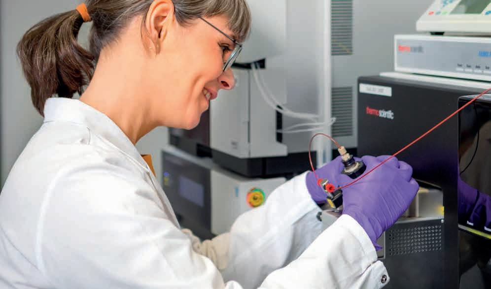
MULTI-OMICS
The aim of the Multi-Omics research programme is to develop bioanalytical and computational technologies for prognostic, diagnostic, and predictive biomarkers. The scientists research methods which can be used to more effectively detect markers in complex biological matrices. These biological markers are intended for cardiovascular diseases (CVDs), cancer, and metabolic disorders.
Most diseases are caused by the deregulation of metabolic and signalling pathways at different molecular levels, from genes to lipids to proteins and metabolites. The regulation of metabolic pathways and their interaction with environmental factors requires the use of multiple analytical methods for the detection of proteins, lipids, metabolites, and their dynamics. A single
analytical technology is neither sufficient for a comprehensive understanding of selected biological model systems, nor for identifying biomarkers. In view of the huge number of potential analytes in biological systems, all measurments need to be carried out to a high degree of precision. That is why a multi-omics approach, a combination of different omics – meaning
One of the technologies that plays a central role in the Multi-Omics research programme is mass spectrometry.
Bioimaging Research Group
Prof. Dr Anika Grüneboom
T: +49 (0)231 1392-239
E: anika.grueneboom@isas.de
Miniaturisation Research Group
PD Dr Joachim Franzke
T: +49 (0)231 1392-174/199
E: joachim.franzke@isas.de
Proteomics Research Group
Prof. Dr Albert Sickmann
T: +49 (0)231 1392-100
E: albert.sickmann@isas.de
Standardisation
Research Group
PD Dr Dirk Janasek
T: +49 (0)231 1392-202
E: dirk.janasek@isas.de
Lipidomics
Junior Research Group
Prof. Dr Sven Heiles
T: +49 (0)231 1392-4202
E: sven.heiles@isas.de
Multidimensional Omics
Data Analysis
Junior Research Group
Prof. Dr Robert Heyer
T: +49 (0)231 1392-271
E: robert.heyer@isas.de
Spatial Metabolomics
Junior Research Group
Dr Karl Smith
T: +49 (0)231 1392-4210
E: karl.smith@isas.de
addressing the entire characterisation of all genes (genomics), metabolites (metabolomics) or proteins (proteomics) – approach is needed.
Multi-omics strategies: indispensable for precision medicine
Omics technologies are an important starting point in personalised therapies (precision medicine). On the one hand, they produce multi-dimensional data sets (in unprecedented quality), which bring insights into disease processes and potential treatment approaches. On the other hand, multiomics data sets for non-directional analyses can be used to demonstrate new correlations (generating new hypotheses) between various molecule classes. However, these large and complex data sets also need to be managed adequately.
One of the main focus areas: omics approaches for CVDs
Regarding CVDs, many factors including genetic predisposition, gut microbiome, lifestyle, and environmental factors, can have an impact. In addition, current therapeutic approaches to tumour diseases and inflammations can have side effects on the cardiovascular system. ISASʼ Multi-Omics programme therefore particularly focuses on developing multi-omics technologies and assays with regard to heart attacks (myocardial infarction), heart failure, cardiotoxicity, and cardio-oncology. With omics-integrative models and the combination of lipidomic, proteomics, and metabolomic data, and by using graph databases and AI, researchers at ISAS aim to gain comprehensive insights into CVDsʼ complex molecular aspects. The analytical challenges that the scientists are tackling include molecular coverage, analytical sensitivity, data
integration, and interpretation, as well as issues related to data quality, reproducibility, and standardisation.
Besides developing technologies to shed light on molecular mechanisms and identify biomarkers, the programme addresses the identification of new therapeutic targets. Therefore, unravelling the molecular mechanisms underlying CVDs is crucial. Systems biology approaches using multiomics data play a critical role in identifying cellular changes and signalling events associated with disease genesis and progression.
High-throughput / High-resolution technologies with new bioinformatic strategies
In general, multi-omics technologies comprise analytical methods which can be used to investigate biomolecules from tissue samples or other biological samples like blood at a global level. Scientists at ISAS devote their time to developing such tools for integrating multi-omics data sets. They combine various analysis techniques such as electrospray ionisation mass spectrometry (ESI-MS), MALDI (matrix-assisted laser desorption/ionisation), light and fluorescence microscopy, and they develop new bioinformatic strategies for data analyses.
(SR)
Mass Spectrometry: Precise Drug Monitoring for Improved Treatment with Biologicals
They are indispensable for patients, but optimum treatment with this group of medicines is complex and fraught with challenges. Biologicals (► p. 54) have become a cornerstone of personalised medicine and are primarily used in autoimmune diseases and cancer. Treatment with these drugs can be difficult at times, pushing conventional laboratory methods for therapeutic monitoring to their limits. Together with other authors, Dr Yvonne Reinders from the ISAS Proteomics research group and Prof. Dr Peter Findeisen from MVZ Labor Dr. Limbach & Kollegen report in the journal Clinical Proteomics on how new analytical techniques can be used to optimise treatment with biologicals for patients. In this interview, the two researchers – a chemist and a specialist in laboratory medicine – advocate for a combination of complementary laboratory techniques.
What is therapeutic drug monitoring and why is it particularly important for chronic inflammatory diseases?
Findeisen: Therapeutic drug monitoring, or TDM for short, refers to measuring the concentration of medicines in the blood. There are certain target values that were determined in clinical trials. If the concentration is too high, side effects occur. If itʼs too low, the medicine is not effective. This is always a balancing act, and you have to monitor the target value regularly, particularly in the case of chronic inflammatory diseases.
TDM is particularly challenging with biologicals. Why?
Findeisen: Biologicals are now used as a standard treatment in gastrointestinal diseases such as Crohn’s disease and ulcerative colitis. Their use is intended to prevent acute flare-ups by interrupting inflammatory processes in a targeted manner and without triggering major side effects. The drugs are also used for other inflammatory diseases such as multiple sclerosis and in oncology. In this context, it’s always important that the drug be administered within the target range. Although dosing is based on body weight, every patient breaks biologicals down differently – and TDM is therefore
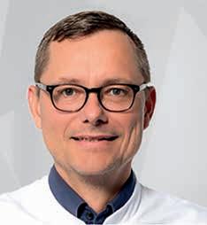
Hentschel, A., Piontek, G., Dahlmann, R., Findeisen, P., Sakson, R., Carbow, P., Renné, T., Reinders, Y. & Sickmann, A. (2024) Highly sensitive therapeutic drug monitoring of infliximab in serum by targeted mass spectrometry in comparison to ELISA data. Clinical Proteomics, 21, 16.
https://doi.org/10.1186/s12014-024-09464-x
Prof. Dr Peter Findeisen is Head of laboratory medicine at Heidelberg-based MVZ labor Dr. limbach & Kollegen.
vital. At the same time, biologicals are very large molecules, ruling out some of the traditional measurement techniques. That’s what makes TDM of biologicals such a complex undertaking.
Until now, biologicals have often been monitored using immunological tests –particularly ELISA, which stands for enzymelinked immunosorbent assay. How suitable is this method for TDM of biologicals?
Findeisen: We can imagine ELISA as being like a sandwich: between the two pieces of bread is the filling – in our case, the drug we want to measure. While the bottom slice of bread captures the drug, the top slice makes it visible by triggering a colour reaction. The more colour is produced, the more drug is present in the sample. The problem is that, for every new drug, it’s first necessary to develop specific antibodies – that is, the suitable slices of bread – and this process takes years. New biologicals are constantly appearing on the market, and the number of ELISA tests lags far behind. In some cases, there’s also a phenomenon known as “cross-reactivity”, whereby the ELISA test also reacts to substances that are not the target drug but resemble it –potentially leading to false positives.
This is where mass spectrometry comes in. How does this alternative technique work in TDM?
Reinders: In the case of mass spectrometry, the large proteins are cut up, so to speak, into peptides. We do this using an enzyme known as trypsin, which only severs the proteins at certain amino acids. This gives us peptides with a length of eight to 20 amino acids – an ideal size for analysis with mass spectrometry. The trick is that we break the large antibodies down into fragments that are nevertheless unique to the respective protein. This then allows us to identify and quantify the whole antibody. Where traditional techniques such as ELISA have limitations when it comes to TDM, with mass spectrometry, the lack of cross-reactions means we can achieve more specific results.
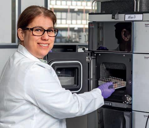
What are the respective strengths of the two techniques?
Findeisen: Although ELISA is capable of a high throughput, as well as being an easier technique to perform and more widespread than mass spectrometry in clinical laboratories, it also suffers from certain problems: not only the cross-reactions I mentioned before, but also the fact that most ELISA formats only detect the free form of biologicals. After all, biologicals are foreign proteins and can therefore trigger an immune response if administered for a long time. In other words, patients develop “anti-drug antibodies” against biologicals as time goes by. These antibodies bind to the drug, meaning that ELISA can often no longer detect it. In contrast, the mass spectrometric test detects both the free form and the form that is potentially bound to anti-drug antibodies
Reinders: Mass spectrometry can respond to new drugs much, much faster. We can draw up a new mass spectrometric method relatively quickly, whereas ELISA needs years of development work to generate specific antibodies. Moreover, a mass spectrometric technique allows to detect multiple substances at the same time,
Dr Yvonne Reinders has been working at ISAS since 2018. She analyses her samples using, among other things, the Orbitrap Astral mass spectrometer.
using a method known as multiplexing, when patients are receiving combinations of different drugs simultaneously.
Are these combined therapies becoming increasingly important in clinical practice?
Reinders: Yes. Particularly in oncology, two or more monoclonal antibodies are often administered together with chemotherapy drugs in order to attack various signalling pathways and boost efficacy. If a patient is receiving personalised combined therapy, you obviously want to measure all of the administered drugs at the same time.
W hat specific developments are you working on at ISAS ?
Reinders: The focus is on the rapid expansion of these techniques. We want to create a platform for the mass spectrometric measurement of as many antibodies as possible, that can compete with ELISA. When new biologicals reach the market, we want to be able to respond quickly. For this, we need unique peptides that allow us to identify each antibody.
W hat are the practical challenges?
Findeisen: Mass spectrometers are very expensive. You also need robots for sample preparation, and very highly trained personnel. These are things that only a few specialised laboratories can afford. It’s likely that the tests will be concentrated at a small number of centres – as is the case with other specialist analyses. However, once the investment costs are covered, the ongoing costs per measurement are significantly lower than with ELISA.
BIOLOGICALS
Biologicals are drugs produced using biotechnology with the help of living cells from microorganisms, animals or plants. They are made up of complex protein compounds, among other things, and intervene in disease mechanisms in a targeted manner with fewer side effects than conventional medicines. One of the most important subgroups of biologicals are antibodies – especially monoclonal antibodies such as infliximab.
Do you see mass spectrometry as a complement to or replacement for ELISA?
Findeisen: The two techniques can certainly complement one another. Mass spectrometry should be seen as a reference method – it’s more reliable and provides a more complete image than ELISA on its own.
Proteomics Research Group
Prof. Dr Albert Sickmann
T: +49 (0)231 1392-100
E: albert.sickmann@isas.de
(The interview was conducted by UE.)
What’s invaluable in your project?
This commercially available column for mass spectrometry! It has been designed for specific chiral separation needs and plays a crucial role in our collaborative research project »PIPMet – Phosphoinositides in Metabolic Disease«. Together with our collaborators at the Leibniz-Forschungsinstitut für Molekulare Pharmakologie (FMP) and the German Institute for Human Nutrition Potsdam-Rehbrücke (Deutsches Institut für Ernährungsforschung Potsdam-Rehbrücke, DIfE), we aim to find out if the altered signalling of phosphoinositides (PIPs), a type of membrane lipids, underlies metabolic disease and related disorders like obesity and diabetes. However, since the seven regioisomers of PIPs differ in their phosphorylation pattern and fatty acyl chains, their separation and accurate profiling remains technically challenging with conventional liquid chromatography mass spectrometry (LC-MS) analysis.
To address this problem, we are currently implementing a chiral chromatography approach at ISAS using specialised columns like the CHIRALPAK IB-U by Daicel and CHIRAL ART Cellulose-SB by YMC, paired with high-resolution mass spectrometry.
The IB-U column is currently installed on our ultra-high-
performance LC-system which is coupled to an Orbitrap Exploris mass spectrometer. By employing chemical derivatisation of PIP standards and improving LC conditions, we successfully enhanced the resolution of the regioisomers, building upon and optimising previously published methods.
With these promising results, we are confident in applying this method to diverse biological samples, including genetically modified cell lines, isolated organelles and tissues from obesity mouse models, and thereby resolving the unmet questions in the context of PIPMet.
(Protocol: CP)
The l eibniz Association is funding this project under the reference K509/2023.

This is a chiral stationary phase column which is widely used to separate enantiomers, meaning molecules that are non-superimposable mirror images of each other.
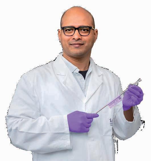
100mm
CHIRALPAK IB-U from Daicel
Laxmikanth Kollipara, Research Associate in the Lipidomics research group
Collecting Valuable Data through FAIR Research Data Management
Modern medicine and the research on which it is based generate an unprecedented flood of data. The average hospital today produces around 50 petabytes of patient data per year – the equivalent of almost 20 billion average e-books. And a single laboratory device can generate tens of thousands of measurement results every day. All this data needs to be managed, preferably in such a way that researchers and clinicians can make full use of it. But often this does not happen. The European Union has estimated that inadequately managed research data costs Europe around ten billion euros a year – in other words, almost 30 million euros a day. In a position paper drawn up with ISAS participation, the German Cardiac Society (Deutsche Gesellschaft für Kardiologie, DGK) and the German Centre for Cardiovascular Research (Deutsches Zentrum für Herz-Kreislauf-Forschung, DZHK) have therefore advocated better research data management in cardiovascular science.
“If research data is only partially accessible, this means, for example, that scientists run the risk of duplicating measurements and observations that already exist,” says Prof. Dr Albert Sickmann, Chair of the ISAS Board of Directors and one of the authors of the position paper. “That wastes resources and, of course, makes no sense.” In order to prevent this, the DGK and DZHK advocate data management in accordance with the FAIR principles. According to these principles, data should be Findable, Accessible, Interoperable and Reusable. In other words, researchers should store their data in such a way that it can be easily found and accessed by others. Furthermore, they should be able to use the data with their own systems for new research projects without complicated conversions.
UnFAIR cardiology?
The position paper, which was published in the journal Clinical Research in Cardiology in 2024, identifies four central reasons why this is still too rarely the case in cardiology and other fields. First and foremost, it’s down to the researchers themselves. They often lack the time, money and incentives to make their data publicly accessible. “If I need 80 per cent of the time to generate the data and 20 per cent for the documentation, that is a considerable amount of time for measurement projects that last a year,” says Sickmann. Furthermore, there is frequently a lack of standards and data formats that regulate how researchers should store data so that other scientists can use it. “It’s not coordinated. Data management at one research location doesnʼt
Among other things, Prof. Dr Albert Sickmann is responsible for the Multi-Omics research programme at ISAS, including the associated analyses and evaluations. He advocates following the FAIR principles when using generated data in science.
necessarily have much in common with that at another location,” says Sickmann. This makes it difficult for researchers from different institutions to exchange information, both at national and international level.
A third challenge in research data management – or RDM – is organising the sheer mass of data in such a way that scientists can see what measurements and evaluations already exist. And fourthly, particularly when it comes to sensitive patient data, data protection regulations make many researchers uncertain as to how they can share the information across national borders in a legally compliant manner.
Open-source software as a solution
Even quite pragmatic hurdles can hinder data utilisation in accordance with the FAIR principles. The mass spectrometers at ISAS, for example, automatically record measurements in a file format specified by the device manufacturer. “The measurement data can then initially only be opened using the manufacturerʼs software,” says Sickmann. This means that another laboratory must first purchase the software in order to be able to use the measurement data. That is cost-intensive and many researchers are reluctant to generate additional expenses. “At ISAS, we have therefore switched to converting our measurements into file formats that can also be processed using open-source software,” explains Sickmann.
At ISAS we have switched to converting our measurements into file formats that can also be processed using open-source software.
The researchers at ISAS even go a step further. They upload the measurements from every paper they publish to the freely accessible database of the European Institute of Bioinformatics in Hinxton, UK, including additional information on how the data was collected: what questions the experiment was intended to answer, how it was set up, which software was used to interpret the measurements, etc. Often, it is only metadata such as this that provides the crucial information that allows other scientists to utilise the measurement data effectively for their own research. “During proteome studies, for example, we identify thousands of proteins,” says Sickmann. “Itʼs possible that only ten of these proteins are of interest for the question we are studying. But
others can find the 20 proteins they need themselves in the entire data set and donʼt have to repeat the measurement.”
A FAIR cookbook and more awareness of RDM
Transparent and standardised RDM also lays the foundation for research breakthroughs that may only be possible in the future. For example, scientists will not only be able to analyse the proteome, metabolome or lipidome individually – as has been the case up to now – but also the complex interactions through which these systems influence one another. “That requires huge amounts of data and innumerable experiments,” explains Sickmann. “I believe that this will be possible in the future thanks to a new way of handling data.” In various collaborations, the expert in clinical proteome research has observed that the awareness of the need for effective RDM is growing in cardiology Tools such as FAIRsharing, a searchable register for data standards, and the “FAIR Cookbook” with practical guidelines help researchers to archive measurement data in such a way that it is accessible to others. Funding is also being increasingly linked to requirements for FAIR data management.
In the long run, change is needed from the inside out
However, the authors of the position paper argue that further steps are needed. They urge that RDM should be made an integral part of scientific training and that the know-how and motivation for this should be created in graduate programmes and post-doctoral training courses. Researchers themselves benefit because the field is continuing to develop. And it should not be forgotten that a study 1 has shown that research papers whose data sets are public are cited significantly more often than publications that are not, increasing the impact of a scientist’s own research. The DGK/DZHK position paper recommends that scientific journals and publishers should specifically demand that research data should be made available. At the same time, universities, funding organisations and the research community should develop overarching data standards for cardiology and improve the digital infrastructure, for example by creating more public data repositories. Only when the data is optimally networked cardiovascular research can realise its full potential – to the benefit of both science and the patient.
Steffens, S., Schröder, K., Krüger, M., Maack, C., Streckfuss-Bömeke, K., Backs, J., Backofen, R., Baeßler, B., Devaux, Y., Gilsbach, R., Heijman, J., Knaus, J., Kramann, R., Linz, D., Lister, A.L., Maatz, H., Maegdefessel, L., Mayr, M., Meder, B., Nussbeck, S.Y., Rog-Zielinska, E.A., Schulz, M.H., Sickmann, A., Yigit, G., Kohl, P. (2024) The challenges of research data management in cardiovascular science: a DGK and DZHK position paper— executive summary. Clinical Research in Cardiology, 113, 672-679.
https://doi.org/10.1007/s00392-023-02303-3
1 https://doi.org/10.7717/peerj.175
J UNIOR SCIENTISTS
From Intern to Postdoc –Supporting Junior Researchers
To support young researchers, ISAS has set up programmes covering all stages of a career in science. The programmes are aimed at bachelorʼs and masterʼs students, including interns, and enable them to spend time in the research groups. Science journalism students also have the opportunity to complete an internship in the Communications team. In addition, the institute offers a graduate programme for doctoral candidates and further training opportunities for postdocs.
In the first three years of the PhD course, the curriculum of the structured doctoral study programme includes ten workshops, an information event on career planning, an internal lab rotation and an optional doctorate-related research visit abroad. In the final stage, the focus shifts to completing the work and the PhD paper. How long a PhD takes at ISAS depends on the specialisation and averages between three and a half and four and a half years.
S cience communication for doctoral candidates and postdocs
To give them the skills they need in order to share their knowledge and research with society in the best possible way, ISAS regularly organises training courses on science communication for doctoral candidates and postdocs. For example, the “Postdoc Pitch Day” is a career development tool that provides a forum for presenting initial scientific ideas and receiving feedback from experienced
researchers. The event aims firstly to motivate the participants to expand their skills in order to communicate research topics in a generally comprehensible way. Secondly, it serves to help them further develop their own research ideas in such a way that they lead to support measures, third-party funding applications or interdisciplinary collaborations with internal or external partners, or a patent application.
E xchange with universities
ISAS promotes the career opportunities of excellent young scientists by entrusting them with the management of research projects in junior research groups. Giving them management responsibility as early as possible is intended to support young scientists aspiring a career in research. In the training of junior researchers, the institute takes part in a regular exchange of ideas and information with the universities with which it cooperates in research and teaching.
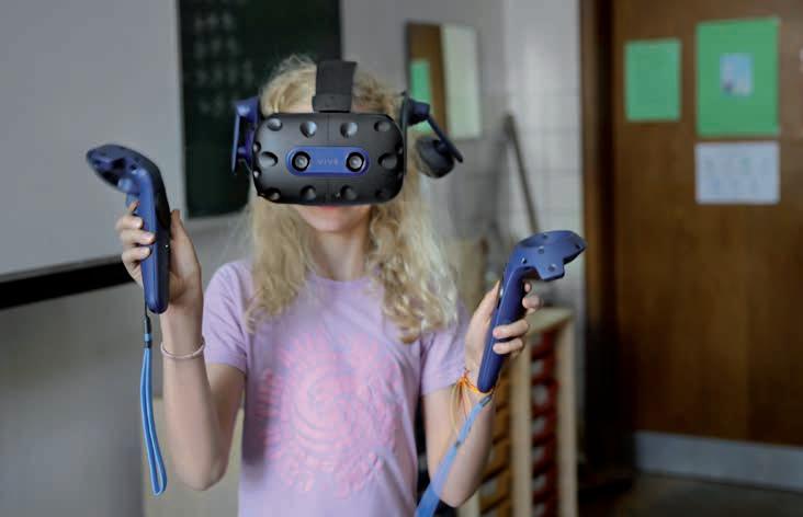
Fascinating Insights: Virtual World in the Classroom
“I havenʼt even arrived back in reality yet,” says Marie (9) laughing as she stumbles slightly around the room. Until a few seconds ago, the primary school student was wearing virtual reality (VR) goggles and looking into a purely digital world. While she slowly finds her way around the classroom at Don Bosco primary school in Bochum, Dr Kathrin Krieger helps the next students to immerse themselves in the virtual world. Together with Dr Jianxu Chen, Head of the AMBIOM –Analysis of Microscopic BIOMedical Images research group at ISAS, the scientist visits class 3a before summer break.
At ISAS in Dortmund, the two researchers are working on analysis methods for health research. More specifically, they are focusing on artificial intelligence (AI) for the evaluation and visualisation of microscopic images. Talking to primary school students about their work is a rare opportunity for them to transfer knowledge in a child-friendly way. The visit is also a new experience for the children. The topic of “extended reality” (XR ► p. 63) initially leaves many question marks on their faces. But Chen and Krieger have anticipated this and prepared intensively for the two-hour lesson with the class.
Only seven things at once
The children are listening attentively to Krieger who is standing in front of them at the whiteboard. The researcher starts at the very beginning: What is reality? What does extended reality mean? What can digital 3D models do that photos cannot? To answer these questions, the scientist has prepared a short memory test. The children are asked to memorise various symbols and later recognise which one is missing. They all agree when it comes to just three symbols: the laptop! But then it gets trickier, more and more pictures and sym-
Marie explores a digital world with VR goggles. The controllers in her hands allow her to interact with the world and grab things, for example.
bols appear and the children find it increasingly difficult to recognise the missing object
The test shows: Humans can only remember things to a limited extent. On average, the short-term memory can memorise seven things at once. With that, Krieger reveals a challenge for researchers in the microscopy laboratory to the children. A light sheet fluorescence microscope produces images of a single sample such as an entire organ (e.g. the jaw, knee or heart of a mouse) layer by layer – with an average of more than 500 images. Biologists later compile the individual images into a 3D model on the computer for analysis. This 3D model already helps the human brain to process the information from the many individual images in a focused manner. However, with the help of XR goggles, these models can be used to literally grasp the examined organ and thereby analyse it even more precisely.
How do vision and information processing actually work? And how do humans perceive colours in the first place? In order to find the answers together, the 3a class continues with a transparent box full of colourful coloured discs. Each child is allowed to choose a colour – a decision that is visibly difficult: blue, red or yellow? The students curiously grab the transparent colour discs and hold them in front of their eyes. The classroom suddenly appears in new colours. Some are thrilled to discover that they can create unexpected colours by combining discs.
V irtual insight into the human body
Krieger rings a small bell and immediately all the childrenʼs eyes are on her again. The researcher uses the attention to show what she and Chen have brought with them: Apple Vision Pro and HTC Vive Pro 2 – AR and VR goggles (► p 63), which all the children are allowed to try out. The researchers brought these XR goggles with them so that the third-graders can experience for themselves what it feels like to be in virtual reality.
Johanna is taking the whole body apart.
The virtual space Krieger has chosen for today contains a model of a human body. She has developed the virtual environment at ISAS to better visualise 3D research results. Instead of other scientists, today the students can use controllers to grasp and reposition the individual organs. Some of the children are particularly engaged: “Johanna is taking the whole body apart,” exclaims Finn (8) enthusiastically, looking at Kriegerʼs laptop. The screen shows how Johanna is cheerfully removing all the organs from the body and distributing them in the virtual space

lily and Charlotte listen attentively to Krieger.
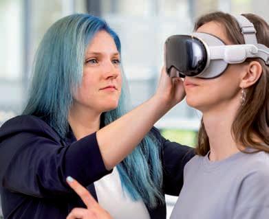

Augmented reality – self-made for a change!
While the students wait to put on the XR goggles one by one, they make their own versions. Each child receives a pair of cardboard glasses onto which they can stick their own coloured tracing paper. Julian (8), who is still in the process of attaching the paper to his glasses, already has a clear idea: “I canʼt see anything special yet, I have to colour in both lenses first, then Iʼll see something nice.” Those like Julian, who have listened carefully to Kriegerʼs explanations at the beginning, are aware of this: Only if they paint an identical image on both lenses, they will be able to see something in the end. Just like in an AR world, they view their classroom as a real environment paired with their own artificially and artistically created elements. After just a short time, children are running around the classroom with their colourful glasses. Some have drawn figures on the tracing paper, others have created imaginative colourful worlds
K nowledge transfer through hands-on experience
ISAS researchers like Chen and Krieger are keen to take part in activities like this. As AI experts, they want to playfully familiarise children with the topic of AI in microscopy. “Technologies like these are important tools. XR is ideal for children, as it combines theoretical knowledge transfer with hands-on experimentation,” says Chen. The two researchers also benefit from the exchange: “To explain something to children, you have to focus on the most basic information. Dealing with my own research in this way and breaking it down to the essentials was an exciting process for me,” summarises Krieger.
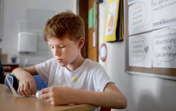
left: Dr Kathrin Krieger works with various VR and XR goggles. Here she puts the Apple Vision Pro on Clara Manthey, an intern in the Communications team.
Right: Dr Jianxu Chen heads the AMBIOM junior research group at ISAS and provided the impetus for the primary school visit.
Julian paints the same picture on both spectacle lenses.
(CM / CP)
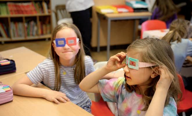
XR is ideal for children, as it combines theoretical knowledge transfer with hands-on experimentation.
EXTENDED REALITY
Extended reality (XR) is an umbrella term for immersive technologies that encompass virtual reality (VR), augmented reality (AR) and mixed reality (MR). The former creates a new, independent world for users. The VR world is a purely digital, computergenerated image that is completely independent of the physical environment. Instead of completely replacing the real world, AR visually overlays it with digital elements. These can be digital images, graphics or even animations. MR combines elements of VR and AR so that digital and real objects can interact with each other.
Biofluorescence
Research Group
Prof. Dr Matthias Gunzer
T: +49 (0)231 1392-100
E: matthias.gunzer@isas.de
AMBIOM – Analysis of Microscopic BIOMedical Images
Junior Research Group
Dr Jianxu Chen
T: +49 (0)231 1392-217
E: jianxu.chen@isas.de
Communications Team
Sara Rebein
T: +49 (0)231 1392-234
E: sara.rebein@isas.de
Anna and Greta look through their self-painted cardboard glasses.
The MSCoreSys associated junior research group AMBIOM – Analysis of Microscopic BiOMedical Images is funded by the Federal Ministry of Education and Research (Bundesministerium für Bildung und Forschung, BMBF) under the funding reference 161 l 0272.
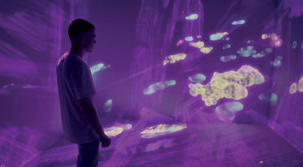
Science Meets Art: Immersion in the Secret World of the Immune System
On September 27, 2024, the city of Dortmund was transformed into a meeting place for the curious and science enthusiasts. Over 2,500 visitors participated in experiments, science shows and lectures throughout the city at the first Science Night in Dortmund. ISAS was also part of this special premiere: together with the storyLab kiU of Fachhochschule Dortmund, University of Applied Sciences and Arts, researchers presented an immersive 3D experience in the Dortmund U. Visitors were able to immerse themselves in "The Secret World of the Immune System" –specifically in the context of a heart attack.
The immersive space of storyLab kiU, located in the foyer of the Dortmund U, measures four by four meters. Anyone who enters the room will step into new worlds involving all their senses with the help of high-performance projectors and a 32-channel sound system. Normally, these are based on works of art from the collections of Dortmundʼs museums - but at the Science
Night, visitors were taken on a journey into the smallest structures of a heart after a heart attack. Visitors were able to marvel at real microscope images from the ISAS, including a 3D reconstruction of a mouse heart and immune cells that migrate into the damaged tissue after a heart attack. These cells, including macrophages for example, are actually supposed to support healing.
In the immersive space, visitors can experience immune cells in heart tissue up close. Here you can see how the macrophages (yellow) slowly move towards their target cells (pink). A special feature of the room: a tracking system records the position of the person so that the optical perspective adjusts accordingly. The projection therefore always appears three-dimensional to the viewer.
LIGHT SHEET FLUORESCENCE MICROSCOPY
One of the imaging techniques used by the Bioimaging research group is fluorescence microscopy. Fluorescence describes the property of substances to absorb short-wave light and re-emit it at a longer wavelength. In order to recognise certain structures under the microscope, researchers stain samples with target structure-specific fluorescent dyes that have these properties. This form of light microscopy includes light sheet fluorescence microscopy. Here, a laser illuminates only a thin layer of the sample, for example a heart, without destroying it. From the many individual images of the tissue layers, researchers later create a 3D model of the entire sample on the computer.
However, in the “clean-up frenzy” after a heart attack, they cause scarring in the heart muscle structure. This can have a long-term negative impact on heart function.
A rt meets immunology
The microscope images on which the immersive experience is built come from the Bioimaging research group at ISAS. The scientists use various microscopic methods, including light sheet fluorescence microscopy (► info box), to better understand the immune response to a heart attack and the resulting consequential damage. In the long term, they hope this will pave the way for new heart attack therapies. “This is the first time we have shown our research data in an artistic context,” reports Prof. Dr Anika Grüneboom, Head of the research group. This required intensive preparation: the artists and scientists worked together for several months to bring the research from the ISAS laboratory to the Dortmund U. How fast do the macrophages move? Where is there room for artistic freedom? And what about the sound
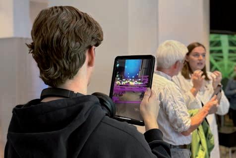
For the Science Night, the storylab kiu team has prepared tablets that project the immune cells into the foyer of the Dortmund u in greatly enlarged form using augmented reality. The macrophages (yellow) move with the visitors through the muscle tissue (purple). In the background, lara Janz, PhD student at ISAS, explains the images to a visitor.
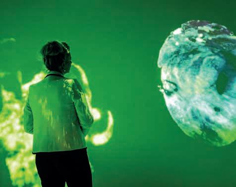
design? Grüneboom thinks the work was worthwhile:
“The cooperation with storyLab kiU shows that science can fascinate children and adults and still amaze even us researchers.”
Researchers in dialogue
While some visitors gazed at the images in the immersive space, others took the opportunity to engage in dialogue with the researchers and designers outside. At the joint FH Dortmund and ISAS stand, curious guests were also able to project the immune cells into the centre of the foyer of the Dortmund U using tablets.
“The dialogue with the visitors was very rewarding for us researchers, too. It gave us new perspectives on our work, which we will take back to ISAS,” summarises Dr Malte Roeßing, research associate in the Bioimaging working group. The biologist was not only in dialogue with the visitors. In the kiU talk “Fact and Fiction” in the evening, he discussed the interplay between art and science with Lennart Oberscheidt and Tobias Bieseke from storyLab kiU.
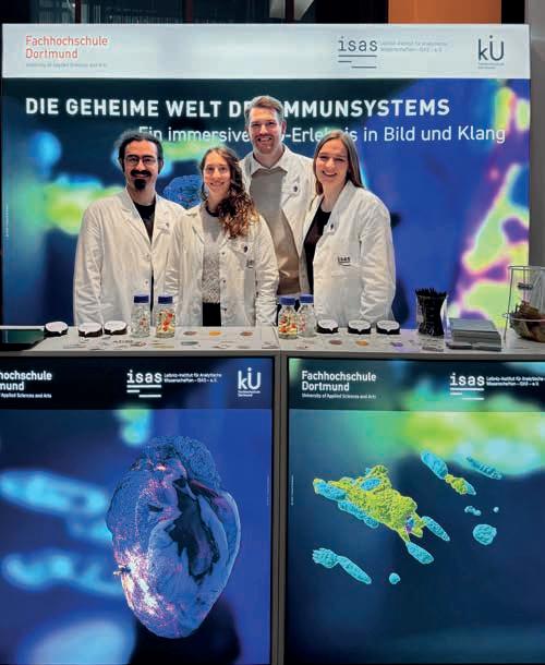
(CP)
Ina Brandes, Minister for Culture and Science of the state of North Rhine-Westphalia, also attended the Science Night and got a firsthand impression of the immersive experience. In the picture, she is looking at an image of a heart after myocardial infarction taken with a light sheet fluorescence microscopy. The green rays in the background are reminiscing of the microscopeʼs laser.
Dr Ali Ata Tuz, Flora Weber, Dr Malte Roeßing and lara Janz (from left to right) provided information at the joint stand of Dortmund university of Applied Sciences and Arts and ISAS and, depending on the age of the visitors, different levels of detailed scientific background information on the cooperation project.

AFTERMOVIE OF THE 2024 SCIENCE NIGHT
https://youtu.be/4Qy0NDzu5ek?si =v3v0o25C5iPmH9SL
Bioimaging Research Group
Prof. Dr Anika Grüneboom
T: +49 (0)231 1392-239
E: anika.grueneboom@isas.de
Communications Team
Sara Rebein
T: +49 (0)231 1392-234
E: sara.rebein@isas.de
“We essentially ask ourselves the same questions”
Lennart Oberscheidt is a research associate and visual effects (VFX) supervisor at storyLab kiU, the digital research and presentation centre of the Dortmund University of Applied Sciences and Arts. Here, the 38-year old focuses on current issues relating to digital society. These include, for example, new narrative strategies in combination with innovative technologies and forms of presentation. In the ISAS cooperation project “The Secret World of the Immune System”, Oberscheidt and the team led by artistic director Harald Opel incorporated microscope data from ISAS into an experience in an immersive space for the first time. In this interview, he talks about the special features of the project and the similarities between art and research.
What is the immersive space at storyLab kiU and what is the goal behind it?
Oberscheidt: Our immersive space at the Dortmund U consists of four walls on which images are displayed all around using high-performance projectors. A tracking system records the positions of the person so that the three-dimensional world of image and sound
lennart Oberscheidt provided creative collaboration on the project.
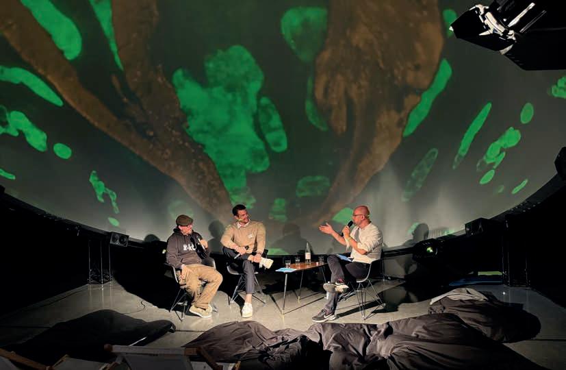
changes with the correct perspective as the person moves. Itʼs a bit like virtual reality or VR goggles – but without the glasses.
The space was created as part of our Page 21 project funded by the Ministry of Culture and Science of North Rhine-Westphalia. The idea behind it was to create a virtual environment from the works of art in Dortmundʼs museums and make it possible for visitors to experience them. To do this we explore storytelling in various new media – both practically and from a research perspective. The aspiration to create completely new narrative worlds is a common thread through all our work at storyLab kiU: whether itʼs façade mapping or, for example, the cooperation project with ISAS.
It was the first time your team had worked with scientific research data. Was there anything special or were there surprises?
Oberscheidt: Simply the fact that the structures on the images are so incredibly small is totally crazy. Taking something that was previously only visible under a microscope or on a screen and allowing people to experience it, as if they were part of it, was completely new for us. Especially at the beginning, we literally had to find our way around the data and understand what was relevant from a biological perspective for the story we wanted to bring to life with ISAS. To do this, we had a lot of discussions with the communicators, asked the researchers lots of questions and visited one
Moderated by Tobias Bieseke (right), lennart Oberscheidt (both from storylab kiu, left) and Dr Malte Roeßing, research associate in the Bioimaging research group at ISAS, discussed the interaction between art and science. During the interview, the three talk about the interdisciplinary collaboration in the project, the role of aesthetics in biomedical imaging and the boundaries of fact and fiction in science.
another. Particularly for the students for whom our projects provide an opportunity to use and expand their skills under real conditions, visiting the laboratory was something special.
„ Just like the researchers at ISAS, we build up our 3D models from different layers. “
We quickly found common ground from a technical perspective. Just like the researchers at ISAS, we build up our 3D models from different layers. How many layers are there and how far apart are they? How can we convert this data into a volume model? We essentially ask ourselves the same questions during our work.
How did you manage to mix artistic storytelling with research data for the experience?
Oberscheidt: Art thrives on fantastic, i.e. unreal, stories that someone has made up. But almost every story has a kernel of truth in it somewhere. The difference when working on scientific topics is that this fact-based core always has to be retained. Myocardial infarction or heart attack is a topic that affects a lot of people and is very present in society. For the Dortmund Science Night we therefore wanted to create a narrative that accurately depicts the scientific facts and provides
an accurate insight into the previously unknown research regarding new therapies.
At the same time, the aim is to create something aesthetically appealing that involves the audience, because simply presenting a collection of scientific facts could quickly become overwhelming – you have to keep a balance. During the course of the project, we gradually worked towards this both visually and through the sound design. In the end we were able to create an experience together that was as entertaining as possible, but still naturally exciting and visually impressive.
The installation was created for the Dortmund Science Night 2024. How can anyone who is interested view the immersive experience now?
Oberscheidt: The immersive space is open to the public on Saturdays and Sundays. Anyone who is interested can simply drop by at the Dortmund U and experience one of the many narrative worlds for themselves. Whether you then find yourself in the microscope images from ISAS or in a work of art from the Dortmund museums is pure chance.
storyLab kiU, Fachhochschule Dortmund, University of Applied Sciences and Arts
Harald Opel
Artistic Director
T: +49 (0)231 99777 941
E: opel@fh-dortmund.de
(The interview was conducted by CP.)
Red Alert: Students Research the Immune System at Girlsʼ Day
For half a day at Girlsʼ Day 2024, twelve schoolgirls were able to find out in their own experiments how bacteria can be hunted down and kept in check, why organs become transparent with the help of cinnamic acid ethyl ester, and what can be seen under the microscope during a heart attack.
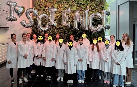
Together with PhD students Antonia Fecke and Luisa Speicher, who accompanied the event as scientific guides, the seventh to ninth graders gained practical insights into the immune system – and learned a lot about bacterial and sterile inflammation. Under the guidance of Dr Christina Sengstock and Dr Christiane Stiller, the pupils learned how to multiply bacterial cultures in the laboratory. They also investigated the effect of silver acetate on Escherichia coli. With Prof. Dr Anika Grüneboom and Luisa Röbisch, everything revolved around the light sheet fluorescence microscope. Before they treated organs with cinnamic acid ethyl ester – a com-
So many bright and curious minds took part in the Girlsʼ Day event at ISAS. At the end of the day, the twelve students gave positive feedback on the day at the institute. And who knows, maybe the photo also shows the next generation of women in research? In any case, the motto "I love science" applies to all those present. Luisa Röbisch, Prof. Dr Anika Grüneboom, Dr Christiane Stiller, Cheyenne Peters (Communications team), Clara Manthey (intern in the Communications team), Luisa Speicher and Antonia Fecke (from left to right) also had a lot of fun carrying out the experiments and presenting their work. (Editorʼs note: Dr Christina Sengstock and Sara Rebein are missing from the photo.)
ponent of cinnamon flavour – and analysed pre-treated samples, the participants had conducted an experiment with glass beads to gain a better understanding of the procedure for optical clearing.
The Communications team coordinated the Girlsʼ Day project at ISAS in 2024, and this time received active support with the organisation and implementation from intern Clara Manthey (student at TU Dortmund University). In the run-up to the event, Luisa Becher, also a student at TU Dortmund University, had already played a key role in the conception and preparation.

In the bacteria lab, Jule (13) is practising how to use a pipette. The work is taking place next to a Bunsen burner the whole time. It forms a sterile circle around its flame, in which it is possible to work in a sterile environment. Within this sterile zone, Jule is training how to pipette, first with water and then with a prepared Luria-Bertani medium. The latter is a nutrient medium for cultivating bacteria. For this, the eighth grader has already placed a new sterile plastic tip onto the pipette before she applies the medium to the prepared agar plate. The aim of this experiment is for Jule and the other pupils to learn how to handle bacterial cultures (E. coli) and how to multiply them to continue working with them afterwards.
Once the students have familiarised themselves with pipetting, they move on to the "inhibition zone experiment". Dr Christina Sengstock (on the right in the picture) is standing by to help them. Sara (14, photo: centre) has already labelled the Petri dish with the culture medium. She is now holding an agar plate and a pipette in her hands. She uses the pipette to drip Luria-Bertani medium onto the plate. The student makes sure that she uses the exact amount of liquid required. The girls have previously taken the liquid from a test tube using the pipette. They will then spread the medium on the plate by spreading it out with a glass spatula they had bent themselves. Sophie (13) is watching attentively before pipetting herself next. The whole exercise serves as preparation for the following experiment. For this, the pupils will make holes into the agar on the plate. They will then pipette silver acetate into the punched-out area. Agar plates with E. coli- cultures are used in real daily life in the
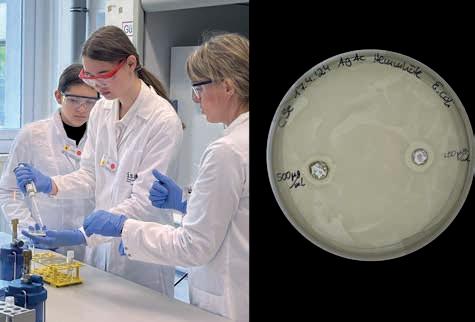
laboratories. The silver ions have an antimicrobial effect – they inhibit the growth of bacteria. That is why inhibition zones around the area treated with silver acetate can later be seen with the bare eye.
The young researchers have moved from the bacteria lab to one of the microscopy labs. To understand how organs become transparent for the analysis under the light sheet fluorescence microscope, the students have been given cups with glass pearls. They are part of an experiment conducted by Prof. Dr Anika Grüneboom and Luisa Röbisch. The two have chosen this experiment to illustrate the principle of optical clearing. What Sophie (left) and Sara cannot see at first because the glass beads do not let any light through, and the beaker appears opaque: there are small red plastic hearts between the glass beads. The pupils are slowly pouring cooking oil into the beaker. The oil displaces the air between the glass beads. The similar refractive index of glass and oil ensures that the hearts in the beakers become visible.
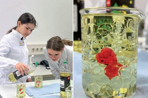
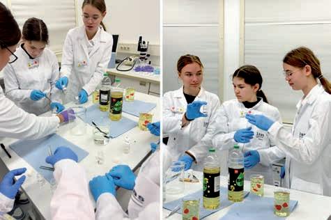
Sara (right) and Sophie are working with organ samples. They are carefully preparing the delicate samples for later use under the light sheet fluorescence microscope. They are using tweezers to pick up the hearts and thymus glands of mice, and take them out of an ethanol solution. Prof. Dr Anika Grüneboom and Luisa Röbisch have prepared samples of the heart, bowel, and thymus for the experiment in advance and placed them in an ethanol solution. This alcohol solution is used to dehydrate the samples. The students are carefully placing the samples in small containers filled with ethyl cinnamate before they will seal the jar. The cinnamic acid ethyl ester is part of Prof. Dr Anika Grüneboomʼs patented clearing process.
Jule (left) is taking a close look at the bowel. She can already see that it is transparent. The lab is darkened with roller blinds because the students will next go to the light sheet fluorescence microscope. Because clearing some of the organs takes several days, biotechnologist Luisa Röbisch had prepared some samples for Girlsʼ Day a few days before the event. Immunologist Prof. Dr Anika Grüneboom will later place a transparent heart under the light sheet microscope. The device fans out the dot-shaped laser beam like a sheet of paper. The thin sheet of light created in this way illuminates each individual layer of the sample. Because the samples are transparent, the laser can penetrate them almost unhindered – and take an image of each layer. At Girlsʼ Day, Grüneboom will later discuss the individual images of a heart after a heart attack with the students. And she will then assemble these images into a 3D model on the computer.
(Editorʼs note: The samples were already available; no organ removals were carried out for the event.)
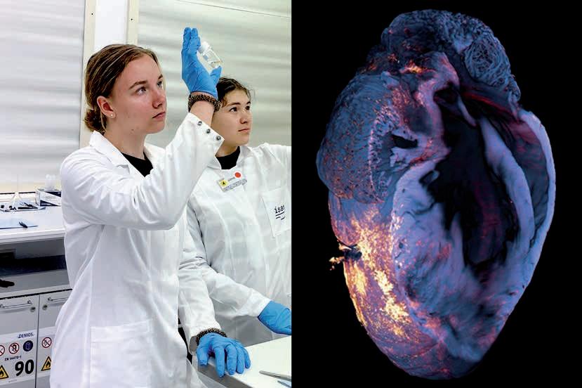
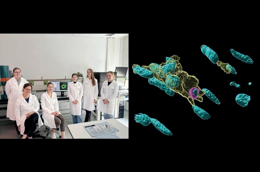
Bioimaging Research Group
Prof. Dr Anika Grüneboom
T: +49 (0)231 1392-239
E: anika.grueneboom@isas.de Communications Team
Sara Rebein
T: +49 (0)231 1392-234
E: sara.rebein@isas.de
The last part of the programme is about how pathogens – in this case E. coli bacteria – multiply and look on the agar plates after a few days. Another topic: immune cells such as macrophages which exaggerate with “cleaning up” after a heart attack and therefore damage the body (sterile inflammation). At this part of the Girls’ Day, the researchers have also shown images that they had taken under a confocal microscope. They revealed a section of the heart tissue with an accumulation of macrophages. Afterwards, Dr Christiane Stiller (from left to right), Luisa Röbisch and Prof. Dr Anika Grüneboom are answering the students’ questions about their day-to-day work at ISAS.
(CM)
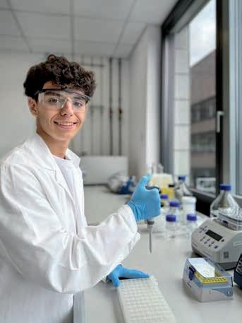
What are you doing at ISAS, Marcos?
For Marcos Nadales Neira (18), a trainee laboratory technician in Zaragoza, Spain, one thing was clear early on during his training: before deciding on a degree programme after his traineeship, he first wanted to gain practical experience – preferably abroad. His passion for chemistry and biology finally led him to ISAS in May 2024. Here he spent four weeks looking over the shoulders of the biological technical assistants. To get an insight into his internship, the editorial team asked Marcos to complete the following sentences.
At ISAS I ...
shadow the work in the laboratory as part of the Technical Service Bioanalytics team. I learn a lot of new things here, for example how the synthesis of proteins works. My colleagues introduce me to many procedures on the mass spectrometers. Soon I will also have the chance to help with sample preparation.
I found ISAS ...
because my teacher supported me in my search for an internship. It was clear to me from the start that I would like to work in a research institution abroad to get to know other working cultures and people. ISAS fits in very well with my interest in natural sciences. I was already fascinated by chemistry in school.
After my internship ...
I would like to enrol at a university. Pathological anatomy and microbiology fascinate me in particular. But I can also imagine studying chemistry. I hope that my time at ISAS will help me to decide on a particular field of study.
Marcos Nadales Neira supported the Technical Service Bioanalytics Team during his internship at ISAS. He assisted with sample preparation in the laboratory, for example.
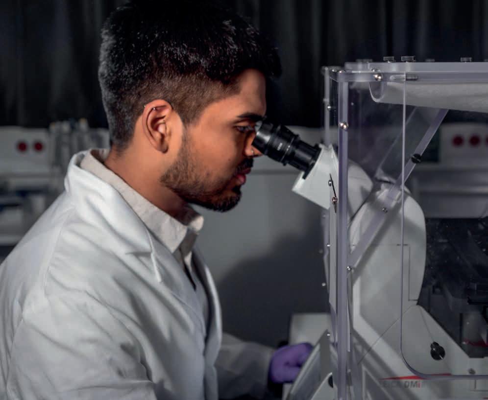
PATHOMECHANISMS
The research programme Pathomechanisms concentrates on the analysis of disease mechanisms with focus on cardiovascular diseases (CVDs), in particular heart failure due to myocardial infarction (heart attack), pathological cardiac hypertrophy (heart growth) or cardiotoxic cancer therapies. The overall goal of the programme is to identify molecular changes that are causative for the development of these diseases and are suitable as targets – and possibly as biomarkers.
Within the multiple pathomechanisms that can underlie for example heart failure, the scientists address the ones that
are of translational potential like cardio-safe targeting of certain kinase (specific enzymes) cascades,
represent common pathomechanisms or events in the heart and in case of cancer, for example cellular growth mechanisms and coagulation (clotting of blood),
involve toxic side effects of drugs like cardiotoxicity of cancer drugs, and
those that are relevant for the further optimisation of analytical methodologies.
Until today, the molecular causes and the course of many diseases of the cardiovascular system are still largely not understood. Many CVDs have multi-factorial causes – genetic constellations play a part in addition to environmental as well as nutritional factors, platelet disorders or cardiotoxic cancer therapies. To obtain a multidimensional picture of the pathomechanisms underlying cardiovascular diseases, to enable doctors to diagnose these illnesses earlier in future, and to carry out individual therapies more effectively and with fewer side effects, researchers at ISAS apply methods that
In addition to regular fluorescence images, researchers are able to use the confocal microscope with a temperature-controlled, transparent incubator unit to examine, for example, cardiomyocytes (heart muscle cells) from mice using live cell imaging.
Biofluorescence
Research Group
Prof. Dr Matthias Gunzer
T: +49 (0)231 1392-100
E: matthias.gunzer@isas.de
Cardiovascular Pharmacology
Research Group
Prof. Dr Kristina l orenz
T: +49 (0)231 1392-103
E: kristina.lorenz@isas.de
Miniaturisation Research Group
PD Dr Joachim Franzke
T: +49 (0)231 1392-174/199
E: joachim.franzke@isas.de
NMR Metabolomics
Research Group
Dr Roland Hergenröder
T: +49 (0)231 1392-178
E: roland.hergenroeder@isas.de
Proteomics Research Group
Prof. Dr Albert Sickmann
T: +49 (0)231 1392-100
E: albert.sickmann@isas.de
AMBIOM – Analysis of Microscopic BIOMedical Images
Junior Research Group
Dr Jianxu Chen
T: +49 (0)231 1392-217
E: jianxu.chen@isas.de
Multidimensional Omics
Data Analysis
Junior Research Group
Prof. Dr Robert Heyer
T: +49 (0)231 1392-271
E: robert.heyer@isas.de
comprise genomic, proteomic and metabolomic parameters. They develop, combine, or optimise various analytical technologies to identify disease mechanisms and potential target molecules for the treatment of different cardiovascular diseases.
Development of analytical tools and combination of new methods
The scientists involved in this research programme combine traditional molecular genetic and biochemical methods with high throughput mass spectrometry methods and spectroscopic approaches. This enables them to cover the entire bandwidth of the analysis – from detailed investigation of individual components through to analysis of entire cellular systems.
They aim, for example, to work out spectroscopic and metabolic characteristics of certain diseases with protein or lipid deposition like amyloidosis or Fabry disease in close collaboration with clinician scientists at the Julius-Maximilians-Universität of Würzburg and University of Duisburg-Essen (University Hospital Essen). The scientists at ISAS continue to press ahead intensively with the applications of biospectroscopic analyses, in particular coherent anti-Stokes Raman scattering (CARS) and Raman spectroscopy combined with vibrational microscopy and MALDI (matrix-assisted laser desorption/ionisation) imaging. Moreover, they develop AI methods to optimise the analyses of the obtained data in order to identify early metabolic or structural changes within the myocardium (cardiac muscle).
Gained insights into metabolic events are supplemented by research that aims to optimise nuclear magnetic resonance (NMR) for a longitudinal monitoring of metabolic fluxes with high sensitivity and spatial as well as temporal resolution. It is an analytical method that is of great relevance for a better understanding of cytotoxicity (quality of being toxic to cells) mechanisms of certain drugs.
Application of different model systems
The researchers use cell- and mouse-based model systems that are able to recapitulate central features of CVDs. For example: They work with platelets that can agglomerate and thereby simulate thrombi or cardiomyocytes that beat and thereby serve for the readout of contraction and relaxation. The scientists also work with genetic mouse models that represent a phenotype of pathological heart growth in children or due to lipid depositions in the heart.
Precision medicine: potential of insights into platelet activation
In heart attacks, which are among the top two causes of death worldwide 1, platelet aggregation plays a central role. ISAS has many years of analytical expertise regarding research into thrombocytes (blood platelets). This includes the comprehensive investigations into the population-based proteome of thrombocytes and in-depth research of thrombocyte malfunctions. More insights into platelet activation/inhibition have the potential to further precision medicine for CVDs. Therefore, the researchers at ISAS have been creating a standard platelet data base. It allows the application of machine learning models to predict platelet aggregation and ultimately haemostasis (blood clotting) in patients with heart failure or stroke – a strategy that is being further advanced as a blueprint for other blood cells.
1 http://www.who.int/news-room/fact-sheets/detail/the-top-10-causes-of-death
(SR)
Stronger Together: Methodological Diversity in Fabry Research
Fabry disease (►p. 81) is a rare but serious hereditary disease – early diagnosis is crucial for successful treatment. At ISAS, Johann Dierks and Dr Eike Brockmann employ different imaging techniques in order to gain a better understanding of the disease, and thus to develop new possibilities for early diagnosis. Dierks conducts his research in the Pathomechanisms programme, Brockmann in the MS-Based Imaging programme (►p. 18). They each pursue different methodological approaches, but work closely together. In their statements, they explain the findings from their respective research and how the combination of their methods provides new insights into Fabry disease.
In the Cardiovascular Pharmacology research group we study the hereditary Fabry disease (► p. 81) in the context of cardiovascular diseases. We want to develop a workflow with which we can detect the changes in heart tissue that are typical of this glycolipid storage disease at an early stage. In this way, we hope to improve the possibility of early diagnosis of this genetic disease.
In our research project we work with Raman spectroscopy. We use it to analyse cells and heart tissue from mice with and without Fabry disease to find out more about the chemical composition of lipids and proteins in the tissue. During Raman spectroscopy, we excite the chemical bonds in the molecules in the samples with a laser and visualise, for example, lipid and protein distributions. We use a special form of Raman spectroscopy, more specifically coherent anti-Stokes Raman scattering, CARS for short, to be able to detect the differences in the tissues of healthy and diseased mice in the spectrometer. Compared with traditional Raman spectroscopy, CARS allows us to visualise different protein-to-lipid ratios in the sample more quickly. However, by contrast with Raman spectroscopy, this method provides less information about the detailed chemical composition of the samples, which means that
crucial disease-specific information may be lost. We are therefore planning to use the two technologies in a complementary manner
The combination of methods is an important aspect, which is why I work closely with Dr Eike Brockmann from the Lipidomics research group when examining mouse heart tissue samples. Our aim is to obtain specific information
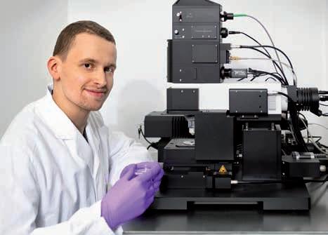
Johann Dierks is a PhD student in the Cardiovascular Pharmacology research group.
Conventional histological methods based on microscopic examinations are often not reliable enough for the early detection of Fabry disease (► info box). Other imaging methods, such as matrixassisted laser desorption/ionisation mass spectrometry, or MALDI-MS for short, offer an alternative here. This allows us to measure the accumulation of a specific lipid class even in the early stages of the disease. In Fabry disease, the so-called glycosphingolipids are typically deposited in the heart and other organs. We are therefore investigating various stages of Fabry disease in the mouse model, specifically in the heart tissue. Using MALDIMS, we can measure the mass-to-charge ratio of lipids in the tissue in a spatially resolved manner. We then use lipid databases and other experiments to determine the spe-
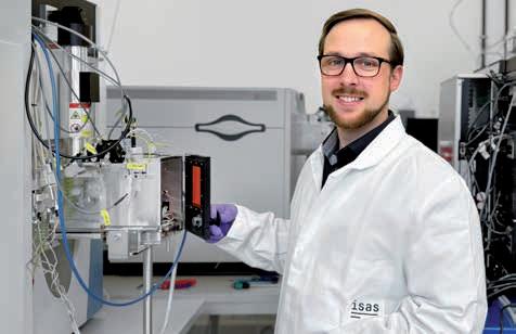
Eike Brockmann is a research associate in the Lipidomics research group.
cific molecules. We have adapted a number of parameters for our analyses, including sample preparation, for example. This includes the way in which we apply the matrix –
on the disease-relevant lipids. As soon as I have finished my Raman measurements, he uses mass spectrometry imaging, more precisely matrix-assisted laser desorption/ ionisation mass spectrometry, or MALDI-MS for short, to further investigate the same samples. By combining the two technologies, we aim to bring together the information that one method alone does not cover. Raman spectroscopy and thus CARS are particularly well suited for such a multimodal approach. Their great advantage is that the samples require little preparation so that they are not destroyed, but are kept intact. As Fabry disease is a rare disease and the number of samples is low, such a method with low sample wear is very suitable for further analyses.
In the next step we want to analyse human tissue samples in addition to mouse samples. In the longer term, we want to combine Raman spectroscopy, CARS and MALDIMS in one workflow in order to investigate the disease in other organs affected by Fabry disease, such as the kidneys. Another key focus in the future will be to apply this multimodal workflow to other cardiac diseases.
(Protocol: LK)
FABRY DISEASE
Fabry disease is a rare genetic glycolipid storage disorder that affects around 8,000 people in Germany, according to estimates by the Fabry self-help group. Persons suffering from this multi-organ disease have no or insufficient enzyme called α-galactosidase A (α-GAL). This normally breaks down certain fatty substances, the glycosphingolipids, in the cells. Without α-GAL, the lipids involved in building the cell membrane accumulate in the tissue and thus damage the cells in the longer term. In addition to organs such as the heart and kidneys, blood vessels and the nervous system are often also affected. The symptoms differ widely, depending on the organ involved, which makes early diagnosis difficult. At the same time, it is crucial for those affected that the disease is diagnosed before major tissue damage occurs: most pharmacological therapies, which can alleviate or even reverse the course of the disease, are only effective if used from an early stage.
an organic salt that we need to separate and ionise the molecules. We have succeeded in developing a workflow with which we can measure a significant accumulation of disease-specific lipids in the tissue in a spatially resolved manner.
In this research project, we are cooperating with the Cardiovascular Pharmacology research group. If we combine our spatially resolved – poorly so compared to microscopy – but very specific molecule measurements with MALDI-MS with the results of Raman spectroscopy, we obtain much more complex and detailed images than with just one method alone. Thatʼs why I carry out my analyses after Johann Dierks has done his measurements (► p. 80). Because we use the same sample, we can combine the images afterwards and correlate the results. With MALDI-MS, I measure larger areas of the sample with a different spatial resolution than Johann does with Raman spectroscopy. Thatʼs why the superimposition of
the images is still our biggest challenge at the moment. Currently this step is still carried out semi-automatically with the help of an algorithm; in future, the support of artificial intelligence would also be ideal.
Our multimodal workflow helps us to develop a better basic understanding of Fabry disease. For example, we can determine where in the tissue the lipid accumulations frequently occur or which cell types are particularly affected. Our MS method is admittedly complex, but it does offer a very good starting point for basic research –also into other diseases, because it allows us to visualise even more classes of molecules than has previously been possible. In the future, we may be able to find biomarkers that can help with early diagnosis of the disease.
(Protocol: AB)
Heart Failure Rarely Occurs on Its Own: ISAS Researchers Develop New Treatment Pillars
Cardiac insufficiency (heart failure) is one of the most common chronic diseases in Germany, affecting around four million people. In general practice, barely a day goes by for doctors without patients presenting with the symptoms of heart failure and the complications and treatment challenges that come with this disease. In recent years we have seen an arsenal of drugs being developed to combat it, including ACE inhibitors, beta blockers, SGLT2 inhibitors and mineralocorticoid receptor antagonists (MRAs). However, the clinical reality is that many patients also have additional disorders, especially chronic kidney disease (CKD), which makes using these drugs more difficult or in some cases even impossible. ISAS researchers are therefore focussing on broadening the range of treatments for heart failure specifically.
Heart failure is one of the leading causes of death in Germany. Doctors distinguish between two main forms. In systolic heart failure, the heart muscle is no longer strong enough to pump enough blood into the circulatory system. In the other form, diastolic heart failure, the organ pumps as usual, but the heart has become stiff or thickened and, as a result, does not fill sufficiently with blood. This also leads to less blood than necessary entering the circulatory system. People affected by both types often feel short of breath and weak. Fluid often accumulates in their lungs, arms and legs. Cardiac arrhythmias can also occur.
A multi-organ vicious circle
However, it is rare for patients to suffer from heart failure alone. Most of those affected also have other chronic disorders. Around half of all people with heart failure also have CKD. This often means that the kidneys are less effective at removing toxins and fluid from the body. In combination with heart failure, this creates a vicious circle: because the weakened heart is pumping less blood, blood flow to the kidneys is reduced, which further impairs how they work. This results in fluid and harmful metabolic products accumulating in the tissue, which in turn puts additional strain on the heart. “CKD patients have an increased risk of cardiovascular disease, including structural heart disease, heart failure and sudden cardiac arrest,” write Prof. Dr Kristina Lorenz, Head of the Cardiovascular Pharmacology research group at ISAS, and general practitioner (GP) Dr Jonas Knaup in an article published in 2024 in the journal MMW – Fortschritte der Medizin
Shaky ground for treatment plan pillars
When treating heart failure, doctors currently rely on a “four-pillar therapeutic approach” consisting of various medications: ACE inhibitors or angiotensin receptor blockers, angiotensin receptor-neprilysin inhibitors, beta blockers, MRAs and SGLT2 inhibitors. The last of these is the only class of active substances that has proven to be effective against both systolic and diastolic heart failure. Ideally, these four pillars are used simul-
taneously to not only alleviate the symptoms of heart failure, but also to slow the progression of the disease. However, in patients with comorbidities – CKD in particular – this treatment plan quickly falls apart if the weakened kidneys are overloaded and can no longer effectively flush the active substances out of the body. “Sometimes one pillar falls, sometimes three,” reports Knaup from his everyday practice.
In some cases, the only remaining option for multimorbid patients is to combat the symptoms with diuretics, i.e. medications that increase urine production. These at least reduce the accumulation of fluid in the body that is typical of heart failure. However, in advanced kidney failure, even this strategy eventually reaches its limits. “The balancing act becomes ever more difficult,” says Knaup. For GPs like him, there is long-term hope on the horizon at ISAS, where pharmacologist Lorenz and her team are currently working on several promising new medicinal approaches.
A stress-resistant heart thanks to proteins?
For a start, researchers at ISAS are developing strategies to combat the pathological remodelling processes in the heart muscle associated with heart failure. When the strength of the heartbeat drops, so too does the blood supply to the organs, causing the body to release stress hormones. These cause the heart to beat faster and
Prof. Dr Kristina lorenz heads the Cardiovascular Pharmacology research group and the Translational Research department at ISAS. She is also Head of the Institute of Pharmacology and Toxicology at Julius-Maximiliansuniversität Würzburg.
harder. In the short term, this actually boosts physical performance – which in the past helped humans evolve to run away from acute dangers, such as a bear on the attack. In the long term, however, these stress hormones bring about changes in the heart by stimulating the growth of heart cells and connective tissue. This ultimately makes the heart thicker and stiffer – and even less able to pump blood.
Lorenzʼs Cardiovascular Pharmacology research group has identified various proteins that counteract this dynamic. “These include a peptide agent that acts directly on the signalling pathway that inhibits pathological heart growth. This enables us to stop this development,” says the researcher. A second protein, in turn, appears to be able to increase the beat strength of the weakened heart muscles without the mechanism of heart growth, which is so dangerous in the long term, even starting. A third protein, the investigation of which is still in the early stages, appears to have the potential to improve the elasticity of the heart muscle and to do so in both systolic and diastolic heart failure.
New therapy pillars for a broader range of treatment options
If the identified molecular points of attack prove effective and new drugs can be developed from them, they would significantly expand the existing arsenal of available treatments. “These would be entirely new pillars of
therapy,” explains Lorenz. In other words, the proteins that the ISAS team is working on are not simply variants of the four existing approaches to treatment, but active principles used to treat heart failure through novel mechanisms that have not yet been deployed.
GP Knaup would welcome these additional treatment options. He sees heart failure patients in his practice almost every day, whose comorbidities often make treating them a challenge. “You just try to somehow hold the rickety system together,” says the doctor. In view of the prevailing demographic trend, physicians must prepare to be confronted with this problem even more frequently in the future. The number of people affected is rising, says Knaup. He goes on: “The older the population gets, the more common this disease becomes.”
Lorenz, K., Knaup, J. (2024) Nach Krankenhausaufenthalt: Wie die Behandlung weiterführen?
MMW – Fortschritte der Medizin, 166, 44–47.
https://doi.org/10.1007/s15006-024-4117-7
Cardiovascular Pharmacology Research Group
Prof. Dr Kristina l orenz
T: +49 (0)231 1392-103
E: kristina.lorenz@isas.de
Dr Jonas Knaup is a specialist in general practice at the Burgbernheim Medical Care Centre.
(UE)
Immune Cell Analysis in Inflamed Tissue: The Less
the Better
Neutrophil granulocytes, a type of immune cell, are indispensable for the defense against pathogens. However, if they migrate into injured tissue, for example into the brain after a stroke, they can promote chronic inflammation and cause long-term damage. To analyse the proteins of neutrophils in sites of inflammation and thus their functional dynamics, it takes millions of these immune cells – a problem, because often only few cells are found in these sites. Researchers at ISAS, the University Hospital Essen and the University of Münster have therefore developed a method that enables mass spectrometric analysis using just 1,000 neutrophils. Their work opens up new possibilities for the evaluation of immune responses within individual inflammatory foci. The researchers recently published their results, including a freely accessible database and a neutrophil proteome web service, in the journal Molecular & Cellular Proteomics.
Analysis of the neutrophil proteome, i.e. the majority of all expressed proteins, can provide valuable insights into the functional dynamics of neutrophils under disease conditions. However, although neutrophils make up around 60 per cent of leukocytes (white blood cells) in healthy people, there are often fewer than 1,000 cells in inflammatory foci. The availability of sample material can therefore become a problem, particularly with rare populations of neutrophils, such as from tumours. Until now, scientists had to pool cells from several people or animals. “With our approach, we can analyse the proteome of neutrophils using only

1,000 cells from a single inflammatory focus in a single person or laboratory animal. This not only allows more specific neutrophil analyses, but also conserves sample resources, making it especially beneficial for keeping animal numbers low,” reports Susmita Ghosh, first author of the study and PhD student at ISAS.
Susmita Ghosh uses the mass spectrometer to analyse neutrophils granulocytes. The 28-yearold joined ISAS in 2021 as a doctoral student in the Biofluorescence research group.
NEUTROPHIL PROTEOME WEB SERVICE
Bioinformatics researchers at ISAS have interactively visualised all proteins and their copy numbers. (Please note that data will be transmitted to Zenodo after activating the link).
https://zenodo.org/records/10891186
P ublicly accessible and comprehensive data collection as a reference value
In order to reduce the number of cells required for proteome analysis using liquid chromatography mass spectrometry (LC-MS), the ISAS researchers optimised protein digestion during sample preparation as well as the LC-MS data acquisition method. This enabled the scientists to create comprehensive, species-specific spectral libraries using neutrophils from the blood of five healthy humans and five healthy mice. The data collections respectively comprise approx. 5,300 human or approx. 6,200 mouse proteins and provide valuable insights into the proteomic differences between human and murine neutrophils. The spectral libraries and neutrophil proteome web service are publicly accessible, allowing the scientific community to use them as reference values for their own neutrophil analyses.
Biofluorescence
Research Group
Prof. Dr Matthias Gunzer
T: +49 (0)231 1392-100
E: matthias.gunzer@isas.de
Bioimagin g Research Group
Prof. Dr Anika Grüneboom
T: +49 (0)231 1392-239
E: anika.grueneboom@isas.de
Proteomics Research Group
Prof. Dr Albert Sickmann
T: +49 (0)231 1392-100
E: albert.sickmann@isas.de
Multidimensional Omics
Data Analysis
Junior Research Group
Prof. Dr Robert Heyer
T: +49 (0)231 1392-271
E: robert.heyer@isas.de
Ghosh, S., Tuz, A.A., Stenzel, M., Singh, V., Richter, M., Soehnlein, O., Lange, E., Heyer, R., Cibir, Z., Beer, A., Jung, M., Nagel, D., Hermann, D., Hasenberg, A., Grüneboom, A., Sickmann, A., Gunzer, M. (2024) Proteomic Characterization of 1000 Human and Murine Neutrophils Freshly Isolated From Blood and Sites of Sterile Inflammation. Molecular & Cellular Proteomics, 31(11), 100858. http://doi.org/10.1016/j.mcpro.2024.100858
Tabasco sauce proves: method is suitable for various inflammation models
The researchers validated their method by examining neutrophils in two different scenarios. First, they analysed neutrophils from the brains of mice after a stroke. The cells responded to the low-glucose environment by increasing their mitochondrial activity and producing more reactive oxygen species. In addition, the scientists examined human neutrophils. To simulate a transient inflammation in the oral cavity triggered by the nervous system, seven test subjects rinsed their mouths with a Tabasco-containing solution at the project partner in Münster. This caused neutrophils to migrate from the bloodstream into the inflamed tissue. There was evidence that these transmigrated cells also underwent functional changes after migration which the researchers also validated by flow cytometry. The two
very different scenarios show: the method, including the spectral library, is suitable for various inflammation models.
E ven lower cell numbers planned
The scientists are already working on further improving their method, reports Ghosh: “In the long term, we want to enable proteomic analyses for a handful of neutrophil numbers, for example for particularly small subpopulations or even individual cells. We’ ve already seen promising initial results with just five cells.”
(CP)
Funded by the German Research Foundation (Deutsche Forschungsgemeinschaft, DFG), project number - 449437943.
»COLLABORATIVE
RESEARCH CENTRE TRANSREGIO 332 –NEUTROPHILS: ORIGIN, FATE & FUNCTION«
At a Collaborative Research Centre (CRC) of the German Research Foundation (Deutsche Forschungsgemeinschaft, DFG), scientists work together in an interdisciplinary research programme on a long-term basis for up to twelve years.
In a CRC / Transregio (TRR), several participants from different locations conduct joint research.
The TRR 332 »Neutrophils: Origin, Fate & Function« is a collaboration led by the University of Münster together with the Ludwig-Maximilians-Universität München, the University of Duisburg-Essen, TUD Dresden University of Technology and ISAS. The project began in July 2022 and was initially scheduled to run for four years. The goal is to develop a better understanding of the biology of neutrophil granulocytes. TRR 332 aims to clarify how the tissue environment influences the production and phenotype of neutrophils, how the intracellular regulation of their activity takes place and how these immune cells work in different disease contexts. In this context, TRR 332 strives to help improve the treatment of patients with rheumatoid arthritis, as neutrophils play a central role in the pathogenesis of this autoimmune disease.
The TRR consists of three project areas which deal with extracellular signalling (A), intracellular regulation (B) and the neutrophil response (C). Six to seven subprojects each are subordinate to these areas. https://neutrophils.de/en
ORGANISATION
Dr C. Fricke (State of North Rhine-Westphalia)
Schwagenscheidt
Günther
Prof. Dr Albert Sickmann
Proteomics Research Group
(Prof. Dr Albert Sickmann)
Standardisation Research Group
(PD Dr Dirk Janasek)
Lipidomics Junior Research Group
(Prof. Dr Sven Heiles)
Multi-dimensional Omics Data Analysis Research Group
(Prof. Dr Robert Heyer)
NMR Metabolomis Junior Research Group
(Dr Roland Hergenröder)
Spatial Metabolomics Junior Research Group (Dr Karl Smith)
Research Departments
Prof. Dr Matthias Gunzer
Biofluorescence Research Group
(Prof. Dr Matthias Gunzer)
Bioimaging Research Group
(Prof. Dr Anika Grüneboom)
AMBIOM – Analysis of Microscopic
BIOMedical Images Junior Research Group (Dr Jianxu Chen)
Technical Services Bioanalytics (Ingo Feldmann) Application Labs
PD Dr Karsten Hinrichs
Prof. Dr Kristina Lorenz
Cardiovascular Pharmacology Research Group
(Prof. Dr Kristina lorenz)
Miniaturisation Research Group (PD Dr Joachim Franzke)
Preclinical Metabolomics Research Group
(Prof. Dr Dr Alpaslan Tasdogan)
ERC-Sulfaging
(Dr habil. Miloš Filipović)
C. Müschenborn
J. Bethke †
D. Schrot
BOARDS
Executive Board
Prof. Dr Albert Sickmann Chairman
Jürgen Bethke †
Chief Financial Officer
Scientific Advisory Board
Prof. Dr Ronen Alon
Weizmann Institute of Science, Department of Immunology
Israel
Dr Anne K. Bendt
Singapore Lipidomics Incubator (SLING), Life Sciences Institute (LSI), National University of Singapore
Singapore
Prof. Dr Jörg Feldmann
Institute of Chemistry, University of Graz
Austria
Prof. Dr Denise Hilfiker-Kleiner Philipps University of Marburg Marburg
Prof. Dr Ina Koch
Institute of Computer Science, Department of Molecular Bioinformatics, Goethe University Frankfurt am Main
Prof. Dr Andreas Radbruch
German Rheumatology Research Center
Berlin
Prof. Dr Markus Sauer
Department of Biotechnology and Biophysics, Biozentrum, Julius Maximilians University Würzburg
Prof. Dr Andrea Urbani
Faculty of Medicine and Surgery, Università Cattolica del Sacro Cuore
Italy
Board of Trustees
Appointed members
Berlin Technical University
represented by Prof. Dr Geraldine Rauch
City of Dortmund
Economic Development Agency Dortmund, represented by Heike Marzen
Federal Republic of Germany
Federal Ministry of Education and Research, represented by Dr Torsten Geißler Berlin
Ruhr University Bochum
represented by Prof. Dr h.c. Martin Paul
State of Berlin
Senate Department for Higher Education and Research, Health and Long-Term Care, represented by Dr Björn Maul
State of North Rhine-Westphalia (Chair) Ministry of Culture and Science of the state of NRW, represented by Dr Christiane Fricke Düsseldorf
Senate Department for Higher Education and Research, Health, and Long-Term Care of the State of Berlin
represented by Dr Björn Maul
TU Dortmund University (Deputy Chair)
represented by Prof. Dr Gerhard Schembecker
Elected members
Prof. Dr Dr h.c. Ursula Gather
Alfried Krupp von Bohlen und Halbach-Stiftung Essen
Dr Joachim Richert
BASF SE (until October 2023)
ludwigshafen / Weinheim
Prof. Dr Dr med Thomas Thum
Medizinische Hochschule Hannover Hannover
Members of the Association
BASF SE
City of Berlin
City of Dortmund
City of Nordrhein-Westfalen
Federal Republic of Germany
Fraunhofer Institute for Toxicology and Experimental Medicine (ITEM)
Industrie- und Handelskammer zu Dortmund
Merck KGaA
Münster University
OBLF Gesellschaft für Elektronik und Feinwerktechnik mbH
Ruhr University Bochum
SENTECH Instruments GmbH
Shimadzu Europa GmbH
TechnologieZentrumDortmund GmbH
Thermo Fisher Scientific GmbH (Bremen)
Thermo Fisher Scientific GmbH (Dreieich)
TU Dortmund University
AK TIVITÄTEN 2024 ACTIVITIES 2024
Publikationen
Publications
Publikationen in referierten Zeitschriften*
Peer-reviewed Papers
Ababneh, R., Telfah, A., Al Bataineh, Q. M., Tolstik, E., Dierks, J. & Hergenröder, R. (2024) 1H, 31P NMR, Raman and FTIR spectroscopies for investigating phosphoric acid dissociation to understand phosphate ion kinetics in body fluids.
Spectrochimica Acta A - Molecular and Biomolecular Spectroscopy, Jg. 307, S. 123594. https://doi.org/10.1016/j.saa.2023.123594
Ahmad, A. A., Al-Bataineh, Q. M., Bani-Salameh, A. A., Al Omari, R. H. & Telfah, A. (2024) Self-cleaning antireflected surfaces based on treated PEO/SiO2 nanocomposite films.
Journal of Applied Polymer Science, Jg. 141, Nr. 17. https://doi.org/10.1002/app.55275
Ahmad, A. A., Migdadi, A. B. & Al-Bataineh, Q. M. (2024) Structural, optical, and electrical properties of strontium-doped tin dioxide films for high photoconductivity.
Thin Solid Films, Jg. 796, 140312. https://doi.org/10.1016/j.tsf.2024.140312
Al-Bataineh, Q. M., Telfah, A. D., Tavares, C. J. & Hergenröder, R. (2024) Wide-field surface plasmon resonance microscope based on polyethylene oxide/ polyacrylic acid brushes.
Applied Surface Science, Jg. 649, 159189. https://doi.org/10.1016/j.apsusc.2023.159189
Al-Bataineh, Q. M., Telfah, A. D., Tavares, C. J. & Hergenröder, R. (2024) Modeling and analysis of discrete particle detection in wide-field surface plasmon resonance microscopy. Sensors and Actuators A: Physical, Jg. 370, 115266. https://doi.org/10.1016/j.sna.2024.115266
Al-Bataineh, Q. M., Ahmad, A. A., Alakhras, L. A. & Telfah, A. (2024) pH-responsivity and photoconductivity for the organic mixed ionic electronic conductor of PANI/PSS composite films. Journal of Applied Polymer Science, Jg. 141, Nr. 29. https://doi.org/10.1002/app.55656
Al-Bataineh, Q. M., Migdadi, A. B., Ahmad, A. A., Brincoveanu, O., Mocanu, A., Toader, G. & Telfah, A. D. (2024) Cobalt-doped SnS2 nanoplates for high-efficiency catalysis applications. Materials Chemistry and Physics, Jg. 317, 129184. https://doi.org/10.1016/j.matchemphys.2024.129184
Al-Bataineh, Q. M., Ahmad, A. A., Migdadi, A. B., Bahti, A. & Telfah, A. D. (2024) Plasmon-exciton interactions in ZnO/AuNPs heterostructure film for high photoconductivity. Physica B: Condensed Matter, Jg. 685, 415970. https://doi.org/10.1016/j.physb.2024.415970
Al-Bataineh, Q., Ahmad, A. A., Migdadi, A. B. & Telfah, A. (2024) Effect of Ionic-Electronic Coupling on the Percolation Phenomenon of Polymer/ Reduced Graphene Oxide Nanocomposite Films. Polymers for Advanced Technologies, Jg. 35, Nr. 12, e70024. https://doi.org/10.1002/pat.70024
Alebrahim, M. A., Ahmad, A. A., Migdadi, A. B. & Al-Bataineh, Q. M. (2024) localize surface plasmon resonance of gold nanoparticles and their effect on the polyethylene oxide nanocomposite films. Physica B: Condensed Matter, Jg. 679, 415805. https://doi.org/10.1016/j.physb.2024.415805
Alsaad, A., Al-Hmoud, M., Marashdeh, M., Tolstik, E., Houshmand, M. & Telfah, A. (2024) Highly Sensitive Silver/Tin Selenide/ Graphene Multilayer SPR Sensor for Hemoglobin and Glucose levels Monitoring in Biological Fluids. Plasmonics, Jg. 2024.
https://doi.org/10.21203/rs.3.rs-4566105/v1
Al-Sawalmih, A., Al-Bataineh, Q. M., Abu-Zurayk, R., Tavares, C. J., Etzkorn, J., Foadian, F. & Telfah, A. (2024) Optical, electrical and structural properties of iron doped zinc oxide nanostructures.
Journal of Materials Science: Materials in Electronics, Jg. 35, Nr. 17, 1159.
https://doi.org/10.1007/s10854-024-12826-8
Alwahsh, M., Abumansour, H., Althaher, A. R. & Hergenröder, R. (2024) Metabolic Profiling Techniques and Their Application in Cancer Research. Current Pharmaceutical Analysis. https://doi.org/10.2174/0115734129317614240 809053901
Alwahsh, M., Al-Doridee, A., Jasim, S., Awwad, O., Hergenroeder, R. & Hamadneh, L. (2024) Cytotoxic and molecular differences of anticancer agents on 2D and 3D cell culture. Molecular Biology Reports, Jg. 51, Nr. 1, 721. https://doi.org/10.1007/s11033-024-09669-1
Alwahsh, M., Hamadneh, Y., Marchan, R., Dahabiyeh, L. A., Alhusban, A. A., Hasan, A., Alrawabdeh, J., Hergenröder, R. & Hamadneh, L. (2024) Glutathione and Xanthine Metabolic Changes in Tamoxifen Resistant Breast Cancer Cell lines are Mediated by Down-Regulation of GSS and XDH and Correlated to Poor Prognosis. Journal of Cancer, Jg. 15, Nr. 13, S. 4047–4058. https://doi.org/10.7150/jca.96659
*sowie KI-Konferenz-Publikationen plus AI Conference Papers
Al-Wahsh, M. I., Nimer, R. M., Dahabiyeh, L. A., Hamadneh, L., Hasan, A., Alejel, R. & Hergenröder, R. (2024) NMR-based metabolomics identification of potential serum biomarkers of disease progression in patients with multiple sclerosis. Scientific Reports, Jg. 14, Nr. 1, S. 14806. https://doi.org/10.1038/s41598-024-64490-x
Antipenko, S., Mayfield, N., Jinno, M., Gunzer, M., Ismahil, M. A., Hamid, T., Prabhu, S. D. & Rokosh, G. (2024) Neutrophils are indispensable for adverse cardiac remodeling in heart failure. Journal of Molecular and Cellular Cardiology, Jg. 189, S. 1–11. https://doi.org/10.1016/j.yjmcc.2024.02.005
Athamneh, M., Daya, N., Hentschel, A., Gangfuss, A., Ruck, T., Marina, A. D., Schara-Schmidt, U., Sickmann, A., Güttsches, A-K., Deschauer, M., Preusse, C., Vorgerd, M. & Roos, A. (2024) Proteomic studies in VWA1related neuromyopathy allowed new pathophysiological insights and the definition of blood biomarkers.
Journal of Cellular and Molecular Medicine, Jg. 28, Nr. 8, e18122, S. e18122. https://doi.org/10.1111/jcmm.18122
Auger, J-P., Zimmermann, M., Faas, M., Stifel, U., Chambers, D., Krishnacoumar, B., Taudte, RV., Grund, C., Erdmann, G., Scholtysek, C., Uderhardt, S., Ben Brahim, O., Pascual Maté, M., Stoll, C., Böttcher, M., Palumbo-Zerr, K., Mangan, MSJ., Dzamukova, M., Kieler, M., Hofmann, M., Blüml, S., Schabbauer, G., Mougiakakos, D., Sonnewald, U., Hartmann, F., Simon, D., Kleyer, A., Grüneboom, A., Finotto, S., Latz, E., Hofmann, J., Schett, G., Tuckermann, J. & Krönke, G. (2024) Metabolic rewiring promotes antiinflammatory effects of glucocorticoids. Nature, Nr. 8010, S. 184–192. https://doi.org/10.1038/s41586-024-07282-7
Bertino, F., Mukherjee, D., Bonora, M., Bagowski, C., Nardelli, J., Metani, L., Venturini, D. I. Z., Chianese, D., Santander, N., Salaroglio, I. C., Hentschel, A., Quarta, E., Genova, T., Mckinney, AA., Allocco, AL., Fiorito, V., Petrillo, S., Ammirata, G., De Giorgio, F., Dennis, E., Allington, G., Maier, F., Shoukier, M., Gloning, K-P., Munaron, L., Mussano, F., Salsano, E., Pareyson, D., di Rocco, M., Altruda, F., Panagiotakos, G., Kahle, K. T., Gressens, P., Riganti, C., Pinton, P. P., Roos, A., Arnold, T., Tolosano, E. & Chiabrando, D. (2024) Dysregulation of FlVCR1a-dependent mitochondrial calcium handling in neural progenitors causes congenital hydrocephalus. Cell Reports Medicine, Jg. 5, Nr. 7, 101647, S. 101647. https://doi.org/10.1016/j.xcrm.2024.101647
Bouza, M., Foest, D., Brandt, S., García-Reyes, J. F. & Franzke, J. (2024) Enhanced Compound Analysis using Reactive Paper Spray Mass Spectrometry: leveraging Schiff Base Reaction for Amino Acid Detection.
Analytical Chemistry, Jg. 96, Nr. 13, S. 5289–5297. https://doi.org/10.1021/acs.analchem.4c00215
Brand, T., Baumgarten, B. T., Denzinger, S., Reinders, Y., Kleindl, M., Schanbacher, C., Funk, F., Gedik, N., Jabbasseh, M., Kleinbongard, P., Dudek, J., Szendroedi, J., Tolstik, E., Schuh, K., Krüger, M., Dobrev, D., Cuello, F., Sickmann, A., Schmitt, J. P. & Lorenz, K. (2024) From Ca2+ dysregulation to heart failure: β-adrenoceptor activation by RKIP postpones molecular damages and subsequent cardiac dysfunction in mice carrying mutant PlNR9C by correction of aberrant Ca2+-handlingʼ. Pharmacological Research, Jg. 211, S. 107558. https://doi.org/10.1016/j.phrs.2024.107558
Brand, T., Lukannek, A-K., Boivin-Jahns, V., Jahns, R. & Lorenz, K. (2024) From “contraindicated” to “first line” –Current mechanistic insights beyond canonical β-receptor signaling.
Current Opinion in Pharmacology, Jg. 2024, Nr. 76. https://doi.org/10.1016/j.coph.2024.102458
Burhenn, S., Golda, J., Kratzer, J., Brandt, S. & Held, J. (2024) Characterization of a co-planar dielectric barrier discharge design as a plasma source for trace element detection by atomic spectrometry.
Spectrochimica Acta Part B-Atomic Spectroscopy, Jg. 213, 106884. https://doi.org/10.1016/j.sab.2024.106884
Chiu, C., Küchler, A., Depienne, C., Preusse, C., Della Marina, A., Reis, A., Kaiser, F. J., Nolte, K., Hentschel, A., Schara-Schmidt, U., Kölbel, H. & Roos, A. (2024) Skeletal muscle vulnerability in a child with Pitt-Hopkins syndrome. Skeletal Muscle, Jg. 14, Nr. 1, 15. https://doi.org/10.1186/s13395-024-00348-0
Cibir, Z. & Gunzer, M. (2024) ComplexEye: a multi-lens array microscope for high-throughput cell migration analysis. Nature Reviews Immunology, Jg. 24, Nr. 233. https://doi.org/10.1038/s41577-024-01009-5
Cnudde, S., Brand, T., Fender, J., Prever, L., Murabito, A., Russo, M., Logrand, F., Gulluni, F., Lorenz, K., Hirsch, E. & Ghigo, A. (2024) PI3KC2α controls cardiac contractility through regulation of β2-adrenergic receptor recycling.
Vascular Pharmacology, Jg. 155, S. 107313. https://doi.org/10.1016/j.vph.2024.107313
Costanzo, M., Cevenini, A., Kollipara, L., Caterino, M., Bianco, S., Pirozzi, F., Scerra, G., DʼAgostino, M., Pavone, L. M., Sickmann, A. & Ruoppolo, M. (2024) Methylmalonic acidemia triggers lysosomal-autophagy dysfunctions.
Cell & Bioscience, Jg. 2024, Nr. 14, 63. https://doi.org/10.1186/s13578-024-01245-1
Della Marina, A., Hentschel, A., Czech, A., Schara-Schmidt, U., Preusse, C., Laner, A., Abicht, A., Ruck, T., Weis, J., Choueiri, C., Lochmüller, H., Kölbel, H. & Roos, A. (2024) Novel Genetic and Biochemical Insights into the Spectrum of NEFl-Associated Phenotypes.
Journal of Neuromuscular Diseases, Jg. 11, Nr. 3, S. 625–645.
https://doi.org/10.3233/JND-230230
Della Marina, A., Hentschel, A., Stenzel, M., Schara-Schmidt, U., Osmanovic, A., Ruck, T., Grüneboom, A., Röbisch, L., Beygo, J., Kölbel, H., Gangfuss, A., Kaiser, F. J., Schänzer, A., Kale, D. & Roos, A. (2024) lipid and protein imbalances in muscle of a FAR1-patient with a heterozygous de novo variant.
Journal of Neuropathology and Experimental Neurology, Jg. 2024, Nr. Volume 83, Issue 11, S. 979–983. https://doi.org/10.1093/jnen/nlae071
Dobelmann, V., Roos, A., Hentschel, A., Della Marina, A., Leo, M., Schmitt, L-I., Maggi, L., Schara-Schmidt, U., Hagenacker, T., Ruck, T. & Kölbel, H. (2024) Thrombospondin-4 as potential cerebrospinal fluid biomarker for therapy response in pediatric spinal muscular atrophy. Journal of Neurology, Jg. 271, Nr. 10, S. 7000–7011. https://doi.org/10.1007/s00415-024-12670-0
Dobersalske, C., Rauschenbach, L., Hua, Y., Berliner, C., Steinbach, A., Grüneboom, A., Kokkaliaris, K. D., Heiland, D. H., Berger, P., Langer, S., Tan, C. L., Stenzel, M., Landolsi, S., Weber, F., Darkwah Oppong, M., Werner, R. A., Gull, H., Schröder, T., Linsenmann, T., Buck, A. K., Gunzer, M., Stuschke, M., Keyvani, K., Forsting, M., Glas, M., Kipnis, J., Steindler, D. A., Reinhardt, H. C., Green, E. W., Platten, M., Tasdogan, A., Herrmann, K., Rambow, F., Cima, I., Sure, U. & Scheffler, B. (2024) Cranioencephalic functional lymphoid units in glioblastoma. Nature Medicine, Jg. 30, Nr. 10, S. 2947–2956. https://doi.org/10.1038/s41591-024-03152-x
Dzyubenko, E., Chen, J. & Willig, K. (2024) Editorial: 15 years of Frontiers in Cellular Neuroscience: super-resolution microscopy in the healthy and the injured brain. Frontiers in Cellular Neuroscience, Jg. 18, Nr. 2024, S. 1448206. https://doi.org/10.3389/fncel.2024.1448206
Egger, J., Gsaxner, C., Luijten, G., Chen, J., Chen, X., Bian, J., Kleesiek, J. & Puladi, B. (2024) Is the Apple Vision Pro the ultimate Display? A First Perspective and Survey on Entering the Wonderland of Precision Medicine. JMIR Serious Games, Jg. 12, S. e52785. https://doi.org/10.2196/52785
Fender, J., Klöcker, J., Boivin-Jahns, V., Ravens, U., Jahns, R. & Lorenz, K. (2024) “Cardiac glycosides”—quo vaditis?— past, present, and future?.
Naunyn-Schmiedebergʼs Archives of Pharmacology.
https://doi.org/10.1007/s00210-024-03285-3
Gangfuß, A., Rating, P., Ferreira, T., Hentschel, A., Marina, A. D., Kölbel, H., Sickmann, A., Abicht, A., Kraft, F., Ruck, T., Böhm, J., Schänzer, A., Schara-Schmidt, U., Neuhann, T. M., Horvath, R. & Roos, A. (2024) A Homozygous NDuFS6 Variant Associated with Neuropathy and Optic Atrophy. Journal of Neuromuscular Diseases, Jg. 11, Nr. 2, S. 485–491.
https://doi.org/10.3233/JND-230181
Gangfuß, A., Goj, G., Polz, S., Della Marina, A., Hentschel, A., Ahlbory, K., Deba, T., Kotzaeridou, U., Schuler, E., Pechmann, A., Diebold, U., Kurlemann, G., Heinzkyll, L., Schmitt, D., Rostasy, K., Ruck, T., Böhm, J., Roos, A. & Schara-Schmidt, U. (2024) Giant axonal neuropathy (GAN): crosssectional data on phenotypes, genotypes, and proteomic signature from a German cohort. Journal of Neurology, Jg. 2025, Nr. 272 (1), 63. https://doi.org/10.1007/s00415-024-12744-z
García-Martínez, J., Caño-Carrillo, I., Gilbert-López, B., Bouza, M., Beneito-Cambra, M., Franzke, J., Molina-Díaz, A. & García-Reyes, J. F. (2024) Miniaturized flexible micro-tube plasma ionization source for the effective ionization of non-easily ionizable pesticides in food with liquid chromatography/mass spectrometry. Talanta, Jg. 274, 126011. https://doi.org/10.1016/j.talanta.2024.126011
Gazeli, O., Elia, E. A., Argirusis, N., Lazarou, C., Anastassiou, C., Franzke, J., Garcia-Reyes, J. F., Georghiou, G. E. & Agapiou, A. (2024) low-cost heat assisted ambient ionization source for mass spectrometry in food and pharmaceutical screening. Analyst, Jg. 149, Nr. 17, S. 4487–4495. https://doi.org/10.1039/d4an00901k
Geist, D., Hönes, S., Grund, S., Pape, J., Siemes, D., Spangenberg, P., Tolstik, E., Dörr, S., Spielmann, N., Fuchs, H., Gailus-Durner, V., Hrabě de Angelis, M., Mittag, J., Engel, D. R., Führer, D., Lorenz, K. & Moeller, LC. (2024) Canonical and noncanonical contribution of thyroid hormone receptor isoforms alpha and beta to cardiac hypertrophy and heart rate in male mice. Thyroid, S. 1–27.
https://doi.org/10.1101/2023.11.24.568041, https://doi.org/10.1089/thy.2023.0683
Ghosh, S., Tuz, AA., Stenzel, M., Singh, V., Richter, M., Soehnlein, O., Lange, E., Heyer, R., Cibir, Z., Beer, A., Jung, M., Nagel, D., Hermann, DM., Hasenberg, A., Grüneboom, A., Sickmann, A. & Gunzer, M. (2024) Proteomic characterization of 1000 human and murine neutrophils freshly isolated from blood and sites of sterile inflammation. Molecular & Cellular Proteomics, Jg. 2024, Nr. 23 (11), 100858.
https://doi.org/10.1016/j.mcpro.2024.100858
Gilglioni, E. H., Li, A., St-Pierre-Wijckmans, W., Shen, T-K., Pérez-Chávez, I., Hovhannisyan, G., Lisjak, M., Negueruela, J., Vandenbempt, V., Bauzá-Martinez, J., Herranz, J. M., Ezerina, D., Demine, S., Feng, Z., Vignane, T., Otero Sanchez, L., Lambertucci, F., Prašnická, A., Devière, J., Hay, D. C., Encinar, J. A., Singh, S. P., Messens, J., Filipović, M., Sharpe, H. J., Trépo, E., Wu, W. & Gurzov, E. N. (2024) PTPRK regulates glycolysis and de novo lipogenesis to promote hepatocyte metabolic reprogramming in obesity.
Nature Communications, Jg. 15. https://doi.org/10.1038/s41467-024-53733-0
Hagemann, N., Qi, Y., Mohamud Yusuf, A., Li, A., Squire, A., Tertel, T., Giebel, B., Ludewig, P., Spangenberg, P., Chen, J., Mosig, A., Gunzer, M. & Hermann, D. M. (2024) Microvascular Network Remodeling in the Ischemic Mouse Brain Defined by light Sheet Microscopy.
Arteriosclerosis, Thrombosis, and Vascular Biology, Jg. 44, Nr. 4, S. 915–929. https://doi.org/10.1161/ATVBAHA.123.320339
Hagemann, N., Qi, Y., Mohamud Yusuf, A., Li, A., Zhang, X., Spangenberg, P., Squire, A., Doeppner, T. R., Jin, F., Zhao, S., Chen, J., Mosig, A., Gunzer, M. & Hermann, D. M. (2024) Arterial specification precedes microvascular restitution in the peri-infarct cortex that is driven by small microvessels. Journal of Cerebral Blood Flow and Metabolism, Jg. 45, Nr. , S.171–186. https://doi.org/10.1177/0271678X241270407
Hellwig, P., Dittrich, A., Heyer, R., Reichl, U. & Benndorf, D. (2024) Detection, isolation and characterization of phage-host complexes using BONCAT and click chemistry. Frontiers in Microbiology, Jg. 15, Nr. 2024, S. 1434301.
https://doi.org/10.3389fmicb.2024.1434301
Hellwig, P., Kautzner, D., Heyer, R., Dittrich, A., Wibberg, D., Busche, T., Winkler, A., Reichl, U. & Benndorf, D. (2024) Tracing active members in microbial communities by BONCAT and click chemistrybased enrichment of newly synthesized proteins. ISME Communications, Jg. 2024, Nr. 4(1). https://doi.org/10.1093/ismeco/ycae153
Hentschel, A., Piontek, G., Dahlmann, R., Findeisen, P., Sakson, R., Carbow, P., Renné, T., Reinders, Y. & Sickmann, A. (2024) Highly sensitive therapeutic drug monitoring of infliximab in serum by targeted mass spectrometry in comparison to ElISA data. Clinical Proteomics, Jg. 21, Nr. 1, 16, S. 12. https://doi.org/10.1186/s12014-024-09464-x
Hinrichs, K., Shetty, N., Kubatkin, S., Malmberg, P., Lara-Avila, S., Furchner, A. & Rappich, J. (2024) Cover: Field Manipulation of Band Properties in Infrared Spectra of Thin Films. Advanced Photonics Research, Jg. 5, Nr. 1. https://doi.org/10.1002/adpr.202470002
Holzer, M-T., Uruha, A., Roos, A., Hentschel, A., Schänzer, A., Weis, J., Claeys, K. G., Schoser, B., Montagnese, F., Goebel, H-H., Huber, M., Léonard-Louis, S., Kötter, I., Streichenberger, N., Gallay, L., Benveniste, O., Schneider, U., Preusse, C., Krusche, M. & Stenzel, W. (2024) Anti-Ku + myositis: an acquired inflammatory protein-aggregate myopathy. Acta Neuropathologica, Jg. 148, Nr. 1, 6. https://doi.org/10.1007/s00401-024-02765-3
Höving, S., Akermann, M., Schiller, A., Franzke, J., Schwendemann, D. & Brandt, S. (2024) Functionalization of Cyclic Olefin Copolymer for Enhanced Electrical Conductivity in Material Extrusion 3D-Printing: Potential Applications in laboratory Environments and Small-Scale Experiments. 3D Printing and Additive Manufacturing. https://doi.org/10.1089/3dp.2023.0304
Höving, S., Schomacher, J., Schiller, A. & Franzke, J. (2024) Setting the Separation Factor α for Ketone Monomers and Dimers by the use of Different Drift Gases. Journal of the American Society for Mass Spectrometry, Jg. 35, Nr. 7, S. 1622–1628. https://doi.org/10.1021/jasms.4c00215
Höving, S., Dörr, S., Akermann, M., Schiller, A., Lorenz, K., Schwendemann, D., Franzke, J. & Brandt, S. (2024) Enhancing Biocompatibility: 3D-Printed Cyclic Olefin Copolymer Structures for Advanced laboratory Applications. 3D Printing and Additive Manufacturing. https://doi.org/10.1089/3dp.2023.0261
Höving, S., Ahlmann, J., Schomacher, J., Schiller, A. & Franzke, J. (2024) Continuous fiber printing of a modular heater for ion mobility spectrometry. Applied Materials Today, Jg. 2024, Nr. 41. https://doi.org/10.1016/j.apmt.2024.102501
Höving, S., Song, H., Speicher, L., Schiller, A. & Franzke, J. (2024) Compact Plasma Ionization for Ion Mobility Spectrometry using a 4.3 MHz Miniature Tesla Coil.
Journal of the American Society for Mass Spectrometry, Jg. 35, Nr. 12. https://doi.org/10.1021/jasms.4c00360
Kahler, J. P., Ji, S., Speelman-Rooms, F., Vanhoutte, R. & Verhelst, S. H. L. (2024) Phosphinate Esters as Novel Warheads for Quenched Activity-Based Probes Targeting Serine Proteases.
ACS Chemical Biology, Jg. 19, Nr. 7, S. 1409–1415. https://doi.org/10.1021/acschembio.3c00203
Kasarla, S. S., Flocke, V., Saw, N. M. T., Fecke, A., Sickmann, A., Gunzer, M., Flögel, U. & Phapale, P. (2024) In-vivo tracking of deuterium metabolism in mouse organs using lC-MS/MS. Journal of Chromatography A, Jg. 1717, S. 464691. https://doi.org/10.1016/j.chroma.2024.464691
Kerp, H., Gassen, J., Grund, S., Hönes, G. S., Dörr, S., Mittag, J., Härting, N., Kaiser, F. J., Lorenz, K. & Führer, D. (2024) Cardiac recovery from pressure overload is not altered by thyroid hormone status in old mice. Frontiers in Endocrinology, Jg. 15, Nr. 1339741, S. 1339741. https://doi.org/10.3389/fendo.2024.1339741
Khesali Aghtaei, H., Heyer, R., Reichl, U. & Benndorf, D. (2024) Improved biological methanation using tubular foam-bed reactor.
Biotechnology for Biofuels and Bioproducts, Jg. 17, Nr. 1, 66. https://doi.org/10.1186/s13068-024-02509-1
Kleefeld, F., Horvath, R., Pinal-Fernandez, I., Mammen, A. L., Casal-Dominguez, M., Hathazi, D., Melchert, S., Hahn, K., Sickmann, A., Muselmann-Genschow, C., Hentschel, A., Preuße, C., Roos, A., Schoser, B. & Stenzel, W. (2024) Multi-level profiling unravels mitochondrial dysfunction in myotonic dystrophy type 2. Acta Neuropathologica, Jg. 147, Nr. 1, 19, S. 12. https://doi.org/10.1007/s00401-023-02673-y
Kraft, F., Rodriguez-Aliaga, P., Yuan, W., Franken, L., Zajt, K., Hasan, D., Lee, T-T., Flex, E., Hentschel, A., Innes, A. M., Zheng, B., Julia Suh, D. S., Knopp, C., Lausberg, E., Krause, J., Zhang, X., Trapane, P., Carroll, R., McClatchey, M., Fry, A. E., Wang, L, Giesselmann, S, Hoang, H, Baldridge, D., Silverman, G. A, Radio, F. C, Bertini, E, Ciolfi, A, Blood, K. A., de Sainte Agathe, J-M., Charles, P., Bergant, G., Čuturilo, G., Peterlin, B., Diderich, K., Streff, H., Robak, L., Oegema, R., van Binsbergen, E., Herriges, J., Saunders, C. J., Maier, A., Wolking, S., Weber, Y., Lochmüller, H., Meyer, S., Aleman, A., Polavarapu, K., Nicolas, G., Goldenberg, A., Guyant, L., Pope, K., Hehmeyer, K. N., Monaghan, KG., Quade, A., Smol, T., Caumes, R., Duerinckx, S., Depondt, C., Van Paesschen, W., Rieubland, C., Poloni, C., Guipponi, M., Arcioni, S., Meuwissen, M., Jansen, A. C., Rosenblum, J., Haack, T. B., Bertrand, M., Gerstner, L., Magg, J., Riess, O., Schulz, J. B., Wagner, N., Wiesmann, M., Weis, J., Eggermann, T., Begemann, M., Roos, A., Häusler, M., Schedl, T., Tartaglia, M., Bremer, J., Pak, S. C., Frydman, J., Elbracht, M. & Kurth, I. (2024) Brain malformations and seizures by impaired chaperonin function of TriC Science, Jg. 386, Nr. 6721, S. 516–525. https://doi.org/10.1126/science.adp8721
Krieger, K., Egger, J., Kleesiek, J., Gunzer, M. & Chen, J. (2024) Multisensory Extended Reality Applications Offer Benefits for Volumetric Biomedical Image Analysis in Research and Medicine.
Journal of Imaging Informatics in Medicine. https://doi.org/10.1007/s10278-024-01094-x
Krishnacoumar, B., Stenzel, M., Garibagaoglu, H., Omata, Y., Sworn, R. L., Hofmann, T., Ipseiz, N., Czubala, M. A., Steffen, U., Maccataio, A., Stoll, C., Böhm, C., Herrmann, M., Uderhardt, S., Jenkins, R. H., Taylor, P. R., Grüneboom, A., Zaiss, M. M., Schett, G., Krönke, G. & Scholtysek, C. (2024) Caspase-8 promotes scramblasemediated phosphatidylserine exposure and fusion of osteoclast precursors
Bone research, Jg. 12, Nr. 1, S. 40. https://doi.org/10.1038/s41413-024-00338-4
Lang, A., Oehler, D., Benkhoff, M., Reinders, Y., Maike, B., Shahrjedi, K., Kaldirim, M., Sickmann, A., Dannenberg, L., Polzin, A., Pfeiler, S., Kelm, M., Grandoch, M., Jung, C. & Gerdes, N. (2024) Mitochondrial Creatine Kinase 2 (Ckmt2) as a Plasma-Based Biomarker for Evaluating Reperfusion Injury in Acute Myocardial Infarction. Biomedicines, Jg. 12, Nr. 10. https://doi.org/10.3390/biomedicines12102368
Lange, E., Kranert, L., Krüger, J., Benndorf, D. & Heyer, R. (2024) Microbiome modeling: A beginnerʼs guideʼ, Frontiers in Microbiology, Jg. 15, Nr. 15, S. 1368377. https://doi.org/10.3389/fmicb.2024.1368377
Li, J., Zhou, Z., Yang, J., Pepe, A., Gsaxner, C., Luijten, G., Qu, C., Zhang, T., Chen, X., Li, W., Wodzinski, M., Friedrich, P., Xie, K., Jin, Y., Ambigapathy, N., Nasca, E., Solak, N., Melito, G. M., Vu, V. D., Memon, A. R., Schlachta, C., De Ribaupierre, S., Patel, R., Eagleson, R., Chen, X., Mächler, H., Kirschke, J. S., de la Rosa, E., Christ, P. F., Li, HB., Ellis, D. G., Aizenberg, M. R., Gatidis, S., Küstner, T., Shusharina, N., Heller, N., Andrearczyk, V., Depeursinge, A., Hatt, M., Sekuboyina, A., Löffler, M. T., Liebl, H., Dorent, R., Vercauteren, T., Shapey, J., Kujawa, A., Cornelissen, S., Langenhuizen, P., Ben-Hamadou, A., Rekik, A., Pujades, S., Boyer, E., Bolelli, F., Grana, C., Lumetti, L., Salehi, H., Ma, J., Zhang, Y., Gharleghi, R., Beier, S., Sowmya, A., Garza-Villarreal, E. A., Balducci, T.,
Angeles-Valdez, D., Souza, R., Rittner, L., Frayne, R., Ji, Y., Ferrari, V., Chatterjee, S., Dubost, F., Schreiber, S., Mattern, H., Speck, O., Haehn, D., John, C., Nürnberger, A., Pedrosa, J., Ferreira, C., Aresta, G., Cunha, A., Campilho, A., Suter, Y., Garcia, J., Lalande, A., Vandenbossche, V., Van Oevelen, A., Duquesne, K., Mekhzoum, H., Vandemeulebroucke, J., Audenaert, E., Krebs, C., van Leeuwen, T., Vereecke, E., Heidemeyer, H., Röhrig, R., Hölzle, F., Badeli, V., Krieger, K., Gunzer, M., Chen, J., van Meegdenburg, T., Dada, A., Balzer, M., Fragemann, J., Jonske, F., Rempe, M., Malorodov, S., Bahnsen, F. H., Seibold, C., Jaus, A., Marinov, Z., Jaeger, P. F., Stiefelhagen, R., Santos, A. S., Lindo, M., Ferreira, A., Alves, V., Kamp, M., Abourayya, A., Nensa, F., Hörst, F., Brehmer, A., Heine, L., Hanusrichter, Y., Weßling, M., Dudda, M., Podleska, L. E., Fink, M. A., Keyl, J., Tserpes, K., Kim, M-S., Elhabian, S., Lamecker, H., Zukić, D., Paniagua, B., Wachinger, C., Urschler, M., Duong, L., Wasserthal, J., Hoyer, P. F., Basu, O., Maal, T., Witjes, M. J. H., Schiele, G., Chang, T-C., Ahmadi, S-A., Luo, P., Menze, B., Reyes, M., Deserno, T. M., Davatzikos, C., Puladi, B., Fua, P., Yuille, A. L., Kleesiek, J. & Egger, J. (2025 / epub 2024) MedShapeNet – a largescale dataset of 3D medical shapes for computer vision.
Biomedizinische Technik / Biomedical engineering, Jg. 70, Nr. 1, S. 71–90. https://doi.org/10.1515/bmt-2024-0396
Llovera, G., Langhauser, F., Isla Cainzos, S., Hoppen, M., Abberger, H., Mohamud Yusuf, A., Mencl, S., Heindl, S., Ricci, A., Haupeltshofer, S., Kuchenbecker-Pöls, L., Gunzer, M., Hansen, W., Hermann, D. M., Gelderblom, M., SchmidtPogoda, A., Minnerup, J., Kleinschnitz, C., Magnus, T. & Liesz, A. (2024) Stroke of Consistency: Streamlining Multicenter Protocols for Enhanced Reproducibility of Infarct Volumes in Preclinical Stroke Research.
Stroke, Jg. 55, Nr. 10, S. 2522–2527. https://doi.org/10.1161/STROKEAHA.124.047232
Lorenz, K. & Knaup, J. (2024) Nach Krankenhausaufenthalt: Wie die Behandlung weiterführen? Multimorbider Patient mit eingeschränkter Nierenfunktion. MMW Fortschritte der Medizin, Jg. 166, Nr. 13, S. 44–47.
https://doi.org/10.1007/s15006-024-4117-7
Luh, D., Heiles, S., Roderfeld, M., Grevelding, C. G., Roeb, E. & Spengler, B. (2024) Hepatic Topology of Glycosphingolipids in Schistosoma mansoni-Infected Hamsters. Analytical Chemistry, Jg. 96, Nr. 16, S. 6311–6320. https://doi.org/10.1021/acs.analchem.3c05846
Luh, D., Ghezellou, P., Heiles, S., Gramberg, S., Häberlein, S. & Spengler, B. (2024) Glycolipidomics of liver Flukes and Host Tissues during Fascioliasis: Insights from Mass Spectrometry Imaging. ACS Infectious Diseases, Jg. 10, Nr. 12, S. 4233–4245. https://doi.org/10.1021/acsinfecdis.4c00551
Lyu, Y-X., Fu, Q., Wilczok, D., Ying, K., King, A., Antebi, A., Vojta, A., Stolzing, A., Moskalev, A., Georgievskaya, A., Maier, A. B., Olsen, A., Groth, A., Simon, A. K., Brunet, A., Jamil, A., Kulaga, A., Bhatti, A., Yaden, B., Pedersen, B. K., Schumacher, B., Djordjevic, B., Kennedy, B., Chen, C., Huang, C. Y., Correll, C. U., Murphy, C. T., Ewald, C. Y., Chen, D., Valenzano, D. R., Sołdacki, D., Erritzoe, D., Meyer, D., Sinclair, D. A., Chini, E. N., Teeling, E. C., Morgen, E., Verdin, E., Vernet, E., Pinilla, E., Fang, E. F., Bischof, E., Mercken, E. M., Finger, F., Kuipers, F., Pun, F. W., Gyülveszi, G., Civiletto, G., Zmudze, G., Blander, G., Pincus, H. A., McClure, J., Kirkland, J. L., Peyer, J., Justice, J. N., Vijg, J., Gruhn, J. R., McLaughlin, J, Mannick, J, Passos, J, Baur, J. A, Betts-LaCroix, J, Sedivy, J. M, Speakman, J. R, Shlain, J., von Maltzahn, J., Andreasson, K. I., Moody, K., Palikaras, K., Fortney, K., Niedernhofer, L. J., Rasmussen, LJ., Veenhoff, L. M., Melton, L., Ferrucci, L., Quarta, M., Koval, M., Marinova, M., Hamalainen, M., Unfried, M., Ringel, M. S., Filipović, M., Topors, M., Mitin, N., Roy, N., Pintar, N., Barzilai, N., Binetti, P., Singh, P., Kohlhaas, P., Robbins, P. D., Rubin, P., Fedichev, P. O., Kamya, P., Muñoz-Canoves, P., de Cabo, R., Faragher, R. G. A., Konrad, R., Ripa, R., Mansukhani, R., Büttner, S., Wickström, S. A., Brunemeier, S., Jakimov, S., Luo, S., Rosenzweig-Lipson, S., Tsai, S-Y., Dimmeler, S., Rando, T. A., Peterson, T. R., Woods, T., Wyss-Coray, T., Finkel, T., Strauss, T., Gladyshev, V. N., Longo, V. D., Dwaraka, V. B., Gorbunova, V., Acosta-Rodríguez, V. A., Sorrentino, V., Sebastiano, V., Li, W., Suh, Y., Zhavoronkov, A., Scheibye-Knudsen, M. & Bakula, D. (2024) longevity biotechnology: bridging AI, biomarkers, geroscience and clinical applications for healthy longevity. Aging-US, Jg. 16, Nr. 20, S. 12955–12976. https://doi.org/10.18632/aging.206135
Manis, C., Casula, M., Roos, A., Hentschel, A., Vorgerd, M., Pogoryelova, O., Derksen, A., Spendiff, S., Lochmueller, H. & Caboni, P. (2024) Ion Mobility QTOF-MS untargeted lipidomics of Human Serum Reveals a Metabolic Fingerprint for GNE Myopathy. Molecules, Jg. 2024, Nr. 29. https://doi.org/10.3390/molecules29215211
Michaud, S. A., Pětrošová, H., Sinclair, N. J., Kinnear, A. L., Jackson, A. M., McGuire, J. C., Hardie, D. B., Bhowmick, P., Ganguly, M., Flenniken, A. M., Nutter, L. M. J., McKerlie, C., Smith, D., Mohammed, Y., Schibli, D., Sickmann, A. & Borchers, C. H. (2024) Multiple reaction monitoring assays for large-scale quantitation of proteins from 20 mouse organs and tissues.
Communications Biology, Jg. 7, Nr. 1, 6, S. 12. https://doi.org/10.1038/s42003-023-05687-0
Migdadi, A., Al-Bataineh, Q. M., Ahmad, A. A., Al-Khateeb, H. M. & Telfah, A. (2024) Titanium dioxide/reduced graphene oxide nanocomposites as effective photocatalytic for hazardous 4-nitrophenol. Journal of Alloys and Compounds, Jg. 971, 172794. https://doi.org/10.1016/j.jallcom.2023.172794
Mittermüller, D., Otto, L., Kilian, A. L., Schnormeier, A-K., Littwitz-Salomon, E., Hasenberg, A., Dittmer, U. & Gunzer, M. (2024) PD-1 knockout on cytotoxic primary murine CD8+ T cells improves their motility in retrovirus infected mice. Frontiers in Immunology, Jg. 15, 1338218. https://doi.org/10.3389/fimmu.2024.1338218
Mondal, R., Ignatova, E., Walke, D., Broneske, D., Saake, G. & Heyer, R. (2024) Clustering graph data: the roadmap to spectral techniques.Discover Artificial Intelligence, Jg. 4, Nr. 1, 7. https://doi.org/10.1007/s44163-024-00102-x
Neubert, T. J., Rösicke, F., Hinrichs, K., Nickel, N. & Rappich, J. (2024) Quantum dot modification of large area graphene surfaces via amide bonding. Advanced Materials Interfaces, Jg. 11, Nr. 15, 6. https://doi.org/10.1002/admi.202301073
Neubert, T. J., Walter, K., Schröter, C., Guglielmotti, V., Hinrichs, K., Reinicke, S., Taden, A., Balasubramanian, K. & Börner, H. G. (2024) Redox-Triggered Debonding of MusselInspired Pressure Sensitive Adhesives: Improving Efficiency Through Functional Design. Angewandte Chemie – International Edition, Jg. 63, Nr. 44, e202408441. https://doi.org/10.1002/anie.202408441
Nguyen, V. B. C., Reut, J., Rappich, J., Hinrichs, K. & Syritski, V. (2024) Molecularly Imprinted Polymer-Based Electrochemical Sensor for the Detection of Azoxystrobin in Aqueous Media. Polymers, Jg. 16, Nr. 10. https://doi.org/10.3390/polym16101394
Panagaki, T., Janickova, L., Petrovic, D., Zuhra, K., Ditrói, T., Jurányi, E. P., Bremer, O., Ascençao, K., Philipp, T. M., Nagy, P., Filipović, M. R. & Szabo, C. (2024) Neurobehavioral dysfunction in a mouse model of Down syndrome: upregulation of cystathionine β-synthase, H2S overproduction, altered protein persulfidation, synaptic dysfunction, endoplasmic reticulum stress, and autophagy. GeroScience, Jg. 46, Nr. 5, S. 4275–4314. https://doi.org/10.1007/s11357-024-01146-8
Pfnür, H., Tegenkamp, C., Sanna, S., Jeckelmann, E., Horn-von Hoegen, M., Bovensiepen, U., Esser, N., Schmidt, W. G., Dähne, M., Wippermann, S., Bechstedt, F., Bode, M., Claessen, R., Ernstorfer, R., Hogan, C., Ligges, M., Pucci, A., Schäfer, J., Speiser, E., Wolf, M. & Wollschläger, J. (2024) Atomic wires on substrates: Physics between one and two dimensions.
Surface Science Reports, Jg. 79, Nr. 2, 100629. https://doi.org/10.1016/j.surfrep.2024.100629
Plaickner, J., Petit, T., Bärmann, P., Schultz, T., Koch, N. & Esser, N. (2024) Surface termination effects on Raman spectra of Ti3C2Tx MXenes: an in situ uHV analysis. Physical Chemistry Chemical Physics, Jg. 26, Nr. 31, S. 20883–20890. https://doi.org/10.1039/d4cp02197e
Provenzale, I., Solari, FA., Schönichen, C., Brouns, S. L. N., Fernández, D. I., Kuijpers, M. J. E., van der Meijden, P. E. J., Gibbins, J. M., Sickmann, A., Jones, C. & Heemskerk, J. W. M. (2024) Endothelium-mediated regulation of platelet activation: Involvement of multiple protein kinases.
FASEB Journal, Jg. 38, Nr. 4, S. e23468. https://doi.org/10.1096/fj.202300360RR
Raabe, J., Wittig, I., Laurette, P., Stathopoulou, K., Brand, T., Schulze, T., Klampe, B., Orthey, E., Cabrera-Orefice, A., Meisterknecht, J., Thiemann, E., Laufer, S. D., Shibamiya, A., Reinsch, M., Fuchs, S., Kaiser, J., Yang, J., Zehr, S., Wrona, K. M., Lorenz, K., Lukowski, R., Hansen, A., Gilsbach, R., Brandes, R. P., Ulmer, B. M., Eschenhagen, T. & Cuello, F. (2024) Physioxia rewires mitochondrial complex composition to protect stem cell viability. Redox Biology, Jg. 2024, Nr. 77, 103352. https://doi.org/10.1016/j.redox.2024.103352
Reinke, A., Tizabi, M. D., Baumgartner, M., Eisenmann, M., Heckmann-Nötzel, D., Kavur, A. E., Rädsch, T., Sudre, C. H., Acion, L., Antonelli, M., Arbel, T., Bakas, S., Benis, A., Buettner, F., Cardoso, M. J., Cheplygina, V., Chen, J., Christodoulou, E., Cimini, B. A., Farahani, K., Ferrer, L., Galdran, A., van Ginneken, B., Glocker, B., Godau, P., Hashimoto, D. A., Hoffman, M. M., Huisman, M., Isensee, F., Jannin, P., Kahn, C. E., Kainmueller, D., Kainz, B., Karargyris, A., Kleesiek, J., Kofler, F., Kooi, T., Kopp-Schneider, A., Kozubek, M., Kreshuk, A., Kurc, T., Landman, B. A., Litjens, G., Madani, A., Maier-Hein, K., Martel, A. L., Meijering, E., Menze, B., Moons, K. G. M., Müller, H., Nichyporuk, B., Nickel, F., Petersen, J., Rafelski, S. M., Rajpoot, N., Reyes, M., Riegler, M. A., Rieke, N., Saez-Rodriguez, J., Sánchez, C. I., Shetty, S., Summers, R. M., Taha, A. A., Tiulpin, A., Tsaftaris, S. A., Van Calster, B., Varoquaux, G., Yaniv, Z. R., Jäger, P. F. & Maier-Hein, L. (2024) understanding metric-related pitfalls in image analysis validation.
Nature Methods, Jg. 21, Nr. 2, S. 182–194. https://doi.org/10.1038/s41592-023-02150-0
Reyat, J. S., Sommerfeld , L. C., OʼReilly, M., Cardoso, V. R., Thiemann, E., Khan, A., O’Shea, C., Harder, S., Müller, C., Barlow, J., Stapley, R. J., Chua, W., Kabir, SN., Grech , O., Hummel, O., Hübner, N., Kääb, S., Mont, L., Hatem, S. N., Winters, J., Zeemering, S., Morgan, N. V., Rayes, J., Gehmlich, K., Stoll , M., Brand, T., Schweizer, M., Piasecki, A., Schotten , U., Gkoutos, G. V., Lorenz, K., Cuello, F., Kirchhof , P. & Fabritz , L. (2024) PITX2 deficiency leads to atrial mitochondrial dysfunction.
Cardiovascular Research, Jg. 120, Nr. 15. https://doi.org/10.1093/cvr/cvae169
Rezaei, A., Heiles, S., Spengler, B. & Schindler, S. (2024) Mass spectrometric analysis of transition metal complexes formed through contact of artificial sweat with circulating Euro coins.
Zeitschrift für anorganische und allgemeine Chemie, Jg. 650, Nr. 4, e202300213. https://doi.org/10.1002/zaac.202300213
Roos, A., Schmitt, L-I., Hansmann, C., Hezel, S., Salmanian, S., Hentschel, A., Meyer, N., Marina, A. D., Kölbel, H., Kleinschnitz, C., Schara-Schmidt, U., Leo, M. & Hagenacker, T. (2024) Alteration of lARGE1 abundance in patients and a mouse model of 5q-associated spinal muscular atrophy.
Acta Neuropathologica, Jg. 147, Nr. 1, 53, S. 53. https://doi.org/10.1007/s00401-024-02709-x
Roos, A., Häusler, M., Kollipara, L., Topf, A., Preusse, C., Stucka, R., Nolte, K., Strom, T., Berutti, R., Jiang, X., Koll, R., Lochmüller, H., Schacht, S. M., Zahedi, RP., Weis, J. & Senderek, J. (2024) HNRNPA1 de novo Variant Associated with Early Childhood Onset, Rapidly Progressive Generalized Myopathy. Journal of Neuromuscular Diseases, Jg. 11, Nr. 5, S. 1131–1137. https://doi.org/10.3233/JND-240050
Schaiter, A., Hentschel, A., Kleefeld, F., Schuld, J., Umathum, V., Procida-Kowalski, T., Nelke, C., Roth, A., Hahn, A., Krämer, H. H., Ruck, T., Horvath, R., van der Ven, P. F. M., Bartkuhn, M., Roos, A. & Schänzer, A. (2024) Molecular composition of skeletal muscle in infants and adults: a comparative proteomic and transcriptomic study. Scientific Reports, Jg. 14, Nr. 1, 22965. https://doi.org/10.1038/s41598-024-74913-4
Schroeter, C. B., Nelke, C., Stascheit, F., Huntemann, N., Preusse, C., Dobelmann, V., Theissen, L., Pawlitzki, M., Räuber, S., Willison, A., Vogelsang, A., Marina, A. D., Hartung, H-P., Melzer, N., Konen, F. F., Skripuletz, T., Hentschel, A., König, S., Schweizer, M., Stühler, K., Poschmann, G., Roos, A., Stenzel, W., Meisel, A., Meuth, S. G. & Ruck, T. (2024) Inter-alpha-trypsin inhibitor heavy chain H3 is a potential biomarker for disease activity in myasthenia gravis. Acta Neuropathologica, Jg. 2024, Nr. 147, 102. https://doi.org/10.1007/s00401-024-02754-6
Sink, A., Gerwe, H., Hübner, H., Boivin-Jahns, V., Fender, J., Lorenz, K., Gmeiner, P. & Decker, M. (2024) "Photo-Adrenalines": ß2-Adrenergic Receptor Agonists as Molecular Probes for the Study of Spatiotemporal Adrenergic Signaling. Chemistry-A European Journal, Jg. 30, Nr. 11, S. e202303506. https://doi.org/10.1002/chem.202303506
Song, H., Tian, C., Speicher, L., Ahlmann, N., Brandt, S., Niu, G. & Franzke, J. (2024) Elucidation of discharge mechanisms in He- and Ar-flexible μ-tube plasmas by temporally and spatially resolved plasma optical emission phoresis spectroscopy. Spectrochimica Acta Part B-Atomic Spectroscopy, Jg. 219, 107014. https://doi.org/10.1016/j.sab.2024.107014
Song, H., Tian, C., Speicher, L., Ahlmann, N., Foest, D., Höving, S., Brandt, S., Niu, G. & Franzke, J. (2024) Excitation and ionization of a diagnosis gas in front of the flexible μ tube plasma and in a diagnosis tube.
Spectrochimica Acta Part B-Atomic Spectroscopy, Jg. 2024, Nr. 221, 107052. https://doi.org/10.1016/j.sab.2024.107052
Sonneck, J., Zhou, Y. & Chen, J. (2024) MMV_Im2Im: an open-source microscopy machine vision toolbox for imageto-image transformation. GigaScience.
https://doi.org/10.1093/gigascience/giad120
Speicher, L., Song, H., Ahlmann, N., Foest, D., Höving, S., Brandt, S., Niu, G., Franzke, J. & Tian, C. (2024) Soft ionization mechanisms in flexible µ-tube plasma-from FµTP to closed µ-tube plasma. Analytical and Bioanalytical Chemistry, Jg. 416, Nr. 22, S. 4919–4927.
https://doi.org/10.1007/s00216-024-05420-8
Squarcina, A., Maier, P., Senft, L., Vignane, T., Filipović, M. R. & Ivanovic-Burmazovic, I. (2024) unlocking Selective Anticancer Mechanisms: Dinuclear Manganese Superoxide Dismutase Mimetics Combined with Pt(II) Complexes.
Chemistry-A European Journal, Jg. 30, Nr. 56, S. e202402685.
https://doi.org/10.1002/chem.202402685
Steffens, S., Schröder, K., Krüger, M., Maack, C., Streckfuss-Bömeke, K., Backs, J., Backofen, R., Baeßler, B., Devaux, Y., Gilsbach, R., Heijman, J., Knaus, J., Kramann, R., Linz, D., Lister, AL., Maatz, H., Maegdefessel, L., Mayr, M., Meder, B., Nussbeck, S. Y., Rog-Zielinska, E. A., Schulz, M. H., Sickmann, A., Yigit, G. & Kohl, P. (2024) The challenges of research data management in cardiovascular science: a DGK and DZHK position paper-executive summary. Clinical research in Cardiology, Jg. 113, Nr. 5, S. 672–679.
https://doi.org/10.1007/s00392-023-02303-3
Stoltzfus, A. T., Ballot, J. G., Vignane, T., Li, H., Worth, M. M., Muller, L., Siegler, M. A., Kane, M. A., Filipović, M. R., Goldberg, D. P. & Michel, S. L. J. (2024) Chemoselective Proteomics, Zinc Fingers, and a Zinc(II) Model for H2S Mediated Persulfidation.
Angewandte Chemie - International Edition, Jg. 63, Nr. 27, S. e202401003. https://doi.org/10.1002/anie.202401003
Telfah, A., Al-Bataineh, Q. M., Ahmad, A. A., Bani-Salameh, A. A., Alsaad, A. M. & Sabirianov, R. F. (2024) Modulated transparent conductive zinc oxide films for efficient water splitting. Applied Physics A: Materials Science and Processing, Jg. 130, Nr. 1. https://doi.org/10.1007/s00339-023-07176-x
Telfah, A., AL-Akhras, M-A., Alshheamat, H., Mousa, M. S., Jumʼh, I., Albawab, A. Q., Tolstik, E., Dierks, J. & Hergenroeder, R. (2024) Dissociation Kinetics and Antimicrobial Activity of Ofloxacin Antibiotic in Artificial Tears Via 1H-NMR, Raman, and uV-Vis Spectroscopic Analysis.
Journal of Ocular Pharmacology and Therapeutics, Jg. 40, Nr. 1, S. 78–88. https://doi.org/10.1089/jop.2023.0019
Telfah, A., Al-Bataineh, Q. M., Ahmad, A. A., Abu-Zurayk, R., Tavares, C. J., Etzkorn, J. & Foadian, F. (2024) Organic mixed ion-electron conductive composite films based on polyacrylic acid/ polyaniline.
Organic Electronics, Jg. 124, 106933. https://doi.org/10.1016/j.orgel.2023.106933
Telfah, A., Abu-Zurayk, R., Al-Bataineh, Q. M., Tavares, C. J., Foadian, F. & Etzkorn, J. (2024) Optical and electrical analysis of polyethylene oxide/disodium hexachloroplatinate complex composite films. Journal of Applied Polymer Science, Jg. 141, Nr. 14. https://doi.org/10.1002/app.54980
Telfah, A., Charifi, Z., Latelli, N., Qattan, I. A., Baaziz, H., Al-Bataineh, Q. M., Alsaad, A. M. & Sabirianov, R. (2024) Formation of hydrogen bonding network of methane sulfonic acid at low degree of hydration (MSA)m·(H2O)n (m = 1-2 and n = 1-5). Scientific Reports, Jg. 14, Nr. 1, S. 11252. https://doi.org/10.1038/s41598-024-61364-0
Telfah, A. D., Al-Bataineh, Q. M., Ahmad, A. A., Aljarrah, I., Al-Essa, K., Houshmand, M., Etzkorn, J. & Appel, T. (2024) Photoconductivity of explosive percolation in conductive polymer/graphene oxide nanocomposite films. Polymers for Advanced Technologies, Jg. 35, Nr. 7, e6494. https://doi.org/10.1002/pat.6494
Telfah, A., Al Bataineh, Q. M., Al-Essa, K., Al-Sawalmih, A., Telfah, M., Gogiashvili, M., Bahti, A., Majer, G. & Hergenröder, R. (2024) 1H and 13C NMR and FTIR Spectroscopic Analysis of Formic Acid Dissociation Dynamics in Water.
Journal of Physical Chemistry B, Jg. 128, Nr. 46, S. 11417–11425. https://doi.org/10.1021/acs.jpcb.4c04701
Tian, C., Song, H., Ahlmann, N., Brandt, S., Foest, D., Niu, G., Franzke, J. & Speicher, L. (2024) Soft ionization mechanisms in flexible μ-tube plasma-elucidation of He-, Ar-, Kr-, and Xe-FμTP.
Analytical and Bioanalytical Chemistry, Jg. 416, Nr. 22, S. 4907–4918.
https://doi.org/10.1007/s00216-024-05419-1
Tolstik, E., Lehnart, S. E., Soeller, C., Lorenz, K. & Sacconi, L. (2024) Cardiac multiscale bioimaging: from nano- through micro- to mesoscales.
Trends in Biotechnology, Jg. 42, Nr. 2, S. 212–227. https://doi.org/10.1016/j.tibtech.2023.08.007
Tuz, A. A., Ghosh, S., Karsch, L., Ttoouli, D., Sata, S. P., Ulusoy, Ö., Kraus, A., Hoerenbaum, N., Wolf, J-N., Lohmann, S., Zwirnlein, F., Kaygusuz, V., Lakovic, V., Tummes, H-L., Beer, A., Gallert, M., Thiebes, S., Qefalia, A., Cibir, Z., Antler, M., Korste, S., Haj Yehia, E., Michel, L., Rassaf, T., Kaltwasser, B., Abdelrahman, H., Mohamud Yusuf, A., Wang, C., Yin, D., Haeusler, L., Lueong, S., Richter, M., Engel, D. R., Stenzel, M., Soehnlein, O., Frank, B., Solo-Nomenjanahary, M., Ho-Tin-Noé, B., Siveke, J. T., Totzeck, M., Hoffmann, D., Grüneboom, A., Hagemann, N., Hasenberg, A., Desilles, J-P., Mazighi, M., Sickmann, A., Chen, J., Hermann, D. M., Gunzer, M. & Singh, V. (2024) Stroke and myocardial infarction induce neutrophil extracellular trap release disrupting lymphoid organ structure and immunoglobulin secretion.
Nature Cardiovascular Research, Jg. 3, Nr. 5, S. 525–540.
https://doi.org/10.1038/s44161-024-00462-8
Weintraut, T., Heiles, S., Gerbig, D., Henss, A., Junck, J., Düring, R-A. & Rohnke, M. (2024) lipid-related ion suppression on the herbicide atrazine in earthworm samples in ToF-SIMS and matrix-assisted laser desorption ionization mass spectrometry imaging and the role of gas-phase basicity. Biointerphases, Jg. 19, Nr. 2, 021003. https://doi.org/10.1116/6.0003437
Yao, J., Hagemann, N., Xiong, Q., Chen, J., Hermann, D. M. & Chen, C. (2024) Topological Analysis of Mouse Brain Vasculature via 3d light-Sheet Microscopy Images.
2024 IEEE International Symposium on Biomedical Imaging (ISBI).
https://doi.org/10.1109/ISBI56570.2024.10635226
Yin, D., Wang, C., Singh, V., Tuz, A. A., Doeppner, T. R., Gunzer, M. & Hermann, D. M. (2024) Delayed DNase-I Administration but Not Gasdermin-D Inhibition Induces Hemorrhagic Transformation After Transient Focal Cerebral Ischemia in Mice.
Stroke, Jg. 55, Nr. 11, S. e297–e299. https://doi.org/10.1161/STROKEAHA.124.047862
Young, S. A. E., Heller, A-D., Garske, D. S., Rummler, M., Qian, V., Ellinghaus, A., Duda, G. N., Willie, B. M., Grüneboom, A. & Cipitria, A. (2024) From breast cancer cell homing to the onset of early bone metastasis: The role of bone (re)modeling in early lesion formation.
Science Advances, Jg. 10, Nr. 8, S. eadj0975. https://doi.org/10.1101/2023.01.24.525352, https://doi.org/10.1126/sciadv.adj0975
Zhang, P., von Ungern-Sternberg, S., Hastenplug, L., Solari, F., Sickmann, A., Kuijpers, M., Heemskerk, JW., Walter, U. & Jurk, K. (2024) Multi-phased kinetics and interaction of protein kinase signaling in glycoprotein VIinduced platelet αIIbβ3 integrin activation and degranulation. Thrombosis and Haemostasis. https://doi.org/10.1055/a-2311-0117
Zhang, S., Dai, G., Huang, T. & Chen, J. (2024) Multimodal large language models for bioimage analysis.
Nature Methods, Jg. 21, Nr. 8, S. 1390–1393. https://doi.org/10.1038/s41592-024-02334-2
Zhou, Y., Cao, J., Sonneck, J., Banerjee, S., Dörr, S., Grüneboom, A., Lorenz, K., Zhang, S. & Chen, J. (2024) EfficientBioAI: making bioimaging AI models efficient in energy and latency.
Nature Methods, Jg. 21, Nr. 3, S. 368–369. https://doi.org/10.1038/s41592-024-02167-z
Zhou, Y., Zhao, S., Sonneck, J. & Chen, J. (2024) 2D label-Free Prediction of Multiple Organelles Across Different Transmitted-light Microscopy Images with Bag-of-Experts. 2024 IEEE International Symposium on Biomedical Imaging (ISBI). https://doi.org/10.1109/ISBI56570.2024.10635298
Zhou, Y., Sollmann, J. & Chen, J. (2024) Deep-learning-based image compression for microscopy images: An empirical study. Biological Imaging, Jg. 4, S. e16. https://doi.org/10.1017/S2633903X24000151
Zou, J., Zhang, P., Solari, F., Schönichen, C., Provenzale, I., Mattheij, N. J. A., Kuijpers, M. J. E., Rauch, J. S., Swieringa, F., Sickmann, A., Zieger, B., Jurk, K. & Heemskerk, J. W. M. (2024) Suppressed ORAI1-STIM1-dependent Ca2+ entry by protein kinase C isoforms regulating platelet procoagulant activity. Journal of Biological Chemistry, Jg. 300, Nr. 12. https://doi.org/10.1016/j.jbc.2024.107899
Andere Publikationen
Other Publications
Della Marina, A., Hentschel, A., Stenzel, M., Schara-Schmidt, U., Osmanovic, A., Ruck, T., Grüneboom, A., Röbisch, L., Beygo, J. & Kölbel, H. (2024) lipid and protein imbalances in muscle of a FAR1-patient with a heterozygous de novo variant.
Journal of Neuropathology and Experimental Neurology, Volume 83, Issue 11, 979–983. https://doi.org/10.1093/jnen/nlae071
Dobelmann, V., Roos, A., Hentschel, A., Della Marina, A., Leo, M., Schmitt, L.-I., Maggi, L., Schara-Schmidt, U., Hagenacker, T. & Ruck, T. (2024) Thrombospondin-4 as potential cerebrospinal fluid biomarker for therapy response in pediatric spinal muscular atrophy. Journal of Neurology, 271/ 10, 7000–7011. https://doi.org/10.1007/s00415-024-12670-0
Dzyubenko, E, Chen, J & Willig, K. (2024) Editorial: 15 years of Frontiers in Cellular Neuroscience: super-resolution microscopy in the healthy and the injured brain. Frontiers in Cellular Neuroscience. https://doi.org/10.3389/fncel.2024.1448206
Walke, D., Micheel, D., Schallert, K., Muth, T., Broneske, D., Saake, G. & Heyer, R. (2024) Correction to: The importance of graph databases and graph learning for clinical applications. Database.
https://doi.org/10.1093/database/baae006
Yin, D., Wang, C., Singh, V., Tuz, A. A., Doeppner, T. R., Gunzer, M. & Hermann, D. M. (2024) Delayed DNase-I Administration but Not Gasdermin-D Inhibition Induces Hemorrhagic Transformation After Transient Focal Cerebral Ischemia in Mice Stroke, 55/11, e297-e299. https://doi.org/10.1161/STROKEAHA.124.047862
Vorträge lectures
Konferenzvorträge Conference Talks
Johann Dierks
Raman Imaging of Fabry Disease-specific Lipid Accumulations in Cardiac Cells
International Conference on Raman Spectroscopy
Rom, Italien
Stefanie Dörr
Thyroid hormone (TH) action in ischemic heart disease
9th German Pharm-Tox Summit: 90. Jahrestagung der Deutschen Gesellschaft für Experimentelle und Klinische Pharmakologie und Toxikologie (DGPT) in Zusammenarbeit mit der ASTOX, APHAR und der AGAH München
Annika Fechner
Development of a non-radioactive IMS device for the detection of food germ in slaughterhouses
33rd International Conference on Ion Mobility Spectrometry
Miami, uSA
Kevin Hau
Small Samples, Big Insights: Advances Spatial
Proteomic Profiling of central carbon metabolism in clinical tissues
ItPA 2024 XVIII International Annual Meeting Rom, Italien
Sven Heiles
Lipid structures and lipid distributions elucidated by high-performance mass spectrometry
IBBI 2024 – 13th Conference on Isolated Biomolecules and Biomolecular Interactions
Timmendorfer Strand
Visualizing cellular lipid distributions by AP-SMALDI mass spectrometry imaging Mass Spectrometry and Proteomic Analysis 2024 lille, Frankreich
Visualizing cellular lipid distributions by mass spectrometry imaging analytica China conference 2024 Shanghai, China
Visualizing cellular lipid distributions by mass spectrometry imaging
BMSS Imaging & MAlDI SIG Meeting 2024 Sheffield, uK
Karsten Hinrichs
Field manipulation of bands in IR spectra of thin films - invited talk
International Congress on Biophotonics 2024 Jena
Infrared and Raman spectroscopic analysis of functionalized graphene and Mxene layers
87th Annual Meeting of the DPG and DPG Spring Meeting 2024 of the Condensed Matter Section (SKM) Berlin
Simon Höving
Setting the separation factor Alpha for ketone monomers and dimers by the use of different drift gases
33rd International Conference on Ion Mobility Spectrometry Miami, uSA
Siva Swapna Kasarla
Enhanced Sensitivity of Spatially resolved
Metabolite Imaging to map metabolic activity in Mouse Organs and Tumor Tissue
Spatial Omics 2024 Gent, Belgien
Kristina Lorenz
Conclusion
90. Jahrestagung der Deutschen Gesellschaft für Kardiologie Mannheim
ERK1/2 im Herzen und bei Tumoren
90. Jahrestagung der Deutschen Gesellschaft für Kardiologie Mannheim
Kardiotoxizität von Arzneistoffen
Jahrestagung der Scheele-Gesellschaft & Apothekertag Mecklenburg-Vorpommern 2024 Warnemünde
Pathomechanismen von Herzerkrankungen pharmacon Meran, Italien
RKIP – a regulator of β-adrenergic signaling in the heart
International Symposium: THE DIFFERENT FACETS OF GuANINE NuClEOTIDE SIGNAlING Herrsching am Ammersee
Targeting of pathological ERK1/2 signaling 9th European Congress of Pharmacology Athen, Griechenland
Hao Song
Excitation and ionization of a diagnosis gas in front of the Flexible µ Tube Plasma and in a diagnosis tube
33 rd International Conference on Ion Mobility Spectrometry
Miami, uSA
Luisa Speicher
New ionisation sources based on a Flexible μ-Tube Plasma
33rd International Conference on Ion Mobility Spectrometry
Miami, uSA
Christiane Stiller
Ultrasmall nanoparticles as possible prevention strategy of implant-associated infections: Biological effects on hMSCs and antimicrobial activity
Jahrestagung der Deutschen Gesellschaft für Biomaterialien 2024 Berlin
Caiyan Tian
Elucidation of discharge mechanism in He-, Ne-, Ar-, Kr-, Xe-Flexible µ Tube Plasmas
33rd International Conference on Ion Mobility Spectrometry
Miami, uSA
Veranstaltungen Events
Mitorganisation & Organisation wissenschaftlicher Veranstaltungen
Co-organisation & Organisation of Scientific Events
„ISAS Skyline“ Course
04.03.2024 – 07.03.2024
Dortmund
Workshop „MS Imaging“ bei der 55th Annual Conference of the German Society for Mass Spectrometry (Deutsche Gesellschaft für Massenspektrometrie, DGMS)
13.03.2024
Freising
Workshop „Metaproteomics and Systems Biology“ bei der de.NBI Spring School 2024 20.03.2024
Bielefeld
Sessions „Analytics for Spatial Biology –Metabolite / Lipid Imaging; DNA / RNA Imaging; Protein Imaging“ bei der analytica conference
11.04.2024
München
Session „Research Data Management: Current State and Practices of Data Management in Modern Analytics – Part I“ bei der analytica conference 11.04.2024
München
Workshop „Bioinformatic metagenome and metaproteome analysis for improved microbiome understanding“ bei der German Conference on Bioinformatics 2024 30.09.2024
Bielefeld
„Applied Metaproteomics“ Workshop 09.12.2024 – 13.12.2024
Magdeburg
Leibniz Delegationsreise nach Australien 05.02.2024 – 14.02.2025
Melbourne; Canberra; Sydney; Brisbane, Australien
Workshop „Health Technologies: Molecular and Visual Approaches“ bei der Leibniz Delegationsreise nach Australien 12.02.2024
Sydney, Australien
Dagstuhl Seminar „24042 – The Emerging Issues in Bioimaging AI Publications and Research“
21.01.2024 – 24.01.2024
Wadern
UA-RUHR: „BIGS – Biomedical Image Analysis
Graduate Seminar“ 21.06.2024
Dortmund
„I2K 2024 – From Images to Knowledge“ 23.10.2024 – 25.10.2024
Mailand, Italien
Session „Unlocking the rationale of GPCR targeting“ beim 9th German Pharm-Tox Summit: 90. Jahrestagung der Deutschen Gesellschaft für Experimentelle und Klinische Pharmakologie und Toxikologie (DGPT)
15.03.2024
München
Wissenstransfer & Öffentlichkeitsarbeit
Knowledge Transfer & Public Relations
„Wenn der Körper sich wehrt – warum kommt es auch ohne Eindringlinge von außen zu Infektionen?“
(Girlsʼ Day 2024 am ISAS) 25.04.2024
Dortmund
„Immersives 3D-Erlebnis: Die geheime Welt des Immunsystems“ (Kooperation mit dem kiu storylab der Fachhochschule Dortmund zur Science Night) 27.09.2024
Dortmund
kiUTalk!#2 – „Fact and Fiction: Interdisziplinäres Wirken von Immunologie und künstlerischer Forschung“ (Science Night 2024) 27.09.2024
Dortmund
„Warum reagiert unser Immunsystem nach einem Herzinfarkt über – und was bedeutet das für Patient:innen?“ (Book a Scientist) 15.10.2024 online
„Tierversuche – wofür brauchen wir sie und wie können wir sie reduzieren?“ (leibniz im landtag) 16.10.2024 online
Lehrveranstaltungen
Teaching Activities
Norbert Esser
Festkörperspektroskopie: Grundlagen und Methoden
Technische universität Berlin, Wintersemester 23/24
Oberflächenphysik
Technische universität Berlin, Sommersemester 24
Joachim Franzke
Angewandte Spektroskopie
Technische universität Dortmund, Wintersemester 24/25
Angewandte Laserspektroskopie
Technische universität Dortmund, Sommersemester 24
Angewandte Plasmaphysik
Technische universität Dortmund, Wintersemester 24/25
Sven Heiles
Analytische Chemie
Universität Duisburg-Essen, Wintersemester 23/24
Analytische Chemie
Universität Duisburg-Essen, Sommersemester 24
Analytische Chemie
Universität Duisburg-Essen, Wintersemester 24/25
Lipidomics – Biochemical Importance and Analytical Methods
universität Duisburg-Essen, Sommersemester 24
Modern Analytical Methods for Systems Medicine
universität Duisburg-Essen, Wintersemester 23/24
Modern Analytical Methods for Systems Medicine
universität Duisburg-Essen, Wintersemester 24/25
Robert Heyer
Graphdatenbanken und Wissensgraphen in den Lebenswissenschaften
universität Bielefeld, Wintersemester 23/24
Graphdatenbanken und Wissensgraphen in den Lebenswissenschaften
universität Bielefeld, Wintersemester 24/25
Omics Data Analysis universität Bielefeld, Sommersemester 24
Reaktionstechnik universität Bielefeld, Wintersemester 23/24
Reaktionstechnik
universität Bielefeld, Wintersemester 24/25
Karsten Hinrichs Ellipsometry
Technische universität Dresden, Wintersemester 23/24
Interpretation von IR-mikroskopischen Molekülspektren
Technische universität Berlin, Wintersemester 23/24
Anika Grüneboom
Auge und Ohr (Physiologie) universität Duisburg-Essen, 09.07.2024
Endokrines System (Physiologie) universität Duisburg-Essen, 25.06.2024
Fortpflanzung (Physiologie) universität Duisburg-Essen, 02.07.2024
Immunologie – Allergische Reaktionen universität Duisburg-Essen, 23.01.2024
Modern Microscopy – Lightsheet Fluorescence Microscopy universität Duisburg-Essen, 17.07.2024
Kristina Lorenz
Grundlagen der Organtoxikologie und -pathologie Teil 1: Experimentelle
Kardiotoxizitätsprüfung; Pathophysiologie; Physiologie; Toxikologie Deutsche Gesellschaft für Pharmakologie und Toxikologie e.V., 21.02.2024
Albert Sickmann
Biochemie I
Ruhr-universität Bochum, Wintersemester 23/24
Biochemie II
Ruhr-universität Bochum, Sommersemester 2024
Proteomik und Metabolomik
Hochschule Hamm-lippstadt, Wintersemester 23/24
Albert Sickmann, Joachim Franzke
Chemische Analytik
Technische universität Dortmund, Sommersemester 24
Albert Sickmann, Fiorella Solari
Bioanalytik
Technische universität Dortmund, Wintersemester 24/25
Kolloquien Colloquia
Prof. Dr. Ralf Küppers
Molecular biology of Hodgkin lymphoma universitätsklinikum Essen, Institut für Zellbiologie (Tumorforschung) 25.01.2024
Prof. Dr. Dr. Alpaslan Tasdogan
Targeting Metabolic Liabilities in Cancer Metastasis
universitätsklinikum Essen, Institut für Tumor Metabolismus, Klinik und Poliklinik für Dermatologie, Allergologie und Venerologie 22.02.2024
Prof. Dr. Almut Schulze
Targeting Metabolic Reprogramming in Cancer Deutsches Krebsforschungszentrum in der Helmholtz-Gemeinschaft, Abteilung Tumor Metabolismus und Microenvironment, Heidelberg
30.04.2024
Prof. Dr. Harald Kolmar
Antibody Engineering for Cancer Therapy: Opportunities and Challenges
Technische universität Darmstadt, Abteilung Biochemie 13.06.2024
Prof. Dr. Wilfried Weber
Molecular Optogenetics – Programming Cells and Materials with Light Otto-von-Guericke-universität, Institut für Molekulare und Klinische Immunologie, leibniz-Institut für Neue Materialien (INM), Magdeburg 18.07.2024
Prof. Dr. Martin Hofmann-Apitius
All Data, All Knowledge, plus Algorithms: Holistic Approaches towards understanding the Co-Morbidity between SARS-CoV-2 and Neurodegenerationargeting Metabolic Fraunhofer-Institut für Algorithmen und Wissenschaftliches Rechnen SCAI, Abteilung Bioinformatik, Sankt Augustin 25.07.2024
Dr. Grit Baier
Bioanalysis at BASF liquid Chromatography, Bioanalysis & Chemometrics, BASF SE, ludwigshafen 22.08.2024
Prof. Dr. Eicke Latz
Sensing the environment with innate immune receptors
Deutsches Rheuma-Forschungszentrum Berlin (DRFZ)
21.11.2024
Drittmittelprojekte Third-Party-Funded Projects
B2B-RARE: Bench to Bedside – Mechanismen seltener Erkrankungen verstehen und personalisiert behandeln
September 2024 – August 2027
EFRE NRW
Decoding Protein Persulfidation Signaling (SULFAGING)
Oktober 2020 – Februar 2025
Eu ERC Consolidator Grant
Developing a Multispectral Organelle Imaging Platform to Unveil Orga-Metabolic Signatures of Neutrophils in Acute and Chronic Inflammation (Strategieprojekt im Transregio 332)
Mai 2024 – Dezember 2024
DFG
Dissecting the Neutrophil-Tumor Cell Interactome Using SILAC-Labelling (Pilotprojekt im Transregio 332)
Mai 2024 – Dezember 2024
DFG
Entwicklung eines schnellen und kostengünstigen Detektionssystems zum Nachweis der zoonotischen Erreger Campylobacter und Salmonella in der Schlachtindustrie (FastMeatControl, FMC)
Juli 2022 – Juni 2025
BMBF
Entwicklung von Verfahren zur Unterscheidung und örtlichen Darstellung von Glycerophospholipidisomeren in Gewebeschnitten mittels bildgebender Massenspektrometrie
Oktober 2024 – September 2027
DFG
Establishment of Guidelines for Metaproteomics Research Data Management and Quality Control (MetaProtRDM) – NFDI4Microbiota
Use Case
Januar 2024 – Dezember 2024
DFG
Graduiertenkolleg GRK 2989: TCI repAMI –Targeting Cellular Interfaces in repAMI; Teilprojekt P9: Unbiased Screening for Cell-State Reversal; Teilprojekt P2: Targeting Cardiac Tissue-Resident and MonocyteDerived Macrophages
April 2024 – März 2029
DFG
Nachwuchsgruppe AMBIOM – Analysis of Microscopic BIOMedical Images
Oktober 2020 – März 2026
BMBF
Nachwuchsgruppe Spatial Metabolomics
Oktober 2020 – März 2026
BMBF
National Research Data Infrastructure for Microscopy and Bioimage Analysis (NFDI4BIOIMAGE)
März 2023 – Februar 2028
DFG
NephrESA – Modellbasierte Optimierung der Anämiebehandlung für den einzelnen Patienten mit chronischer Nierenerkrankung
Juni 2019 – November 2025
BMBF
Phosphoinositide-Mediated Nutrient Response in Metabolic Disease (PIPMet)
Januar 2024 – Januar 2028 leibniz-Gemeinschaft
Quality Bioimage SEGmentation: Erweiterung des Bildanalyse-Tools Allen Cell and Structure Segmenter und Verbreitung von KI-Vorlagen und Testsuiten zur Qualitätssicherung und Wiederverwendbarkeit in einer breiteren Bioimaging-Gemeinschaft (QBSEG)
September 2024 – August 2027
DFG
Sonderforschungsbereich / Transregio 296: Lokale Kontrolle der Schilddrüsenhormonwirkung (LocoTact); Teilprojekt P10: Local TH Action in Acute and Chronic Ischaemic Heart Disease
Juli 2020 – Juni 2025
DFG
Sonderforschungsbereich / Transregio 332: Neutrophils – Origin, Fate, and Function; Teilprojekt C05: Phagocytic Crosstalk between Neutrophils and Macrophages in Rheumatoid Arthritis
Juli 2022 – Juni 2026
DFG
Sonderforschungsbereich / Transregio 369: DIONE – DegeneratIon of Bone Induced by Inflammation; Teilprojekt C04: Differentiation and Fate of Osteoclasts during Health and Disease
April 2024 – Dezember 2027
DFG
Synthese, Struktur und biologische Effekte von ultrakleinen (1-2 nm) bimetallischen Silber-Platin-Nanopartikeln
Dezember 2021 – April 2025
DFG
The Role of Zinc Fingers in H2S Signaling September 2020 – Juli 2024 university of Maryland
Schutzrechte Industrial Property Rights
Patente Patents
Anordnung zur Erfassung von Reflexions-Anisotropie
EP-Patent: EP3035034 (validiert in Deutschland)
Detektor für die kernmagnetische Resonanzspektroskopie „Mehrfachresonanzkopf mit Hilfsinduktivität“
DE-Patent: DE102014115572
Echelle-Spektrometer mit verbesserter Detektorausnutzung durch die Verwendung zweier Spektrometeranordnungen „Aryelle“
EP-Patent: EP1754032
(validiert in Deutschland, Frankreich, Großbritannien und Österreich)
uS-Patent: uS7804593
Au-Patent: Au2005252809
CN-Patent: CN101014841
Probenkopf für die kernmagnetische Resonanzspektroskopie „DoppelresonanzProbenkopf auf Mikrostreifenleiterbasis für die kernmagnetische Resonanzspektroskopie an massen- und volumenbegrenzten Proben“
DE-Patent: DE102014107296
Probenkopf für die kernmagnetische Resonanzspektroskopie „Mikrostreifenleiter Probenkopf mit dreiecksförmiger Einschnürung“
EP-Patent: EP3350610 (validiert in Deutschland)
Probenkopf für die kernmagnetische Resonanzspektroskopie „MikrostreifenleiterProbenkopf zur Erzeugung von Gradienten des äußeren Magnetfeldes in kernresonanzspektroskopischen Messungen“
DE-Patent: DE102015115996
Spektrometer
DE-Patent: DE102016110210
Spektrometeranordnung „SuZee“
EP-Patent: EP2516975
(validiert in Deutschland, Frankreich und Großbritannien)
uS-Patent: uS8873048
CN-Patent: CN102656431
Verfahren zur Analyse des Metaboloms dreidimensionaler lebender Zellkulturen mittels NMR-Spektroskopie „SLRO-NMR“
DE-Patent: DE102021103574
NMR-Verfahren zur Detektion und Quantifizierung von einzelnen Analyten in flüssigen Analytgemischen „Pocket-NMR“
EP-Patent: EP3555603
(validiert in Deutschland, Frankreich, Italien und Spanien)
uS-Patent: uS10782256
Verfahren zur hochaufgelösten Erfassung von Nanopartikeln auf zweidimensionalen Messflächen
DE-Patentanmeldung: DE102009003548
uS-Patent: uS8587786
Verfahren zur Identifizierung von Markerproteinen zur Diagnose und Risikostratifizierung von Störungen der Blutgerinnung
EP-Patent: EP3295177
(validiert in Deutschland, Frankreich, Großbritannien, Italien, Österreich, Schweiz und Spanien)
uS-Patent: uS10948496
CN-Patent: CN107709996, CN111596066
JP-Patent: JP6959143
HK-Patent: HK40034506
Verfahren zur Ionisierung von gasförmigen Proben mittels dielektrisch behinderter Entladung und zur nachfolgenden Analyse der erzeugten Probenionen in einem Analysegerät „FµTP“
EP-Patent: EP3636048 (validiert in Deutschland)
uS-Patent: uS11043368
Verfahren zur Ionisierung von gasförmigen Proben mittels Gasentladung
DE-Patent: DE102022121736
Verfahren zur Messung der Thrombozytenfunktion „Blutplättchenmesssytem“
EP-Patent: EP2990787
(validiert in Deutschland, Frankreich, Großbritanien, Italien, Österreich und Spanien)
uS-Patent: uS9778248
JP-Patent: JP6590589
CN-Patent: CN105388202
Vorrichtung zur Detektion und Charakterisierung von organischen Molekülen in einem flüssigen Probenvolumen
DE-Patent: DE102016101001
Absolvent:innen Graduates
Dissertationen
Dissertations
Mais Jamil Amin Ahmad
Characterization of bio-hybrid interfaces under ambient condition.
Technische universität Berlin
Pengyu Zhang
Multisite phosphorylation in platelets stimulated via glycoprotein vi and g protein-coupled receptors: Interactions and functional impact.
Johannes Gutenberg-universität Mainz, Maastricht university
Thibaut Vignane
Investigating persulfidation-related protective mechanisms in aging and age-related disease. universität zu Köln
Dunja Petrovic
The role of protein persulfidation in cellular stress response. universität zu Köln
Abschlussarbeiten
Degree Theses
Ajo Ahmad, B.Sc.
Massensprektrometrische Analyse der Lipide aus menschlichem Blutplasma durch Verwendung verschiedener Extraktionsprotokolle.
Westfälische Hochschule, Campus Recklinghausen
Jacqueline Bender, M.Sc.
In vitro Investigation of Proteome Changes Induced by Interferon- Gamma Using DIA-Based Quantitative Proteomics. Hochschule Fresenius
Carla Bröckers, B.Sc.
Analysis of diet-dependent changes in the global lipid composition of heart and liver tissue of mice using qualitative and quantitative HPLC-MS.
Westfälische Hochschule, Campus Recklinghausen
Ashik Iqbal Emon, M.Sc.
Enhancing Deep Learning Based Tabular Data Analysis for Missing Value imputation in Omics Applications. Fachhochschule Aachen
Elsa Gusseinov, B.Sc.
Etablierung und Evaluierung verschiedener Matrices und ihrer Applikationsmethoden zur Optimierung der Signalintensitat und Lipidabdeckung im MALDI-MSI.
Westfälische Hochschule, Campus Recklinghausen
Editha Jasniewicz, M.Sc.
The impact of adversarial attacks on microscopic image classification problems. Technische universität Dortmund
Kevin Hau, M.Sc.
Quantification of central carbon metabolism enzymes from laser microdissected tumor samples using targeted LC-MS/MS. universität Duisburg-Essen
Lara Janz, M.Sc.
Morphological analysis of the murine epiphyseal-synovial axis. universität Duisburg-Essen
Alisa Muminovic, B.Sc.
Effekte von nicht-fibrillärem und fibrillärem Transthyretin auf H9c2 und Huh7-Zellen. Westfälische Hochschule Gelsenkirchen, Bocholt, Recklinghausen
Lukas Seehagel, B.Sc.
Effekte von TRa Varianten auf das mitochondriale Netzwerk von H9c2-Zellen. Westfälische Hochschule Gelsenkirchen, Bocholt, Recklinghausen
Jos Schomacher, M.Sc.
Verbesserung der Trennleistung eines 3D-gedruckten Ionenmobilitätsspektrometers für Zweistoff-Keton-Mischungen. Hochschule Hamm-lippstadt
Valentin Trögel, M.Sc. Entwicklung 3D-gedruckter Komponenten für die Ionenmobilitätsspektrometrie. Technische universität Dortmund
Anna Maria Wegenaer, B.Sc. Spurenanalytik im Wasserstoff mittels unterschiedlicher Messverfahren. universität Duisburg-Essen
Vera Werner, B.Sc.
Untersuchung der Effekte von Kurz- und Langzeitstimulation mit T3 auf Kardiomyozyten unter Ischämie/Reperfusion. Hochschule Hamm-lippstadt
Stipendat:innen Scholarship Holders
Qais Al Bateineh
Jordan university of Science and Technology, Jordanien
September 2021 – Dezember 2024
Ahmed Bahti
An Najah National university, Palästinensische Gebiete Januar 2023 – Juni 2024
Zhang Ye
Harbin Institute of Technology, China
September 2024 – September 2025
Auszeichnungen Awards
Kevin Hau
Feralco Water Award for the Best Master Thesis (Water Science Programme)
16.12.2024
Darleen Hüser
Best Poster (DFG SFB TRR 332 Retreat 2024) 16.09.2024
Yvonne Reinders
National Society Travel Award of the DGPF at the HUPO World Congress 2024 20.10.2024
Ali Ata Tuz
Paper of the Year (DFG SFB TRR 332 Retreat 2024) 15.09.2024
ISAS-Mitgliedschaften in Fachverbänden
ISAS Memberships in Scientific Associations
Deutsche Gesellschaft für Klinische Chemie und Laboratoriumsmedizin e. V. ( DGKL ) Bonn
German Society for Extracellular Vesicles (GSEV ) e. V. Freiburg
Gesellschaft Deutscher Chemiker e. V. (GDCh)
Frankfurt / Main
Gesellschaft für Biochemie und Molekularbiologie e. V. (GBM)
Frankfurt / Main
idw Informationsdienst Wissenschaft e. V. Bochum
Leibniz-Gemeinschaft e. V. Berlin
MedEcon Ruhr e. V. im Innovationszentrum Gesundheitswirtschaft Bochum
NanoMikroWerkstoffePhotonik e. V. –NMWP. NRW Düsseldorf
windo e. V. – Arbeitsgemeinschaft der Wissenschaftsinstitutionen
c / o Technische Universität Dortmund Dortmund
Wissenschaftsforum Ruhr e. V. Arbeitsgemeinschaft der Forschungsinstitute Ruhrgebiet Essen
Fördermittelgeber
Funding Sources
Das ISAS wurde 2024 institutionell gefördert durch den Bund und seine Länder.
Das ISAS hat Standorte in NRW und Berlin.

Weitere Fördermittelgeber:
IMPRESSUM IMPRINT
Herausgeber | Editor
Leibniz-Institut für Analytische Wissenschaften – ISAS – e. V.
Amtsgericht (Local Court) Dortmund VR 1724
St.-Nr. (Tax No.) 317 / 5940 / 0866
USt.-Id.-Nr. (VAT ID) DE 124913007
Postfach 101352, 44013 Dortmund
Bunsen-Kirchhoff-Straße 11, 44139 Dortmund
P +49 (0) 231 1392 - 0
F +49 (0) 231 1392 - 120 presse@isas.de · www.isas.de
Vorstand | Executive Board
Prof. Dr. Albert Sickmann
Dorit Günther
Chefredaktion | Chief editorship
Cheyenne Peters (CP)
Redaktion | Editorial staff
Anna Becker (AB), Ute Eberle (UE), Lena Kantert (LK), Clara Manthey (CM), Sara Rebein (SR) presse@isas.de
Gestaltung | Design labor b designbüro · www.laborb.de
Illustrationen | Illustrations visuellverstehen GmbH
Layout | layout
SLOE KommunikationsDESIGN · www.sloe.de
Fotografien | Photos
Sofern nicht anders angegeben | If not mentioned differently Hannes Woidich, Visuelle Konzepte für Industrie, Wissenschaft und Kultur · www.hanneswoidich.de
ISAS Team Kommunikation | Communications team
S. 12 unten | P. 12 below: Universitätsklinikum Essen
S. 16 | P. 16: Privat | Private
S. 24 | P. 24: Dr. Daniel Foest
S. 28 | P. 28: Prof. Dr. Sven Heiles
S. 36 oben | P. 36 above: Prof. Dr. Anika Grüneboom / Yu Zhou
S. 41 | P. 41: Schloss Dagstuhl – LZI GmbH
S. 52 | P. 52: Privat | Private
S. 64 | P. 64: storyLab kiU
S. 66 oben | P. 66 above: roland baege fotografie
S. 67 | P. 67: Privat | Private
S. 84 | P. 84: Privat | Private
Der ISAS-Jahresbericht wurde klimaneutral auf mattem RecyclingOffset-Papier aus 100% Altpapier gedruckt. | ISASʼs annual report has been printed climate neutrally on matte offset paper from 100% recycled paper.