ANNUAL REPORT 2022
advancing analytics
ANNUAL REPORT 2022 2 ←

To achieve future-proof analytics, we need innovative technologies with even more sensitive, more specific and faster measurements than have been possible in the past, as well as new, intelligent strategies for handling the huge volumes of data involved. We at ISAS are convinced that we will only succeed in this through interdisciplinary cooperation from an early stage. This is imperative if our research findings are to be successfully translated into clinical practice.
In addition to the AMBIOM (Analysis of Microscopic BIOMedical Images) junior research group, who have continued to expand their research activities relating to AI software to great effect, the Multidimensional Omics Data Analysis junior research group commenced their work at our institute in 2022. In cooperation with Bielefeld University, we won Prof Dr Robert Heyer for this junior professorship. A further junior professorship, together with the University of Duisburg-Essen, was filled by Prof Dr Sven Heiles, an analytical chemist who heads the Lipidomics junior

research group. We currently have twelve research groups, four of which are junior research groups. For us, this ratio of established to young leaders is a clear commitment to forward-looking science. We laid the foundations for further expanding our research and further deepening our translational cooperation with University Hospital Essen in 2022 in the form of two new appointment procedures together with the University of Duisburg-Essen.
On the following pages, we would like to give you an insight into events at ISAS and present some successful research outcomes, members of staff and cooperation partners.
Happy reading!

Large devices such as various mass spectrometers and microscopes, minor technical equipment, fume cupboards, refrigerators and freezers at minus 80 degrees Celsius – this is only a brief taste of all the technology for which ISAS needs electricity. In addition, there are other areas outside the laboratories which similarly require electrical power to operate. At the ISAS City location alone, electricity consumption amounted to 743,360 kilowatt hours in 2020. This is the same amount consumed on average by approximately 150 households of three or more persons in 2020.

“The more highly developed the technology, the greater the information output. This results in increasing volumes of data but unfortunately also in more computing power being required to process it,” says Prof Dr Matthias Gunzer, head of the Biospectroscopy department at ISAS and Director of the Institute for Experimental Immunology and Imaging at University Hospital Essen. In order to drive the ISAS forward in terms of sustainability, the previous year ISAS had put a cogeneration unit into operation, set everything in motion for a photovoltaic system and decided to use liquefied gas. But do these measures alone suffice in the interests of performing climate-friendly research that is fit for the future? Gunzer’s answer is this:
“It is also important to reduce the energy consumption of the technologies used in research. But at the same time, we would still like to increase their performance.” He went on to say that what initially sounds like a
contradiction in terms can be implemented with clever planning and actually involves fascinating research and development work.
It is also important to reduce the energy consumption of the technologies used in research.
An example from the area of imaging makes one thing clear: technical progress goes hand in hand with ultra-high-resolution microscope images that have a high information content. These images generate large quantities of data. Storing and making this data available uses a lot of energy. In addition, analysis of the data using artificial intelligence (AI) requires substantial
Biofluorescence Research Group
Prof Dr Matthias g unzer
T: +49 (0)231 1392-1403
E: matthias.gunzer@isas.de
AMBIOM – Analysis of Microscopic BIOMedical Images Junior Research Group
Dr Jianxu Chen
T: +49 (0)231 1392-217
E: jianxu.chen@isas.de

computing power, which in turn causes high power consumption. For this reason, AI specialists at ISAS are working towards reducing the energy consumed by data storage while still increasing the analysis quality of the images. To this end, they are first developing methods that make it possible to compress the data without losing key information. Less energy is consumed storing smaller files than larger ones. “We are also developing new software that extracts the maximum amount of image information from a kilowatt hour of electricity for the analysis calculations and, despite this low energy consumption, facilitates even more accurate image analyses than before,” adds Dr Jianxu Chen, head of the AMBIOM –Analysis of Microscopic BIOMedical Images – research group.
But not only the processing and analysis of data play a role in reducing electricity consumption. For the ISAS researchers, what happens beforehand during microscopy work in the laboratory is crucial too.
In order to track individual cell movements and cell shapes in real time, researchers at University Hospital Essen and ISAS have developed the ComplexEye. The prototype brings together in a single measuring device 16 microscopes (96 are planned for the future) that can take images simultaneously over a certain period of time of migrating immune cells such as neutrophil granulocytes (► p. 25) for example. The researchers then combine these images into image sequences (so-called movies) of hundreds of individual migrating immune cells to create a timelapse video. “Although it generates more images in a shorter time than conventional
Although it quickly generates more images than conventional microscopes, the ComplexEye currently consumes around 30 times less energy for the same amount of information.
microscopes, the ComplexEye currently consumes around 30 times less energy than a conventional system for the same amount of information,” Gunzer explains.
Immune cells are constantly searching the body for infectious intruders or incipient malignant diseases. However, migrating immune cells can themselves cause damage as well. For example, infiltration of growing tumours with neutrophils is associated with a poor prognosis for patients. The ComplexEye makes it possible to achieve a high throughput analysis of the migration of immune cells and provides important information that researchers were previously not able to gather. For example, the new microscope could help discover new kinds of active agent for cancer treatment, the effectiveness of which is based on stopping neutrophils migrating into tumours.
In order to find out how existing pharmaceutical active ingredients influence the migration of neutrophil granulocytes, the Essen-based researchers associated the samples with different substances via the Lead Discovery Center, Dortmund, in each case. For the subsequent analysis of the immune cells, the Dortmund-based AI specialists programmed a tailored application (► p. 08) in 2022. “We developed a software based on various methods of artificial intelligence because common computer programmes for biomedical research reach their limits with this large number of movies,” says Chen. This information gathered using ComplexEye and evaluated with the help of AI also makes new means of diagnostics possible – for example, to detect sepsis (blood poisoning) earlier and thus be better able to treat it.
The Federal Ministry of Education and research is funding the MSCoreSys-associated junior research group AMBIOM
– Analysis of Microscopic
BIOMedical Images under the funding code 161L0272.
Wherefore, for what reason, why? It’s stupid not to ask AI
Prior to working with the AMBIOM research group and the new AI-based software, I used different online software for tracking the neutrophil granulocytes. But that software had snags in terms of quality and costs: neutrophils with modified morphology (shape) sometimes posed an unsolvable problem for the software. Even a minor morphological deviation resulted in tracking errors. In such cases, I had to make tedious readjustments by hand. Another downside to the former software was the cost. Evaluation cost around one US dollar per video. Which does not sound much to begin with, but it soon mounts up over time. After all, the large number of movies we are able to generate with the ComplexEye, which we then also have to evaluate, means the amount quickly adds up. But software that nevertheless delivers faulty results is unacceptable for a valid analysis of our data.
In our research project, we wanted to examine the influence of known pharmaceutical ingredients on the movement behaviour of neutrophils. Because some of these active substances have a strong influence on the morphology of the neutrophils, one thing quickly became clear: we need intelligent software that is able to track the immune cells without error. For me, it was very exciting to be involved for the first time in developing AI-based software right from the initial concept through to completion. During development, Justin Sonneck (AMBIOM) was the contact for us working in the lab. He was the link between us biologists at the Institute for Experimental Immunology and Imaging at the University of DuisburgEssen and the AI specialists at ISAS.
In this project, the paths taken by the neutrophils and their speed are decisive for our biomedical analysis. The objective of the AI specialists at ISAS was to teach the software to segment and track the cells for the analysis, quickly and without errors. The exchange of ideas and information during the development and testing phase was instructive for me and proved more than worthwhile: we ultimately obtained software that is tailored to precisely segmenting and tracking cells such as neutrophils. Thanks to the great coordination between everyone and the helpful instructions on how to use the tracking system, I am now able to fully exploit the software’s potential in the lab – and am happy to have error-free evaluations.


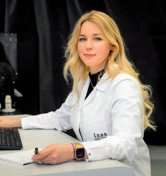
Zülal Cibir is a PhD student at the University of Duisburg-Essen. She conducts research at the Institute for Experimental Immunology and Imaging of University Hospital Essen.

The most important factors for biologists in the tracking of immune cells, such as neutrophil granulocytes, and what AI-based software can make possible in analyses were topics under constant discussion between the researchers at ISAS and University Hospital Essen. great communication, including between the doctoral candidates Justin Sonneck and Zülal Cibir, paid off in the form of powerful, free software.
Working on software for the ComplexEye was a special experience for me because it involves a new microscope that is not available anywhere on the market. My task was to act as the interface between the biologists like Zülal and the programmers in our team. What do the researchers in the lab consider to be important when evaluating the movies? What challenges arise specifically from the analysis of neutrophil granulocytes? How can we use AI to understand the motion paths of the individual immune cells?
Our brain is able to understand and process individual images, but it is not able to compare them accurately or objectively. For example, we see on the ComplexEye videos that the light patches are the neutrophil granulocyte immune cells. But our eyes are not able to compare the innumerable cells with each other. In addition, the quantity of data is simply too big: on average, one movie consists of several hundred ComplexEye images.
We have developed software which is able to segment the neutrophils as the first step. This makes it possible to differentiate between the individual cells and the background. The software then identifies the trajectories, meaning paths, of the individual immune cells. For example, we are able to determine the speed of the neutrophils in a sample. We can also objectively compare a large number of videos to each other, each one consisting of various images from the ComplexEye. Every movie shows the neutrophils in contact with a pharmaceutical ingredient. To date, the researchers have examined around 1,000 different active substances. What’s more, our software is open source. It is capable of identifying within a short period of time active agents that, for example, significantly reduce the speed of the neutrophils. For each video, such evaluation takes only a few minutes on average.
The following principles apply to our work: We always want to harvest the maximum amount of information from an image or a video. At the same time, we would like to maintain a low level of energy consumption, for instance, during data processing. That is why we have developed further open-source software with which we are able to optimise the energy consumption of AI models. Even if the number of kilowatt hours saved by this seems small, it all adds up in the end. After all, in biomedical research we are dealing with a very large number of high-resolution images and videos. The data volumes are huge – and in future we will be faced with even greater ones.
AMBIOM – Analysis of Microscopic BIOMedical Images
Junior Research Group
Dr Jianxu Chen
T: +49 (0)231 1392-217
E: jianxu.chen@isas.de
Biofluorescence Research Group
Prof Dr Matthias g unzer
T: +49 (0)231 1392-1403
E: matthias.gunzer@isas.de

Justin Sonneck is a PhD student in the AMBIOM Junior Research Group.
There are myriad medical reasons to perform research into fat. For Sven Heiles, it is the metabolism of fats, specifically lipids, that is of primary interest. “If it changes, this might be an indication of illness,” explains the head of the Lipidomics junior research group. With Heiles’s appointment, ISAS and the University of Duisburg-Essen reinforced their cooperation in 2022; the professorship is awarded according to the Jülich Model.
Lipids are substances that are insoluble in water and fulfil many tasks in the human body: they form the membrane of the cells (lipid membranes), store energy and are eliminated as soon as they have been utilised (lipid metabolism). These metabolic products can vary greatly in healthy and ill individuals in terms of quantity and chemical structure.
“We would like to find out how cardiovascular diseases affect lipids, for example, in order to fully understand the biochemical relationships within the body. If we succeed in precisely identifying lipid signatures, they could be used as biomarkers for early testing for various cardiac diseases,” says Heiles. In order to identify changes in lipids associated with illnesses faster and more accurately, the 39-year-old is developing new analysis methods at ISAS. He wants to obtain information on various molecule

classes, their quantity and how they are distributed throughout a sample, all at the same time if possible. To achieve this, he uses a combination of mass spectrometry imaging and microscopy.
In addition, the chemist is conducting research into the role lipids play in cancer: “Lipids can be used in examinations as tumour markers. Their being present in the blood or tissue of patients may provide information on the aggressiveness of tumours.” The findings of the lipid analyses are to be used in conjunction with those of other researchers looking into enzymes, hormones and genes in order to make holistic statements relating to individual patients. Consequently, Heiles’s team works closely with other research groups at ISAS, the University of Duisburg-Essen and University Hospital Essen.
(University of Duisburg-Essen, SR)
Lipidomics
Junior Research Group
Prof Dr Sven Heiles
T: +49 (0)231 1392-4202
E: sven.heiles@isas.de
The term omics refers to the holistic characterisation of all genes (genomics), metabolites (metabolomics) or proteins (proteomics). Omics data are an important starting point in precision medicine, because they provide insights into disease processes and possible therapeutic approaches.
Nowadays, analytical methods, including mass spectrometry, deliver increasingly more sensitive, more specific and faster measurement data. In order to analyse these large and in the future even more complex amounts of data on corresponding genes, metabolites and proteins adequately, new bioinformatics strategies are required.
In 2022, ISAS established the junior research group Multidimensional Omics Data Analysis (MdOA). The group aims to develop open source software for data analysis. Furthermore, the researchers want to process and visualise the measurement data using biostatistical methods and machine learning – so that the data can then be interpreted in cooperation with experts for health research and clinical application. To do this, the scientists at ISAS first link individual omics data sets with each other and with information from clinical studies, databases and scientific publications.
The findings from their multidimensional data analyses can be used, for example, to reveal biochemical pathways – actions between molecules in a cell – that interact with each other as biological networks. Uncovering these networks provides important information for individual strategies for the prevention, diagnosis and therapy of diseases. Thus, the researchers can identify potential biomarkers, for example for the prognosis of cardiovascular diseases or for monitoring the progression and therapy of chronic inflammatory bowel diseases. In addition, they can use the omics data to develop mathematical models that will assist physicians with diagnostic and therapeutic decisions in the future.
The junior research group at ISAS is a cooperation with Bielefeld University based on the Jülich model. There, robert Heyer holds a junior professorship in bioinformatics.
Multidimensional Omics
Data Analysis
Junior Research Group
Prof Dr r obert Heyer
T: +49 (0)231 1392-271
E: robert.heyer@isas.de
With his research on the structure and spatial distribution of lipids, Sven Heiles successfully habilitated in the field of analytical chemistry at Justus Liebig University (JLU) Giessen. A talk on this subject within the field of palaeontology concluded the habilitation procedure last Wednesday. Palaeontology refers to the scientific study of prehistoric beings, for example on the basis of fossils. How can analytical methods tell us more about the life of dinosaurs? In front of the Faculty Council as well as about 60 other guests, Heiles answered this question in an entertaining presentation.

Heiles has been at ISAS since August 1, 2022. In Dortmund, he heads the Lipidomics junior research group. Prior to his move, he worked at the University of California, Berkeley and then at JLU.
In Giessen, Heiles submitted his habilitation thesis in analytical chemistry in May 2022.
(SR)

Lipidomics
Junior Research Group
Prof Dr Sven Heiles
T: +49 (0)231 1392-4202
E: sven.heiles@isas.de
With Albert Sickmann, the Göttingen Academy of Sciences & Humanities gained a (corresponding) member of the Mathematical and Natural Sciences Class in spring 2022.
Since then, Prof. Dr. Albert Sickmann, Chairman of the Board at ISAS, has been contributing to the traditionrich community and exchanging ideas with about 380 other members.
ISAS congratulates PD Dr Dirk Janasek on his successful habilitation in the field of „Applied Analytics and Microfluidics“ at the department of Biochemical and Chemical Engineering at TU Dortmund University. The biochemist has been conducting research on microfluidic systems for 20 years, of which he has spent the last 19 at ISAS.
Microfluidics refers to the transport and analysis of low volumes of fluids. In his research, Janasek uses free-flow electrophoresis: an electric field separates injections in a chamber filled with an electrolyte solution into different fractions. In diagnostics, this method isolates different analytes, for example proteins or nucleic acids, from samples like blood or saliva within milliseconds.
In his habilitation lecture in mid-July, Janasek talked about paper-based microfluidic test systems. Blood sugar or haemostasis tests are well-known examples for this type of diagnostics. “What is special about microfluidic systems is that non-professionals can also use them to diagnose diseases,” Janasek explained. In his lecture, he demonstrated that especially in developing countries, these point-of-care tests (POC tests) could be an economic alternative

to conventional laboratory diagnostics.
“Paper-based POC tests are not only fast and reliable, but can also be produced at low costs and are easy to handle. Because their main component is the renewable resource timber, they can be produced and disposed of with a relatively small ecological footprint,” Janasek summed up.
Translational Analytics Research Group
PD Dr Dirk Janasek
T: +49 (0)231 1392-202
E: dirk.janasek@isas.de
Born in the city of Waldheim, Dirk Janasek obtained his doctorate on enzymebased sensors at Martin Luther University (MLU) of Halle-Wittenberg in 1999. Equipped with a Leopoldina fellowship, his path as a postdoc led him from MLU to Imperial College London. Since 2003, Janasek has been working at ISAS, where he heads the Translational Analytics research group.
Modern imaging methods are regarded as a key technology in firstclass medical research. At ISAS, the Bio-Imaging research programme focusses on temporal and spatial high-resolution visualisation and measurement of physiological states in whole organs, the cell and tissue structures they are made of up to the molecules which are essential to the function of the cells.
Using Light Sheet Fluorescence Microscopy (LSFM), high-resolution Confocal Laser Scanning Microscopy (CLSM) and Raman Microscopy for example, the scientists validate biomarkers to accelerate

the early detection of various diseases such as cardiovascular or autoimmune diseases. In order for the results of this fundamental research to be subsequently translated into clinical practise – i.e. transferred from the laboratory to patient care – there is close collaboration with the Institute for Experimental Immunology & Imaging at Essen University Hospital among others. The researchers also develop new microscopic measurement techniques which are designed to massively increase the throughput of samples, and therefore the speed of the analyses. Furthermore, by experimenting with animals and using human samples, ISAS researchers carry out measurements on intact organs and integrate artificial intelligence (AI) into their image analyses. Depending on the microscope used, one individual sample can produce hundreds of images. Without AI, an in-depth rapid analysis of the information in these images would not be possible, nor would it be possible to administer it efficiently. Microscopy is only one of many areas of application in medical imaging where AI is continuously revolutionising the processing of huge quantities of data.
Different research groups in various research projects at ISAS work to clarify the molecular and cellular processes that form the basis of what are referred to as immuno-vascular interactions under inflammatory conditions. During this work, the researchers investigate these cell interactions both in acute inflammatory processes as in case of heart attack or stroke, and also in chronic autoimmune disorders as well as rheumatoid arthritis.
In addition to LSFM and CLSM, Two-Photon Laser-Scanning Microscopy (TPLSM) is also used as an imaging method. With this combination of methods, it is possible to carry out a three-dimensional analysis of biological samples from macroscopic to subcellular level. However, in order to be able to characterise morphological and functional changes in inflammatory tissue with their fundamental molecular mechanisms over a period of time, scientists at ISAS combine LSFM, CLSM and TPLSM with complementary analytical technologies such as mass spectrometry (MS) and high-dimensional flow cytometry.
Localisation of osteoclasts (green) along the osseous blood vessels (red) in the murine mandible (lower jaw of the mouse). The image was taken using Confocal Laser Scanning Microscopy.As a disease mechanism is not only decisively influenced by the quantity of a biomolecule in a system but also its precise spatial concentration, combining microscopic methods with general and locally-resolved MS paves the way for entirely new diagnosis options in future. At present, many of the stated imaging methods still inevitably lead to destruction of the samples. This means that analyses are restricted to using individual techniques, which may also be mutually exclusive. This is problematic, especially with infrequent samples, such as human tissue biopsies for example, because comprehensive analyses are not possible. In the Bio-Imaging programme, ISAS therefore works on harmonising and combining complementary imaging and analytical methods with the aim of obtaining new non-destructive integrative measurement strategies. The purpose of developing this kind of cross-scale multi-method concept – in the form of 4D analysis – is to enable the location- and time-resolved, quantitative, in-vivo analysis at cellular to molecular level. The technical innovations required for this are crucial for comprehensive multimodal and multidimensional analysis, and therefore an overall understanding of biomedically-relevant processes. In the long term, these emerging new analytical technologies are to be integrated into clinical diagnostics which will in turn improve prevention and early diagnosis as well as personalised approaches to therapy.
(SR)
AMBIOM – Analysis of Microscopic BIOMedical Images
Junior Research Group
Dr. Jianxu Chen
T: +49 (0)231 1392-217
E: jianxu.chen@isas.de
Biofluorescence Research Group
Prof Dr Matthias g unzer
T: +49 (0)231 1392-1403
E: matthias.gunzer@isas.de
Bioimaging Research Group
Prof Dr Anika g rüneboom
T: +49 (0)231 1392-239
E: anika.grueneboom@isas.de
The Federal Ministry of Education and r esearch is funding the MSCoreSys-associated junior research group AMBIOM – Analysis of Microscopic BIOMedical Images under the funding code 161L0272.
When the highest alarm level has been triggered by an infection, phagocytes are on the scene in no time: as the body’s own defence and part of the congenital (unspecific) immune response, this group of white blood cells – including neutrophil granulocytes, neutrophils for short, and macrophages –arrive as the first line of defence against infectious agents.
But it is not always bacteria or viruses that are involved in an infection: with rheumatoid arthritis, for example, the body’s own processes are the trigger, so the autoimmune disease is considered to be a sterile infection. This chronic inflammatory joint disease is the most common of all the autoimmune diseases. It may have severe effects on patients and extend from loss of quality of life to occupational disability. Despite decades of research, the mechanisms that lead to this disease are still not fully understood, which means targeted treatment remains difficult. How neutrophils and macrophages communicate with each other in rheumatoid arthritis and exactly what this means for the course of the disease is one of the topics being looked at by the Collaborative Research Centre / Transregio 332 “Neutrophils: origin, fate and function” (► p. 18).
Since July 2022, researchers from the Bioimaging research group at ISAS and Münster University have been running the sub-project “C5: Phagocytic Crosstalk between Neutrophils and Macrophages in Rheumatoid Arthritis” to investigate an unresearched hypothesis on the development of the disease. This might facilitate new ways of treatment. The objective of the researchers in Dortmund and Münster is to find out exactly which immunological reactions in rheumatoid arthritis cause neutrophils to trigger inflammatory responses by macrophages. This research is intended to provide important insights into the disease mechanisms and, ultimately, new treatments for rheumatoid arthritis.
With rheumatoid arthritis, the neutrophils travel in a targeted manner into various anatomic niches such as joint cavities, where they die from various forms of cell death such as programmed cell death (apoptosis) and NETosis. In the latter, the neutrophils dissolve their cell and nucleus membranes and form a net-like structure from the DNA of their nuclei in order to bind and kill pathogens using these neutrophil extracellular traps (NETs). Scavenger cells such as macrophages ultimately dispose of the remaining DNA residues. As NETs activate the immune system, they may contribute to the formation of autoantibodies in rheumatoid arthritis. However, the extent to which neutrophils infiltrate what is known as the synovial tissue in the joint cavities and form NETs there has been unclear to date.
All in all, the sub-project involves the use of multimodal imaging methods, including Light Sheet Fluorescence Microscopy and confocal microscopy as well as what are known as multi-omics analyses. The researchers analyse neutrophils both from diseased mice and from patients.
The german research Foundation (Deutsche Forschungsgemeinschaft, DFg) is funding the research alliance “neutrophils: origin, fate and function” to the tune of around EUr 11.5 million over an initial four-year period. The alliance’s spokesperson is Prof Dr Oliver Söhnlein from Münster University. As for the projects run from Dortmund and Essen, the spokesperson for these is Prof Matthias gunzer, head of the Biospectroscopy department at ISAS and Director of the Institute for Experimental Immunology and Imaging/Imaging Centre at University Hospital Essen. For more information, see:
https://neutrophils.de

Bioimaging Research Group
Prof Dr Anika g rüneboom
T: +49 (0)231 1392-239
E: anika.grueneboom@isas.de
What are your tasks as a doctoral candidate in Transregio 332?
Hüser: It is known that neutrophils differ in rheumatoid arthritis depending on their location in the knee joint, whether they are in the synovial cavity or the synovial fat pad, for example. However, the composition of these sub-populations of neutrophils – i.e. what the immune cell fingerprint looks like in rheumatoid arthritis – is still unclear. I am testing the hypothesis that the various neutrophil sub-types trigger different
Darleen Hüser performs scientific work in the Bioimaging research group. Her PhD topic relates to neutrophil granulocyte immune cells.“My research is literally hard work”
Darleen Hüser is one of the ISAS researchers who are carrying out research as part of Transregio 332. The neutrophils and imaging techniques encountered in this project were entirely new to the 27-year-old when she first began working on it. In this interview, the biologist tells us why she expects to come up against hard work in uncharted territory.
inflammatory responses in the macrophages. This information might be of great importance for a future targeted treatment of the illness. The objective is to find out how many neutrophil sub-types there are, exactly which sub-populations the entire neutrophil infiltrate is composed of and precisely where the individual sub-types act in the inflamed joint. For this purpose, I am using various imaging technologies, such as Confocal Microscopy and Light Sheet Fluorescence Microscopy, to examine the knee joints of mice with rheumatoid arthritis.

Why are you carrying out some of your research using the confocal microscope?
Hüser: We would like to find out how the neutrophil sub-types that have migrated into the synovial cavity differ from those in the synovial fat tissue of the knee joint. This spatial information is crucial in answering our questions. We want to use this information to learn the reason for the different infiltration profiles. After I have examined the knee joint under the light sheet fluorescence microscope, I prepare it for further analysis under the confocal microscope. The latter features a very high resolution at the sub-cellular level and provides two advantages: firstly, I am able to identify which neutrophil sub-types are present in which anatomical niche. Secondly, confocal microscopy enables me to perform an analysis of cell-cell interactions between the various neutrophils and the macrophages located in the tissue.
membrane. The cells marked in green are the neutrophils. The image taken using a confocal microscope shows that, in a healthy state, no immune cells can be seen in the synovial cavity.
What challenges do you face in this research project?
Hüser: The mix of imaging methods is very exciting, but at the same time my research is hard work. For light sheet fluorescence microscopy, I need the knee joint to be a transparent intact joint. This means I first deal with clearing (► p. 21) the bones. I subsequently reverse this step and then manually cut the joint for confocal microscopy into thin slices of between ten and 14 micrometres on the cryostat microtome. What sounds easy in theory is tricky in the laboratory. Firstly, because there are only a few established methods of performing microscopic analyses on bones, so I first had to develop a suitable protocol for the treatment of the joints. Secondly, I require even and consistent cuts – which presents a challenge when it comes to cutting a knee joint comprised of bone, soft synovial tissue, tendons and fat pads. I have to work with a razor-sharp
tool and apply exactly the right amount of pressure to the blade so that the cuts are accurate and there are no bone fragments. The morphology, meaning shape and structure, of the tissue should be maintained to the greatest possible extent.
“There is so much we have no idea about when it comes to neutrophils. ”
Is there one aspect of your work that you find especially satisfying?
Hüser: It was not until the project began that I started to become aware of how extensive and heterogeneous the world of neutrophils is and how important they are for our immune system in so many ways. But most of all: there is so much we have no idea about when it comes to neutrophils. Apart from plunging into a new thematic area, when I first started working on the project most of the imaging methods were new to me too. I find it fascinating what information we can gather from a knee joint with the help of a mix of imaging procedures and which details suddenly become visible when doing so.
(The interview was conducted by SR.)
As tissue and bone can absorb, reflect or scatter light, they need to be chemically treated to see deep inside them beyond the surface. The clearing method developed for this purpose by Prof Dr Anika Grüneboom, leader of the Bioimaging research group, is used in light sheet fluorescence microscopy at ISAS and worldwide. With this method, researchers use cinnamic acid ethyl ester, a natural flavouring, to make the samples transparent. Grüneboom’s clearing can be reversed, meaning that no samples are destroyed and the same bones or the same tissue can be subsequently examined under the confocal microscope, for example.
Funded by the g erman research Foundation (Deutsche Forschungsgemeinschaft, DF g ) –Project number 449437943.

I like to look back at this moment: the photo shows Prof Dr Ronen Alon of the Weizmann Institute of Science in Israel and me in one of our laboratories at ISAS. My esteemed colleague is also an immunologist and a member of the scientific advisory board of ISAS. We met for the first time in person when he was visiting Dortmund in August 2022. First of all, Ronen spoke at the colloquium about his work and “LFA-1-ICAM-1 Signals for Leukocyte Differentiation & Effector Functions: Findings & Puzzles”. Subsequently, we were able to give him a little insight into our research during a tour of the two locations. When this photo was taken, the topic was my clearing method (► p. 21) for light sheet fluorescence microscopy. Ronen is holding in his hand a sample with cleared leg bones from a mouse. I no longer recall what made us laugh in that moment. But I do remember that we both had a lot of fun during our exchange of information and ideas. I am particularly pleased that Ronen now uses my clearing protocol for his research projects. We remain in contact, for instance, when questions arise about this method of creating transparent organs. I am very happy that his visit has developed into a regular exchange.
Prof Dr Anika Grüneboom, Head of Bioimaginghttps://www.isas.de/kompakt/ isas-wissenschaftspodcast-folge-7

Bioimaging Research Group
Prof Dr Anika grüneboom
T: +49 (0)231 1392-239
E: anika.grueneboom@isas.de
Team Communications
Sara r ebein
T: +49 (0)231 1392-234
E: sara.rebein@isas.de
Hepatitis B and C, but also a fatty liver caused by high alcohol consumption or being overweight, are the most common causes of cirrhosis of the liver. Diseases of the liver often take a gradual course and are frequently asymptomatic to begin with. But one thing is clear: the earlier cirrhosis of the liver is treated and complications are recognised, the higher the patients’ chances of survival. A new approach to diagnosing life-threatening deteriorations, such as infections and organ failure, at an early stage has been developed by ISAS immunologist Professor Dr Matthias Gunzer. The mobility of certain immune cells within the human body could help predict an imminent deterioration in a patient’s health.
When the human body is no longer able to compensate for the gradual failure of the liver, patients face an acute decompensation (AD) of the cirrhosis of the liver (► p. 24). This rapidly occurring complication arises from inflammatory reactions and defective immune responses. Some patients develop a sepsis (blood poisoning) or quickly suffer an acute-on-chronic liver failure (ACLF), which sees further organs such as the kidneys or brain fail too. As there are currently hardly any therapeutic options for ACLF, some patients die within days.
In the past, research has found that immunological and inflammatory mechanisms play a decisive role in how cirrhosis of the liver progresses. Severe dysregulation of the immune system is a consequence of cirrhosis of the liver and the reason that sufferers are highly susceptible to infections. The life expectancy of patients with cirrhosis of the liver depends on whether and which diseaserelated complications occur and are identified at an early stage.


„So far, we in the medical sector have no way whatsoever to predict complications such as infections or organ failure. And that is a huge problem because it means we are always running the risk of being overtaken by events and losing patients,” says Prof Dr Christian Lange, head of the Liver Centre at the University Hospital of Munich. He explains: “To be able to act in time, for example, by administering antibiotics or even performing a liver transplant, we would have to find out as early as possible about a further deterioration in vital functions such as organ failure or infections. But such a marker was lacking to date.”
The functional analysis of specific immune cells might be useful in monitoring the health of patients with cirrhosis of the liver and in detecting impending complications as early as possible. The blood stem cells in the bone marrow produce more neutrophil granulocytes as a reaction to infections (► p. 25), which then migrate in the direction of the source of infection. And it is precisely this migration that immunologist Prof Dr Matthias Gunzer has in his sights. He heads the Biospectroscopy department at ISAS and is Director of the Institute for Experimental Immunology and Imaging at University Hospital Essen. “For more than one hundred years, we have known that neutrophils move. And now we even know down to molecular detail how the immune cells do this and which proteins are responsible for this process inside the cells,” he explains.
Although the liver is a vital organ, its central importance for the human body is often underestimated. It is not only responsible for detoxification and for digesting fat, but also for storing energy. While it’s true that the liver is the only organ capable of regenerating itself, this is only possible up to a certain degree of damage. Cirrhosis of the liver constitutes one of the leading problems for the global health system and even in industrialised countries such as Germany it is not uncommon. With this disease, the liver tissue is increasingly destroyed and replaced by connective tissue. The tissue hardens, becomes scarred and shrinks as well, which is why the disease also has the colloquial name “shrunken liver”. The liver is then no longer able to fulfil its vital tasks at all or only incompletely.
Prof Dr Matthias gunzer heads the Department of Biospectroscopy and the Biofluorescence research group at ISAS. He is Director of the Institute for Experimental Immunology and Imaging at the University Hospital Essen.In medical practice, this knowledge has to date remained unused, however; there is a lack of functional examinations of human blood. Depending on the clinical picture, practitioners may have a look at the number of immune cells, but they do not investigate whether they are functioning normally or not. This means that it is possible for the blood count to be in the normal range in terms of numbers – but the function of the neutrophils, such as their movement, is nevertheless impaired.
We woud have zu find out as early as possible about a further deterioration in vital functions such as organ failure or infections.
Gunzer has developed an assay (laboratory test) that analyses the migration behaviour of the neutrophils. The findings could permit conclusions to be drawn about a patient’s health and act as markers to facilitate the early detection of complications. In brief: routine and regular measurement of the movement of neutrophils could act as an early warning system for medical professionals.
This was how the idea arose of Lange and Gunzer collaborating. The big question was this: can the movement behaviour of the neutrophil granulocytes predict whether the health of patients with cirrhosis of the liver will deteriorate within a few days or weeks? Lange and Gunzer decided to deploy their teams to use the standardised migration assay to characterise how the neutrophils migrate in the blood of sufferers.
The researchers used immunomagnetic separation to isolate the neutrophils from the blood of 125 patients with cirrhosis of the liver at the Liver Centre at different stages of severity of the disease and from the blood of 24 healthy individuals. In the experiment, the researchers added three different active agents (the chemotactic formyl peptide f-Met-Leu-Phe and the chemokines CXCL1 and CXCL8), which are known to trigger the migration of the neutrophils. With a cell culture microscope, they took images of the neutrophils every eight seconds for one hour. The researchers then combined these images to make videos that allowed them to automatically evaluate the movement of the cells over the course of time. Furthermore, they used flow cytometry (► p. 26) to examine
These immune cells perform important tasks in the human body. Neutrophil granulocytes are primarily responsible for being the first line of defence against infectious agents such as bacteria and fungi. They are able, for example, to identify and kill or eat up microorganisms and other structures that are foreign to the body.
the relationship between the neutrophil migration and the expression of chemokine receptors and activation markers on neutrophils.
The study ultimately succeeded in actually discovering special migration patterns amongst the neutrophils of patients with cirrhosis of the liver who have a high risk of developing complications. The researchers under Lange and Gunzer therefore came to the conclusion that a large proportion of immobile neutrophils and a high average velocity of the moving neutrophils are particularly characteristic of a high risk of developing a sepsis or an ACLF, or even dying, over the next seven to 30 days.
The new method of analysing neutrophils has ultimately shown that it is possible to regularly observe the behaviour of these immune cells in patients with cirrhosis of the liver and to establish whether pathological migration patterns are developing. But until the examination can become standard clinical practice for sufferers, the experimental approach will have to be simplified and automated using machine-based algorithms so that the blood test can be performed quickly and with little intervention by staff on a routine basis. “Transposing this procedure into clinical practice would make it possible for the first time to detect infections and organ failure at an early stage in patients with cirrhosis of the liver. This would enable us as medical practitioners to put therapeutic measures in place at an early stage and to save the lives of sufferers,” says Lange.
Incidentally, the mobility test of neutrophil granulocytes is not restricted to use in patients with cirrhosis of the liver. Gunzer: “As far back as 2018 we were able to demonstrate that the severity of a preliminary stage of leukaemia can be evidenced through the migration behaviour of the neutrophil granulocytes. Early detection in diseases other than leukaemia and cirrhosis of the liver would also be conceivable.” (CK)
Langer, M.-M., Sichelschmidt, S., Bauschen, A., Bornemann, L., Guckenbiehl, S., Gunzer, M., Lange, C.M. (2022) Pathological neutrophil migration predicts adverse outcomes in hospitalized patients with liver cirrhosis.
Liver International, 43(4), 896–905.
https://doi.org/10.1111/liv.15486
With the help of a flow cytometer, it is possible to determine, for example, molecules such as proteins on the surface of and inside individual cells quantitatively and at high speed. In this respect, the device follows a physical-chemical principle: a stream of liquid transports cells. They flow quickly past a laser beam and are analysed by means of the scatter of visible light or by fluorescence. Over and above this, the cells influence the laser light depending on their size, the structure of their membrane or the content of intracellular structures.
Over the course of one year, the Chan Zuckerberg Initiative (CZI) will fund the development of the Dortmund software for the napari image analysis platform: At ISAS, the research groups AMBIOM – Analysis of Microscopic BIOMedical Images and Spatial Metabolomics will develop new plug-ins. Thus, scientists worldwide will be able to better analyse microscopic and chemical images – free of charge.


Cell movements provide important insights in biomedicine, for example regarding the development of various forms of cancer. To find out how tumour cells spread in the body in different types of cancer, researchers among other things examine their movement under the microscope. The resulting time-lapse recordings provide information, for example, about the motility of the cells. For this quantitative analysis, cells that overlap must first be separated from one another (segmentation). Then, their paths can be tracked.
Image analysis has improved significantly in recent years. Nevertheless, the technology currently still reaches its limits when it comes to cell tracking. „Right now, there are many microscopy scenarios in which automated analysis does not provide satisfactory results – and manual curation is required. The images of 50 cells

generate large amounts of data which researchers can hardly analyse manually,“ explains Dr Jianxu Chen, an artificial intelligence (AI) expert and head of AMBIOM. That is why the computer scientist and his team want to develop specific software in the napari ecosystem. This software will enable biomedical scientists to intervene directly in the automatic analysis and correct errors, for example during segmentation or tracking.
The „Human-in-the-loop Cell Tracking“ software, which has received funding of 20,000 US dollars, will include a total of three modules (segmentation, tracking and analysis). Its goal using AI: first, to improve the result of napari image analysis, and second, to train the algorithm with the human-input information using machine learning. To this end, the AMBIOM team will cooperate with immunologists at ISAS, University Hospital Essen and the University of Duisburg-Essen.
AMBIOM – Analysis of Microscopic BIOMedical Images
Junior Research Group
Dr Jianxu Chen
T: +49 (0)231 1392-217
E: jianxu.chen@isas.de
Spatial Metabolomics
Junior Research Group
Dr Prasad Phapale
T: +49 (0)231 1392-4244
E: prasad.phapale@isas.de
AMBIOM is also involved in the programming of the second software for biochemical imaging, which is funded with another 20,000 US dollars. This project is the first napari plug-in for the evaluation and annotation of mass spectrometry imaging data. A mass spectrometer can be used to identify substances such as metabolites in a sample based on their masses. Using mass spectrometry imaging (MSI), scientists can, for example, examine tumour tissue for metabolic differences at subcellular resolution. In this way, they obtain information about the spatial distribution of the molecules and can compare the results with morphological abnormalities in the tissue.
Dr Prasad Phapale, a chemist and head of Spatial Metabolomics, is striving to improve the multiplexing (integration) of MSI data with other image formats. For example, the „Biochemical Annotations of Mass Spectrometry Imaging Data“ plug-in for napari will allow biochemical annotation of MSI data with image co-registration (image fusion). In short, it will enable scientists worldwide to match
their MSI results with metabolite databases, as well as match spatial information from their imaging with that of complementary analytical methods, such as microscopy. This would improve the sharing of knowledge within the research community.

napari is an open source tool that enables a powerful visualisation of multidimensional images, for example from microscopy. napari is based on the Python programming language. It is being continuously expanded with the help of a worldwide growing community of researchers and software developers. CZI promotes napari through its Imaging Program with the goal of facilitating biologists’ access to new methods of image analysis based on machine learning.
The MSCoreSys associated junior research group AMBIOM – Analysis of Microscopic BiOMedical Images is funded by the Federal Ministry of Education and research (funding reference 161L0272). The Federal Ministry for Education and r esearch is also funding the MSCoreSys associated junior research group Spatial Metabolomics (funding number 161L0271).
This project has been made possible in part by a grant from Chan Zuckerberg Initiative, an advised fund of Silicon Valley Community Foundation.
Dr Prasad Phapale heads the Spatial Metabolomics junior research group.Individuals in the western world who become infected with HIV (human immunodeficiency virus) are usually spared the onset of AIDS thanks to anti-retroviral therapy (ART). To date, sufferers have had to take medication on a daily basis for life. One important pillar of ART consists of active agents from the substance class of integrase inhibitors, including dolutegravir and elvitegravir. What influence these drugs have on certain immune cells belonging to the body’s own defence system, such as CD8⁺ T cells, was examined jointly by Dr Enrico Richter from Prof Dr Hendrik Streeck’s research group at the Institute of Virology at the University of Bonn and ISAS researchers in 2022.
To replicate, HIV requires host cells that carry a certain receptor, the CD4+ receptor, on their surface. CD4+ is a glycoprotein that is mainly found on the surface of immune system cells. Such cells enable the viruses to dock onto the cells and to advance into the cell interior. The majority of these are helper T cells carrying CD4+ (CD4+ cells). Helper T cells belong to the cellular part of the immune system and perform their helper function by releasing effector molecules such as cytokines – proteins that act as messenger substances. Helper T cells play a significant role in many immunobiological processes. They support the function of leukocytes (white blood cells) and are involved in maturing immune cells such as macrophages (scavenger cells) or activating cytotoxic T lymphocytes. The latter are also known as CD8+ lymphocytes. They fight degenerated cells just as much as they eliminate cells infected with pathogens when they detect their antigens that are typical of pathogens.

In HIV-infected patients, there is always a certain portion of the host cells that is latently infected. This means the cellular DNA of these cells contains integrated virus DNA but they do not produce any virus particles. However, under suitable conditions, the virus DNA may be reactivated again at any time and help to synthesise new virus components. To date, the body’s immune defence has been only partially successful in detecting and eliminating such latently infected cells. This is one of the main reasons why patients
remain infected with HIV for life even though medicine now has a large number of anti-retroviral drugs at its disposal.
Only a small proportion of CD4+ T cells is located in the body’s lymph follicles. However, it has become apparent that it is exactly these follicular helper T cells that contain the majority of HIV in infected individuals. These latently infected, dormant CD4+ helper T cells, also known as memory T cells, are something akin to a long-lasting reservoir for HIV that may be activated at any time. Tracking down these cells in a targeted manner and effectively eliminating them is a central challenge in translational HIV research.
The focus was on a potential limiting effect of integrase inhibitors on various functions of the CD8+ T cells.
An infection with HIV triggers a sustained and permanent response in the form of more CD8+ T cells. But they are not able to eradicate the small proportion of CD4+ T cells latently infected with HIV. Not even simultaneous application of ART or a so-called “shock and kill” strategy is successful in this respect. The latter is based on active agents with a small molecular size that can reverse the viral latency status in the cells by initiating synthesis of the virus components. This makes the cells vulnerable to the immune system. Nevertheless, the small pool of latently infected cells remains in place. This situation is not changed by HIV-specific CD8+ T cells either, not even in the presence of additionally applied anti-viral medications. Does this therefore mean that the ability of the CD8+ T cells to kill latently infected cells is impaired by the active substances?
“The focus of our experiments was on a potential limiting effect of integrase inhibitors on various cellular functions of the CD8+ T cells,” says Richter. In an experimental approach CD4+ T cells were cultivated together with CD8+ T cells that had previously been incubated with various anti-retroviral substances. The quantity of HIV replicating in the CD4+ cells was subsequently determined using ELISA (► see box on the right).
An enzyme-linked immunosorbent assay (ELISA) is a test method for establishing the concentration of antigens (molecules that are able to bind to the respective specific antibodies) or antibodies in liquids

“However, we were unable to provide evidence for the differences in the cytotoxic activity of CD8+ T cells,” explains the virologist. So what could be the reason for the elimination of the latent reservoir not working in vivo? Investigations were then conducted into the other biological capabilities of CD8+ T cells under the influence of anti-retroviral drugs. In addition to the cytotoxic activity, the parameters examined included the functioning, the replication (proliferation), the cell metabolism and the migration behaviour of the cells.
Biofluorescence Research Group
Prof Dr Matthias g unzer
T: +49 (0)231 1392-1403
E: matthias.gunzer@isas.de
In order to exercise their cytotoxic effect, immune cells, and thus also CD8+ T cells, must be able to actively move. Defects in this migration can explain poor functioning. In order to determine potential negative effects of ART active agents on the migration of the immune cells, the researchers incubated CD8+ T cells from healthy volunteers with various anti-retroviral active agents for one day. After the end of the 24-hour period, the researchers stimulated one part of the samples with a buffer, the other part with the protein SDF-1α. They then determined the migration behaviour of the cells using video microscopy. At 20-fold magnification, the microscope took images every 15 seconds for a period of three hours. The researchers then combined the individual images into a video. There followed an automatic segmentation of all cells in every video image, which made it possible to reconstruct the paths taken by all the individual cells (tracking).
We had to try out a lot of things, even just to find the right molecule to get the CD8+ T cells running around in the Petri dish.
Prof Dr Matthias Gunzer and his team worked on the migration assay for around one year. Here is his summary: “The development was a challenge. We had to try out a lot of things, even just to find the right molecule to get the CD8+ T cells running around in the
HIV attacks the body’s own defences directly by infecting host cells of the immune system. Human immunodeficiency viruses, which belong to the family of retroviruses, thus weaken and destroy the organism’s protective defences. After HIV has been smuggled into the human host cell, the virus’ own proteins come into play in the cytoplasm: the reverse transcriptase (rT) enzyme rewrites the virus’ genome, which consists of two rnA strands, into viral DnA. This is smuggled into the cell nucleus by the integrase, another viral enzyme, and there it is integrated into the cellular DnA. Starting with the viral DnA and later with the integrated DnA, also known as provirus, HIV proteins are produced via transcription and translation. In turn, these can form new viruses with the viral DnA replicated in parallel. The rT of the HIV generates a lot of errors and the double strands of the viral DnA it produces consequently contain a large number of mutations. This is one of the reasons why HIV infections are difficult to treat. Dolutegravir and elvitegravir belong to the class of integrase strand transfer inhibitors (InSTIs) that prevent the virus genome from being integrated into that of the host cells.
Petri dish.” In the end, the analysis of the migration behaviour of the immune cells revealed the following: dolutegravir and elvitegravir actually do interfere with the mobility of the CD8+ T cells.
Furthermore, the researchers found out that treatment with the two active agents impairs cytokine expression, proliferation, new synthesis of effector molecules and respiratory metabolism. The findings make it clear that there is a significant negative impact on the most important functions of CD8+ T cells being treated with dolutegravir and elvitegravir. Now further investigations are necessary to fully understand the mechanism of action of this observation and its potentially serious long-term toxicity.
Richter, E., Bornemann, L., Korencak, M., Alter, G., Schuster, M., Esser, S., Boesecke, C., Rockstroh, J., Gunzer, M., & Streeck, H. (2022). Reduction of CD8 T Cell Functionality but Not Inhibitory Capacity by Integrase Inhibitors. Journal of Virology, 96(5), e01730-21.
https://doi.org/10.1128/JVI.01730-21
First of all, one has to say that we in the western world are very lucky to only have to speak of HIV, the human immunodeficiency virus, and not of AIDS. The reason for this is our access to excellent medical care with so-called highly active antiretroviral therapies (HAART). These medications ensure that HIV-positive patients do not develop acquired immunodeficiency syndrome (AIDS). Unfortunately, the situation for those affected by HIV in other countries, such as in large parts of Africa, is worse. That is why we definetely need a vaccine in order to protect them from HIV.
Vaccination is the process of confronting the body with specific antigens; in the case of viral diseases such as COVID-19 or AIDS, these are components of the virus that causes the disease. The aim is for our body to produce antibodies and defence cells against the antigens. The result of a successful vaccination is then a protective immune response that can last for months or years.
Scientists have been researching HIV vaccines for decades. But of more than 400 clinical studies on possible vaccines that have taken place since 1987, none has been convincing in terms of the final result. However, this is by no means because too little research is being done on HIV. We can clearly see this in the successful treatment options that now exist, such as HAART. We should not
Biofluorescence Research Group
Prof Dr Matthias g unzer
T: +49 (0)231 1392-1403 E: matthias.gunzer@isas.de
forget that many years of research on HIV have also led to the fact that we now know a great deal about how antibodies work, for example – or about how to induce them particularly efficiently with a vaccine.
Compared to the coronavirus, however, the HI virus is incredibly variable: tens of thousands of new copies are created every day – in a single person. On average, each of these new copies carries at least one unique mutation. Therefore, over the years, a single person carries
countless variants in their body, but only a few of these variants can be transmitted to others. The main problem that these variants pose for vaccines is that some
Compared to the coronavirus, however, the HI virus is incredibly variable: tens of thousands of new copies are created every day – in a single person.
mutations are located precisely in the parts of the virus that are usually attacked by the immune system. Therefore, mutations like these can help the virus remain unrecognised. A successful vaccine needs to elicit an immune response that can deal with this diversity in order to provide full protection against an infection.
In addition, unlike SARS-CoV-2, the HI virus is a true concealment artist. Parts of its surface are covered with a dense layer of sugar molecules – the glycan shield. This shield covers possible points of attack for antibodies. Although the coronavirus has such a sugar layer as well,
the crucial areas of its spike protein remain uncovered. Thus, in the case of SARS-CoV-2, antibodies can recognise its spike protein and bind to it, thus neutralising the virus. The second hiding tactic that the HI virus uses is also tricky: the HI virus inserts its genetic blueprint into the DNA of its host, i.e. humans, and thus creates a hidden reservoir in the immune cells. This makes the HI viruses invisible to the immune system.
At present, six phase III trials are being conducted to investigate the efficacy and safety of potential HIV vaccines in large patient populations. Among the vaccine candidates are new variants such as those designed to elicit broadly neutralising antibodies and several based on mRNA molecules. Many people are already familiar with the latter because of the highly effective vaccines against COVID-19. That said, the research on HIV is ongoing – which is why, despite the challenges, we should by no means give up hope for an HIV vaccine.
(Protocol: CP, SR)Originally, Dr Jianxu Chen (AMBIOM – Analysis of Microscopic BIOMedical Images) had invited Dr Rita Strack to ISAS. But on Wednesday, in the packed lecture hall at the ISAS campus, Strack was the hostess. With her talk, the senior editor at Nature Methods opened the usually closed doors of the journal and provided insights into the internal editorial processes. The change in perspective that the biochemist demonstrated to the participants was at the same time a key for successful publications in the future – also in other journals besides Nature Methods.

Strack has read more than 4,000 manuscripts in the past eight years at Nature Methods. The journal receives more than 200 manuscripts per month. Ten to 15 percent of them make it to the review stage, and 60 percent of these peer-reviewed manuscripts are ultimately published. Anyone who followed Strack quickly realised that the
former researcher is driven by her enthusiasm for new research topics and successful publications. Strack revealed what is important for a successful paper with the help of numerous tips and examples. For instance, the US-American gave the advice to consider the content and target group of a journal carefully and to clarify open questions with the editors before submitting.
“Is the topic relevant to the readership and therefore to this journal? How transparent and accessible are the submitted data? Are the results reproducible?” Much of the background information and advice that Strack shared with the scientists in the audience and online can be applied to other journals. “We learned what really matters and heard a lot about common mistakes, practical tips and even an extensive list of trending topics. This is all very helpful for future publications,” says Chen. What surprised him was the way Strack and her colleagues approached their work: To support researchers, to get the best out of their manuscripts and to ensure that the publications meet the journal’s standards. One aspect that besides Chen may have surprised many of the more than 70 participants: As long as the science part is good, according to Strack, formatting, text length or number of figures or tables do not play any role at all in deciding whether a manuscript will be reviewed.
After a lively discussion, Strack finished by inviting those present to stay in touch. “I am a person,” she encouraged the scientists to talk to her or to editors of other journals in the future.
Biofluorescence Research Group
Prof Dr Matthias g unzer
T: +49 (0)231 1392-1403
E: matthias.gunzer@isas.de
Bioimaging Research Group
Prof Dr Anika g rüneboom
T: +49 (0)231 1392-239
E: anika.grueneboom@isas.de
AMBIOM – Analysis of Microscopic BIOMedical Images
Junior Research Group
Dr Jianxu Chen
T: +49 (0)231 1392-217
E: jianxu.chen@isas.de


This research programme focusses on analysing the molecular mechanisms associated with various illnesses, for example cardiovascular diseases. Many diseases have multi-factorial causes – genetic constellations also play a part in addition to environmental and nutritional factors. Because the prognosis varies from patient to patient, they respond to treatment in different ways. In order to obtain a comprehensive understanding of the disease mechanisms and diagnose the diseases earlier in future, with fewer side effects, and to be able to administer individual treatments more effectively, researchers at ISAS identify potential target molecules (targets).
The fundamental research conducted by the scientists involves using methods that are by no means restricted to genome level, but instead also use proteomic and metabolomic parameters. The researchers apply, test and optimise multi-omics methods for this purpose. One of the focal themes of the programme are cardiovascular diseases. Here, the institute has many years of analytical expertise to fall back on, including comprehensive investigations into the proteomes of thrombocytes (blood platelets) and also in-depth investigation of thrombocyte malfunctions and molecular processes in the event of cardiac insufficiency (commonly known as heart failure).
The molecular causes and the course of many diseases of the cardiovascular system are still largely not understood. The scientists at ISAS work on improving diagnostics for cardiac insufficiency and establishing new treatment approaches. They combine traditional molecular genetic and biochemical methods with high throughput methods. This enables them to cover the entire bandwidth of the analysis – from detailed investigation of individual components through to analysis of entire cellular systems.
The scientists involved in this research programme develop new instruments for diagnosis and treatment which enables them to differentiate between different heart diseases. To achieve this, they work with transgenic mice, for example. The objective is to work out the spectroscopic characteristics of various courses of disease. The scientists have continued to press ahead intensively with biospectroscopy analyses, among other things, using vibration microscopy and high-resolution microscopy with imaging. Using the optical methods, in-vivo and molecular biological investigations, they have succeeded, in collaboration with Julius-MaximiliansUniversität of Würzburg and the University of Duisburg-Essen, in investigating various molecular mechanisms of cardiac insufficiency in different mouse models with genetic diseases and detecting the disease in its early stages using the M. Fabry model, for example.
In order to identify the potential of non-linear spectroscopic imaging instruments and various assays for the identification of cardiac involvement in metabolic disorders and genetically-based storage diseases such as Fabry disease, the researchers used Coherent Anti-Stokes Raman Scattering (CARS) Microscopy (► p. 42) to carry out the investigation with the mouse model. Thanks to the high sensitivity of the acquired spectral information and the computerassisted diagnostics, subtle changes in the protein-lipid content between the heart tissue in Fabry disease and control tissue can be detected with up to 96 percent reliability.
Furthermore, in this research programme the scientists develop and optimise silicon-based nanocontainers which means that drugs can be applied for heart muscle cells specifically, which reduces the side effects. The researchers are also investigating the action mechanism of Cold Atmospheric Plasma (CAP) in the treatment of cardiovascular diseases. Until now, this kind of plasma has mainly been used in the area of tissue repair, for treating infectious skin disease, in dentistry and for cancer treatment. These plasma could increase the concentration of nitrite in the blood and therefore reduce a cardiovascular risk factor.
Cardiovascular Pharmacology Research Group
Prof Dr Kristina Lorenz
T: +49 (0)231 1392-103
E: kristina.lorenz@isas.de
ERC-Sulfaging
Dr habil Miloš Filipović
T: +49 (0)231 1392-4173
E: milos.filipovic@isas.de
Miniaturisation Research Group
PD Dr Joachim Franzke
T: +49 (0)231 1392-174/199
E: joachim.franzke@isas.de
Proteomics Research Group
Prof Dr Albert Sickmann
T: +49 (0)231 1392-100
E: albert.sickmann@isas.de
Translational Analytics
Research Group
PD Dr Dirk Janasek
T: +49 (0)231 1392-202
E: dirk.janasek@isas.d e
Fabry disease is a treacherous illness that gradually destroys the heart and other organs and is often not detected until it is too late to intervene. In future, a new diagnostic method tested at ISAS may help to diagnose thousands of sufferers in Germany alone at an earlier stage. Ultimately, other patients with cardiovascular diseases – the most common cause of death worldwide – might also benefit from the progress being made in the area.

The approach is based on Raman spectroscopy, one of the more recent spectroscopic imaging methods used in biomedicine. Biomedical spectroscopy – including the well-known nuclear magnetic resonance spectroscopy, which is based on measuring the magnetic momentum of nuclei – is becoming increasingly better at identifying illnesses using specific biomarkers, says Prof Dr Kristina Lorenz, head of the Translational Research department and the Cardiovascular Pharmacology research group.
“We deployed a special version of Raman spectroscopy, called Coherent Anti-Stokes Raman Spectroscopy or CARS (► p. 42), to see if we can detect changes in the heart at early stages of the disease, changes that are caused by Fabry disease,” explains physicist Dr Elen Tolstik, who has been conducting research in the Cardiovascular Pharmacology research group since 2018.
The trigger for Fabry disease is a gene defect that causes too little or none of an enzyme named alpha-galactosidase A, or α-GAL A, to be produced in the body. In healthy people, the enzyme is present in virtually all cells of the body, where it breaks down what are known as glycosphingolipids – fats that are involved in creating cell membranes. Without α-GAL A and the breaking down of lipids it initiates, these lipids accumulate – they are “stored” as it were – which damages the cells over time. For this reason, Fabry disease is also called a
Dr Elen Tolstik is a physicist who has been conducting research in the Cardiovascular Pharmacology research group since 2018.“storage disorder”. Alongside organs such as the heart and the kidneys, the blood vessels and nerves are often affected.
If treatment is commenced at an early stage, patients have good chances of survival, but in most cases the disease is diagnosed too late.
The damage to tissue caused by accumulations of lipids becomes apparent during the teenage years in some cases due to severe joint pains and changes in the skin. Typical signs are, for example, reddish-purple lumps and discolourations on the lower body. But this metabolic disorder may also cause quite different symptoms in some individuals, which makes diagnosis more difficult. Many sufferers acquire corneal deposits in the eyes, others develop abnormally tortuous blood vessels in the brain and experience strokes.
Raman spectroscopy uses the phenomenon of Raman scattering, where the light from molecules is scattered inelastically while their wavelength fluctuates. With the help of highprecision measurements, it is possible to obtain specific information from the process on the properties of the light-scattering molecules, for example, their chemical composition, structures and molecular dynamics. In contrast to simple Raman spectroscopy, with CARS two intense laser beams with different wavelengths selectively excite certain molecular vibrations. The resulting coherent scattering uses a significantly higher signal to penetrate deeper into the layers of tissue such that large structures can be analysed more quickly, says Tolstik.
Fabry disease can also lead to diarrhoea, vertigo attacks and tinnitus. In addition, damage to the heart and the kidneys are commonplace, often leading to an irregular heartbeat, a weak heart (cardiac insufficiency) and kidney failure such that regular dialysis may even become necessary. According to statements by the German National Fabry Disease Self-help Group, many Fabry disease patients become fully or partially incapacitated for work even as young adults. An estimated 8,000 people suffer from the disease in Germany. As the responsible gene defect is located on the X chromosome, of which men have one copy and women two, the disease disproportionately affects men.
While there are now a handful of pharmacological therapeutic options that can mitigate or even reverse the course of the disease, they are only effective if they are employed before extensive tissue damage has occurred. “If you begin treatment at an early stage, patients have a good chance of survival, but in most cases the disease is diagnosed too late and the heart has already been destroyed by fibroses, meaning abnormal changes in tissue,” says Tolstik. “For us, late diagnosis was one of the most important reasons to carry out this research.”
In principle, blood and gene tests can reveal whether the key enzyme α-GAL A is present in the body or whether there is a gene defect. But damage that has potentially already been sustained in the body
is nevertheless often only identified at a late stage. “New biomarkers and/ or diagnostic methods for the cardiac consequences of Fabry disease are urgently required,” write Lorenz and Tolstik in a specialist article about their project in the International Journal of Molecular Sciences. Working closely with cardiologist PD Dr Peter Nordbeck of University Hospital of Würzburg, which runs an internationally recognised competence and reference centre for Fabry disease, the two ISAS researchers tested the CARS-based analysis.
To date, the two researchers have tested the CARS procedure in a murine model (α-GAL A knockout mouse ► see box on the right) and in healthy animals. Assisted by a sophisticated computer data processing system, in the biopsies of the knockout mice it was possible to detect even subtle changes in the protein-lipid content of the heart tissue with a sensitivity of up to 96%. This means that the abnormal storage of lipids in the tissue – the molecular signature of Fabry disease – can be detected using CARS even without complex histological investigations, before the organ affected is severely damaged.
For the next step, Lorenz and Tolstik are planning to test the diagnostic method in human samples. If the method continues to prove its worth, it may in principle be used for other similar illnesses too, potentially in combination with further imaging procedures. The researchers at ISAS are thus building up further expertise in this vital new area where imaging procedures are used to find novel biomarkers for cardiovascular diseases. Collectively, these diseases kill around 18 million people annually around the world – more than every other cause of death. As non-destructive tools that provide an insight into the chemical composition of tissue and fluids, Raman and CARS spectroscopy may take on a key role in cardiovascular research.
On account of their biological and genetic similarity to humans, mice are often deployed in research. With a knockout mouse, researchers literally knock out a certain gene or several genes. These genetically modified animals subsequently help them to examine a certain disease model. For the research on Fabry disease, these are knockout mice where the gene responsible for producing α-GAL A has been deactivated.
Cardiovascular Pharmacology Research Group
Prof Dr Kristina Lorenz
T: +49 (0)231 1392-103
E: kristina.lorenz@isas.de
Tolstik, E., Ali, N., Guo, S., Ebersbach, P., Möllmann, D., Arias-Loza, P., Dierks, J., Schuler, I., Freier, E., Debus, J., Baba, H.A., Nordbeck, P., Bocklitz, T., Lorenz, K. (2022). CARS Imaging Advances Early Diagnosis of Cardiac Manifestation of Fabry Disease. International Journal of Molecular Sciences, 23(10), 5345.
https://doi.org/10.3390/ijms23105345
Her colleagues in the ERC group Sulfaging affectionately call Dunja Petrovic (27) the “worm lady”. For she has been spending a lot of time with nematodes since starting her PhD in Bordeaux, France, in 2018, and has been continuing at ISAS since 2020. Petrovic’s daily routine in the lab and her free time revolve around Caenorhabditis elegans, C. elegans for short. In the interview, the PhD student talks about why the mutant roundworms help the Serbian to research ageing processes in humans and which new developments the laboratory change to Dortmund entailed.
What is the subject of your dissertation?
Petrovic: I deal with hydrogen sulfide, H2S for short. We know that it influences many important processes in our body, such as ageing. I pose the question which effect the absence of enzymes that produce H2S in the body has on the organism. For that, I use the model organism C. elegans. For my research, I can “switch off” the genes that contain information for these enzymes in wildtype roundworms. With these so-called mutant worms, I analyse how the lack of a certain protein product influences their development or stress response. For example, I look at their lifespan or examine how sensitively the worms react to different environmental stimuli. That is part of my research work.
The European Research Council awarded the leader of your working group, Dr habil Miloš Filipović, with two million euros in funding.
What is the aim of your working group?
Petrovic: Ageing processes lie at the heart of our group’s research. As we age, the proteins in our body are more prone to irreversible oxidation. The reason for that is the contact with hydrogen peroxide and other oxidising agents, by-products of our metabolism, whose amount increase with ageing. On the other hand, our
defence mechanisms become less efficient. H2S comes into play here. This gas can react with oxidised cysteines and form persulfides. We believe that this process, called persulfidation, prevents protein overoxidation and therefore preserves its function. I want to understand how the persulfidome of a cell and a whole organism changes during ageing. For example we were able to show that the levels of persulfidation decrease
with age in different model systems. This phenomenon might be one of the many reasons why we get old.
Can you transfer the results of your work with worms to humans?
Petrovic: Biologically speaking, the worms are much simpler than humans are, of course. However, we are able to understand some of the fundamental signalling pathways in the body because of them and then build on these findings. Time plays a decisive role here. Unlike mammals, where several years go by before they start ageing, C. elegans only takes three days to develop from an egg to an adult animal. During their two to three week lifespan, we can track their whole ageing process. It is like watching an old person through a microscope. The animals move less than at the beginning of their adulthood and their bodies look almost wrinkly.
„ I want to understand how the persulfidome of a cell and a whole organism changes during ageing.“
What is special about working with these worms?
Petrovic: The worms are easy to keep and with a size of only about one millimetre, they are very small. They
develop fast, so I get results from a multitude of animals in a short amount of time. That is not only convenient, but also makes the reproducibility of findings easier. Another significant advantage is that C. elegans is already well researched. There is a big database, known as WormBase, with almost all possible mutations and relevant information. It is an exciting field of study and a positive way of interacting, because the whole ‘worm community’ works together closely and shares many of the results and insights about C. elegans. For me, the roundworms are a full-time job: when experiments run for several days, I cannot just stop or get on with other things. Often, everything revolves around the worms –even in my free time.
How did you first become interested in your research area?
Petrovic: I wanted to do my PhD abroad from the outset and I heard that my current working group leader had a free doctorate position. The project Sulfaging immediately inspired me, as I had already worked on neurodegenerative diseases in Serbia. I found it fascinating to study the biochemical processes alongside the genetic aspects, which I know well from molecular biology studies. However, I had only worked with human blood samples before. Working with living organisms like C. elegans was a new experience for me. In Bordeaux, where our working group did their research before, there was a worm expert. I learned a lot from her. It took a little time for me to be able to reliably identify the different stages of development and handle the tiny animals. By now, I teach the handling of C. elegans to others.

What did moving to ISAS mean to you and to your research?
Petrovic: When we moved to Dortmund in October 2020, I was at the end of the second year of my PhD. Here, we did not only have to re-accustom ourselves, but also re-built from the ground up. That was difficult at first and cost a lot of time. It was worth it in the end, though, because we have a lot of opportunities at ISAS that we would not have anywhere else. Here, we can use many different methods of analysis and work in collaboration with the other working groups. For example, we have been lately focused on developing and optimising our method for proteomics analyses. With these, we do not only get an insight into the protein persulfidation and its dynamics under different conditions, but also into the total cell proteome. This powerful data enables us to get a much bigger picture of processes happening inside the cell. Thus, we can understand ageing processes on a broader scale. We also use imaging techniques such as fluorescence microscopy. At ISAS City, we use the confocal microscope to localise regions of the cells or worms of interest. With these methods, we can see how pronounced the levels of persulfidation are in different regions of the worms’ bodies.
Petrovic, D., Kouroussis, E., Vignane, T., Filipovic, M. (2021). The Role of Protein Persulfidation in Brain Aging and Neurodegeneration.
Frontiers in Aging Neuroscience. 13: 674135.
https://doi:10.3389/fnagi.2021.674135
Your PhD is drawing toward an end. What are your plans for the time afterwards?
Petrovic: I definitely want to stay in research. Miloš Filipović taught me the importance of loving what you do. I like being in the lab and enjoy my work. My career choice allows me to travel a lot and work in foreign countries. After my PhD, I want to tap into that and gain experience in different laboratories before I settle in one place.
(The interview has been conducted by CP.)
ERC-Sulfaging
Dr. habil. Miloš Filipović
T: +49 (0)231 1392-4173
E: milos.filipovic@isas.de
This project has received funding from the European research Council (E r C) under the European Union’s Horizon 2020 research and innovation programme (grant agreement n o 864921).

Extracellular signalling cascades control many central processes in and outside of cells. They constitute complex signal transmission chains, whose links mainly consist of proteins. The transmission chain usually begins with a signal protein that binds to a receptor in the cell membrane and thus sets off the further signal transmission into the cell interior. With many diseases, such as heart hypertrophy (thickening of the heart muscle) and inflammations, what is known as the Raf-MEK-ERK signalling cascade plays an important role. Protein kinases are crucial here. These are enzymes that are able to transfer a phosphate residue onto other proteins, i.e. to phosphorylate. Through phosphorylation, kinases are able to activate or inhibit the biochemical activity of proteins.
Researchers from the Cardiovascular Pharmacology research group examined the influence of the extracellular signal-regulated kinases (ERK) 1 and 2 on an ischemic cerebral infarction. Cardiovascular diseases and circulatory disorders of the brain are among the most common causes of death worldwide. Whether the stimulation or the inhibition of the ERK1/2 system plays a beneficial or detrimental role is unclear as the findings of previous studies were contradictory. In order to experimentally explore this question, the researchers worked with wild-type and transgenic mice. The transgenic mice were animals with ubiquitous overexpression of ERK2, RKIP and a modified RKIP variant. RKIP stands for Raf kinase inhibitor protein; it regulates many cellular signal paths and can, among other things, inhibit the Raf-MEK-ERK signalling cascade. This made it possible to analyse, in parallel, the effect of activating and inhibiting the ERK1/2 signalling cascade on a cerebral infarction in mice.
Findings: An overexpression of ERK2 led to a larger cerebral infarction and more severe neurological deficits, as well as to permeability of the blood-brain barrier, inflammations and an increase in nerve cell death. In contrast, the condition improved if the RKIP or RKIP variant was expressed. Therefore, stimulation of the Raf-MEKERK1/2 signalling cascade after an ischemic cerebral infarction has a distinctively negative effect, inhibition of the same, on the other hand, has a positive effect.
Schanbacher, C., Bieber, M., Reinders, Y., Cherpokova, D., Teichert, C., Nieswandt, B., Sickmann, A., Kleinschnitz, C., Langhauser, F., Lorenz, K. (2022). ERK1/2 Activity Is Critical for the Outcome of Ischemic Stroke.
International Journal of Molecular Sciences, 23(2), 706.
https://doi.org/10.3390/ijms23020706
Cardiovascular Pharmacology
Research Group
Prof Dr Kristina Lorenz
T: +49 (0)231 1392-103
E: kristina.lorenz@isas.de

Whenever researchers want to investigate complex samples using mass spectrometers – blood samples from patients, for example – they come up against an obstacle. The substances to be analysed therein are often fundamentally different. For instance, some have a polar chemical structure, while others are non-polar. To date, in practice this means that researchers are not able to detect the substances in one single processing step and instead have to spend time evaluating the samples twice. But an ISAS doctoral candidate has now developed a method with which even slightly polar substances – the important biomarker cholesterol, for instance – can be detected at the same time as polar biological substances in a common mass spectrometric analysis.
Mass spectrometry is a central detection technique in medical research, as it makes it possible to accurately examine which biological substances are contained in urine, blood or saliva samples, for example. For this purpose, the ingredients – usually molecules – are transferred into the gas phase and ionised, then accelerated by an electric field and separated according to their mass-to-charge ratio.
From the signals generated, it is possible to read off what was contained in the analyte solution and in what quantities.
“For liquid biological samples, primarily two methods have become established: electrospray ionisation and plasma ionisation techniques,” says Daniel Foest, doctoral candidate in the Miniaturisation research group. With electrospray ionisation, the sample is mixed with a solvent and directed through a metal capillary with an energised tip, which – with the help of a counter electrode – builds up an electric field. The solution escaping from the tip of the needle then disintegrates inside the same into a cloud of fine droplets, which electrostatically repel each other – this is known as the Taylor cone. The droplets disintegrate into ever smaller units, until only hovering, ionised analyte molecules are left and these are directed into the mass spectrometer. With plasma ionisation, in contrast, the solution is evaporated in a thermospray at a temperature of several hundred degrees and subsequently ionised by the plasma. Loosely speaking, with electrospray ionisation the ions are generated in the liquid phase, while with plasma ionisation, this occurs in the gas phase.
When we examine a complex sample using electrospray mass spectrometry, we never have the full picture.
“A complex sample such as a blood or liver sample contains many substances that are soluble in water and thus polar, for which electrospray ionisation works well,” the 37-year-old explains. But where there is any doubt, it must be assumed that the sample also contains non-polar lipids, for which it is plasma ionisation that has proven its worth. In other words: “When we examine a complex sample using electrospray mass spectrometry, we never have the full picture because we see only one part of the substances that are in there.”
For this reason, researchers often have to duplicate their examinations of complex samples: first using the electrospray, then repeating the process with plasma ionisation. “This takes a lot of time,”
Miniaturisation Research Group
PD Dr Joachim FranzkeT: +49 (0)231 1392-174/199
E: joachim.franzke@isas.de
says the chemist. As the mass spectrometer needs to be refitted and recalibrated between the two analysis runs, it can easily take more than half a day to measure one single complex sample. “Or you set up two devices, which may be too expensive, depending on where you are working.” A mass spectrometer can easily cost several hundred thousand euros.
This is why medical researchers have spent decades looking for a way to unify the two methods, says Foest. But as they require contrasting conditions, this has always proved difficult until now. “On the one hand, one must not use heat with electrospray ionisation, as the aerosols in the Taylor cone would evaporate too quickly and the spraying process would be interrupted. On the other hand, we need heat for plasma ionisation,” says Foest, who came to ISAS for the first time around ten years ago in order to complete his bachelor ’s thesis for the chemistry degree he was studying for at a university of applied sciences. He liked the work so much that he decided to work towards doing a PhD at the institute. Foest switched to the University of Wuppertal and returned to ISAS time and time again during his master’s programme.
We are now able to analyse all molecules in a single measurement; previously, these had to be measured using two different approaches.
During his dissertation, the junior researcher noticed something. He was examining a liver sample on the mass spectrometer using paper spray ionisation, a variant of electrospray in which paper replaces the metal capillary otherwise used. “I was performing a long-duration measurement and when the paper eventually ceased spraying because the solvent ran out, I saw a corona discharge being formed, i.e. plasma-based ionisation. To my knowledge, this had never been observed before,” says Foest, who published his discovery in the specialist journal Analytica Chimica Acta.
The signals that Foest observed were initially only weak, but the doctoral candidate found that he was able to amplify them
by adding a plasma source developed during miniaturisation to the paper spray. “This also helped to clarify the ionisation process, as part of the sample is deposited at the inlet of the mass spectrometer and is desorbed again to be subsequently ionised by the plasma,” he explains.
This works very well with cholesterol, for example, as was tested by Foest. This molecule is incredibly important in medicine because it is involved in numerous cell processes and serves as a biomarker in diagnosing many illnesses, from cardiovascular diseases through to prostate cancer. Being a largely non-polar compound, analysing cholesterol with conventional electrospray ionisation for mass spectrometry has provided only poor results in the past. Other volatile compounds can also be analysed using this method as potential biomarkers for various cancers.
In the months of work that followed, Foest developed a procedure in which complex samples are first analysed using a mass spectrometer and electrospray ionisation then, in the same processing step, with a powerful flexible microtube plasma (FμTP). With this procedure, a cooling gas keeps the electrospraying process to a low temperature and a heating element assists in the subsequent plasma ionisation step. In the hybrid ionisation method developed by Foest and his colleagues in the Miniaturisation research group, polar and non-polar lipids are now detected “simultaneously”, says the doctoral candidate. In addition to cholesterol, it is now possible to detect other slightly polar substances, such as diglycerides and triglycerides (neutral fats), using hybrid ionisation. “This means we are now able to analyse all molecules in a single measurement; previously, these had to be measured using two different approaches.”
Foest does not know what will come after his PhD. But he would be pleased if he could remain involved in analytics for health research. “I really do like research that has medical applications in mind.”
Foest, D., Knodel, A., Brandt, S., Franzke, J. (2022). Coupling paper spray ionization with the flexible microtube plasma for the determination of low polar biomarkers in mass spectrometry. Analytica Chimica Acta, 1201, 339619.
https://doi.org/10.1016/j.aca.2022.339619

For her dissertation, Kaja reiffert examines how ultra-small silver particles can prevent infections in implants, for example with bacteria like Escherichia coli (E. coli).
Knee prostheses, hip implants or cardiac pacemakers – due to medical progress, there are numerous implants designed to support the human body. However, despite all sterile preparations, implants also carry a risk of infections. To prevent these, Kaja Reiffert, PhD student in the Bioimaging research group, is developing a strategy using silver.
When bacteria grow on the implant’s surface – most commonly during its insertion in surgery – an infection likely develops. The bacteria build their own environment and thus create the perfect conditions to grow and multiply. A biofilm, a community of bacteria, develops. Once a biofilm has formed, it is very solid and difficult to combat. Antibiotics resistance makes the treatment even more difficult. Inflamed tissue around the implant causes pain and, in the worst case, there is a risk of blood poisoning. The consequence: In case of an infection, the implant has to be removed.
Reiffert is working on a strategy using silver in order to prevent infections from occurring in the first place. Silver ions have an antimicrobial effect. On the implant’s surface, they could protect it from bacterial colonisation. To achieve this antibacterial effect as effectively as possible, Reiffert uses ultra-small silver particles. When they are exposed to an aqueous environment, they
Kaja reiffert is doing her PhD at the University of Duisburg-Essen.
The title of her dissertation is „Ultra-small Mono- and Bi-metallic nanoparticles as a Possible Prevention Strategy of Implantassociated Infections“ . Her work contributes to the research project „Synthesis, structure, and biological effects of ultrasmall (1-2 nm) bimetallic silverplatinum nanoparticles“.
release antimicrobial silver ions. The size of the particles is of great importance, because during contact the following applies: The smaller the particles, the bigger is their surface and thus the greater the rate of release of silver ions. “Imagine a cube. If you divide a big cube into eight small ones, you have a much bigger surface area in total. This increase in surface area can ensure that more ions are released,” the 24-year-old explains. The junior scientist works with ultra-small particles that are just one to two nanometres in size. With these, she hopes to increase the antimicrobial effect on the implant.
The effect of silver particles that are the size of one to two nanometres on the human body is yet unknown.
“The first question is whether toxicity can occur in the body; in other words, whether the silver particles are toxic for humans. It is not enough to study only the effect on the bacteria. We also have to observe whether the silver ions affect human cells – and how,” the biologist points out. She analyses the effects on so-called mesenchymal stem cells, cells of the supporting and connective tissue.
We also have to observe whether the silver ions affect human cells and how.
nanoparticles are particles smaller than 100 nanometres (nm). One nanometre corresponds to one millionth of a millimetre. That is about 1000 times smaller than the diameter of a human hair. In the case of ultra-small nanoparticles, scientists usually speak of a size between 1-3 nm.
The special thing about these stem cells is that they can develop into different types of cells. In the human body, they can become bone, cartilage or fat cells. Researchers refer to this development as stem cell differentiation. Platinum has an osteogenic promotion effect; that means it promotes the differentiation of bone cells. This effect could strengthen the connection between bone and implant and thus prevent inflammation. That is why the young researcher also wants to study the effect of silver in combination with platinum in her dissertation.
The same is true for the combination of silver and gold. Promotive effects on osteogenic differentiation have already been observed: “Gold nanoparticles are already being intensively studied for biomedical research. That is why we know more about their chemical and physical characteristics than those of silver and platinum.
However, the nanoparticles used in those studies are much bigger than the ones we use. The exact effects of ultra-small gold nanoparticles are actually still unknown. As with silver and platinum, we expect an increased ion release and thus a stronger effect.

Even
We want to use this to optimise the osteogenic promotion effect.”
C ombination of analytical methods required
During the first steps of her research, Reiffert wants to find answers to the following questions: How do the silver particles get into the cell? And what happens once the cells have taken them in? For her analysis, Reiffert uses various imaging techniques. “With the help of the confocal microscope, I want to find out whether nanoparticles can penetrate the cells and where they are localised within the cell. For this, I make different cell compartments visible with fluorescent dyes or antibodies as a marker and use fluorescence-marked nanoparticles,” the PhD student explains. Since it is not yet clear whether the nanoparticles’ fluorescence marker influences the images or the localisation,
Reiffert also uses a transmission electron microscope. Transmission electron microscopy does not require fluorescent dyes. Contrasting makes definable cell structures, the organelles, visible. With the transmission electron microscope, Reiffert takes high-resolution images, which are necessary for the depiction of ultrasmall nanoparticles. Later, Reiffert also wants to use a flow cytometer to characterise the cell populations and determine how effectively the cells take in the nanoparticles and how the ultra-small particles affect the cells’ vitality and differentiation. The researcher sums up: “Only the combination of different analytical methods yields all the information we need to study the nanoparticles’ effect on bacteria and human cells and evaluate their suitability for implants comprehensively.”
(NG)
Bioimaging Research Group
Prof Dr Anika g rüneboom
T: +49 (0)231 1392-239
E: anika.grueneboom@isas.de
Project manager
Dr Christina Sengstock
The g erman research Foundation (Deutsche Forschungsgemeinschaft, DF g ) funds the project under the number 452179459.

Kshitij Sinha is a student intern in the Spatial Metabolomics junior research group headed by Dr Prasad Phapale. The 22-year-old is enrolled at the Indian Institute of Technology. In this interview, Sinha talks about how he wants to contribute to the development of effective drugs against tuberculosis. He also reveals how a tweet paved the way to his internship abroad.
Kshitij, you are both an undergraduate and graduate student. How is that possible?
Sinha: I am doing a dual degree in biochemical engineering. My college is located in Varanasi, a city in the state of Uttar Pradesh in North India. After completing a five-year-program, I will receive a bachelor’s as well as a master’s degree. I am now in my fourth year. I am going to finish my master’s thesis on antimicrobial drug resistance in Mycobacterium tuberculosis.
Why did you choose tuberculosis as the research topic of your master’s thesis?
Sinha: According to the World Health Organization, ten million people fall ill with tuberculosis every year. Unfortunately, the bacterial species Mycobacterium tuberculosis can develop resistance to the drugs that are supposed to cure the disease. The result is a form of multidrug-resistant tuberculosis which is difficult to treat. That is why I want to contribute to the development of new, effective drugs against tuberculosis. Therefore, I try to figure out the structural biology and interaction of proteins that are involved in the development of tuberculosis. More precisely, I am looking at the FAS II pathway in the Mycobacterium tuberculosis. FAS II stands for fatty acid synthase type II. This pathway carries out the biosynthesis of mycolic acids. These
long-chain fatty acids form the outer layer of the microorganism that contains the causative agent of tuberculosis. My goal is to inhibit this pathway by identifying a binding molecule for the protein InhA that contributes to the formation of mycolic acids. This protein could be a target for future drugs.
How did you come to pursue an internship at ISAS’ Spatial Metabolomics group?
Sinha: Students in my college usually do a summer internship for some work experience. I was looking for an opportunity to gain research experience in life sciences and came across Dr Phapale’s Twitter account. In one of his tweets, he wrote that he was expanding his lab. That made me curious. When I read one of his research papers on pharmacometabolomics, I was hooked immediately. I then spontaneously emailed him and asked for an internship. To wrap things up: He agreed and I also applied for a DAAD (Deutscher Akademischer Austauschdienst, German Academic Exchange Service) scholarship. The Working Internships in Science and Engineering (WISE) scholarship made obtaining a visa easy, and the funding is also helpful for my three-month-stay.
Prasad Phapale and his team are developing a multi-method approach that will allow the parallel analysis of metabolomics processes under spatial and temporal aspects. What are your tasks in his research group?
Sinha: My first task is to compare workflows of the metabolomics data analysis by using two software types: Compound Discoverer and Progenesis QI. Both are automated analysis software for small molecule data. The data sets I am using are metabolites which we have analysed by using liquid chromatography mass spectrometry, short LC-MS. The computational process of analysing these LC-MS data consists of a few steps. Whereas Progenesis QI automates most steps with
default parameters, Compound Discoverer allows the user to choose suitable parameters.
“ The research I do in the Spatial Metabolomics group, is essential for my further studies. ”
My second task is to identify metrics for the software comparison through literature search. As both have an adequate default workflow, one of them could be used as a reference standard when comparing other software in the future.
To what extent is your internship at ISAS helpful for your own research and further career?
Sinha: The research I do in the Spatial Metabolomics group is essential for my further studies. For example, I worked with various programming languages that I had not used before. From Dr Phapale and his team, I learnt a lot about data visualisation. That was completely new for me. In addition, this internship helps me decide what possible research area to choose for my PhD. Metabolomics, protein biochemistry or bioinformatics –I still have to figure out which one I want to focus on. After spending time in Dortmund, Germany also became an option for my doctorate. Since I got a first glimpse of how scientists work here, I really came to appreciate the research culture in Germany.
(The interview was conducted by BD.)
Why is it that germs end up in hospitals? Where are bacteria even present? And what is the role played by proteins in our bodies when we are fighting off infections, for example? To find answers to these questions, 20 schoolgirls from grades eight to ten paid a virtual visit to the laboratory during the 2022 Girls’ Day at ISAS.
The digital participation day was fully booked within a few days. Great interest was also shown during the event, with participants really keen to get involved. Together with junior ISAS researchers from the Bioimaging and Cardiovascular Pharmacology research groups, the schoolgirls tracked down bacteria, examined the effect of silver acetate on Escherichia coli and made proteins visible. Girls’ Day at ISAS also included an experiment kit for home use, which the pupils were sent beforehand. This meant that they were not only able to interactively participate in the action in the laboratory but also to carry out live experiments themselves. At the end of the event, the ISAS researchers were available to answer questions from the participants about their careers.
(SR)

Bioimaging Research Group
Prof Dr Anika g rüneboom
T: +49 (0)231 1392-239
E: anika.grueneboom@isas.de
Cardiovascular Pharmacology Research Group
Prof Dr Kristina Lorenz
T: +49 (0)231 1392-103
E: kristina.lorenz@isas.de
Team Communications
Sara r ebein
T: +49 (0)231 1392-234
E: sara.rebein@isas.de


Konrad Krug is a mathematician and never wanted to work for an insurance company. Since September 2022, he has been an intern in the Communications department of the Leibniz Association’s headquarters, where his main role is to edit “leibniz” magazine. Konrad, 32, composes articles, prepares social media posts and researches images. He is also involved in other internal and external communications activities.
In these times of pandemics, species extinction and climate crises, communication between scientists and the general public is, for Konrad, more important than ever. But why, over and above this, is he so enthusiastic about science communication? His answer is that anyone like him, who has an insatiable thirst for knowledge, will find satisfaction in science communication.

I am at ISAS...
because I just happened to be in the area :-). Just joking, I always appreciate opportunities to expand my horizons. In addition, I want to use my internship to see as many Leibnizians as possible “in action” so I can experience the world of science not only from my desk.
In the laboratory, I...
most of all came to appreciate again the world of the very, very little things. It is great to see the complexity that microscopes can make visible – and how nice and welcoming the researchers operating them are.
I would never have thought that...
I would wear a lab coat ever again in my life – the last time was probably in my biology or chemistry classes at school. And that is easily 15 years ago.
I will speak about my visit...
to all and sundry – but in particular to my Granny and I’m going to see her tomorrow!
At ISAS, in November 2022, after gaining insight into the Bioimaging research group, Konrad continued outside the laboratory. He looked into the challenges involved in evaluating microscope images and how artificial intelligence significantly improves the analysis of tumour cells, for example, during a visit to researchers from the AMBIOM (Analysis of Microscopic BIOMedical Images) group.
Nineteen doctoral candidates, postdocs and technical assistants had a look into the work procedures and various diagnostic methods used in the central laboratory at University Hospital Essen in September 2022.

With regard to training for junior researchers, ISAS takes part in a regular exchange of ideas and information with the universities and colleges with which it cooperates in research and teaching, which includes the University of Duisburg-Essen. However, the excursions taken by the ISAS doctoral candidates are chosen by the participants themselves. In this case, the PhD students organised a trip to the central laboratory of University Hospital Essen, in order to familiarise themselves with the diagnostic methods used in the hospital.
In addition to laboratory organisation and automation, the agenda included plasma protein or autoimmune diagnostics. The guides accompanying the visitors around the various stations included laboratory physicians Dr Roland Asset (photo: first row), Dr Alexandra Schumann and Dr Janina Kreuz (photo: second row, from left to right) as well as the deputy director of the central laboratory, Dr Marc Wichert, and Dr Bernd Wagner (photo: third row, from left to right). The interesting guided tour and the exciting insights into the diagnostic methods used in the hospital’s day-to-day operations were received very positively by all the junior ISAS researchers. (SR)

In order to understand when and where in the body the biological decision between disease and health is made, analytical methods that simultaneously map information on different classes of molecules and their spatial distribution patterns are needed. The aim of the Biomarkers research programme is to identify biological characteristics in the blood or tissue to facilitate an early diagnosis or therapy tailored to individual requirements using 4D analysis. In modern medicine, reliable markers expand the options for evidence-based diagnostics, which enable differentiated therapy tailored to individual requirements. Marker-based diagnoses make it possible to subdivide diseases into subtypes and therefore adapt treatments to specific patients. Using predictive markers, when administering cancer therapy with immune checkpoint inhibitors for example, it is possible to determine whether the treatment would be effective in individual cases.
Biological markers can be different small or large molecules. Specific conclusions can be drawn about metabolic changes and the modulation of protein functions using amino acids, lipids and metabolites. Proteins serve mainly as markers for the change in cellular structures and signal paths within a cell
or cell cluster. Research work at ISAS involves identifying, investigating and validating biomarkers for different disease profiles and stages. The focus of the research programme is on markers for use in the treatment of cardiovascular diseases in cardio-oncology
and also diseases which increase the risk of cardiovascular disease, such as metabolic syndrome or type 2 diabetes.
The scientists not only devote their time to the discovery and validation of biomarkers, they also research methods which can be used to more effectively detect markers in complex biological matrices. In view of the huge number of potential analytes in biological systems, measurements need to be carried out to a high degree of precision: Omics technologies. The expression ‘Omics’ refers to research into bioanalytical methods, such as genomics, lipidomics, metabolomics or proteomics, which can be used to investigate biomolecules from tissue samples or other biological samples at global level. An important driving force behind this technology and data-intensive method is the possibility of verifying known molecular correlations and also generating new hypotheses if to date unknown correlations have been discovered.
Omics technologies are therefore an important starting point in personalised medicine (precision medicine). On the one hand, they produce multi-dimensional data sets (in unprecedented quality), which enable conclusions to be drawn about disease processes and potential treatment approaches and on the other, multiomics data sets for non-directional analyses can be used to demonstrate new correlations between the various molecule classes. Scientists at ISAS devote their time to developing tools for integrating multiomics data sets. They combine various analysis techniques such as electrospray ionization mass spectrometry (ESI-MS), MALDI (Matrix Assisted Laser
Desorption/Ionization), imaging mass spectrometry and light and fluorescence microscopy.
Furthermore, the researchers use nuclear magnetic resonance (NMR), for example, in order to analyse the metabolome of 3D cell cultures (organoids). Using NMR spectroscopy, they can selectively analyse defined metabolite sets for early diagnosis of diseases or for monitoring the success of treatments. Furthermore, they apply nondirectional analyses in order to investigate metabolic networks.
(SR)
Lipidomics Junior Research Group
Prof Dr Sven Heiles
T: +49 (0)231 1392-4202
E: sven.heiles@isas.de
Multidimensional Omics Data Analysis
Junior Research Group
Prof Dr r obert Heyer
T: +49 (0)231 1392-271
E: robert.heyer@isas.de
NMR Metabol omics Research Group
Dr r oland Hergenröder
T: +49 (0)231 1392-178
E: roland.hergenroeder@isas.de
Proteomics Research Group
Prof Dr Albert Sickmann
T: +49 (0)231 1392-100
E: albert.sickmann@isas.de
Spatial Metabolomics
Junior Research Group
Dr Prasad Phapale
T: +49 (0)231 1392-4244
E: prasad.phapale@isas.de
Translational Analytics
Research Group
PD Dr Dirk Janasek
T: +49 (0)231 1392-202
E: dirk.janasek@isas.de
When and why in the body is the biological decision made between health and disease? What criteria are responsible for the fact that identical therapies are successful in different ways in various patients? In order to clarify the relevant physiological processes, analytical methods are needed that simultaneously capture information from different classes of molecules such as proteins, lipids and metabolites (metabolic products). The technological basis for achieving this is provided by 4D analytics. It describes the quantitative and qualitative analysis of biological systems and the time- and spatially-resolved detection of (bio-)molecules. With 4D analytics, researchers at ISAS aim to determine the quantities and types of different substances, such as proteins or lipids, as well as their location simultaneously within a sample at different points in time

Neutrophil granulocytes – in short neutrophils – are a type of white blood cells (leukocytes) and can promote tumour growth although their task as the body’s police is the exact opposite. To understand why neutrophils cannot only be tumour-inhibiting but also tumour-promoting, Susmita Ghosh wants to unravel the molecular makeup of these immune cells in tumours. However, the 25-yearold has only few samples from mice available for the analysis with a mass spectrometer. In order to avoid mistakes that could damage the biopsy material, a robot could assist Ghosh in the future. Therefore, Julia Rauch, a PhD student in the Proteomics research group, helps with its technical implementation. Together, the junior researchers are working on enabling the robot to complete the entire sample analysis – from pipetting to peptide elution – in a precise and fast manner.
As immune cells of the innate, non-specific immune response, neutrophils are essential for the human organism. For example, they repel infectious agents or fight inflammation. In case of a tumour, the body’s blood stem cells in the bone marrow react, for example, by producing more neutrophils. The fact that these immune cells move fast but
react in a non-specific way, lets them play an important role in carcinogenesis – the process that enables the development and growth of tumours. Recruited by signalling proteins of the cancer cells, neutrophils enter the tumour microenvironment. Due to molecular processes, these tumourassociated neutrophils (TANs) can have tumour-promoting or tumour-inhibiting effects in said microenvironment. The former are especially dangerous for cancer patients. They promote tumour growth, encourage metastases and worsen the prospect of a successful chemotherapy.
The Bravo robot manages 96 samples with less or hardly any mistakes in one run.
The molecular differences between these two types of TANs are still unknown. Therefore, Ghosh is examining the type and amount of proteins in the neutrophils for her doctoral thesis. Her goal is to identify the proteins which are typical for TANs. In this way, the biologist, who has been working as a PhD student at ISAS since October 2021, eventually wants to identify possible targets for new drugs, for example against skin cancer.
The 25-year-old is mainly researching skin cancer by using melanoma samples from mice. However, she finds the biopsy material challenging: “The amount of neutrophils in the mice tumours is quite small. In addition, the number of proteins in the
neutrophils is also low,” Ghosh explains. This is why she has to work carefully and precisely while preparing the samples for the mass spectrometer. After all, the junior researcher not only has to extract the proteins from the neutrophils, but she also has to clean them by dividing the peptides (chains with less than 100 amino acids) from the complete proteins (chains with more than 100 amino acids). The spectra of the peptides in the mass spectrometer later convey which and how many proteins are present in the neutrophils. In order to, figuratively speaking, cut the neutrophils’ proteins into peptides the Indian native uses a manual method. However, this procedure takes time and is sometimes imprecise. The consequence: the samples become unusable.
Robot is supposed to enable an automated proteomics workflow
When Rauch, who has already worked with various robots in the lab, heard about Ghosh’s challenge, she instantly suggested to apply the so-called SP3 protocol: the Bravo robot could take over all steps from pipetting, digestion, to the elution of proteins and peptides with the help of magnetic beads. The machine has several advantages: “A human can process about ten samples of this sensitive biopsy material per day. The Bravo robot manages 96 samples with less or hardly any mistakes in one run”, says Rauch who has been conducting research at ISAS for three years.
It is a helpful coincidence that she and Ghosh share an office: what started as a spontaneous idea in a chat between two colleagues a few months ago, has grown
Rauch’s and Ghosh’s goal is to make the robot fit for using the SP3 workflow automatically including the following steps:
After having extracted the proteins from the neutrophils with a chemical solution (lysis buffer) during sample preparation, the robot is supposed to add tiny magnetic beads to the protein solution. These bind to the proteins. In the next step, the machine will place the 96-well plates – microplates with space for 96 sample containers – on magnetic disks. The magnetic field will concentrate the beads-protein-mixture on one spot. This step helps with the removal of unwanted contaminants, for example remains of the lysis buffer. After that, the enzymatic digestion will split the proteins into peptides. Since the eluted peptides do not bind to the magnetic beads, they can be extracted for the analysis with a mass spectrometer.
into a collaboration between two research groups. Since February 2022, the two junior researchers have been working on transferring the existing manual workflow to the robot. They are currently trying to ensure error-free pipetting results. Even though the volumes differ after pipetting and supply shortages hinder them from getting supplies for the robot, Rauch and Ghosh stay optimistic. Because if they succeed, other researches will also benefit from their efforts.
In the future, the Bravo robot adjusted by Rauch and Ghosh could be of help when it comes to other challenging samples. “An automated, error-free workflow could also optimise the analysis of other leucocytes, for example lymphocytes,” Ghosh explains. For Rauch, the overall advantages go hand in hand: “Frequent manual pipetting puts a strain on the tendons in hands and arms. I know many colleagues who suffer from tenosynovitis because of that. Therefore, the robot could ease the daily lab routine in every respect.”
Biofluorescence
Research Group
Prof Dr Matthias g unzer
T: +49 (0)231 1392-1403
E: matthias.gunzer@isas.de
Proteomics Research Group
Prof Dr Albert Sickmann
T: +49 (0)231 1392-100
E: albert.sickmann@isas.de
For newborn screening, time plays a crucial role in identifying certain endocrine or metabolic disorders. With a few drops of blood shortly taken after birth, the detection of diseases like the so-called adrenogenital syndrome, an endocrine disorder in the adrenal cortex, or the rare genetic disease mucoviscidosis is possible. The faster the babies receive a diagnosis, the better the chances to counteract, for example, organ damage, developmental disorders or mental disabilities. This is why Dr Marcos Bouza Areces from the University of Jaén (Spain) came to the institute. At ISAS, he wants to find out how to speed up the screening with the help of plasma-based ionisation sources. Because the 36-year-old’s project could possibly provide important findings for new screening methods, the European Union (EU) supports his research with a fellowship of its Marie Skłodowska-Curie Actions (► p. 69).

Marcos, why does every minute count for newborn screening?
Bouza Areces: If untreated in newborns, some endocrine or metabolic disorders can lead to mental disability, coma, and some possibly even to death. In case of the adrenogenital syndrome, the adrenal cortex produces, for example, too little aldosterone. Because this hormone plays an important role in regulating the body’s level of salt, this endocrine disorder can lead to shock or even coma. If untreated, coma is also one of the possible outcomes of the so-called maple syrup urine disease. There are also other diseases like mucoviscidosis, where an early diagnosis and therapy can improve the infant’s life quality and expectancy.
How did the idea of speeding up the screening occur to you?
Bouza Areces: Diseases like the adrenogenital syndrome have a short time window for an initial therapy. In case like this, hours are a long time. Today, parents have to wait several hours for the screening results. If my
project is successful, it could reduce the waiting time to a few minutes.
In order to perform the screening, a mass spectrometer is used. For the analysis, the blood samples are ionised, meaning they are converted into charged gaseous particles. This is necessary to analyse the gas particles with the mass spectrometer. The result is information on the types and amount of molecules, for example amino
acids which for example regulate multiple metabolic effects. The information provides physicians with insights into diseases.
Until now, the blood samples have to undergo various preparation steps before their analysis in the mass spectrometer. That is how substances that are irrelevant for the analysis or which would lead to inaccurate measurements can be removed from the sample. This procedure can take several hours. To save time, I want to analyse the complete sample directly on the filter paper – without preparing it in a time-consuming process.
The newborn screening is a blood test that takes place 36 to 72 hours after birth. Therefore, the baby’s heel is pricked. A few drops of blood are then collected on filter paper. The filter paper enables an easy sample storage and transport to the laboratory. There, the blood analysis is conducted to reveal specific endocrine and metabolic diseases. The screening is important because newborns often hardly show any symptoms after birth although they are ill. In Germany, the newborn screening has existed since 1969. In the beginning, the test only included the metabolic disease phenylketonuria – now, 19 diseases are part of the screening.
Why did you choose ISAS for your research stay?
Bouza Areces: For my analysis, I have so far focused on metabolic disorders. One of my target molecules is phenylalanine which is an amino acid. So-called plasma-based ionisation sources are an important part of my method. At ISAS, researchers like PD Dr Joachim Franzke are specialised on miniaturised plasmas for analytical chemistry. I wanted to use this expertise for my research.
Which analytical methods did you apply for the screening?
Bouza Areces: I tested different ionisation methods at ISAS. First, I tried to analyse dried cow blood directly on the filter paper by using electrospray ionisation. However, the results of these experiments were not satisfying. Besides target molecules like phenylalanine, the mass spectrometer detected many other molecules in the sample due to the missing preparation process. After that, I used the Flexible Microtube Plasma (FμTP)
that was developed at ISAS. The initial results were promising. To continue my experiments, I would like to return to ISAS.
Besides the newborn screening: could a direct sample analysis with the FμTP be usable for other diagnostic purposes?
Bouza Areces: Apart from blood, a time-saving analysis of other biological fluids like urine could be possible. That would enable fast doping tests.
(The interview was conducted by BD & SR.)
Miniaturisation Research Group
PD Dr Joachim Franzke
T: +49 (0)231 1392-174/199
E: joachim.franzke@isas.de
The goals of the Marie Skłodowska-Curie Actions (MSCA) are to support European scientists on a personal and professional level and to increase the appeal of Europe as a science location.
Since 2014, “Horizon Europe” has been the EU’s key funding programme for research and innovation. Besides experienced researchers, junior scientists as well as employees in the research and innovation sector can participate in the programme that is open to all domains of research.
Pan-European research stays of the participants are an important aspect of the MSCA. A postdoctoral fellowship supports the researchers, for example, with a monthly salary as well as an allowance for travel expenses.
The exact amount of funding depends on the EU member state.
During the SARS-CoV-2 pandemic, effective and safe vaccines quickly became available for large-scale vaccination programmes against COVID-19 – at least in wealthy industrialised countries. This development was possible only through significant effort and deployment of considerable financial resources on research, production and distribution. With a view to future endemics or pandemics, it would be desirable to also have inexpensive and effective antiviral active agents available worldwide as a prophylaxis and treatment – especially at an early stage of the outbreak.
Natural medicine has known several prophylactic, alleviating or even healing substances, mainly natural substances, for time immemorial. In the case of respiratory infection, for example, people have used infusions for millennia as a home remedy and self-medication or to alleviate symptoms. In Europe, for instance, sage is valued as a plant for medicinal and herb infusions (► p. 72) to combat bacterial colds and respiratory illnesses. Perilla and its variants are popular and widespread primarily in Asia. The antimicrobial effect of extracts from both plants has been described. But what about viral infections? Can infusions from sage (Salvia officinalis) or perilla (Perilla frutescens) be used – whether as a

prevention or an aid to healing – against coronavirus infections? These questions were investigated by an interdisciplinary team headed by Prof Dr Mirko Trilling from the Faculty of Medicine at the University of Duisburg-Essen and researchers at ISAS during the coronavirus pandemic.
“I knew from my mother-in-law that an infusion of perilla leaves is drunk in Vietnam as a tea to combat colds,” says the virologist, recalling the background to the analyses. He goes on: “Even before the COVID-19 pandemic, we investigated whether such an infusion is effective against herpes simplex
viruses. But it did not show any effect against human herpesvirus type 1.” Upon the outbreak of the pandemic, Dr Vu Thuy Khanh Le-Trilling, first author of the study (► p. 73), turned to this topic again. To the researchers’ great surprise, it turned out that perilla and sage infusions are effective against SARS-CoV-2 in cell cultures even at high dilutions. The active agents are therefore capable of protecting human cells from infection. This was interpreted as meaning that heat-stable compounds in a cocktail of substances had to be involved. As became apparent over the course of further analyses, inhibition of viral replication was in no way restricted to one variant of SARS-CoV-2.
Caffeic acid, perilla aldehyde and perilla alcohol
“Our tests with measurements before and after an infection with SARS-CoV-2 were performed in cell cultures. For this purpose, we used both a human cell line and cells from the vervet monkey,” explains Trilling. The cells were pretreated with samples of the infusions for various periods of time, then infected with the new coronavirus and further incubated at different times in turn. In order to detect the antiviral active agents in the plant infusions, the researchers first applied themselves to sorting the biomolecules present in the infusions by size. They then fractionated them and tested whether the fractions inhibited viral replication.
In the fraction with a molecular mass of under 1,000 daltons, the researchers made a discovery. They finally succeeded in iden-
tifying three antiviral compounds in the plant infusions: caffeic acid, perilla aldehyde and perilla alcohol. The combination of the three substances increased the antiviral effect against SARS-CoV-2 even more. This was a synergistic effect, as demonstrated by further experiments. Plant infusions based on both perilla and sage demonstrated
Even before the COVID-19 pandemic, we investigated whether an infusion of sage or perilla is effective against herpes simplex viruses.
The drug Veklury® is a virostatic agent, which means the active agent remdesivir that it contains inhibits the reproduction of viruses such as the Ebolavirus, SArS-CoV-2 and more. The medication has had a conditional approval in Europe for the treatment of COVID-19 since July 2020. Medical professionals may use the drug in adults and young people (minimum weight 40 kilograms) with COVID-19 and pneumonia (inflammation of the lungs) who require additional oxygen but not invasive ventilation.
in vitro an antiviral effect against the coronavirus variants Alpha, Beta, Delta and Omicron. Under the chosen laboratory conditions, these antiviral effects were comparable or even superior to those of interferon beta and the drug Veklury® (remdesivir) (► p. 71) used in certain cases of COVID-19.
In order to assess whether and how the population of proteins in the cells changes with and without the addition of the infusion, as well as before and after the infection
with SARS-CoV-2, a comprehensive mass spectrometry examination was the method of choice. The analysis of the proteome (entire set of proteins in the cell being examined) this makes possible was performed by researchers at ISAS. The point of determining the amounts of protein involved was to clarify the effect of plant extracts on the proteome of the cells. A comparative analysis of the MS findings eventually led the researchers to the protein heme oxygenase (HMOX-1). HMOX-1 is an enzyme that is involved in the cellular response to oxidative stress.
Biofluorescence
Research Group
Prof Dr Matthias g unzer
T: +49 (0)231 1392-1403
E: matthias.gunzer@isas.de
Proteomics Research Group
Prof Dr Albert Sickmann
T: +49 (0)231 1392-100
E: albert.sickmann@isas.de
Spatial Metabolomics
Junior Research Group
Dr Prasad Phapale
T: +49 (0)231 1392-4244
E: prasad.phapale@isas.de
For respiratory illnesses, such as bronchitis or pneumonia, traditional medicine uses a range of plants with healing properties. These include many representatives of the Lamiaceae (labiate) family. Many culinary and seasoning herbs with a high essential oil content belong to this plant genus: basil, lavender, marjoram, (pepper)mint, oregano, rosemary and thyme. The team of researchers headed by Prof Dr Mirko Trilling established antiviral activity not only in perilla and sage but also in infusions from two other labiates such as thyme and mint. The latter two contain a high concentration of caffeic acid. Even infusions made from conventional peppermint teabags demonstrated this effect.
The concentration and activity of HMOX-1 ultimately increased in infected cells that had been cultivated with perilla or sage infusions. “But this was initially only a correlation that we saw,” explains Trilling. The researchers managed to provide evidence of the effectiveness of HMOX-1 by using sulforaphane and fraxetin, substances that induce HMOX-1. Both are known to increase the quantity of HMOX-1 and also demonstrated an antiviral effect. Now combining suboptimal doses of fraxetin with perilla or sage in an infection experiment gives rise to a strong antiviral effect. The findings gained finally make clear that the protein HMOX-1 is mainly responsible for this – and is thus considered to be a mediator of the antiviral
The concentration and activity of HMOX-1 ultimately increased in infected cells that had been cultivated with perilla or sage infusions. But this was initially only a correlation that we saw.
effect of the two plant infusions. With the results of their analyses, the researchers from Essen and Dortmund have discovered a new molecular target for future antiviral therapeutic options.
Le-Trilling, V. T. K., Mennerich, D., Schuler, C., Sakson, R., Lill, J. K., Kasarla, S. S., Kopczynski, D., Loroch, S., Flores-Martinez, Y., Katschinski, B., Wohlgemuth, K., Gunzer, M., Meyer, F., Phapale, P., Dittmer, U., Sickmann, A., & Trilling, M. (2022). Identification of herbal teas and their compounds eliciting antiviral activity against SARS-CoV-2 in vitro.
BMC Biology, 20(1), 264.
https://doi.org/10.1186/s12915-022-01468-z

Dr Christopher Nelke is an assistant physician and research associate at the Clinic for Neurology at Düsseldorf University Hospital (Universitätsklinikum Düsseldorf, UKD). His research interests include neuromuscular diseases such as myasthenia gravis. The medical practitioner participates in the Clinician Scientist programme of Heinrich Heine University Düsseldorf, which is sponsored by the German Research Foundation (Deutsche Forschungsgemeinschaft, DFG). According to the DFG, the focus is on structured training and the scientific qualification of researching physicians. In 2022, Dr Nelke spent two weeks at ISAS as a guest researcher.
During his research stay at ISAS, Dr Christopher nelke, assistant physician at Düsseldorf University Hospital, spent several days in the laboratory solely preparing samples.
During your stay at ISAS, you had thymus samples with you. Why were you analysing them?
Nelke: At UKD, we are addressing myasthenia gravis. This is an autoimmune disease in which antibodies against the connection between nerves and muscles lead to muscle weakness. In all probability, these antibodies are in part formed due to a misdirected immune response against the thymus. The thymus, also known as the thymus gland, is a small lymphatic organ located behind the sternum. Through my stay at ISAS, we wanted to learn how the protein composition of the thymus changes with this disease. The analysis is not particularly easy, as the material is rare and the thymus is different in every individual. The samples stem from patients with myasthenia gravis. Since the disease is rare, these specific samples are also very rare. For this reason, we had to limit ourselves to certain areas of the gland that were present in all samples. Together with colleagues at ISAS, I analysed them using mass spectrometric proteomics.
2
When you got an insight in Dortmund into the proteomics workflow for mass spectrometric examination, did this include all the steps needed to prepare samples? Of what use was the practical insight for your own research?
Nelke: The insight was very valuable, as up to then we had not seen enough really practical problems for a valid analysis. How big may one sample be? How big may the differences between individual samples be? How many samples can be processed and analysed at
once? I believe these insights will help us to better plan upcoming research projects.
3
Fr om your perspective, what is the greatest challenge or the greatest advantage of your work between the patient ’s bed and the research laboratory ?
Nelke: Time is certainly the biggest challenge. It is always a balancing act to meet the demands of patients and simultaneously find time for research. But I also think it is very helpful to be familiar with patients and their problems in order to follow up issues in research in a targeted manner and to then interpret the results.
(The interview was conducted by SR.)
An unbalanced diet and an unhealthy lifestyle are known to boost civilisation diseases and metabolic disorders such as obesity (adiposity), metabolic syndrome or type 2 diabetes. This also includes non-alcoholic fatty liver disease (NAFLD), which may develop over years into non-alcohol related steatohepatitis (NASH) and ultimately into liver cancer (hepatocellular carcinoma, HCC).
An excessively calorific diet may, over time, provoke changes in the liver. As a consequence, what is known as the enterohepatic circulation between the liver, intestines and gall bladder becomes dysfunctional. This dysfunction changes the microbiome (all microorganisms seen as a whole) in the intestines. Could these changes act as a diagnostic marker, a so-called biomarker, allowing for early detection of NASH and HCC?
This question was investigated by an interdisciplinary group of researchers including Prof Dr Robert Heyer and Dr Svenja Sydor. Furthermore, the researchers wanted to find out the extent to which the microbiome of fatty liver patients differs from that of healthy individuals. The question of whether there were any differences between NASH and HCC was also relevant for the researchers.
Sydor, S., Dandyk, C., Schwerdt, J., Manka, P., Benndorf, D., Lehmann, T., Schallert, K., Wolf, M., Reichl, U., Canbay, A., Bechmann, L.P., Heyer, R. (2022). Discovering Biomarkers for Non-Alcoholic Steatohepatitis Patients with and without Hepatocellular Carcinoma Using Fecal Metaproteomics. International Journal of Molecular Sciences, 23(16), 8841.
http://dx.doi.org/10.3390/ijms23168841
The researchers examined stool samples from 19 healthy volunteers as the controls and from 32 NASH and 29 HCC patients. Using proteomics analyses of the samples, they determined the entire population of human proteins and of proteins of the microbial intestinal flora. A spectrum with typical protein patterns became apparent for the individual diseases. In conducting this work, it became clear that there are indeed differences in the composition of the microbiome and the metaproteins. For instance, the NASH and HCC samples increasingly exhibited antibodies and inflammation factors. In this context, an elevated incidence of the three proteins kielin/chordin, E3 ubiquitin ligase and nucleophosmin 1 proved to be valuable faecal biomarkers, which indicate disease-related changes in the liver. However, it was not possible to determine one single biomarker for distinguishing between NASH and HCC at an early stage. Nevertheless, the AI procedure developed for the analysis, which uses classification algorithms to deliver an overview of the faecal proteins, ultimately provides 86% accuracy in distinguishing between stool samples from healthy individuals and samples from NASH and HCC patients.
To summarise: faecal metaproteomics shows great potential for further research into the early detection of NASH and HCC by providing biomarkers and metaprotein panels based on machine learning.
Matrix-assisted laser desorption/ionisation (MALDI) is especially suitable for examining large molecules. It is also possible to use this method to identify and characterise more closely large biomolecules and biopolymers such as proteins from various organisms. For this purpose, the samples are enclosed in a matrix prior to ionisation.
The procedures of mass spectrometry imaging (MSI) will then permit conclusions to be drawn on how the molecules being examined are distributed in cells or tissues. Such examinations are usually performed on biomaterial under normal atmospheric pressure conditions. But the problem with this is pronounced distortions arise in the imaging modalities with all common MSI procedures. In this respect, there is no difference between polar and non-polar molecules in the samples. This relates to the type of desorption and ionisation of the radiation source.
Using a refinement of an infrared MALDI apparatus, researchers at Justus Liebig University Giessen, including Prof Dr Sven Heiles, demonstrated how the challenge presented by this distortion can be overcome. They used a capillary-assisted DBD (dielectric barrier discharge) module to connect to the MALDI ionisation source a further ionisation source that removes the disrupting desorption particles. While the signal strength of polar compounds remained virtually unchanged in the IR-MALDI mass spectra, these increased 10,000 fold for
non-polar compounds. In future this increase will permit significantly better and more precise experimental options, for example relating to the distribution of non-polar metabolites such as compounds from the lipid metabolism in tissues. The research team successfully performed the corresponding procedures with IR-MALDI-DBD on tissue samples from murine brains and the intestines of the monarch butterfly caterpillar.
Schneemann, J., Schäfer, K.-C., Spengler, B., Heiles, S. (2022). IR-MALDI Mass Spectrometry Imaging with Plasma Post-Ionization of Nonpolar Metabolites. Analytical Chemistry, 94 (46), 16086-16094.
https://doi.org/10.1021/acs.analchem.2c03247
Lipidomics
Junior Research Group
Prof Dr Sven Heiles
T: +49 (0)231 1392-4202
E: sven.heiles@isas.de
160
employees
As at December 31, 2022, ISAS had 72 female and 88 male members of staff at its locations.
70
35 researchers (m/f/n-b) were employed at the Institute in 2022, of whom 28 were women and 42 men.
doctoral candidates (m/f/n-b)
Among the 70 researchers, there were 17 female and 18 male PhD students.
32
scientific technical staff members (m/f/n-b)
currently work at ISAS, of whom 16 are women and 16 men.
7.25
impact factor
The average impact factor of the publications in peer-reviewed journals.
121 publications were published in peer-reviewed journals.
62
papers with a lead or corresponding author from ISAS were published in 2022.
The ISAS City location generates 550,000 kilowatts of electricity each year by means of the photovoltaic system and the generation plant. Of this total, 150,000 kilowatt hours of electricity are produced by 338 photovoltaic modules that were mounted on 624 m2 (85%) of the roof area in 2022. In order to be independent of natural gas, the institute converted its City location to liquefied gas (four tanks with a capacity of 2,700 litres each). At this location, ISAS additionally deploys an air heat pump to supplement the gas heating system.
15 poster presentations were held by researchers in 2022.
30 conferences
In 2022, ISAS researchers contributed at 30 specialist conferences.
BSc, MSc
These included two bachelor’s and three master’s students who wrote their final theses at ISAS.* 13
academic qualifications
Of the 28 final theses, 8 were internal papers.* 28
PhD theses
These included 14 PhD dissertations, of which two were written at ISAS.* 14
1
* The other projects were external expert assessments. postdoctoral qualification
45 lectures were given by ISAS researchers at conferences and other institutions.
10
scientific events were (co-)organised by ISAS in 2022.
13.538 million
total funding for ISAS
ISAS received EUr 13,538,000 in institutional funding from the federal and state governments for its core budget.
€€colloquia at ISAS
12 third-party funding
5.783 million
In addition, the institute received EUr 5,783,494 in third-party funding for 2022.

Association
General Meeting
Dr Christiane Fricke (State of north rhine-Westphalia)
Prof Dr Albert Sickmann Chairman
Personnel, IP-Management & Communications
Dorit Günther Grant Management (Dr. Maria gies)
Fundamental Issues (Dorit günther)
Communications (Sara rebein)
Personnel (Sandra Schwagenscheidt)
Executive Office & Services (Heike Schweda)
Prof Dr Matthias Gunzer
Biofluorescence Research Group (Prof Dr Matthias gunzer)
Bioimaging Research Group (Prof Dr Anika grüneboom)
AMBIOM – Analysis of Microscopic BIOMedical Images Junior Research Group (Dr Jianxu Chen)
Executive Board
Dr Dieter Sommer Honorary member
Dr Günther Prescher Honorary member
Jürgen Bethke Chief Financial Officer
Patrick Borkowski, ecoprotec GmbH Erika Pulvermacher
Finance, Procurement, IT & Building Technology
Jürgen Bethke
Procurement (Carolin Müschenborn)
IT (Jens Hinrichs)
Accounting (Christoph nieder)
Technical Services (Dieter Schrot)
Prof Dr Albert Sickmann
NMR Metabolomics Research Group (Dr roland Hergenröder)
Proteomics Research Group (Prof Dr Albert Sickmann)
Translational Analytics Research Group (PD Dr Dirk Janasek)
Lipidomics Junior Research Group (Prof Dr Sven Heiles)
Multidimensional Omics Data Analysis
Junior Research Group (Prof Dr robert Heyer)
Spatial Metabolomics Junior Research Group (Dr Prasad Phapale)
Technical Service Bioanalytics (Ingo Feldmann)
Prof Dr Kristina Lorenz
Cardiovascular Pharmacology Research Group (Prof Dr Kristina Lorenz)
Miniaturisation Research Group (PD Dr Joachim Franzke)
ERC-Sulfaging (Dr habil Miloš Filipović)
Prof Dr Norbert Esser
Broadband Spectroscopy
Application Laboratory (Prof Dr norbert Esser)
In-Situ-Spectroscopy
Application Laboratory (PD Dr Karsten Hinrichs)
Prof Dr Albert Sickmann Chairman
Jürgen Bethke Chief Financial Officer
Prof Dr Ronen Alon
Weizmann Institute of Science, Department of Immunology Israel
Dr Anne K. Bendt
Singapore Lipidomics Incubator (SLING), Life Sciences Institute (LSI), National University of Singapore Singapur
Prof Dr Jörg Feldmann
Institute of Chemistry, University of Graz Austria
Prof Dr Denise Hilfiker-Kleiner University of Marburg Marburg
Prof Dr Ina Koch
Institute of Computer Science, Department of Molecular Bioinformatics, Goethe University Frankfurt am Main
Prof Dr Markus Sauer
Department of Biotechnology and Biophysics, Biozentrum, Julius-Maximilians-Universität Würzburg
Prof Dr Andrea Urbani
Faculty of Medicine and Surgery, Università Cattolica del Sacro Cuore Italy
Prof Dr Heike Walles
Core Facility Tissue Engineering, Otto von Guericke University Magdeburg
Appointed members
Federal Republic of Germany
Federal Ministry of Education and Research, represented by Dr Torsten Geißler Berlin
State of North Rhine-Westphalia (Chair) Ministry of Culture and Science of the state of NRW, represented by Dr Christiane Fricke Düsseldorf
Ruhr University Bochum represented by Prof Dr Martin Paul
Senate Department for Higher Education and Research, Health, Long-Term Care and Gender Equality Department of Higher Education and Research, Berlin represented by Dr Björn Maul
City of Dortmund Wirtschaftsförderung Dortmund, represented by Heike Marzen
Berlin Technical University represented by Prof Dr Geraldine Rauch
TU Dortmund University (Deputy Chair) represented by Prof Dr Gerhard Schembecker
Elected members
Dr Susanne Eickemeier
University of Design Offenbach am Main
Prof Dr Dr h.c. Ursula Gather Alfried Krupp von Bohlen und Halbach-Stiftung Essen
Prof Dr Dieter Häussinger
Heinrich Heine University
Düsseldorf
Dr Joachim Richert
BASF SE
Ludwigshafen
Prof Dr Dr Thomas Thum Institute for Molecular and Translational Therapeutic Strategies, Hannover Medical School (MHH) Hannover
BASF SE
City of Dortmund
Federal Republic of Germany
Fraunhofer Institute for Toxicology and Experimental Medicine (ITEM)
Industrie- und Handelskammer zu Dortmund
Federal State of North Rhine-Westphalia
Merck KGaA
OBLF Gesellschaft für Elektronik und Feinwerktechnik mbH
Ruhr University Bochum
Senate Department for Higher Education and Research, Health, Long-Term Care and Gender Equality
Department of Higher Education and Research, Berlin
SENTECH Instruments GmbH
Shimadzu Deutschland GmbH
Technische Universität Dortmund
TechnologieZentrumDortmund GmbH
Thermo Fisher Scientific GmbH (Bremen)
Thermo Fisher Scientific GmbH (Dreieich)
Münster University