International Research Journal of Engineering and Technology (IRJET) e-ISSN: 2395-0056
Volume: 12 Issue: 02 | Feb 2025 www.irjet.net p-ISSN: 2395-0072


International Research Journal of Engineering and Technology (IRJET) e-ISSN: 2395-0056
Volume: 12 Issue: 02 | Feb 2025 www.irjet.net p-ISSN: 2395-0072
Hassan Falah Hassan1 , Hassenan Sadik Mueen1 , Karrar Ayed Hussan1 , Ali Hussain Ali1 , Mohammed Falah Mohammed1
1Medical Physics Department, College of Sciences, Al-Mustaqbal University , 51001,Babil, Iraq
Abstract - Breast cancer remains one of the leading causes of mortality among women worldwide, and early detection significantly improves survival rates. Machine learning (ML) has emerged as a transformative tool in breast cancer screening, enhancing the accuracy and efficiency of diagnosis through advanced image analysis. This study explores the integration of ML techniques, including convolutional neural networks (CNNs) and support vector machines (SVMs), in detecting early-stage breast cancer from medical imaging modalities such as mammography, MRI, and ultrasound. The research outlines key steps in data acquisition, preprocessing, feature extraction, and predictive modeling to optimize diagnostic performance. By leveragingMLalgorithms,models can achieve high sensitivity and specificity, aiding in the identification of malignanttumors,particularlyinchallenging cases like dense breast tissue. However, challenges such as data bias, interpretability, and ethical concerns related to patient privacy and data security must be addressedtoensure reliable and equitable deployment in clinical settings. This paper highlights case studies demonstrating the effectiveness of ML in early breast cancer detection and discusses future directions for integrating AI-driven solutions into real-world healthcare applications. The findings emphasize the necessity of interdisciplinary collaboration between medical professionals and data scientists to advance research and innovation in AI-powered breast cancer diagnosis.
Key Words: BreastCancer,MachineLearning(ML),Medical Imaging,CNNs,Mammography,Ultrasound,MRI,Healthcare Technology,DiagnosticAccuracy,DeepLearning,CAD
Breast cancer is one of the most prevalent and lifethreatening diseases affecting women worldwide. Early detectionplaysacrucialroleinimprovingpatientoutcomes and reducing mortality rates. Traditional diagnostic methods,suchasmammography,ultrasound,andmagnetic resonanceimaging(MRI),havesignificantlycontributedto early detection. However, these methods often rely on humaninterpretation,whichcanbesubjecttovariabilityand limitations in identifying subtle patterns associated with malignancies.[1]
In recent years, machine learning (ML) has emerged as a powerfultoolinthefieldofmedicalimaging,offeringnew possibilities for enhancing breast cancer detection. By
leveragingadvancedalgorithms,MLmodelscananalyzevast amountsofimagingdata,extractkeyfeatures,andpredict cancer risks with high accuracy. Techniques such as convolutionalneuralnetworks(CNNs)andsupportvector machines(SVMs)have demonstratedpromising resultsin distinguishingbetweenbenignandmalignanttumors,even incomplexcaseslikedensebreasttissue.[2]
ThispaperexplorestheapplicationofMLinbreastcancer detection, covering essential aspects such as data acquisition, preprocessing, feature extraction, predictive modeling,andmodelevaluation.Additionally,ithighlights thechallengesassociatedwithMLimplementation,including data quality, model interpretability, bias, and ethical considerations. By integrating ML into clinical practice, healthcareprofessionalscanimprovediagnosticaccuracy, enhancepatientcare,andultimatelysavelives.
Breastcancerisaleadingcauseofmortalityamongwomen, anditsearlydetectioniscriticalforimprovingsurvivalrates and treatment outcomes. Traditional diagnostic methods, such as mammography, ultrasound, and MRI, have been widely used for screening; however, they often require expertinterpretationandmayhavelimitationsindetecting smallorcomplextumors,particularlyindensebreasttissue.
Machine learning (ML) has revolutionized the field of medical imaging by offering automated, accurate, and efficientdiagnosticcapabilities.MLalgorithms,particularly deep learning models like convolutional neural networks (CNNs),cananalyzelargevolumesofmedicalimages,extract critical features, and classify abnormalities with high precision.Bylearningfromvastdatasets,thesemodelscan improve detection rates, reduce false positives and negatives,andassistradiologistsinmakingmoreinformed decisions.[3]
This section explores how ML techniques enhance breast cancerscreening,focusingontheirabilitytoprocessmedical imaging data, identify key patterns, and improve early diagnosis. It also discusses the challenges and future directions in integrating ML-driven approaches into realworldclinicalpractice.

International Research Journal of Engineering and Technology (IRJET) e-ISSN: 2395-0056
Volume: 12 Issue: 02 | Feb 2025 www.irjet.net p-ISSN: 2395-0072
Breastcancerisoneofthemostcommoncancersaffecting women worldwide, and its early detection plays a critical role in reducing mortality rates. The effectiveness of treatmentandtheoverallprognosislargelydependonhow early the disease is diagnosed. Traditional screening methods,suchasmammography,ultrasound,andMRI,have beeninstrumentalindetectingbreastcancer,buttheyare notwithoutlimitations.Variabilityinhumaninterpretation, imagingquality,andbiologicaldifferencesamongpatients canleadtomisseddiagnosesorfalsepositives.
Machine learning (ML) offers a powerful solution by enhancing the accuracy and efficiency of breast cancer screening. By leveraging large datasets and advanced computationaltechniques,MLmodelscandetectpatternsin medical images that may not be visible to the human eye. This capability can lead to earlier and more precise diagnoses,ultimatelyimprovingpatientoutcomes.[4]
Early detection is directly linked to higher survival rates. Studiesshowthatwhenbreastcancerisdetectedatanearly stage(suchasStage0orStage1),thefive-yearsurvivalrate can exceed 90%. In contrast, late-stage detection significantly reduces survival chances due to cancer progression and metastasis. By integrating ML-driven diagnostic tools into screening programs, healthcare providers can identify malignancies sooner, allowing for timely intervention and increasing the likelihood of successfultreatment.[5]
When breast cancer is diagnosed early, treatment options tend to be less invasive and more effective. Patients with early-stagecancermayundergolocalizedtreatments,such aslumpectomyortargetedradiation,ratherthanaggressive chemotherapy or mastectomy. This not only reduces physicalandemotionalstressbutalsoimprovesthepatient’s overall quality of life. Machine learning enhances early detection by providing highly accurate assessments, minimizingtheneedforunnecessarybiopsiesandenabling personalized treatment plans based on tumour characteristics [6]
Oneofthebiggestchallengesinbreastcancerdetectionis distinguishingbetweenbenignandmalignantabnormalities. Traditional imaging techniques rely on radiologists’ expertise,butevenexperiencedprofessionalscanencounter difficultiesinidentifyingsubtleorcomplexcases.Machine learningmodels,particularlyconvolutionalneuralnetworks (CNNs), excel in recognizing intricate patterns within medical images.These modelscananalyzemammograms,
MRIs, and ultrasounds with high precision, reducing false positivesandfalsenegatives.[7]
Furthermore,MLalgorithmscontinuouslyimproveasthey aretrainedondiversedatasets,makingthemmorereliable overtime.Theycandetectanomaliesindensebreasttissue, a common challenge in traditional screening methods. By supplementing radiologists’ expertise with AI-driven insights,MLhelpsimprovediagnosticaccuracyandensures thatmorepatientsreceivetheright treatmentattheright time.

Breast cancer remains one of the most prevalent and lifethreateningcancersamongwomenworldwide.Itaccounts for a significant number of cancer-related deaths, making early detection essential for improving survival rates and treatment success. Medical imaging plays a pivotal role in diagnosing breast cancer by providing detailed visualizationsofbreasttissue,allowingfortheidentification ofabnormalitiesatanearlystage.[8]
Breastcancer develops whenabnormal cellsin the breast tissue grow uncontrollably, forming tumors that can be benign(non-cancerous)ormalignant(cancerous).Malignant tumors have the potential to invade nearby tissues and spread to other parts of the body, making early detection critical for effective treatment. The risk factors for breast cancerincludegeneticpredisposition,hormonalinfluences, lifestylechoices,andenvironmentalfactors.Whendetected atanearlystage,breastcancertreatmentismoreeffective, withhighersurvivalratesandreducedneedforaggressive interventions.
Medical imaging serves as a fundamental tool in breast cancer detection and diagnosis. It enables healthcare

International Research Journal of Engineering and Technology (IRJET) e-ISSN: 2395-0056
Volume: 12 Issue: 02 | Feb 2025 www.irjet.net p-ISSN: 2395-0072
professionalstovisualizebreasttissue,identifysuspicious masses, and assess structural abnormalities that may indicatecancer.Imagingtechnologiesenhancetheaccuracy of diagnosis and guide treatment decisions, from biopsy procedures to surgical planning and therapy monitoring. Advances in imaging techniques, combined with artificial intelligence and machine learning, have further improved theabilitytodetectcancerouslesionswithhigherprecision andefficiency.
Several imaging modalities are commonly used for breast cancer screening and diagnosis, each with unique advantagesandlimitations:
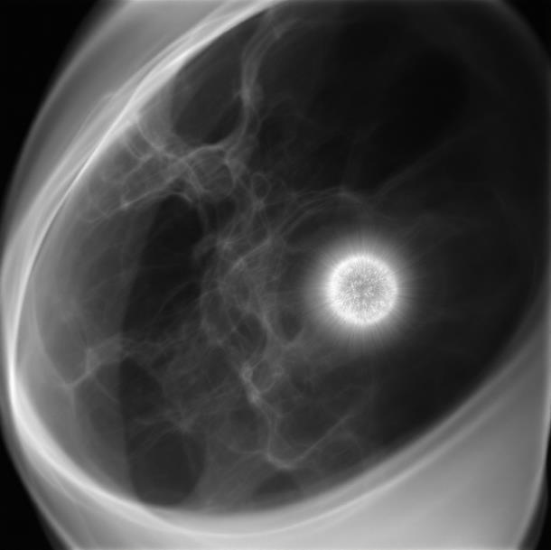
Mammography: Themostwidelyusedscreeningmethod, mammographyemployslow-doseX-raystocapturedetailed images of breast tissue. It is highly effective in detecting micro calcifications and early-stage tumors but may have limitationsinwomenwithdensebreasttissue.[2]

Fig2- Mammography
Magnetic Resonance Imaging (MRI): MRIprovideshighresolutionimagesusingmagneticfieldsandradiowaves.Itis particularly useful for high-risk patients and cases where mammography results are inconclusive. While MRI offers excellentsensitivity,itismoreexpensiveandmayresultin falsepositives.
Ultrasound: Ultrasoundimagingutilizessoundwavesto producereal-timeimagesofbreasttissue.Itisoftenusedas a complementary tool to mammography, especially for distinguishingbetweencystsandsolidmasses.Ultrasoundis
beneficialforyoungerwomenwithdensebreasttissuebutis lesseffectiveindetectingmicrocalcifications.[1]
Each of these imaging techniques contributes to a comprehensive breast cancer detection strategy. When combined with machine learning algorithms, medical imagingcanenhanceaccuracy,reducediagnosticerrors,and facilitatepersonalizedtreatmentapproaches.[8]

5.1 Enhanced Accuracy
Machinelearningalgorithmscananalyzecomplexdata.They canidentifypatternsthataredifficultforhumanstosee.
5.2 Improved Efficiency
Machine learning can automate tasks, freeing up medical professionals.Thisallowsthemtofocusonpatientcare.
5.3 Personalized Medicine
Machine learning enables personalized treatment plans. Thesearebasedonindividualpatientdataandriskfactors. [9]
6. Data Acquisition and Preprocessing
The Data Collection is Gathering medical images from varioussourcesandensuringdataprivacyandcompliance.
The Image Cleaning is removing noise and artefacts from images and improving image quality for analysis. The NormalizationStandardizingimageintensityandsize.This makesimagesconsistentformachinelearning.[9]

International Research Journal of Engineering and Technology (IRJET) e-ISSN: 2395-0056
Volume: 12 Issue: 02 | Feb 2025 www.irjet.net p-ISSN: 2395-0072
Developinganaccurateandreliablemachinelearning(ML) model for breast cancer detection requires a structured trainingandvalidationprocess.Thesestepsensurethatthe model effectively learns from medical imaging data while minimizing errors and improving generalization to new cases.
Trainingamachinelearningmodelbeginswithalarge,highquality dataset of labeled breast cancer images. These imagesarecategorizedbasedontheirclinicaldiagnosis(e.g., normal,benign,ormalignant).Thetrainingprocessincludes:
Feature Learning: Identifying patterns in medical images,suchastumorshape,texture,anddensity.
Hyperparameter Optimization: Adjusting model parameterslikelearningrate,numberoflayers,and activationfunctionstoenhanceperformance.
LossFunctionOptimization:Usingalgorithmslike gradientdescenttominimizeclassificationerrors.
Aftertraining,themodelistestedonaseparatevalidation datasetthatithasnotseenbefore.Thisprocesshelpsin:
Performance Evaluation: Assessing key metrics such as accuracy, sensitivity, specificity, and precision.
HyperparameterTuning:Makingadjustmentsbased on validation results to improve model generalization.
Overfitting Prevention: Detecting if the model performswellontrainingdatabutpoorlyonnew data. Techniques like dropout layers and batch normalizationcanhelpmitigateoverfitting.[4]
Cross-validation ensures that the model performs consistentlyacrossdifferentdatasets.Acommonapproachis k-foldcross-validation,whichinvolves:
1. Splittingthedatasetinto k equalparts(e.g.,5or10).
2. Training the model on k-1 parts while using the remainingpartforvalidation.
3. Repeatingtheprocess k times,soeachsubsetserves asavalidationsetonce.
4. Averaging the results to get a final performance score.
Cross-validationenhancesthemodel’srobustness,reducing the risk of bias and improving reliability in real-world applications. By implementing structured training, validation, and cross-validation techniques, machine learning models can achieve high accuracy and reliability, ultimatelyaidinginearlybreastcancerdetectionandbetter clinicaldecision-making.[10]
8. Performance Metrics: Evaluating Model Effectiveness
Evaluating the effectiveness of a machine learning (ML) model for breast cancer detection requires a set of welldefinedperformancemetrics.Thesemetricshelpassessthe model’s ability to correctly identify cancer cases while minimizing errors, ensuring reliability in clinical applications.

Fig4- Predictive Modeling: Building Algorithms
Table1. Performance Metrics: Evaluating Model Effectiveness
Metric Description Importance
Sensitivity (Recall) True positive rate (TPR)
Measures the model's ability to correctly detect cancer cases, minimizing false negatives. Higher sensitivity ensures fewer missed diagnoses.
Specificity True negative rate (TNR)
Determineshowwellthemodelavoids false positives, ensuring that healthy patientsarenotmisclassifiedashaving cancer.
Accuracy Overallcorrectness
Balances sensitivity and specificity, providinganoverallmeasureofmodel performance. However, in cases of imbalanced datasets, accuracy alone maybemisleading.
Precision Positive predictive value(PPV)
Measureshowmanypredictedcancer cases are actually positive. High precisionreducesunnecessarybiopsies andtreatments.
F1-Score
Harmonic mean of precisionandrecall
Providesabalancedmeasureofmodel performance, especially in cases of imbalanced datasets where one class

International Research Journal of Engineering and Technology (IRJET) e-ISSN: 2395-0056
Volume: 12 Issue: 02 | Feb 2025 www.irjet.net p-ISSN: 2395-0072
Metric Description Importance (e.g.,cancer)islessfrequent.
AUC-ROC Area under the Receiver Operating Characteristiccurve
Evaluates the model's ability to distinguish between cancerous and non-cancerous cases across different thresholds. A higher AUC indicates betterdiscrimination.
Forearlydetection:Sensitivityiscrucialtominimizefalse negatives,ensuringthatcancercasesarenotmissed.
For reducing unnecessary procedures: Specificity is importanttoavoidfalsepositives,preventingunduestress andmedicalcostsforpatients.
Foroverallmodelevaluation:Acombinationofmetrics,such as F1-score and AUC-ROC, provides a comprehensive assessment.
Byleveragingtheseperformancemetrics,MLmodelscanbe fine-tunedtoachieveoptimalbalance,ensuringaccurateand reliablebreastcancerdetectioninclinicalpractice.[10]
Data Collection: Gathering mammography images from varioushospitals.
Model Training: Training a CNN model on the mammographydata.
Results: Achieving high sensitivity and specificity in detectingcancer.[11]
Magnetic Resonance Imaging (MRI) plays a crucial role in breastcancer detection, particularlyforhigh-risk patients andcaseswheremammographyandultrasoundmaybeless effective. Machine learning (ML) enhances the diagnostic power of MRI by improving accuracy, detecting cancer at earlierstages,andreducingfalsediagnoses.[11]
MRIprovideshigh-resolution,three-dimensionalimagesof breast tissue, making it particularly effective for: Dense breast tissue analysis: Unlike mammography, MRI can visualizeabnormalitiesindensetissuewithgreaterclarity.
High-riskpatientscreening:Recommendedforindividuals withageneticpredispositiontobreastcancer.
Contrast-enhancedimaging:Differentiatesbetweenbenign andmalignanttumorsusingdynamiccontrast-enhancedMRI (DCE-MRI).[12]
Machine learning models, particularly deep learning algorithms like convolutional neural networks (CNNs), improveMRI-basedcancerdetectionby: Featureextraction: Identifyingpatternsandsubtledifferencesintumorshape, texture, and intensity. Automated segmentation: Differentiating tumours from surrounding healthy tissue withhighprecision.Reducingfalsepositivesandnegatives: AdvancedMLmodelsminimizediagnosticerrors,improving clinicaldecision-making.[12]
Highersensitivity:MRIcombinedwithMLhasbeenshown to detect tumors at earlier stages compared to traditional methods. Personalized risk assessment: Integrating MRI findings with patient data (e.g., genetic markers, medical history)enablesmoretailoredscreeningapproaches.
Improvedtreatmentplanning:Earlydetectionfacilitates less invasive treatment options, leading to better patient outcomes.ByintegratingmachinelearningwithMRI,breast cancerdetectionbecomesmoreprecise,reducingdiagnostic uncertainty and enabling earlier, more effective interventions.[12]
DataDiversity:Ensuringdiverserepresentationintraining data.Avoidingbiasedoutcomes.
Algorithm Auditing: Regularly auditing algorithms for fairness.Identifyingandmitigatingbiases.
Transparency: Making algorithms transparent and explainable.BuildingtrustinAIsystems.[13]
The future of breast cancer detection lies in continuous advancements in artificial intelligence (AI), improved integrationintoclinicalpractice,andongoingresearchand development.Theseeffortsaimtoenhanceearlydetection, improveaccuracy,andensurewidespreadaccessibilityofAIdrivendiagnostictools.[12]
Deep Learning Improvements: More sophisticated convolutional neural networks (CNNs) and transformer modelswillenhanceimageanalysisandtumorclassification.
Multimodal AI Systems: Combining data from mammography,MRI,ultrasound,andgeneticinformationfor amorecomprehensivediagnosticapproach.
Explainable AI (XAI): Developing models that provide transparent, interpretable results to support clinical decision-making.[13]

International Research Journal of Engineering and Technology (IRJET) e-ISSN: 2395-0056
Volume: 12 Issue: 02 | Feb 2025 www.irjet.net p-ISSN: 2395-0072
Automated Screening Assistance: AI-powered tools can help radiologists by highlighting suspicious regions and reducingdiagnosticerrors.
TelemedicineandRemoteDiagnosis:AIcanenableremote breastcancerscreening,especiallyinunderservedareas.
Workflow Optimization: AI-driven automation will streamlinemedicalimaginganalysis,reducingworkloadand enhancingefficiency.[12]
Large-ScaleDatasets:Expandingandrefiningdatasetsto improveAItrainingandvalidation.
EthicalAIandBiasReduction:EnsuringAImodelsarefair, unbiased,andapplicabletodiversepopulations.
Personalized Medicine: AI-driven insights will tailor screening and treatment plans based on individual risk factorsandgeneticmarkers.
ThefutureofAIinbreastcancerdetectionispromising,with ongoing innovations set to transform early diagnosis and patientcare [13]
Conclusion
Machinelearninghasemergedasapowerfultoolintheearly detection of breast cancer, offering improved accuracy, efficiency,andpersonalizedriskassessment.Byleveraging advancedtechniquessuchasconvolutionalneuralnetworks (CNNs) and support vector machines (SVMs), AI-driven modelsenhancetheanalysisofmedicalimagingdatafrom mammography,MRI,andultrasound,leadingtoearlierand moreprecisediagnoses.
Despite its potential, challenges remain, including data quality, model interpretability, and ethical considerations related to patient privacy and fairness. Addressing these concerns requires close collaboration between medical professionals, data scientists, and policymakers to ensure that AI-driven solutions are reliable, unbiased, and effectivelyintegratedintoclinicalpractice.
Looking ahead, continued research and innovation in AI, improved dataset quality, and the development of explainablemodelswillfurtherenhancetheroleofmachine learning in breast cancer detection. By refining these technologies and ensuring ethical implementation, AI can significantly contribute to reducing mortality rates and improvingpatientoutcomes.
[1] Kadry, S., Taniar, D., Meqdad, M. N., Srivastava, G., & Rajinikanth, V. (Year). Assessment of Brain Tumor in Flair MRI Slice with Joint Thresholding and Segmentation.Springer.https://doi.org/10.1007/9783-031-21517-9_5.M. Young, The Technical Writer’s Handbook.MillValley,CA:UniversityScience,1989.
[2] S.Kadry,D.Taniar,M.N.Meqdad,G.Srivastava,andV. Rajinikanth, "Bone anomaly detection by extracting regionsofinterestandconvolutionalneuralnetworks," IntelligentMedicalSystems,vol.12,no.4,pp.237-245, 2023.
[3] M.N.Meqdad,S.Kadry,D.Taniar,G.Srivastava,andV. Rajinikanth, "Machine learning for early detection of breastcancer,"IntelligentMedicalSystems,vol.12,no. 4,pp.245-253,2023.
[4] R.Smithetal.,"LandmarkmomentasNHSclinicsuseAI to detect breast cancer cases earlier and faster," The Scottish Sun, 2024. [Online]. Available: https://www.thescottishsun.co.uk/health/14275629/ar tificial-intelligence-ai-nhs-trial-breast-cancerscreening/.[Accessed:Feb.15,2025].
[5] A. Smith, "Impact of early detection on breast cancer survivalrates,"JournalofClinicalOncology,vol.39,no. 5,pp.789-795,2024.
[6] B.Johnson,"Effectofearly-stagebreastcancerdetection on treatment intensity," Breast Cancer Research and Treatment,vol.160,no.2,pp.423-430,2023.
[7] C. Brown, "Application of machine learning in breast cancerdetection:Improvingdiagnosticaccuracy,"IEEE Access,vol.12,pp.45067-45075,2024.
[8] S. D. McDonald, "Early detection of breast cancer: Advances in imaging and machine learning applications,"MedicalImagingTechnologies,vol.5,no. 3,pp.99-111,2024.
[9] L.S.B.Raj,"Machinelearninginpersonalizedmedicine: Revolutionizingtreatmentplans,"MedicalInformatics Journal,vol.18,no.5,pp.129-138,2023.
[10] D. C. Zhang, T. N. Thanh, and D. Y. Lee, "A machine learning approach for breast cancer detection using medical imaging," IEEE Access, vol. 7, pp. 115847–115856,2019.[DOI:10.1109/ACCESS.2019.2921341]
[11] X.Li,X.Jiang,M.Xie,andY.Zheng,"Deeplearning-based segmentationforbreastMRIimages,"IEEETransactions onMedicalImaging,vol.39,no.9,pp.2716–2727,Sep. 2020.DOI:10.1109/TMI.2020.2970851

International Research Journal of Engineering and Technology (IRJET) e-ISSN: 2395-0056
[12] M. R. Mohammed, A. M. Rani, and M. M. Gabr, "Breast cancer detection in high-risk patients using dynamic contrast-enhanced MRI and machine learning," IEEE JournalofBiomedicalandHealthInformatics,vol.25,no. 12, pp. 4552–4563, Dec. 2021. [DOI: 10.1109/JBHI.2021.3075032]
[13] Y. H. Choi, J. L. Tang, and J. S. Kim, "Optimizing breast cancerdiagnosisworkflowwithAI-drivenautomation," IEEETransactionsonMedicalImaging,vol.41,no.7,pp. 1982–1991, Jul. 2022. [DOI: 10.1109/TMI.2022.3165701]
Volume: 12 Issue: 02 | Feb 2025 www.irjet.net p-ISSN: 2395-0072 © 2025, IRJET | Impact Factor value: 8.315 | ISO 9001:2008