
Covid Detection Using Lung X-ray Images
1Dept. of Information Technology, Atharva College of Engineering, Maharashtra, Assistant of Information Technology, Atharva of Engineering,
Abstract - In the current times, automated disease detection has become a critical issue in the field of medical science because of the rapid population.Anautomateddisease detection model is required to assist the doctors in the diagnostics of disease and provide consistent, exact and fast results and help in reducing the death rate. This project tackles the problem of covid 19 detection by utilizing deep learning CNN (convolutional neural networks). A critical phase in the battle against COVID 19 is successfully screening contaminated patients. This project aims to detect COVID 19 patients using digital chest x ray pictures whileincreasingthe accuracy of detection using deep convolutional neural networks. Conventional testing is costlyandtakesalotoftime. This leads to excessive time and money expenses on the diagnosis. In this project, we have addressed both problems using the deep neural model. The dataset we’ve used is publicly available on the internet. We used a total of 5837 images for both training and testing our network, out of which 2837 Images were of Covid affected lungs and the remaining 3000 were of normal. Our model achieves a precision of 0.94, F1 score of 0.94, recall of 0.94 and accuracy of 0.94.
Key Words: Machine Learning, COVID 19, VGG16, Augmentation, X Ray, Convolutional Neural Network
1. INTRODUCTION
InrecenttimesCOVID 19hashadatremendouseffectonthe livesofindividualsglobally. Inthepast,there weremany such viruses but nothing as strong as this Coronavirus. Peoplesuredidnotanticipatefacingaviruslikethisinthe presentageoftechnology.Evaluatingthetimerequiredfor thediagnosticprocessandtheexpenseofthekitsneededto test cases, AI and deep learning based application was requiredtohelpsupportdoctorsworkingonconstraining thespreadofthevirusandtreatingpatientscombatingthe illness.
1.1 Motivation
The Covid 19 pandemic has been spreading rapidly throughout the world since last year. The rapid spread of Covid 19andtheintensityofthesymptomshaveclaimedthe COVID19pandemicquarantine.familylivesofnumeroushumans.Individualshavelostemployment,membersandfriends.IthasputsocietyinaOneoftheissueswearefacingwhilefightingthisisthelackofquickandaffordablediagnosis.diagnosiscantakealotoftimeandcanbe
expensiveforsomepeople.Inthisproject,weintendtouse Lung X Ray scans to identifyandclassifythem into COVID andnon COVIDimages.Thisreducesboththecostandtime takenforCOVID19diagnosis.
1.2 Aim
To build a deep learning model that could extract COVID 19'spictorialfeaturesfromtheX rayimageofthelungs,thus savingcrucialtimefordiseasecontrol.Accuracyisthemost significant factor in this problem, so by taking a huge number of pictures for training the network and by increasing the number of iterations, the model's accuracy canbeimproved.
1.3 Scope
The scope of this project is to classify X ray images and distinguishthemasCovidpositiveorNormalusingmachine learningalgorithmsbasedonimagedatasets.Thedatasetof this work has been collected from the Kaggle repository, whichhasbeencollectedfrommultiplesourcesandcontains Chest X Ray scans of Covid 19 affected and normal lungs. Thiscollected dataset is notmeant to claim the diagnostic abilityofanyDeepLearningmodelbuttoresearchvarious possiblewaysofefficientlydetectingCoronavirusinfections usingcomputervisiontechniques
2. LITERATURE REVIEW

Inthispaper[1],theun augmenteddatasetwhichwasused hadachievedanaverageF measureof95%meanwhilethe augmented data set achieved a score of 99%. The experiment initially contained only 128 images, on which theyperformedaugmentationandextendedittoabout1000 images. Paper [2] brought up an interesting observation, where the researchers found out that lung x ray images providedbetteraccuracyincomparisontoCTscanimages usingthesamealgorithm.Withthelimiteddatasetthatthey had at hand, using the VGG19 model they attained 86% accuracyforX rayimagesand84%accuracyforCT scans. CNNmodelscanstruggletoprovideconsistentresultsifthe datasetsaretoosmall.
Theresearchersfor[3]createdatemporaryfixforthelack ofdatabyimplementingtransferlearningintheirproject.In thispaper,multiplemodelshadbeenused,withVGG19and Mobilenet v2 providing the best accuracy. Even though
VGG19 provided a higher accuracy, Mobilenet v2 was selected as the better model due to the lesser number of falsenegativesachievedforthedetectionofCOVID.Afalse negative means that a person carrying COVID has been diagnosedasnormal,whichmaybepotentiallyharmful.
[4]usesaResNet 50modelonarelativelysmalldataset.The algorithmcanbeeasilyre trainedwithnewsetsoflabelled imagestoenhancetheperformancefurther.Inpaper[5],a combined deep CNN LSTM network was developed. The samplesizeisrelativelysmallandneedstobeincreasedin sizetotestthegeneralizabilityofthedevelopedsystem.The CNNarchitectureachieved95.3%AUC.Theresearchersfor [6] employed domain extension transfer learning (DETL) withpre trainedCNNsandusedfivefoldcross validationto estimatethefeasibilityoftheuseofchestX Raystodiagnose COVID 19.Theoverallaccuracywasmeasuredtobe90.13% ±0.14.
3. METHODOLOGY
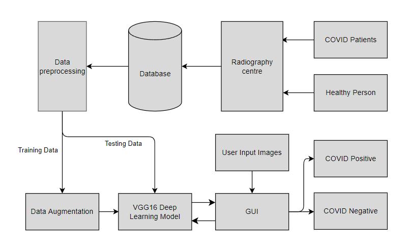
Fig-1:SystemDesign
The proposed method used in this project uses Deep learning algorithm VGG 16 The detection of COVID 19 requiresdifferentstages,asshowninFigure1.Theoriginal X ray image is preprocessed, including size adjustment, widthshift,heightshift,rotation,andscalingandhorizontal flippingoftheimages.Thedatasetisthensplitintotraining andvalidation(test)sets.Thepreprocesseddataisusedto extract the image features from the X ray images. The performance of the method is assessed by indices such as accuracy,recallrate,precision,andF1 score.
3.1 VGGAlgorithm16:
VGG16 is a convolution neural net (CNN) model that was usedtowinILSVR(Imagenet)contestin2014.Itisbelieved tobeoneofthebestvisionmodelarchitecturestodate.The most extraordinary thing about VGG16 is that instead of havingalargenumberofhyper parameterstheyfocusedon havingconvolutionlayersofa3x3filterwithastride1and
alwaysusedthesamepaddingandmaxpoollayerofa2x2 filter of stride 2. It follows this system of convolution and maxpoollayersconsistentlythroughoutthewholemodel. Finally, it has 2 FC(fully connected layers) followed by a softmax for output. The 16 in VGG16 points to the model having16layersthathaveweights.Thisnetworkispretty largeandithasabout138million(approx.)parameters.
4. DATASET
This project is using chest X ray images that are collected from different publicly accessible datasets, online sources and published papers. This dataset is divided into two categoriesCovidandNormal.[7][8][9]
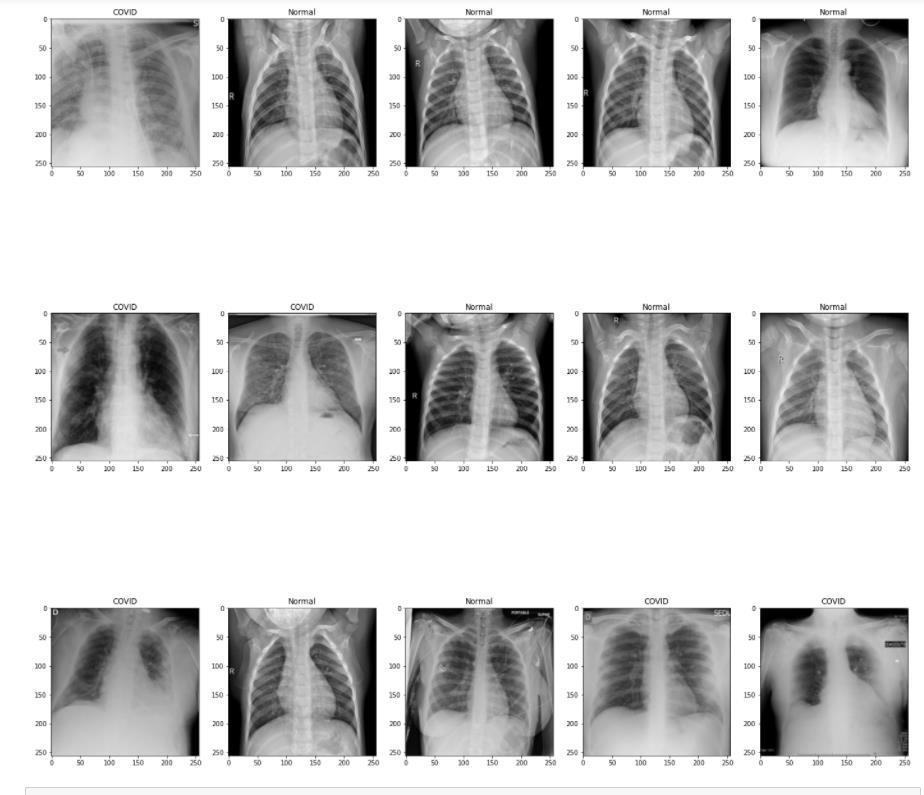
Fig 2: COVIDinfectedandNormalX Rays
Specificationsofthedataset:
Covid TheCoviddatacontainsatotalof3,615images 2,473CXR imagesarecollectedfromthepadchest dataset
183CXRimagesfromaGermanmedicalschool. 559 CXR image from Github, Tweeter, Kaggle and SIRM
400CXRimagesfromanotherGithubsource.
Normal TheNormaldatacontainsatotalof10,192images arecollectedfromtwodifferentdatasets.
8851RSNA 1341Kaggle
5. IMPLEMENTATION
Theimplementationofthisprojectisthecombinationofthe followingsegments:Loadingimagedata,splittingofthedata, dataaugmentation,Trainingofthemodel,Evaluationofthe modelandlastlytheGraphicalUserInterface(GUI).
A. LOADING IMAGE DATA
Firstly,themodelcollectsthepathsofeachoftheimageof the dataset folder, then it stores the image data into a variable,thenitisensuredthattheimagesareaccordingto ourspecifications i.e.all imagesare of thesamecolor and sameinputsize.Normalizingisthenperformedontheimage data.Lastlylabelbinarizeriscalleduponthelabeltoconvert theminto0or1accordingtothelabel.
B. SPLITTING OF THE DATA
Theprocedurewhichistermedtest train splitisemployed to estimate the fulfillment of the machine learning algorithmsoncetheyarerequiredtomakepredictions.20% ofthedatasetisreservedfortestingandtheremaining80% ofthedataisfurthersplitintotrainingdataandvalidation dataintheratioof80:20respectively.
C. DATA AUGMENTATION
Indataanalysis,dataaugmentationreferstoapproachesfor increasing the quantity of data by adding slightly changed copiesofcurrentdata orcreatingnewsynthetic datafrom existing data. When training a machine learning model, it functionsasaregularizerandhelpstominimizeoverfitting Inaugmentationwechangedtheparametersoftheimages suchasheight,rotation,horizontal flipandwidth.
D. TRAINING OF THE MODEL
The model is trained with the augmented dataset and is validatedaftereachepoch.Thismodelusesanearlystopping callback because if the model’s validation accuracy stops improvingwithinthenext5epochs,itwillrevertbacktothe epochwiththebestvalidationaccuracy. Thissavestimeand resourceswhiletrainingofthemodel.
6. EVALUATION OF THE MODEL
Theperformanceofaclassificationalgorithmisshownand summarizedusingaconfusionmatrix.Themodelwasableto predictasfollows:Truepositive 575,Truenegative 526, Falsepositives 25andFalsenegative 42
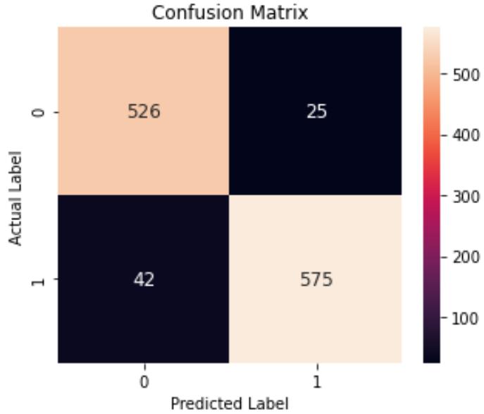
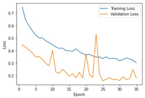
Fig-5: VGG16TrainingandValidationloss
Themodelwasevaluatedonthebasisofparameterssuchas precision,recall,F1score,andsupport.Themodelwasable to achieve the accuracy of 0.9426, sensitivity of 0.9546, specificityof0.9319.
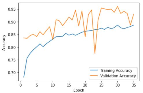
Fig-3:ConfusionMatrix Fig 4: VGG16TrainingandValidationaccuracyTable
7. GRAPHICAL USER INTERFACE (GUI)
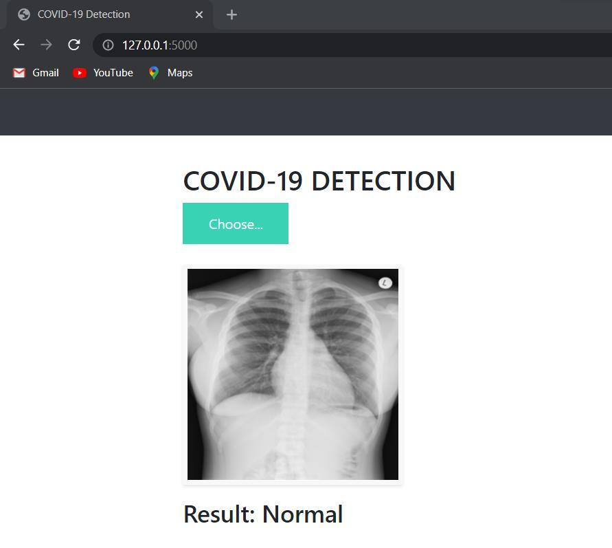
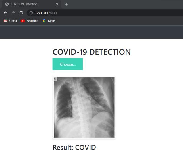
A graphical user interface (GUI) allows people to interact withthesystem.Userswillprimarilybegiventhechoiceto pickafilefromwhichtosubmittheirlungX Rayimage.
On uploading the image model will process the imageand givethepredictionof‘COVID’ ‘Normal’accordingly.

Fig
Fig 6: Interface
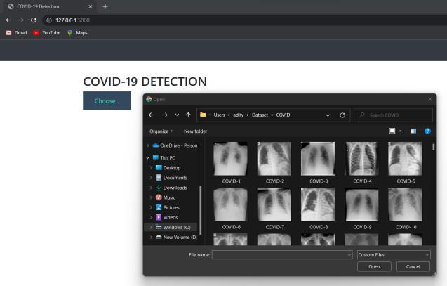
On clickingthe ‘Choose’ button, the user will be prompted withtheirrespectivefilemanagertoselectanduploadthe lungX Ray. theLung Ray
Fig-9: Predicted
8. CONCLUSION
The concept of a deep learning model approach to the augmenteddatasetforCOVID 19detectionispresentedin this research. Our active experimentation is the effective implementationofthesystem,evaluationparameters,and GUIexecution COVID 19wasdetectedutilizingchestX ray picturesusingadiagnosticalgorithmbasedonVGG16.With the use of an augmented dataset, the model was able to detect COVID 19 quickly and reliably, with an F1 score of 0.94
International Engineering Technology (IRJET) ISSN: 2395 0056
Volume: 09 Issue: 04 | Apr 2022 www.irjet.net ISSN: 2395 0072
9. REFERENCES
[1] Alazab,M.,Awajan,A.,Mesleh,A.,Abraham,A.,Jatana,V. andAlhyari,S.,2020.COVID 19predictionanddetection usingdeeplearning.InternationalJournalofComputer Information Systems and Industrial Management Applications,12,pp.168 181.
[2] Horry,M.J.,Chakraborty,S.,Paul,M.,Ulhaq,A.,Pradhan, B.,Saha,M.,and Shukla,N., 2020.COVID 19 detection through transfer learning using multimodal imaging data.IEEEAccess,8,pp.149808 149824.
[3] Apostolopoulos,I.D.,Mpesiana,T.A.Covid 19:automatic detectionfromX rayimagesutilizingtransferlearning withconvolutionalneuralnetworks.PhysEngSciMed 43,635 640(2020).https://doi.org/10.1007/s13246 020 00865 4
[4] DeepLearningforMedicalImaging:COVID 19Detection PostedbyJohannaPingel,March18,2020[BLOG].
[5] Islam,M.Z.,Islam,M.M.andAsraf,A.,2020.Acombined deep CNN LSTM network for the detection of novel coronavirus(COVID 19)usingX rayimages.Informatics inmedicineunlocked,20,p.100412.
[6] Basu, S., Mitra, S. and Saha,N., 2020, December. Deep learning for screening covid 19 using chest x ray images. In 2020 IEEE Symposium Series on Computational Intelligence (SSCI) (pp. 2521 2527). IEEE.
[7] Ozturk,T.,Talo,M.,Yildirim,E.A.,Baloglu,U.B.,Yildirim, O. and Acharya, U.R., 2020. Automated detection of COVID 19casesusingdeepneuralnetworkswithX ray images. Computers in biology and medicine, 121, p.103792.
[8] Hussain,E.,Hasan,M.,Rahman,M.A.,Lee,I.,Tamanna,T. andParvez,M.Z.,2021.CoroDet:Adeeplearningbased classificationforCOVID 19detectionusingchestX ray images.Chaos,Solitons&Fractals,142,p.110495.
[9] Rahman, T., Khandakar, A., Qiblawey, Y., Tahir, A., Kiranyaz,S.,Kashem,S.B.A.,Islam,M.T.,AlMaadeed,S., Zughaier,S.M.,Khan,M.S.andChowdhury,M.E.,2021. Exploringtheeffectofimageenhancementtechniques on COVID 19 detection using chest X ray images. Computersinbiologyandmedicine,132,p.104319.
[10] Poojary, R., Raina, R. and Mondal, A.K., 2021. Effect of data augmentation on fine tuned CNN model performance. IAES International Journal of Artificial Intelligence,10(1),p.84.