
Wewouldgatherimagesfromuser'sdevicesviacameraor system storage and identify skin cancer diseases like melanomatotheirclosestpredictioninthisapplication.It would give a description and symptoms of the condition aftersuccessfulidentification.Theusercouldalsolookup thecontactinformationfornearbydoctors. Itwouldalsoprovidechartdataaswellasapproximations of other nearby conditions via a user friendly interface, allowingconsumerstobedirectedtospecific skindisease results.
Skin Cancer Detection Application
International Research Journal of Engineering and Technology (IRJET) e ISSN: 2395 0056 Volume: 09 Issue: 03 | Mar 2022 www.irjet.net p ISSN: 2395 0072 © 2022, IRJET | Impact Factor value: 7.529 | ISO 9001:2008 Certified

Key Words: Melanoma, TensorFlow, Dermatology, Image Processing,TransferLearning, 1. INTRODUCTION
In the age of apparatuses, machine learning is finding its applicationinallfieldsandclinicaldiagnosisisthenewest calculation to it. . Emerging machine learning (ML) based analytical tools are exciting to build, but they must be chosen carefully. To improve diagnostic accuracy, choose the correct decision making system. Face identification, fingerprint recognition, tumour detection, and segmentation are just a few of the applications of image processing and machine learning. Fungal infection, bacteria, allergies, and viruses, among other things, can causeskinproblems.
1.2 BASIC CONCEPT
1.1 MOTIVATION OF PROJECT
Sheha, Mariam A. On a collection of dermoscopy photos, Mai S.Mabrouk and Amr Sharawy [1] proposed an automated method for melanoma diagnosis. So, while the first exercise, Automatic iteration counter, is faster, the secondpractise,Defaultiterationcounter,ismoreprecise, with a precision of 100% for the training set and 92 percentforthetestset. JonathanBlackledgeetal.[2]proposedtheMoletestonline skingrowthscreeningframework. The half examination of a photograph in terms of its fractal structureand thefractal qualitiesthatcharacterise it.
Tushar Gharge [1], Narayan Gupta [2], Krutik Raut [3], Prof. Varsha Salunkhe [4] [1], [2], [3]Student, Department of Computer Engineering, Atharva College of Engineering, Mumbai [4]Professor, Department of Computer Engineering, Atharva College of Engineering, Mumbai ***
Abstract
The project's purpose is to create and test robust, automated textural feature extraction and selection algorithms, as well as learning algorithms for identifying pigmented skin lesions, in order to improve skin cancer diagnosis in primary care settings. The goal of this initiativeistoenhancethedetectionandtreatmentofskin cancer. One of the project's main objectives is to develop aninteractivesupporttool witha simpleuserinterface to assist primary care practitioners in accurately screening pigmented skin lesions in order to improve early detection, reduce unnecessary hospital referrals, and reducethenumberofmalignantmelanomasmissed.
Journal | Page350
The Project Entitled As “Skin Cancer Detection Application”. Skin cancer is regarded as one of the most dangerous types of cancer seen in humans. Every year, tens of thousands of people in the United States are diagnosed with cancer. As the disease spreads, the rate of survival drops dramatically. Melanoma, Basal and Squamous cell Carcinoma are the most common types of skin cancer, with Melanoma being the most unpredictable. Early detection and identification of the type of cancer can aid in its cure Many technologies have demonstrated that AI and computer science can play a key role in picture scanning. We describe a mobile technique for detecting Melanoma Skin Cancer utilising Image Processing and basic portable equipment in this technical study. The cancer image is fed into the system, which analyses it using unique image processing algorithms to determine the existence of skin cancer. The cancer picture analysis software looks for asymmetry, border, colour, diameter, and other Melanoma properties, as well as size, frequency, and form, to identify the image and feature phases. The scanned feature parameters are used to distinguish between normal skin andmelanomacancer lesions inthe image.
Theimpactoftheproposedprojectistwofoldsignificantly improved understanding of correspondence between automatedextractedfeaturesTheABCDruleofthumband computerisedautomaticidentification(suchassize,shape, colour, and texture information) physicians used to pre screening the early skin cancer; and advances integration of feature extraction and classification in bio medical imaging processing. The overarching goal is to provide transparent support for skin cancer screening by primary care physicians based on advanced bio medical image processinganddecision makingtechniques.
The primary motivation that drives the project is to use theadvancedimageclassificationtechnology for the well being of the people. Computer vision has made good progress in machine learning and deep learning that are scalable across domains. The model's improved accuracy and efficiency may help detect melanoma in its early stagesandpreventunneededbiopsies.
2. BACKGROUND STUDY
et al. [8] proposed an automated approach with typical and anomalous classes for skin cancer identification. The great division execution is accomplishedbyactivecontoursegmentation.Inanorder approach with two classifications (threatening and favorablesores),anaffectabilityof90%,precisionof95% andaspecificityof85%iswatched. 3.
We start by gathering the best dataset containing photographsofskinconditions.Westartedbylookingfor melanoma relatedphotosonKaggleanddataverse.While searching for skin disorders, there were numerous datasets with a large number of good photographs and models, so we choose the one with the most likes or recommends. We discovered Kaggle's SIIM ISIC Melanoma Classification, which included over ten GB of data, and Harvard's data verse HAM10000 images part1 & HAM10000 images part 2 dataset image when searching for the top datasets We chose these two datasetsbecauseoftheenormousnumberofdatasetsand recommendations. Normal and day to day living duties, educators, and psychiatrists might benefit from knowing the distinction between benign and malignant types. Choosing the best photographs from the two datasets, merging them, and categorizing them. Data was framed and stored in csv files from the obtained photos and datasets.
et al. [7] suggested an ingenious and entirely practical advanced mobile phone based programme for determining the type of early disease and its prevention. The first segment is a constant alarm to provide customers with help in preventing skin smoulder caused by sunshine, and the second segment is a new counted statement to know the most of the possibility for skintopointsispresentedalongtheselines.Theproposed framework is effective, according to the output data, groupingtypical,atypical,andmelanomaimageswith96.3 percent, 95.7 percent, and 97.5 percent accuracy, V.respectively.JeyaRamya
3.2 DATA CLEANING AND PREPROCESSING
Capdehouratetal.[5]fromGermanysuggestedamachine learning method for dealing with order melanocytic lesionsinhazardousandgenerous dermatoscopicimages. There are 433 benign sores and 80 benign lesions in the image database. Following a step of image pre processing that includes hair evacuation filtering, each image is then fractured using well known image segmentation techniques.YogendraKumarJainetal. [6]focusesonthedevelopmentofaskincancerscreening frameworkthatmaybeusedaspartofroutinepractiseby non specialists to distinguish between normal and abnormal instances. Highlight Discovery and Order Procedurearetwostepsin theadvancementprocess.The components are separated using wavelet change to decompose images into multiple recurring sub groups. As a result, the characterization framework is based on the use of a Probabilistic Neural System and a Grouping OmarClassifier.Abuzaghleh
The "ELM 7 point agenda," developed by G. Di Leo et al. [4], is a new analytic approach that describes an arrangement of seven components, including texture parameters, that convey the malignancy of a lesion. It has been shown to be faster and more precise than the traditionalABCDcriteriainthedetectionofmelanoma.
3.1 DATA COLLECTION
Maintainingthedataisveryimportantsothatbaddata doesnot coincide with the training and modelling of our main and working model. We focus to pull out the important datathat is required for the process. Before
© 2022, IRJET | Impact Factor value: 7.529 | ISO 9001:2008 Certified Journal | Page351
So, which of these two perspectives, or a combination of both,hasbeenusedtodescribeahandlingandimagetest engine that is unique in its customary technique and entirelynonexclusiveintermsoftheapplicationstowhich itmaybeconnected.
Inlightofformdescriptors,MessadiM.etal.[3]developed an interpretable order technique for skin cancers in dermoscopicimages.Theirresearchdemonstratestheuse of a fuzzy rule based classifier to distinguish between melanoma and other cancers. A flexible Neuro Fuzzy inference System (ANFIS) has a specific purpose in mind: to uncover the fuzzy rules that prompt the correct classification. The technique used in previous stages of developmentwastoreducetheimpactofsmallstructures, hairs, bubbles, and light refraction. In the second step, an unsupervised lesion segmentation algorithm is proposed. In a transparent way, repeating thresholding is linked to the instate level specified. They've also addressed the requirementtofocusalltheascribesemployedtoaddtoa depiction strategy that enables professionals to make the bestdecision.
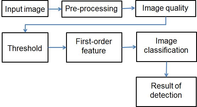
IMPLEMENTATIONFig1
:SystemArchitecture
International Research Journal of Engineering and Technology (IRJET) e ISSN: 2395 0056 Volume: 09 Issue: 03 | Mar 2022 www.irjet.net p ISSN: 2395 0072
Each image must be unique and clear in way of visibility, while maintaining it’s original dimensions and data. For even better results, .dcm files: DICOM files were also added. Most of the images were saved in the "Digitals Image and Communication in Medicine" format. It contains an image from a medical scan, such as an ultrasound or MRI + information about the patient. In ordertopreparethesetsforourclassifierpre processing of these images was required. This includes proper lighting, removing any other visual discrepancies. After thedatawaspre processedandcleaned,itwasaddedinto ourcolumninofourcsvfiles.
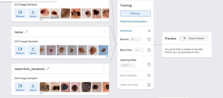
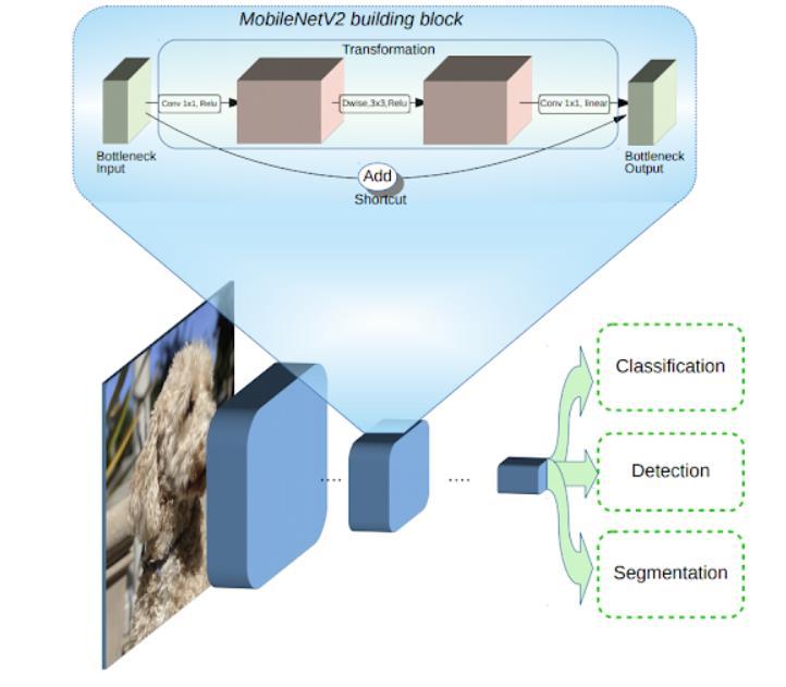
Fig 3:MobileNet v2
3.3
DATA MODELLING
Inthecontextofourproject for training model, we would use the teachable machine’s image classification to train themodelforeaseofhavingbetteraccuracyandresult.We woulddothefollowingsteps: Added the cleaned and preprocessed dataset for classificationasgroupofclassesbasedonthediseasename of the respective data. After all the data was uploaded, it wasreadyforthenextprocessoftraining. Fortrainingthemodel withnearestaccuracypossible, we tried to adjust the models’ parameters of epochs (for numberoftimesdatasetfedfortraining),batchsize(setof samplesfedineachiteration),learningrate(ratetoadjust themodelsunderstanding)accordingly.
The optimized model scored well on our test set, scoring an AUC score of 0.65 ~ 0.75. We would try proceed to know our model a bit better before making final estimationandrecommendations.
© 2022, IRJET | Impact Factor value: 7.529 | ISO 9001:2008 Certified Journal | Page352 beginningwiththecleaning,weconcatenatedthetwosets and added a specialcolumnto indicate the image isfrom whichtypeofcanceritis.Thefirststepincleaningwasto reducethenumberofunnecessarycolumnsfromoutdata framedsets.Mostofthesetshadaboutnumerousamounts of columns, of which only few we considered were important. This included the title of the disease, date of thecontent,resolutionofthecontent, andtheprobability orconfidencecolumnthatweadded.
International Research Journal of Engineering and Technology (IRJET) e ISSN: 2395 0056 Volume: 09 Issue: 03 | Mar 2022 www.irjet.net p ISSN: 2395 0072
To decide on training model, we used TensorFlow lite transfer learning with MobileNet v2 architecture model. training a TensorFlow Lite model with your own custom datasetdecreasesthenumberoftraineddatarequiredand will shorten the overall time So, the pre trained neural networkwouldhelpyoutocreateyourownclasses,andso youcansortofpicturethatyourclassarebecomingthelast layerorstepofourneuralnet. MobileNet v2 is a CNN that is 53 layers deep. People can load a pretrained version of the network trained on more than a million images from the ImageNet database. The pre trainedCNN canclassifyimagesinto mostofthe real time objects categories, such as keyboard, mouse, pencil,andmanytestdata Suchkindofresultisthewhich hasthenetworkandlearnedrichfeaturerepresentations forawiderangeofimages.Theoutputhasanimageinput Fig 2:Trainingmodel sizeof224 by 224.
So, after going multiple trial and run cases, we achieved our accuracy highest accuracy at epochs of 89, batch size of 128 and learning rate of 0.0001. After working our model onmultiplepermutationsoftheabove parameters, we saved the result match and tried to deploy it for the testingstage.
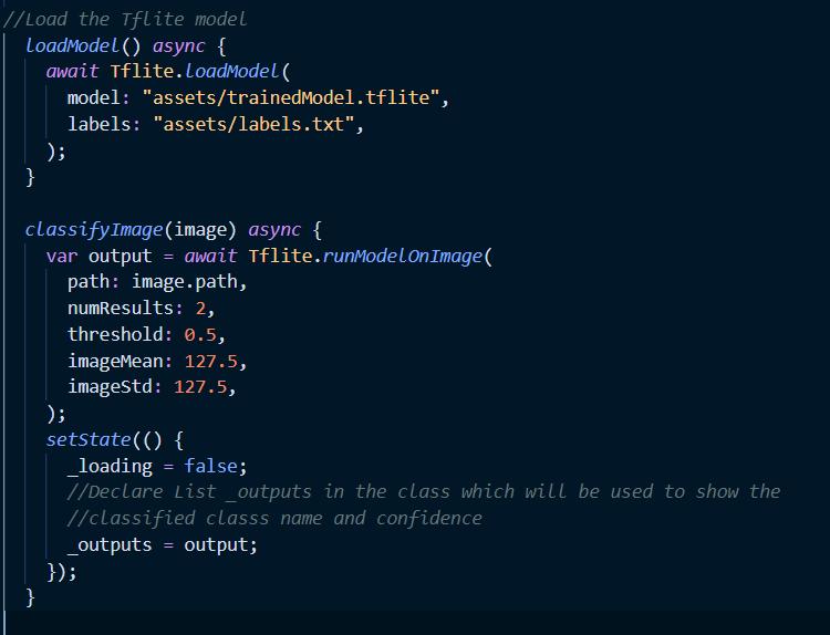
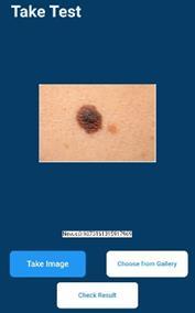
The final phase was to load and test whether our trained modelsubmittedonrealtimedevicesworksaccuratelyor not. After training, the trained model was converted into tflite quantizedmodel astheextensionof.tflite. Afterthe model was converted, it was loaded and integrated into Fluttercodeandwasmadetorunontheliveimagesfrom the devices. The model was made to run on threshold of 0.5,imagemeanandstandardof127.5.
4. LOADING AND TESTING
Fig-5:Testingonrealtimeimages
Our gives average of 70.6 percent accuracy when looking atregularentries(i.e.isnotincludingthevarianceofother diseasesoutsideofmelanoma). Final the level ofaccuracy means that the model might have the potential to be generalizeforusageindaytodaylikeschools,ruralareas and even for just verifying the disease for anyone who downloadtheapplication.
At last, the modelling and testing phase we can conclude that our classifier has an above average rating in terms of beingcorrectandprovidingaccurateresults.Thismeansin thefuturesucha systemcanbeimplementedtocheck ifa person or patient has been experiencing any kind of melanoma cancersthat might lead to possible even bigger problems.Suchasystemcanalsobeimplementedonother web applications, IOS & android devices as well as embedded devices. In the end our model was able to achieve close to 70 percent rate of success, with more research and studies this rate can be increased even furtherandprovidemoreaccurateanddependableresults. Themainissueoftheworkwasthatalotofpre processing and collecting the data, which was required even on the sets on which the models were tested on to provide near results. While training more and more of our model from, datasetswewouldbeabletoknowhowouroutputwould beabletochangeindifferentscenarios
International Research Journal of Engineering and Technology (IRJET) e ISSN: 2395 0056 Volume: 09 Issue: 03 | Mar 2022 www.irjet.net p ISSN: 2395 0072
Testing was done on many real time skin cancer diseases of melanoma from many numbers of individuals and patients.Weachievedtheaccuracyof0.60~0.80approx. The images taken with better illumination and high resolutions showed the higher accuracy of around 0.70 and above, while the images having lower pixels or resolutions and less illumination had little lower result range in between of 0.40 and 0.60. Overall, the model worked accurately from the portable device like mobile trained from the MobileNet v2 architecture. Response time was very quick and was able to identify the diseases eveninthelow enddevices.
5. RESULTS
© 2022, IRJET | Impact Factor value: 7.529 | ISO 9001:2008 Certified Journal | Page353
Fig 4:Loadingmodel
8. CONCLUSION
We owe sincere thanks to our college Atharva College of Engineering for giving us a platform to prepare a project onthetopic"SkincancerDetectionapplication"andwould like to thank our principal Dr. Shrikant Kallurkar for instigatingwithinus theneedforthisresearchandgiving us the opportunities and time to conduct and present researchonthetopic.Wearesincerelygratefulforhaving Prof. Varsha Salunkhe as our guide and Dr. Suvarna Pansambal, Head of Computer Engineering Department, and our project coordinators during our research. So, at last, work of this research wouldn’t have been impossible withoutthecooperationofallthemembers
7. FUTURE SCOPE
This idea and approach that we have implemented does notendhere.Therecanbeafuturescopefordevelopment we can extend this project into multiple platforms.
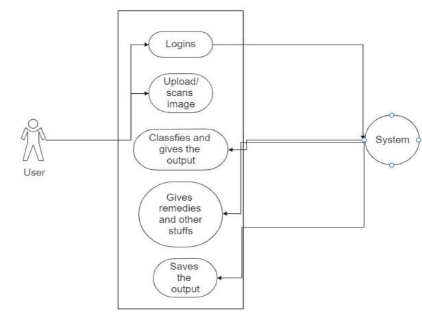
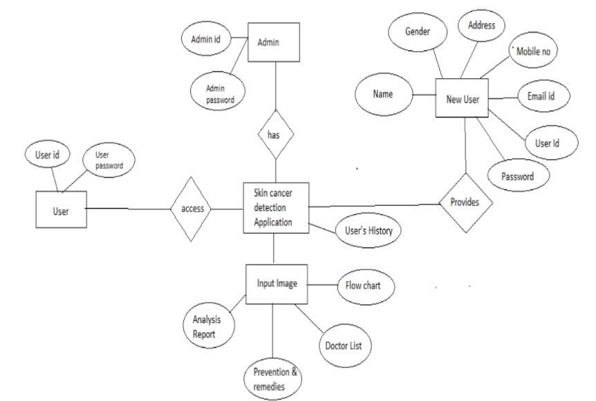
International Research Journal of Engineering and Technology (IRJET) e ISSN: 2395 0056 Volume: 09 Issue: 03 | Mar 2022 www.irjet.net p ISSN: 2395 0072 © 2022, IRJET | Impact Factor value: 7.529 | ISO 9001:2008 Certified Journal | Page354 6. DESIGN DETAIL 6.1 E R DIAGRAM Fig-6.1: E Rdiagram 6.2 USE CASE DIAGRAMFig6.2:Usecasediagram
9. ACKNOWLEDGEMENT
Particularly, we can incorporate additional algorithm togethertomakeismoreappealing. One other scope of improvement in expanding the idea hidingthedata intoa videoformatsothattheapplication canbemoresecuresothatdifferentsectorcanusethisto secure the way of communication between the user and themself. As specially, since the sensitive data can be storedwithoutanyoneknowingaboutit.Suchthatstoring sensitivedatawon’tbeaproblemitasweaddhigherlevel ofsecurity.
Skin cancer detection is very hard to for most of the people to detect without visiting doctors Main project of our diagnosis requires experience of data, in which early stagesmaylookidenticalto benign Detectingthedisease withsuch toolforless experiencephysicians. Ourmodel's project is an automated melanoma classification system thatwasappliedtodermoscopypicturestoaidintheearly diagnosis of malignant melanoma and melanocytic nevi lesions. With main advantage that it contrasts with other methods in medical image analysis segmentation process is avoided using mobile analysis. In this project we had created a mobile application to classify skin cancer. This method consists of data classification, image processing andimageclassificationusingMobilenetmodel.Usingthis technique,wecanclassifyimagesatnearestprobability.
10. REFERENCES
[1] Automatic Detection of Melanoma Skin Cancer Using Texture Analysis, International Journal of Computer Applications (0975 8887) Volume 42 No.20, March 2012. Mariam A.Sheha, Mai S.Mabrouk, Automatic Detection of Melanoma Skin Cancer Using Texture Analysis, International Journal of Computer Applications (0975 8887) Volume 42 No.20, March 2012.
[4] Automatic Diagnosis of Melanoma: a Software System Based on the 7 Point Check List, Proceedings of the 43rd Hawaii International Conference on System Sciences,IEEE,2010.G.DiLeo,A.Paolillo,P.Sommella, andG.Fabbrocini,AutomaticDiagnosisofMelanoma:a Software System Based on the 7 Point Check List, Proceedings of the 43rd Hawaii International ConferenceonSystemSciences,IEEE,2010. [5] German Capdehourat, Andres Corez, Anabella Bazzano, and Pablo Mus, Pigmented Skin Lesions Classification Using Dermatoscopic Images, Springer Verlag Berlin Heidelberg,2009,pp.537 544
[6] Yogendra Kumar Jain, Comparison between Different ClassificationMethodswithApplicationtoSkinCancer, InternationalJournalofComputerApplications(0975 8887)Volume53 No.11,September2012 [7] Dr. J. Abdul Jaleel, Artificial Neural Network Based Detection of Skin Cancer, International Journal of Advanced Research in Electrical, Elcetronics and InstrumentationEngineering,Col.1,Issue3,2012
International Research Journal of Engineering and Technology (IRJET) e ISSN: 2395 0056 Volume: 09 Issue: 03 | Mar 2022 www.irjet.net p ISSN: 2395 0072
[8] DETECTION OF MELANOMA SKIN CANCER USING DIGITAL CAMERA IMAGES, ARPN Journal of Engineering and Applied Sciences, VOL. 10, NO. 7, APRIL 2015. V. Jeya Ramya, J. Navarajan, R. Prathipa, andL.AshokKumar,DETECTIONOFMELANOMASKIN CANCER USING DIGITAL CAMERA IMAGES, ARPN JournalofEngineeringandAppliedSciences,
[3]Ahybridmachine learningapproachforsegmentationof protein localization data, Oxford University Press, Vol. 21 no. 19 2005, pages 3778 3786. Peter M. Kasson1, JohannesB.Huppa,MarkM.Davis,andAxelT.Brunger, A hybrid machine learning approach for segmentation of protein localization data, Oxford University Press, Vol.21no.192005,pages3778 3786.
© 2022, IRJET | Impact Factor value: 7.529 | ISO 9001:2008 Certified Journal | Page355
[2] Moletest: A Web based Skin Cancer Screening System, Dublin Institute of Technology, 2011. ] Jonathan Blackledge, Moletest: A Web based Skin Cancer Screening System, Dublin Institute of Technology, 2011.