2Assistant Professor, Dept. of Information Technology, Atharva College of Engineering, Maharashtra, India ***
International Research Journal of Engineering and Technology (IRJET) e ISSN: 2395 0056
Many cancer cases early are misdiagnosed, resulting in severe consequences, including the patient's death. In this project, both the above problems are addressedusing deep neuralnetworks andtransferlearning architecture. This paper proposes a novel system automatically detecting and classifying Skin Diseases into seven classes. Publicly available HAM10000 databases provided by Harvard were used to train and test the model. First, the proposed system uses the U Net Model for SemanticImageSegmentationfollowedbyastate of the art Transfer Learning InceptionV3 model. The U Net predefined model produced a validation accuracy of 94.88%. The masked image predicted from this model is then merged with the original image, converting it into a disease focused image. The proposed system then extracts 52 features from the state of the art predefined InceptionV3 transfer learning model and combines them with other metadata (CSV file features). Resampling techniques are considered for balancing the dataset, producing 4,200 images contributed from 7 disease classes equally. In the last stage, the system uses an ensemble model combination of metadata and features extracted from InceptionV3 and XgBoost Classifier to predict seven classes based on 74 features. Our model achieves an accuracy of 0.95, precision 0.95, recall 0.95, F1 score 0.95, and ROC AUC 0.99, which is better than the previous state of the art approaches. The individual class f1 scores were: akiec 0.96, bcc 0.96, bkl 0.93,df 1.00,mel 0.92,nv 0.90,vasc 0.99.
Bhavya Bipin Gada1, Shivangkumar Gandhi1, Pruthvi Rathod1, Amruta Sankhe2
In [1], fully Convolutional networks like InceptionV3 and MobileNet were used as standard classification transfer learning techniques on the MNIST HAM10000 dataset to detect skin diseases which achieved an accuracy of 72% with InceptionV3 and 58% with MobileNet. Dermnet research dataset was accumulated in [2], which produced 72.2%averagedataaccuracyusingAlexNetConvolutional Network. The segmentation and classification approach wasusedin[3]onISBI2017datasetwasabletogenerate an IOU of 74.67% on the UNET segmentation model and 91.3% on the FCRN Classification algorithm with a recall [4]of0.22.studied
©
|
propose an image processing based approach to diagnose skin diseases. This method takes the digital image of the diseaseaffectingtheskinareathenusesimageanalysisto identify the type of disease. The proposed approach is swift,simple,andinexpensiveasitonlyrequiresacamera andaprocessortodiagnose,bothofwhicharepresentina mobilephone.
2. LITERATURE REVIEW
p
Certified Journal | Page1236
1Dept. of Information Technology, Atharva College of Engineering, Maharashtra, India
Multi-Class Skin Disease Classification using Pre-Processing and MultiModel Ensemble Techniques
Abstract
| ISO
1. INTRODUCTION
various classification models and data preprocessing techniques that included Noise Removal techniques and GrayScale conversion, discovering accuracies ranging from 80% 89% amongst different availabledatasets.[5]addressesanewframeworkbyfine tuning layers of ResNet152 and InceptionResNet V2 models with a triplet loss function. This framework, first, learns the embedding from input images into Euclidean spacebyusingdeepCNNResNet152andInceptionResNet V2model.ItclassifiestheinputimagesusingL 2distances tolearnthediscriminativefeaturesofskindiseaseimages using the triplet loss function. Human face skin disease images used in this framework are acquired from the TheHospitalinWuhan,China.[6]systemclassifies skin lesions into benign or malignant lesions based on a novel regularizer technique. A binary classifier discriminates between benign or malignant lesions. This achieved an average accuracy of 97.49%. The performance of CNN in terms of AUC ROC with an embedded novel regularizer was tested on multiple use cases. [6] The area under the curve (AUC) achieved was 0.77 for nevus against melanoma and 0.93 forseborrheickeratosisvbcc.Also,seborrheickeratosisv melanoma had obtained 0.85, whereas solar lentigo v
Volume: 09 Issue: 03 | Mar 2022 www.irjet.net ISSN: 2395 0072 2022, IRJET Impact Factor value: 7.529 9001:2008

Skin diseases are more common than other diseases. In general, skin diseases are chronic, infectious, and sometimes may burgeon to be carcinoma. Therefore, skin diseasesmustbediagnosedduringtheearlystagestoslow down their development and spread. However, skin diseasedetectionisacomplicatedprocess,andsometimes, even dermatologists (skin specialists) may find it difficult todiagnoseit.Theadvancementsinlasersandphotonics based medical technology have made it possible to diagnose skin diseases far more quickly and accurately. Butthecostofdiagnosisremainsexpensive.Therefore,we
Key Words: Multi Model Ensemble, ResNet50, InceptionV3 and Xception, Data Point Extraction, Ensemble Data Points, InceptionV3+XGBoost Classifier
Net
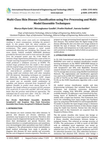
3. METHODOLOGY
International Research Journal of Engineering and Technology (IRJET) e ISSN: 2395 0056
Theproblemisformulatedintofourmainstages:
iii. Feature Extraction and ensembling Data Points: 52 image features are extracted from the InceptionV3 model foranygivenimage.Thisisachievedbypullingoutthelast seven layers of the neural network. The extracted image features are then merged and mapped according to the datapointsofthemetadata intheformofCSVintraining. Whiletesting,thesameparametersareachievedfromthe datatheuserenterswhileuploadingtheimage.
Pre processing of the training and validation data is performed in three parts, data segmentation, data resamplinganddataresizing.
follow up examination (follow_up), expert consensus (consensus),orconfirmationby(confocal).Inaddition,the dataset includes lesions with multiple images, which the lesion_id column within the HAM10000_metadata file can track.
| ISO 9001:2008 Certified Journal | Page1237
Overall implementation is a superset of Pre processing, Model training, comparison between models,modelevaluationandfinally,GUI.
i. Data Segmentation for UNET
3:
Volume: 09 Issue: 03 | Mar 2022 www.irjet.net
melanoma achieved 0.86. Ideas from each of the above mentionedpapersledustogenerateamethodologywhich ismentionedbelow.
4. DATA-SET AND RESOURCES
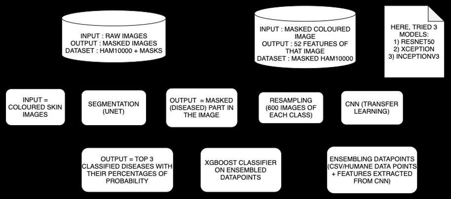
The HAM10000 dataset is a collection of dermatoscopic imagesofskinlesions,foundintheHarvardDataverse[7]. Thedatasetconsistsof10,015imagesthatcanbeusedfor academic deep learning purposes. It is a set of dermatoscopic images acquired and stored by different modalities from different populations. Cases include a representative collection of all critical diagnostic categories in the realm of pigmented lesions: Actinic keratosesandintraepithelialcarcinoma/Bowen'sdisease (akiec), basal cell carcinoma (bcc), benign keratosis like lesions(solarlentigines/seborrheickeratosesandlichen planus like keratosis, bkl), dermatofibroma (df), melanoma (mel), melanocytic nevi (nv) and vascular lesions (angiomas, angiokeratomas, pyogenic granulomas Moreandhemorrhage,vasc).than50%of lesions are confirmed through histopathology (histo), the rest of the cases is either
A. DATA PRE PROCESSING
Fig 2:HAM10000skinlesions
Fig 1:SystemDesign
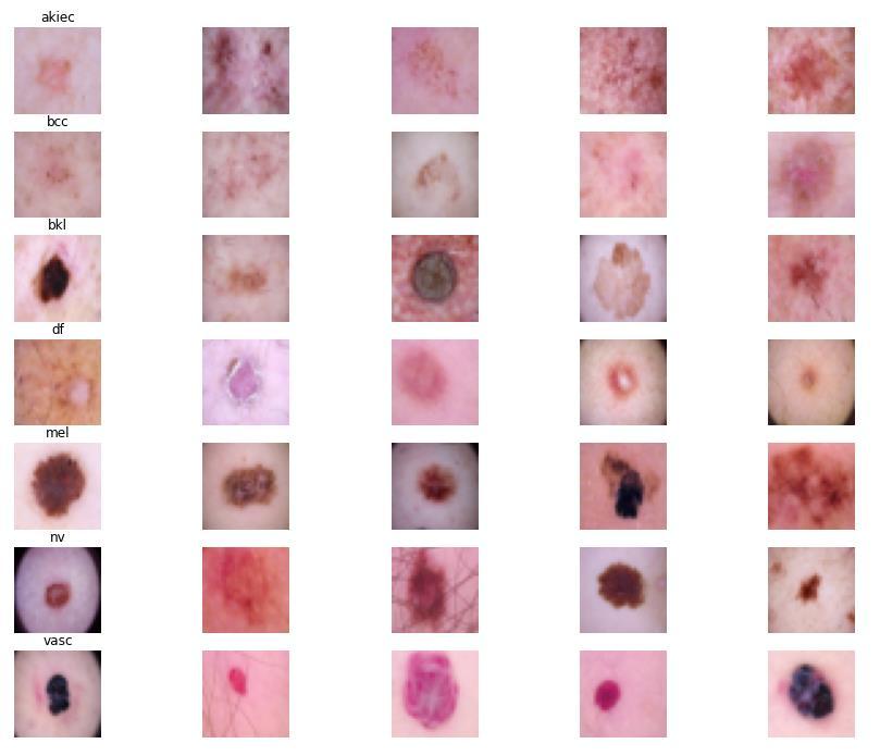
Datasegmentationtargetsaccuratedivisionorpartitionof the skin lesion (diseased area) from the actual dermoscopic image, i.e., division into multiple segments like diseased and healthy areas of skin. This comes under pre processingandformsablackmaskaroundthehealthy part of the skin. This results in hiding inconsistencies like noise and intense colour or illumination effects which reducesartefactsandprovidesbettertraining.Thesystem uses UNet architecture to segment the input images to obtainamaskedimage.
© 2022, IRJET | Impact Factor
iv: XgBoost Classification: Theobtainedensembleddata pointsareusedtotraintheXgBoostmodel.Predictingtop 3diseases,withanaccuracyof95%.
Fig SegmentationofimageusingU
5. IMPLEMENTATION
p ISSN: 2395 0072 value: 7.529
ii. Classification Model: The Segmented skin images are then passed to the classification model, the Inception V3 transfer learning algorithm. It is measured in terms of accuracy.
i. Segmentation Model: U net based architecture is implemented for precise segmentation of the image data fromtheHAM10000images.Themodelisevaluatedover IntersectionOverUnion(IOU)
C. CLASSIFICATION MODELS COMPARISON
International Research Journal of Engineering and Technology (IRJET) e ISSN: 2395 0056
The study showed little difference between the models when compared to the entire spectrum. A contrasting resultwhereResNet50wasthehighestamongstallothers
ii. Resampling
In Fig 3, the first image from the left, titled "Original image", would be the image uploaded by the patient. The middle image titled "Mask" is the predicted mask that covers the healthy part of the skin for better training. Finally, the last image titled "Segmented Output" is the output of the segmentation process used for training. The UNet Segmentation model generates a masked image, concentrating only on the affected part and covering the
The dataset is highly imbalanced, with most data of Melanocytic nevi, Melanoma, and Benign keratosis like lesions.Thesystemusesresamplingtechniquestobalance thedata,reducingthedatasetto600imagesperclasswith techniques of UpSampling and DownSampling. On purpose, the training images were not cleaned, thus still containing some noise. This comes mainly in the form of intensecolours,illuminationeffects,andsometimeswrong labels. All images were rescaled to have a maximum side lengthof(256,192)pixelsfortheUNetModel.
Dermatofibroma 115 600
determined based on the number of data trains(training) that gave the best result when spread across the neural network in each iteration (train steps). Inthistrainingprocess,thebatchsizewasdefinedas256, which means that each step was to be spread to 256 data train to the neural network. The input images were in (128,128,3) shape and were run for 50 epochs for all models. Outputs were generated by a combination of differentlayerslikeconvolutionallayers,activationlayers, pooling layers, batch normalization layers, and concatenation layers, summing up to 59 layers that generated output based on several parameters compared inTable 2
iii. Image Resizing
Certified Journal | Page1238
| ISO
Table-1:
© 2022, IRJET | Impact
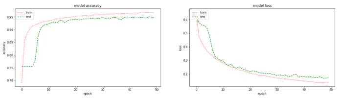
Fig 4: U NetSegmentationModelAccuracyandLoss
Thissurroundingskin.isabinaryclassificationmodel
DatasetSizeComparisonafterResampling Class Data Initial ResamplingAfter Melanocyticnevi 6705 600 Melanoma 1113 600 Benignkeratosis likelesions 1099 600 Basalcellcarcinoma 514 600 Actinickeratoses 327 600 Vascularlesions 142 600
Resizing images is an essential pre processing step in training machine learning models. Models train faster on smaller images, but shrinking can cause the deformation of features and patterns inside the image. Additionally, various deep learning architectures need images of the exact dimensions even though the raw collected images may be different in size. For example, the original dermoscopicimageintheHAM10000datasethasasizeof 600x450 pixels in the RGB format. The images are then resizedto128x128pixelstoalllayers.
B. PRETRAINED MODEL
While working with pre trained models like InceptionV3, ResNet50 and Xception, as our transfer learning models, we had images in our dataset divided into the ratio of Training:Testing:Validation set, which was precisely 2520:1050:630 summing up to a total of 4200 images used in the experiment. The data arrays for training and testing were created using train_test_split function of the BatchSklearnmodel.Sizewas
p
Volume: 09 Issue: 03 | Mar 2022 www.irjet.net ISSN: 2395 0072 Factor value: 7.529 9001:2008
andisabletogenerate masks for test images. The model has an accuracy of 93.74%, with a training accuracy of 97.37%. The f1 Score ROC AUC curve values are 0.94 each, and the Validation accuracywas94.88%asshowninFig 4.
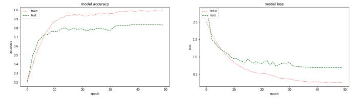
The three models in consideration, namely InceptionV3, ResNet50 and Xception, were then compared based on multipleareasofattention likethe numberofparameters the models generated and a comparison of trainable and non trainable parameters, in Table 3, the accuracy and loss graphsof eachof the modelswere investigatedalong with the ease of learning taken into account, in Fig 5,6,7, and finally, the classification results were examined individuallyforallevaluationparameters,intable 3.
D. Feature extraction and merging CSV data points
OnceitwasestablishedthatInceptionV3wasthemodelto choose for further research, wemoved to the next part of our approach, which was feature extraction. InceptionV3 had 59 layers in our experiment. Out of which, the last seven layers were excluded from extracting 52 vital features the model analyzed from the image. These 52 features were ensembled with 22 data points from the MetaData(CSV file). These included skin type, age, skin localizationandsexaswell.Thisresultedinadatasetof74 data points in which a few data points like gender, localization, cell type was hot encoded to further pass themforboosting.
Volume: 09 Issue: 03 | Mar 2022 www.irjet.net
E. Training Model (Inception V3 + XgBoost)
|
XGBoost, which itself is a scalable ensemble technique based on gradient boosting [10], was chosen to boost the ensembleddata pointsetandtoclassifytheimagesinto7 categories at the question, namely Vascular lesions, Basal cell carcinoma, Benign keratosis like lesions, Actinic keratoses, Melanoma, Dermatofibroma, Melanocytic nevi.AllthedatapointswerecombinedandtakenasX,and the labels(name of diseases for classification) were taken as Y. This Multi Model Ensembled approach led to a surprising accuracy of 0.95 f1 score which is seen in Table 4.
| ISO
Certified Journal | Page1239
Althoughofonly0.788.InceptionV3
ResNet50 1140,441,6 7540,387,0 54,536
InceptionV3 0.81 0.81 0.81 0.81 0.81 0.812 1.000
Total Trainable TrainableNon-
p ISSN: 2395 0072 2022, IRJET Impact Factor value: 7.529 9001:2008
©
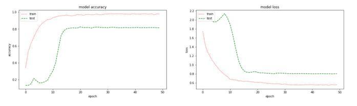
Fig 7: XceptionAccuracyandLoss
Pre Trained Model A B C D E F G
Xception 0.79 0.79 0.79 0.79 0.79 0.788 1.000
Fig 5: InceptionV3AccuracyandLoss Fig 6: ResNet50AccuracyandLoss
Table 2: ComparisonofClassificationModelsbasedona numberofparameters.
InceptionV3 7126,073,7 2326,037,9 35,848 Xception 4730,375,3 0330,319,4 55,944
Table 3: ClassificationResultsofAllModels
Parameters
International Research Journal of Engineering and Technology (IRJET) e ISSN: 2395 0056
Pre-TrainedModel
had a classification accuracy of 0.003 less than that of Resnet50, its learning was smoother and less computationally expensive than Resnet50, with a minimal difference in the ratio of non trainable parameters. Hence, it was taken into account thatInceptionV3isthebesttransferlearningalgorithmfor classifying the seven skin diseases into consideration amongallthreemodelsinthepicture.

ResNet50 0.81 0.81 0.81 0.81 0.81 0.815 1.000
in terms of trainable parameters, particularly difficulties inlearningwhentimeandGPUutilizationweretakeninto account. It also had a non monotonous testing accuracy and loss graph contradicting its monotonous training accuracy and loss graph. When such parameters were considered for comparison of Xception model, it fell back at mostly all starting from classification average accuracy
A=Accuracy,B=Precision,C=Recall,D=F1Score, E=ROC AUC,F=TestAccuracy,G=TestAccuracy
Finally, the model was evaluated on parameters like Precision, Recall, F1 Score and Support which was then compared with the same evaluation parameters achieved fromtheInceptionV3model,calculatedwithout ensemble and boosting. Before boosting, the Accuracy calculated by f1 scorewas0.81whichwenttoasurprisingscoreof0.95 whenboostedwiththeensembleddatapoints.
Accuracy
akiec 0.81 0.97 0.85 0.95 0.83 0.96 156 121 bcc 0.83 0.93 0.84 0.98 0.84 0.96 156 117 bkl 0.64 0.94 0.75 0.91 0.69 0.93 146 112 df 0.91 1.00 0.97 1.00 0.94 1.00 151 120 mel 0.77 0.90 0.66 0.94 0.71 0.92 139 127 nv 0.75 0.93 0.62 0.87 0.68 0.90 152 122 0.97 0.98 0.99 1.00 0.98 0.99 150 121 0.81 0.95 1050 840 avg 0.81 0.95 0.81 0.95 0.81 0.95 1050 840
avgWeighted 0.81 0.95 0.81 0.95 0.81 0.95 1050 840
International Research Journal of Engineering and Technology (IRJET) e ISSN: 2395 0056 Volume: 09 Issue: 03 | Mar 2022 www.irjet.net p ISSN: 2395 0072 © 2022, IRJET | Impact Factor value: 7.529 | ISO 9001:2008 Certified Journal | Page1240
ThemodelwasexecutedinGraphicalUserInterface(GUI) form using [8] Streamlitlibrary,whichis a pythonlibrary that allows developers to make data based web applications quickly and easily. Along with Streamlit, the project hosted the Web Application using [9] remote.it which is a SaaS, allowing remote users to user your web applications via an autogenerate URL. It was decided to use[9]sinceitissaferandbetterthanngrok.
Table-4:
6. RESULTS
8. CONCLUSION
Fig 8: TakingInputsfromtheuser
7. GUI
Macro
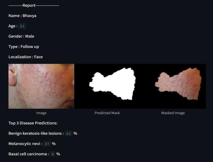
Fig 9: Generatingtheexpectedoutput
Y=InceptionV3X=InceptionV3ClassificationReportof:model+XgBoostmodel / Precision Recall f1 score support X Y X Y X Y X Y
In Conclusion, the proposed solution to the problem at hand considers all the parts of the detection issues and selectivelyresolvesthemsuccessfully.Thepaperpresents theideationofanensembledmulti modelapproachtothe HAM10000 dataset for skin disease detection. The successful implementation of the system, evaluation parameters, and execution via GUI is our active experimentationindefenceoftheproprietarypipeline.
vasc
This Fig 8 shows the GUI of the skin disease classification systemandacceptsuserdata alongwiththetestimage to classify. Whereas, Fig 9 shows the GUI of the skin disease classified system and generates a report with the Top 3 probablediseasesalongwithsegmentedimages

[8] Streamlit, tvst (2019) Streamlit [Source Code] https://github.com/streamlit/streamlit.git
[2] T. Shanthi, R.S. Sabeenian, R. Anand, Automatic diagnosis of skin diseases using convolution neural network, Microprocessors and Microsystems, Volume 76, 2020,103074, ISSN 0141 9331, https://doi.org/10.1016/j.micpro.2020.103074.
[5] B. Ahmad, M. Usama, C. Huang, K. Hwang, M. S. Hossain, and G. Muhammad, "Discriminative Feature Learning for Skin Disease Classification Using Deep ConvolutionalNeuralNetwork,"inIEEEAccess,vol.8, pp. 39025 39033, 2020, doi: 10.1109/ACCESS.2020.2975198.
© 2022,
International Research Journal of Engineering and Technology (IRJET) e ISSN: 2395 0056
|
[3] V. B.N., P. J. Shah, V. Shekar, H. R. Vanamala and V. Krishna A., "Detection of Melanoma using Deep Learning Techniques," 2020 International Conference on Computation, Automation and Knowledge Management (ICCAKM), Dubai, United Arab Emirates, 2020, pp. 391 394, doi: 10.1109/ICCAKM46823.2020.9051495.
[9] remot3.it, accessed on 10th February, 2022, < https://app.remote.it/#devices>.
[11]ionv3https://www.kaggle.com/code/shivanggandhi/inceptxgboostclassification/notebook
[1] K. E. Purnama et al., "Disease Classification based on Dermoscopic Skin Images Using Convolutional Neural Network in Teledermatology System," 2019 International Conference on Computer Engineering, Network, and Intelligent Multimedia (CENIM), Surabaya, Indonesia, 2019, pp. 1 5, doi: 10.1109/CENIM48368.2019.8973303.
[4] L. F.Li,X.Wang,W. J.Hu,N.N.Xiong,Y. X.DuandB. S. Li, "Deep Learning in Skin Disease Image Recognition: A Review," in IEEE Access, vol. 8, pp. 208264 208280, 2020, doi: 10.1109/ACCESS.2020.3037258.
| ISO
[12]segmentation/notebookhttps://www.kaggle.com/code/shivanggandhi/unet
[7] Tschandl, Philipp, 2018, "The HAM10000 dataset, a largecollectionofmulti sourcedermatoscopicimages of common pigmented skin lesions", https://doi.org/10.7910/DVN/DBW86T, Harvard Dataverse, V3, UNF:6:/APKSsDGVDhwPBWzsStU5A== [fileUNF]
Volume: 09 Issue: 03 | Mar 2022 www.irjet.net p ISSN: 2395 0072 IRJET Impact Factor value: 7.529 9001:2008
[6] M. A. Al Bahar, "Skin Lesion Classification Using Convolutional Neural Network With Novel Regularizer," in IEEE Access, vol. 7, pp. 38306 38313, 2019,doi:10.1109/ACCESS.2019.2906241.
Certified Journal | Page1241
9. REFERENCES
[10]Bentéjac, Candice & Csörgő, Anna & Martínez Muñoz, Gonzalo.(2019).AComparativeAnalysisofXGBoost.