An Intracranial Neoplasm or brain tumor is a mass that is
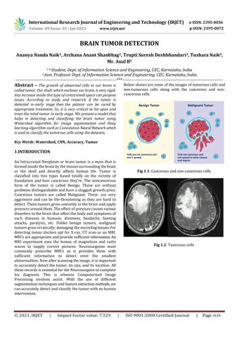
MdtuasdpredaCaprffcoformedinsidethebrainbythetissuessurroundingthebrainrtheskullanddirectlyaffectshumanlifeTumorislassifiedintotwotypesbasedtotallyonthevicinityofoundationandhowcancerousthey'reThenoncancerousormofthetumoriscalledBenignThesearewithoutoblemsdistinguishableandhaveasluggishgrowthpricenceroustumorsarecalledMalignantTheseareveryggressiveandcanbelifethreateningastheyarehardtoetect.Thesetumorsgrowunevenlyinthebrainandapplyssurearoundthem.Theeffectofpressurecausesvariousisordersinthebrainthataffectthebodyandsymptomsofuchdiseasesinhumans;dizziness,headache,faintingttacks,paralysis,etcUnlikebenigntumors,malignantmorsgrowerratically,damagingtheencirclingtissuesForetectingtumordoctorsoptforXray,CTscanoranMRIRI commonly icasPhthtabnormalities.Nowafterscanningtheimage,itisimportantoaccuratelydetectthetumor,itssize,anditslocation.AlleserecordsisessentialfortheNeurosurgeontocompleteisdiagnosis.ThisiswhereinComputerizedImagerocessinginvolvesassist.Withtheuseofdifferentegmentationtechniquesandfeatureextractionmethods,wenaccuratelydetectandclassifythetumorwithnohumanntervention
Key Words:
International Research Journal of Engineering and Technology (IRJET) e ISSN: 2395 0056 p ISSN: 2395 0072Volume: 09 Issue: 01 | Jan 2022 www.irjet.net © 2021, IRJET | Impact Factor value: 7.529 | ISO 9001:2008 Certified Journal | Page 1634 BRAIN TUMOR DETECTION Ananya Nanda Naik1 , Archana Anant Shanbhag2 , Trupti Suresh Deshbhandari3 , Tushara Naik4 , Mr. Anal B5 1 4 Student, Dept. of Information Science and Engineering, CEC, Karnataka, India 5 Asst. Professor Dept. of Information Science and Engineering, CEC, Karnataka, India *** Abstract
1.INTRODUCTION
’sareappropriateandprovidesufficientinformation An MRI experiment uses the homes of magnetism and radio waves to supply correct pictures. Neurosurgeons most
The growth of abnormal cells in our brain is called tumor. Our skull, which encloses our brain, is very rigid. Any increase inside this type of constrained space canpurpose issues. According to study and research, if the tumor is detected in early stage then the patient can be cured by appropriate treatment So, it is very critical to hit upon and treat the mind tumor in early stage We present a model that helps in detecting and classifying the brain tumor using Watershed algorithm for image segmentation and Deep learning algorithm such as ConvolutionNeuralNetworkwhich is used to classify the tumorous cells using the datasets Watershed, CNN, Accuracy, Tumor
prescribe MRI’ s as it provides them with sufficient information to detect even the smallest
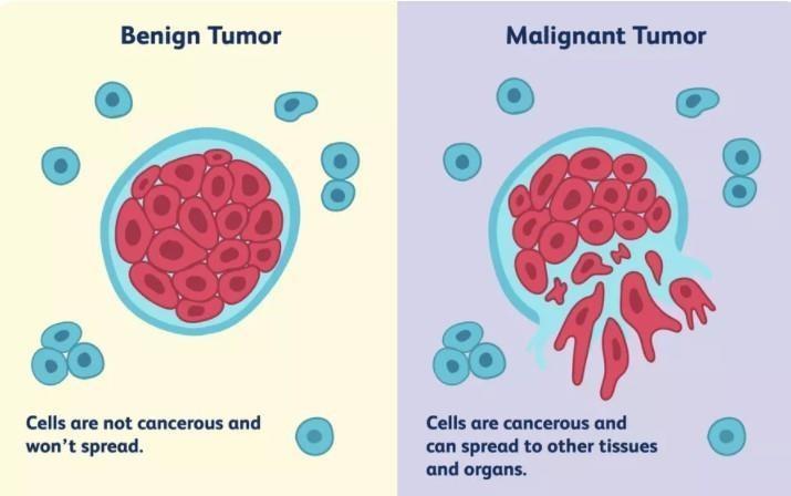
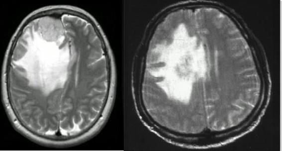
Belowshownaresomeoftheimagesoftumorouscellsand non tumorous cells along with the cancerous and non cancerouscells. Fig 1.1:Cancerousandnon cancerouscells Fig 1 2:Tumorouscells

© 2021, IRJET | Impact Factor value: 7.529 | ISO 9001:2008 Certified Journal | Page 1635
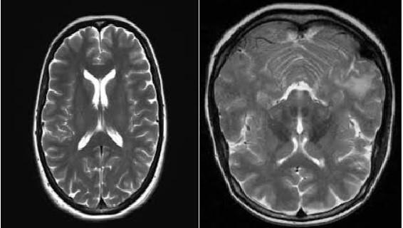
International Research Journal of Engineering and Technology (IRJET) e ISSN: 2395 0056 p ISSN: 2395 0072Volume: 09 Issue: 01 | Jan 2022 www.irjet.net Fig 1 3: Non tumorouscells 2. RELATED WORKS tuprethfmaprecsut90actytuaIeNodDiusfhoIsthireTthIn[1],MilicaM.Badzaetal.,HereaCNNstructureisusedforeclassificationofbraintumoron3tumortypes.echnologicalimprovementsandmachinestudyingcansourceradiologistsintumorprognosiswithouttheuseofnvasivemethods.AnalgorithmformachinelearningthateCNNhasachievedsignificantsuccessinpictureegmentationandclassificationn[2],ImranAshrafetal,Inthispapertheyexplainedaboutwtheclassificationofbraintumorisdonebydoingtheollowingsteps,theproposedapproachincludesfivemiddleteps.Instepone,thelinearcomparisonstretchingishiredsingregionprimarilybasedhistogramequalizationandscreteCosineTransform(DCT)Inthesecondonestep,eeplearningfunctionextractionisachievedBymakingusefswitchmastering,twopreskilledConvolutionalNeuraletwork(CNN)fashionshavebeenusedforfeaturextraction.n[3],Dr.R.C.Sugantheetal.,Theyexplainedaboutdetailboutimageclassificationalgorithmhelpsindetectingthemoratearlystagewithhighaccuracybraintumorandpesandhowdangerousitis,andfordetectionandlassificationtheyproposedadeeplearningmethodCNNndRNNforaccuracybetweenthemtheyprovedRNNhave%accuracyIn[4],RupalRAgravatetal,ThemainfocusofthispaperisosegmenttumorfromBRATS2018benchmarkdatasetandseage,shapeandvolumetricfeaturestopredictoverallurvivalofpatientsandalso,theytackledaproblemoflassifyingtheBraintumortypeandoverallsurvivaldictiontheyusedseveralmethodsandtheyfoundtheccuracyforeachmethodsothattheycanadaptthatethod.Theproposedtechniquemakesuseoffewereatureshoweverachievesbetteraccuracythannationoffeartworkstrategies.Inthistheywilldividethesurvivaldictioninto3partsbasedonfactorslikeageandtypeofmorasshortmid,longsurvivors.
In[5], Axel Davy et al, Inthis paper they proposed a deep neuralcommunitybasedonconvolutionalneuralnetworks andresidualcommunityforphotoinformation So,theCNN exploits both close by capabilities similarly to extra worldwidecontextualfeaturesconcurrently.Also,exclusive from maximum conventional makes use of CNNs, this community makes use of a final layer that may be a convolutional implementation of a totally related layer whichallowsaforty foldaccelerate.Thisalsodescribea2 section schooling manner that lets in us to address difficulties associated with the imbalance of tumor labels. Finally,wediscoveracascadestructurewhereintheoutput of a number one CNN is treated as a similarly supply of IrecordsforasubsequentCNNn[6],LinaChatoetal,Proposed paper automatically predict the survival rate of patients with a Glioma brain tumorbyclassifyingthepatient’sMRIimageusingmachine learning (ML) methods.in this the classes of survivors are dividedasshort term,mid term,andlong term.Toimprove the prediction results. For these Features like volumetric, statistical and intensity texture, histograms and deep features are taken. And used CNN for best accuracy Ipredictionn[7],G. Hemanth et al., Proposes an automatic segmentationtechniquethatreliesuponCNN(Convolution Neural Networks), figuring out small 3 x three kernels By incorporating this single method, segmentation and classificationiscarriedout CNN(aMLtechnique)fromNN (Neural Networks) where it has layer based for results
svthsdIfithmttucaffsLtufIhclassificationandstepsinvolvedtoachievethismethodwithighestaccuracyisalsoexplainedn[8],AMartinetal,ProposedanewvariationalmodelforeaturedetectioninimagesanditsapplicationtobrainmorsegmentationTheyexplainedabouthowDeepearningframeworkforusedforavailableknowledgefromapecificapplicationtooptimizetheparametersoftheenergyunctional.Theyusedonlyglioblastomapatientsdatabaseordetectionhenceitscommontypeofbraintumor.In[9],MuhammadWaqasNadeemetal.,Thisstudyisassessmentthatsummarizesamassivenumberofclinicalontributionstothesubject(i.e.,Deeplearninginthoughtsmoranalysis),byusingwayofmappingacoherentaxonomyofresearchlandscapefromtheliterature),byappingacoherenttaxonomyofresearchlandscapefromeliteratureThepredominantcharacteristicsofthisneweldhadbeenexaminedandanalyzedn[10],LiSunetal,UsingmultimodalMRIscans,theyemonstratedadeeplearningbasedsystemforbraintumoregmentationandsurvivalpredictioningliomaThen,tofindemosteffectivefeatures,decisiontreesandcrossalidationareutilizedFinally,topredictpatientoverallurvival,arandomforestmodelistrained.
© 2021, IRJET | Impact Factor value: 7.529 | ISO 9001:2008
Fig 3.1:Methodology Step 1: Input MRI image • Input the data set from Kaggle website and it consists of 153 tumorous images and 94 non tumorous images. Augmentationtothedata setis donewhichmeansincreasingthedatasettomake the training effective. Some of the augmentation techniques we followed are rotation, translation etc. • ThetrainingdataaresplitintovalidationandtraindatasetbyusingKerasImagedatagenerator
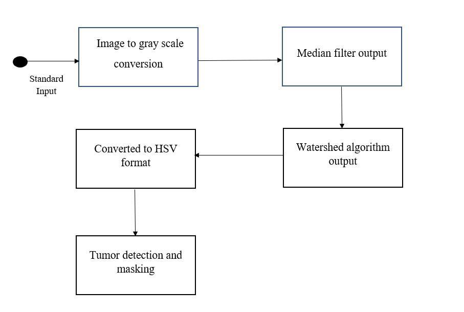
• Oneofthemostcommonpre processingpracticesis the conversion of the RGB image to a gray scale image A gray scale picture includes sunglasses of graywithoutaobviouscolour Thiswaythatevery pixel represents the intensity fee at that pixel withoutshowinganycolour Also,notlikea black and white picture, a gray scale picture has many splendidsolarshadeswithwhitebeingthelightest coloration and black being the darkest Also, in a grayscaleimagetheintensityvalues ata pixel are notabsoluteandcanbeinfractions.
• Theinput imagefor thesegmentationalgorithmis the image which is free from noises. We perform median filter for noise removal Morphological watersheds providea complementary approach to the segmentation of objects of an image so it is especially useful for segmentation of objects that aretouchingeachother
Certified Journal | Page 1636
International Research Journal of Engineering and Technology (IRJET) e ISSN: 2395 0056 p ISSN: 2395 0072Volume: 09 Issue: 01 | Jan 2022 www.irjet.net
• dMothPerformthesegmentationtechniquetosegmentoutetumorusingWatershedalgorithmwhichusesrphologicalopeningandDilationtechniquestoetecttheboundariesintheimage
• Inwatershedmethodweviewa grayscalepicture asa topologicalsurface,wherethevaluesof f(x, y) corresponds to heights. This algorithm also finds catchment basins and ridge lines.to get proper segmentationof imagepost processing of imageis ftobedone,sotheoutputimageisconvertedtoHSVormattomaskoutthetumorpartintheimage.
• Gray scaling is important as it provides a more accurate colour information which aids during segmentation Thisisthenumberonesteptakenby ficmeansofalltheresearchers.Oncethephotographisonvertedtoagrayscalephotograph,it'smilesthenlteredtogetridofextranoise
• The important intention of the segmentation is to changetheillustrationofan imageintosomething ithatisextrasignificantandlesscomplicatedsothisslesscomplicatedtoanalyse
Step 3: Segmentation
• Image segmentation is a part of digital image processing and computer vision, segmentation is the process of dividing a digital image into a numberofsegments.
3. PROPOSED SYSTEM
• Filtersareoftwotypes,onethatallowsthelow end frequenciestopassorfiltersthatallowthehigh end frequencies to pass. A filter can either flatten the imageorsharpentheimage Whenafilterisusedto flatten the image, the noise is blurred leaving pratqisthbehindasmoothimagehoweverthefinerdetailsofeimagearelostIfthephotographistobeharpenedthanthefilteroutenhancesthefinernformation,howeverthisresultsinanmultiplieduantityofnoisewithinthephoto.Thisnoisehaveobeclippedbeforeinadditionprocessingasitisbletointervenewiththeaccuracyofthedetectionogram.
• Pre processing involves processes like conversion to gray scale, noise reduction and noise removal, image reconstruction, Photo enhancement and regarding clinical pics may additionally contain stepslikeskullremovalfromanMRIimage
• Obtain 1444 training image with 310 validation imageandaround310testimagestoevaluateour trainedmodelson Step 2: Data Pre-processing
• Itisessentialthingforthehigh qualityresults.The fundamental part of pre processing of picture information consists of removal of undesirable statisticsandpictureoughttobeinproperformat forprocessingsothateffectsarecorrect
REFERENCES
• Weusedbinarycross entropy as our lossfunction mThebandsoftmaxlayerwasusedtolimittheprobabilityetween0and1inlastlayeroftheneuralnetwork.activationfunctionusedisReLuwhichistheostcommonlyusedfunction.
’
Step 6: Identification
Step 5: Classification
• In this step the MR images categorized into two classes as normal and tumor. The accuracy is obtained by CNN. As the user inputs the image of theMRI image, theoutputwill bedisplayed tothe useralongwithitsstageofthetumordescription.
▪ The proposed model detects the tumor at early stage using the deep learning techniques such as convolution neural networks and watershed algorithms.
▪ Infuturestudies,wehopetoproposeamodelthat willmakeitpossibleto useit on different medical imagesandindifferentfields.
• The Models that gives state of art results have a huge number of parameters. For a model to train hsoisproperlynumberofsamplesrequiredinthedatasethouldbecomparabletothenumberofparametersnthemodelHowever,inmedicaldomaingettingmanydatapointsisneartoimpossiblehence,toandlethisissueweusepretrainedmodels.
ThetrainingwasusingKeraslibraryoftensorflowWeusedAdamoptimizerfortrainingwithalearningrateof1e4Learningrateplateauwasappliedtoreducethelearningrateaccordingtovallossimprovement
• Inthisweloadtheweightsforatraininginwhich the model is trained on a very large data set, so dowhiletrainingonsmallnumberofmedicaldata,wen t have to start from the scratch The basic features like edge, corners are already covered in thepre trainedweights,hencetrainingontheother imageissimple
[1] Milica M. Bad•za; Marko C. Barjaktarovi´c (2020).”Classification ofBrain Tumors from MRI Images Using a ConvolutionalNeural Network” . [2]Muhammad Attique Khan;Imran Ashraf; Majed Alhaisoni; Robertas Dama•sevi•cius; Rafal Scherer; Amjad Rehman; Syed Ahmad Chan Bukhari(2020), ”Multimodal Brain Tumor Classification Using Deep Learning” [3]Dr.R.CSuganthe; GRevathi; SMonisha; RPavithran (2020), ”Deep Learning Based BrainTumor Classification Using Magnetic Resonance Imaging” [4] Rupal R. Agravat; Mehul S Raval, ”Prediction of Overall Survival ofBrain Tumor Patients”, Inproceedings ofthe 2019IEEE Region 10Conference (TENCON 2019).
© 2021, IRJET | Impact Factor value: 7.529 | ISO 9001:2008 Certified Journal | Page 1637
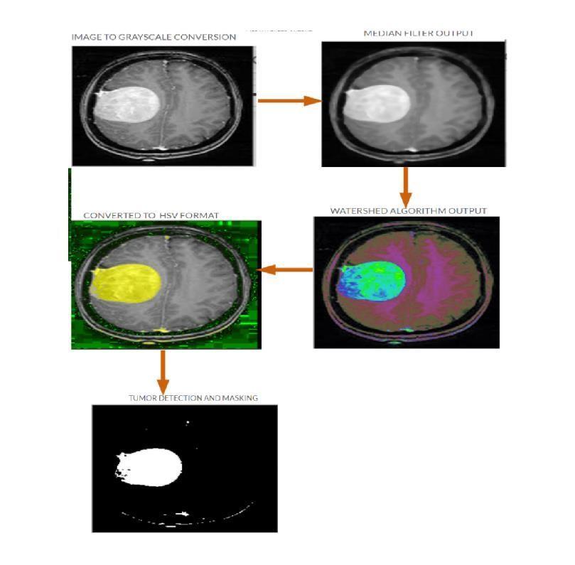
Fig 3.2:Flowofimagethroughdifferentprocesses
•
Step 4: Training Parameters
International Research Journal of Engineering and Technology (IRJET) e ISSN: 2395 0056 p ISSN: 2395 0072Volume: 09 Issue: 01 | Jan 2022 www.irjet.net
• Classification is done using CNN algorithm: mcveimneTheConvolutionalNeuralNetwork(CNN)isadeepuralstructureusedtodecodevisualsymbolsTheostcommonusesareinrecommendersystems,mageandvideorecognition,andclinicalimagexaminationCNNisamultilayerperceptronersionusedinnaturallanguageprocessing,imagelassification,braincomputerinterfaces,andonetarytimeseries.
4. CONCLUSIONS
[5] Mohammad Havaei;Axel Davyb; David Warde Farley; Antoine Biard;Aaron Courville; Yoshua Bengio; Chris Pal;Pierre Marc Jodoin; Hugo Larochelle(2016), ”Brain Tumor Segmentation with DeepNeural Network” [6]Lina Chato; Shahram Latifi (2017), ” MachineLearning and Deep Learning Tech niques to Predict Overall Survival ofBrain Tumor Patients using MRI Images” , In proceedings of the 2017 IEEE 17th International Conference on Bioinformatics and Bioengineering [7] GHemanth; MJanardhan; LSujihelen, ” Design And Implementing Brain Tumor Detection Using Machine Learning Approach” , In Proceedings of the Third Interna tional Conference on Trends in Electronics and Informatics (ICOEI 2019).
[8] I Ram´ırez;A Mart´ın;E Schiavi, ”Optimization ofa variational modelusing deeplearning: An application to brain tumor segmentation” , In proceedings ofthe 2018 IEEE 15th International Symposium on Biomedical Imaging (ISBI 2018). [9] Muhammad Waqas Nadeem; Mohammed A Al Ghamdi; Muzammil Hussain;Muhammad Adnan Khan; Khalid Masood Khan;Sultan H.Almotiri; Suhail Ash faq Butt (2020), ”Brain Tumor Analysis Empowered with Deep Learning:A Re view,Taxonomy, and Future Challenges” . [10]Li Sun; Songtao Zhang; Hang Chen; Lin Luo (2019), ”Brain Tumor Segmentation and Survival Prediction Using Multimodal MRI Scans with Deep Learning” © 2021, IRJET | Impact Factor value: 7.529 | ISO 9001:2008 Certified
International Research Journal of Engineering and Technology (IRJET) e ISSN: 2395 0056 p ISSN: 2395 0072Volume: 09 Issue: 01 | Jan 2022 www.irjet.net
Journal | Page 1638