
DISEASE DETECTION AND
The eye is a complicated system that serves as a vital component of our bodies. The retina, iris, pupil, and optic nerve are all interconnected elements of the eye. Glaucoma,trachoma, age relatedmaculardegeneration, pathological myopia, retinitis pigmentosa, and diabetic retinopathy are all disorders that have an influence on theeye.Patientsare sometimes unawareoftheseverity of theirillness.It isincrediblydifficulttoappropriately impair eyesight due to late identification of eye
1M.Phil Research Scholar, Department of Computer science and ApplicationsSambalpur University, Jyotivihar Burla
Subhashree
2Associate Professor & Head, Department of Computer science andApplications Sambalpur University, Jyotivihar Burla ***
Afterdisorders.[1].aneyeexam with various technologies such asa slit lamp, retinal examination, or visual acuity tests,an ophthalmologist or optometrist diagnoses eyeproblems [3]. A printed chart or a sequence of smaller characters read with a viewing device are used to perform visual acuity tests [3]. The ophthalmologist examines the eye under magnificationwithastronglineoflightusingaslit lamp[4]. Duringaretinalexamination,thepupilis dilatedusing dropstohelpwiden the eyelens and analyseeyemovementsandpapillaryresponse.
Abstract- Among the most common blindness hasseveral causes that is cataract, especially among the elderly. According to WHO/NPCB surveys, India has approximately 12 million blind persons, with cataract accounting for 80.1 percent of those cases. Therefore detecting a cataract in the early stage is a very important as well as difficult task. There are different types of image models currently being used by researchers for problems related to eye. The utilization of five different imaging modalities specifically for automated cataract detection is presented. More focus is given to the retinal images for the cause of cataract detection as it is considered better for extraction of the macula, lesion, blood vessels, optic disc, fovea, and macula are all components of the macula are all featuresto look for. Detecting these features accurately can solvemany problems related to eye disease including diagnosis and grading of cataracts automatically. In this research article we explore the potential of different retinal imaging methodologies currently employed by many of the researchers along with their advantages and disadvantages. Through the research an automated approach for segmenting retinal images has been developed. The segmentation is achieved by an iterative region expanding approach that merges the contents of numerous binary images produced by vessel width dependent morphological filters. Finally, the suggested work's experimental results and evaluation are shown, which reveal promising results.
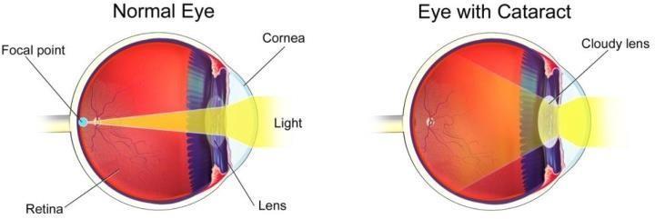
However, these procedures need the use of expensive medical equipment that can only be used by ophthalmologists with extensive experience. Because these processes are manual, they are both time consuming and subjective based on ophthalmologist experience. As a result, academics have made various attempts in recent years to automatetheidentificationofeyediseases.Fig1:NormaleyeversusCataracteye[7]Cataractsarethecauseof47.8%ofallblindnesscasesintheworld.Advancedageisthemostsignificantriskfactorforcataractdevelopment.Over314millionpeopleareblindorpartiallyblindaroundtheworld.Sixtysevenpercentofthosewhoareblindareblindowingtocataracts.
International Research Journal of Engineering and Technology (IRJET) e-ISSN: 2395-0056 Volume: 09 Issue: 01 | Jan 2022 www.irjet.net p ISSN: 2395 0072 © 2022, IRJET | Impact Factor value: 7.529 | ISO 9001:2008 Certified Journal | Page1273
1. INTRODUCTION
Keywords: Cataract, retinal image, feature extraction, segmentation,morphologicalfilter
IMAGE
CATARACT CLASSIFICATION USINGRETINAL MODEL Sarangi1 , Dr. Chandra Sekhar Panda2

The use of digital camera photos for cataract screening becomes intriguing when considering thehealth services in underdeveloped countries. A digital camera is also a simple and easy to use instrument when compared to a slit lamp and othercomplexmedicalequipmentusedforcataract diagnosis.
Fig3:Sampleimagesofeachimagemodel
Nuclear cataract is typically detected using slit lamp imaging. Because nuclear cataract affects the nucleus of the eye lens, characteristics from the nucleus area are extracted for automated identificationandgrading.[9,10]. (a) (b) (c) (d) (e)
2.4 Ultrasonic Images
International Research Journal of Engineering and Technology (IRJET) e ISSN: 2395 0056
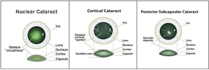
More than 90 percent of the world's blind population live in developing countries. The World Health Organization (WHO) defines a cataract as a clouding of the eye's lens that inhibits light from passing through due to protein clumping together as people age. Theseproteinsgrowuponlywhentheoldercells compress into the lens's centre, resulting in a fuzzy viewoftheretina. [5]. Early diagnosis of cataracts is a little discussed area forongoingresearch,despitethefactthatitisoneof the most common eye illnesses and the main cause of blindness worldwide. Nuclear cataract, cortical cataract, and posterior subcapsular cataract are the three types of cataracts that can arise depending on where they form or how they form [2]. The most frequenttypeofcataract produced byageisnuclear cataract (NC). The hardening and yellowing of the nucleus,thecentrecomponentoftheeyelens,isthe most common reason [8]. Cortical cataract (CC) is a type of cataract that develops in the lens cortex and appears as white wedged shaped and directed opacities that move from the lens's border to the centre in a spoke like pattern [8]. PSC (posterior subcapsularcataract) appears as little breadcrumbs or sand particles beneath the lens capsule. It is especially common in steroid addicted and diabetic people [8]. The numerous types of eye cataracts are depictedin
Volume: 09 Issue: 01 | Jan 2022 www.irjet.net p ISSN: 2395 0072 © 2022, IRJET | Impact Factor value: 7.529 | ISO 9001:2008 Certified Journal | Page1274
CataractsFigFigure2.2:TypesofEyeCataractarediagnosedbyanophthalmologist
A non stereoscopic image obtained using a "Neitz CT R cataract camera to concentrate on the anterior/posterior cortex of the lens"[10] is recognized as retro illumination. Cortical and posterior subcapsular cataracts are examined usingaretroimaging.
by examining the changes in the eye lens. After cataract detection, grading can be done by comparing slit lamp pictures to a collection of standard images using various grading systems. The most often usedgrading techniques are the Lens Opacities Classification System III (LOCS III) [9]andtheWisconsinGradingSystem(WGS)[10].
2. IMAGEMODELS
2.1 Digital Images
(a)Digitalimage (b) Slit lampimages (c) Retro illuminationimage (d) Retinalimage (e) Ultrasonic image
2.3 Retro illumination Images
Ultrasound is a popular technique for diagnosing ocular disorders. For cataract diagnosis, ultrasound A scan signals are collected using an ultrasound scanner equipped with 30 60MHz ultrasonic transducers from porcine lenses. B scan and Nakagami images are created using the acousticparameters velocity, attenuation,andbackscatteringsignals[22].
2.2 Slit-Lamp Images

International Journal of Engineering Technology (IRJET) e ISSN: 2395 0056
Volume: 09 Issue: 01 | Jan 2022 www.irjet.net p ISSN: 2395 0072 © 2022, IRJET | Impact Factor value: 7.529 | ISO 9001:2008 Certified Journal | Page1275
2.5 Retinal Images
The diagnosis of eye related disorders such as macular degeneration, diabetic retinopathy, and glaucoma has long relied on retinal fundus imaging. [11 13]. Although retinal (fundus) scans of the eye haverarely been used for cataract diagnosis and grading, several studies have attempted to identify cataractusingtheseimagesandhaveachievedahigh Theaccuracyrate.funduscamera
3. RELATEDWORKS Automatic cataract detection using retinal images has attracted significant attention, and was discussed in [26 28]. Various methods which can be used for cataract detection and classification using retinal image model is discussedfurther.
Research
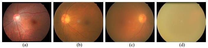
Fig4.Retinalimages:(a)non cataract,(b)mildcataract, A(c)moderatecataract,and(d)severecataract.cataractisaslowgrowingeyecondition that can be partial or total, stationary or progressing. Patients with various degrees of cataracts require different therapies. Patients with light cataracts might delay degeneration by wearing antiglare sunglasses [24], whereas intermediate and severe cataract patients require surgical intervention [25]. It is feasible to discern the different levels of severity by extracting suitable information fromretinalpictures.
Figure 4(b) shows an image with a mildcataract, withthemainvesselsandopticdiscvisible,butthe choroid and capillary vessels only faintly visible. Only the main blood veins and the optic discare seen in Figure 4(c). Furthermore, in Figure 4, there are essentially no retinal structures visible (d).It can be assumed that more severe cataracts revealfewerretinalstructures.
3.1 Transformation based approach: [29] offereda study for cataract detection utilising retinal pictures and the Fourier transform. Due to variable amounts of blurriness, the high frequency component distributions of retinal pictures differ. The high frequency component distribution curveis used to assess the cataract severity. The results show that the performance of this method has a good association with the LOCS III score, indicating thattheretinalimage basedmethodisgenuine.
is simple to use and can be operated by technologists or even patients. Retinal pictures have been employed widely in the diagnosis of ophthalmological illnesses such as glaucoma [16 17], age related macular degeneration [18 19], and diabetic retinopathy [20 21] since the invention of the fundus camera in 1910. Furthermore, this technique has been investigated in an attempt to automatically quantify cataractseverity [22 23]. Some attempts at detection in smartphone based systems have also been made [15]. Figure 4 shows four typical retinal pictures, exhibiting non cataract, mild, moderate,andseverecataracts.Figure4(a)shows a healthy retina with clearly visible main vessels, optic disc, choroid, and even capillary vessels.
3.3 Local feature based approach: In [34], the local standard deviation was employed. The localstandarddeviationsofthecoefficientsand the other eight features were extracted after the bloodvessels were strengthened by match filtering in different orientations. Retinal pictures with various cataract severities were effectively categorised using a decision tree [35].
and
3.2 Global feature-based approach: In [30 31], the Haar wavelet and discrete cosine transformation (DCT) were utilised to investigate four class categorization using retinal pictures. In both the discriminant analysis and the supervised technique, the results showed that the Haar wavelet feature achieves higher accuracy than the DCT feature. [32] employed texture andmorphological data, and the findings showed that based on the BP net, this strategy also produced pretty good accuracy. TheperformanceoftheHaarwavelet, DCT, and texture features were summarised in [33], which suggested an ensemble learning based technique. The results showed thatthe Haar wavelet feature outperforms theDCT and texturefeaturesintermsofaccuracy.
International Research Journal of Engineering and Technology (IRJET) e-ISSN: 2395-0056
2 approachbasedfeaturesGlobal Haarwavelet Fan et al.[30] Low cost running detection procedures. This method can achieve the highest accuracy by applying different global features in less time and less memoryoverhead. It cannot be used for some particular applications wherethe illuminationlevelofimagesis very low.Discrete cosine transformation Guo et al. [31]
Volume: 09 Issue: 01 | Jan 2022 www.irjet.net p ISSN: 2395 0072 © 2022, IRJET | Impact Factor value: 7.529 | ISO 9001:2008 Certified Journal | Page1276
NoSl Approaches Methods Authors Advantages Disadvantages 1 approachonTransformatibased
Decisiontreebased forhighordertasks. method 4 Deep features approachbased Deep CNN Zhang et al.[36]
Gives a slight higher accuracy than above methods. Needs a large no of data set for training purpose thus use more cost, Systemtimeandoverhead.
3 Local Localstandard Xiong et al.[34] Many features can be Thelocalstandarddeviation features deviation extracted locally so alone gives lowaccuracy, it needs based thatitcanbeusedasa someothermethodstobefused approach goodnumericalability together. Zhang et al.[35]
3.4 Deep feature-based approach: In [36], deep CNN (DCNN) based categorization was used, and the DCNN achievedsomewhatbetter accuracythantheother approaches. However, whenthetrainingsamplesarenearto 2000,theaccuracyreaches arelativelyhigh andsteadylevel,necessitatingsignificantlymorephotographsthan otherrelevantefforts.
FourierTransform RahmanAbdul et al.[29] Exhibits strong correlation with the LOCSIIIscore Frequency component may differfor variouslevelsofblurriness
Texture and featuremorphological based method Yang et al.[32] Ensemble learning basedmethod Yang et al.[33]
Table 1: Literature review table of methods usedin retinal image model for cataract detection andclassification
3)Retrievedfromhttp://www athenseyecarenet/co nditions/cataracts/? 4)Retrievedfromhttp://www parentyourparentsc om/pyp_article/cataracts/?
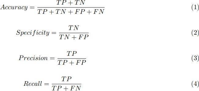
To evaluate how accurate a system is, i.e. to measure its performance, many evaluation metrics can be used like Accuracy, Specificity, Precision, Recall, F1 score and mean square errorrate.
4. PERFORMANCEEVALUATIONMETRICS
False Negative (FN: f + −): The number of instances that were positive (+) and incorrectly classifiedasnegative( ). Itisalsoknownas Type 2Error.
True Positive (T P: f + +): The number of instances that were positive (+) and correctly classifiedaspositive(+).
6. REFERENCES
7)Retrievedfromhttp://wwweyecentercomph/what wedo.html#ripen.©Copyright2011. American EyeCenter.
International Research Journal of Engineering and Technology (IRJET) e ISSN: 2395 0056 Volume: 09 Issue: 01 | Jan 2022 www.irjet.net p ISSN: 2395 0072
False Positive (FP: f − +): The number of instances that were negative ( ) and incorrectly classifiedaspositive(+).Itisalsoknownas Type 1Error.
Also,forevaluatingcataractseverity.ifweconsideramongalltheapproachesused inretinal image model then global feature based approach specially texture based and morphological method is much suitable for cataract detection and classification as it is a low cost running detection procedure. Also, this method can achieve the highest accuracy by applying different global features in less time and lessmemoryoverhead.
2)Retrievedfromhttp://www medindianet/patient s/patientinfo/cataract.htm
8)Delcourt, C., Cristol, J. P., Tessier, F., Léger, C. L., Michel, F., Papoz, L., & POLA Study Group. (2000). Risk factors for cortical, nuclear, and posterior sub capsular cataracts: the POLA AmericanJournalofEpidemiology,151(5),497study.
© 2022, IRJET | Impact Factor value: 7.529 | ISO 9001:2008 Certified Journal | Page1277
6) Pizzarello,L.,Abiose,A.,Ffytche,T.,Duerksen, R., Thulasiraj, R., Taylor, H., & Resnikoff, S. (2004). VISION 2020: The right to sight: A global initiative to eliminate avoidable blindness. Archives of Ophthalmology, 122(4),615 620.
5) Seddon, J., Fong, D., West, S. K., & Valmadrid, C. T. (1995). Epidemiology of risk factors for age related cataract. Survey ofOphthalmology,39(4),323 334.
The strain on ophthalmologists and clinicians is reduced by automatic cataract identification and grading. It also offers an objective approach to assess the severity of cataracts and aids in the reduction of visual loss through quick and precise diagnosis. This document provides an overview of the procedures and approaches used to identify and grade cataracts. The use of five different imaging modalities for automated cataract diagnosis using digital image processing is demonstrated. Slit lamp images, retro illumination images, retinal images, ultrasonic images, and digital eye images are examples of these sorts.. Retinal images are considered good forfeatureextraction so features like lesion, blood vessels, optic disc, fovea, and macula can be detected which can beused for automatic cataract detection and grading. The scale of the visible retinal structure is the most valuable information
5. CONCLUSION
True Negative (TN: f − −): The number of instances that were negative ( ) and correctly classifiedasnegative( ).
1)Zhang, Z., Srivastava, R., Liu, H., Chen, X., Duan, L., Wong, D. W. K., Kwoh, C. K., Wong, T. Y.,& Liu, J. (2014). A survey on computer aided diagnosis for ocular diseases. BMC Medical Informatics and DecisionMaking.
20) M. R. K. Mookiah, U. R. Acharya, C. K. Chua, C. M. Lim, E. Y.K. Ng and A. Laude, “Computer aided Diagnosis of Diabetic Retinopathy: A Review,” Computers in Biology & Medicine, Vol. 43, No. 12, pp. 2136 2155,2013.
24) K. Pesudovs and D. B. Elliott, “Cataract Morphology, Classification, Assessment and Referral,” CE Optometry, Vol. 4, pp. 55 60, 2001.
13) Akram, M. U., Khalid, S., Tariq, A., & Javed, M. Y.(2013). Detection of neovascularization in retinal images using multivariate m Mediods based classifier. Computerized Medical Imaging and Graphics,37(5),346 357.
16) S.Dua,U.R.Acharya,P.ChowriappaandS.V. Sree, “Wavelet Based Energy Features for Glaucomatous Image Classification,” IEEE Transactions on Information Technology in Biomedicine, Vol. 16, No. 1, pp. 80 87, 2012.
17) S. Yousefi, M. H. Goldbaum, M. Balasubramanian et al., “Glaucoma Progression Detection Using Structural Retinal Nerve Fiber Layer Measurements and Functional Visual FieldPoints,” IEEE Transactions on Biomedical Engineering, Vol. 61, No.4, pp. 1143 1154, 2014.
21) C. Sinthanayothin, J. F. Boyce, T. H. Williamson et al., “Automated Detection of Diabetic Retinopathy on Digital Fundus Images,” Diabetic Medicine, Vol.19, No.2, pp.105 112,2002.
22) O. Hockwin, V. Dragomirescu and H. Laser, “Measurements of Lens Transparency or Its Disturbances by Densitometric Image Analysis of Scheimpflug Photographs,” Graefes Archive for Clinical & Experimental Ophthalmology,Vol.219,No.6,pp. 255 262, 1982.
© 2022, IRJET | Impact Factor value: 7.529 | ISO 9001:2008 Certified Journal | Page1278 504.
9)Chylack, L. T., Wolfe, J. K., Singer, D. M., Leske,M.C.,Bullimore,M.A.,Bailey,I.L., Friend, J., McCarthy, D., & Wu, S. Y. (1993). The lens opacities classification system III. Archives of Ophthalmology, 111(6),831 836.
International Research Journal of Engineering and Technology (IRJET) e ISSN: 2395 0056 Volume: 09 Issue: 01 | Jan 2022 www.irjet.net p ISSN: 2395 0072
14) Tsui, P. H., Huang, C. C., Chang, C. C., Wang, S.H.,&Shung,K.K.(2007).Feasibilitystudy of using high frequency ultrasonic Nakagami imaging for characterizing the cataract lens in vitro. Physics in Medicine andBiology, 52(21), 6413.
19) S. Kankanahalli, P. M. Burlina, Y. Wolfson, D. E. Freund and N. M. Bressler, “Automated Classification of Severity of Age Related Macular Degeneration from Fundus Photographs,” Investigative Ophthalmology & Visual Science, Vol. 54, No. 3, pp. 1789 1796, 2013.
11) Akram, M. U., Tariq, A., Khan, S. A., & Javed, M. Y. (2014). Automated detection of exudates andmaculafor grading of diabetic macular edema. Computer Methods and ProgramsinBiomedicine,114(2), 141 152.
23) S. K. West, F. Rosenthal, H. S. Newland and H. R. Taylor, “Use of Photographic Techniques toGrade Nuclear Cataracts” Investigative Ophthalmology & Visual Science,Vol.29,No.1,pp.73 77,1988.
10)Panchapakesan, J., Cumming, R. G., & Mitchell, P. (1997). Reproducibility of the Wisconsin cataract grading system in the Blue Mountains Eye Study. Ophthalmic Epidemiology,4(3),119 126.
15) B. Raju, N. S. D. Raju, J. D. Akkara and A. Pathengay, “Do It Yourself Smartphone Fundus Camera DIYretCAM,” Indian Journal of Ophthalmology, Vol.64, No.9, pp.663 667,2016.
12) Akram,M.U.,Khalid,S.,Tariq,A.,Khan,S.A., &Azam, F. (2014). Detection and classification of retinal lesions for grading of diabetic retinopathy. Computers in BiologyandMedicine,45,161 171.
18) L. Giancardo, F. Meriaudeau, T. P. Karnowski et al., “Exudate Based Diabetic Macular Edema Detection in Fundus Images Using Publicly Available Datasets,” Medical Image Analysis, Vol. 16, No.1, pp.216 226, 2011.
© 2022, IRJET | Impact Factor value: 7.529 | ISO 9001:2008 Certified Journal | Page1279
30) W. Fan, R. Shen, Q. Zhang, J. Yang and J. Li, “Principal Component Analysis Based Cataract Grading and Classification,” in 2015 IEEE 17th International Conference on e Health Networking, Applications and Services (Healthcom): Short and Demo Papers, pp. 459 462, 2015.
31) L. Guo, J. Yang, L. Peng, J. Li and Q. Liang, “A Computer Aided Healthcare System for Cataract Classification and Grading Based on Fundus ImageAnalysis,” Computers in Industry,Vol.69,No.C,pp.72 80,2015.
27) I.ShaheenandA.Tariq,“SurveryAnalysisof Automatic Detection and Grading of Cataract Using Different Imaging Modalities,” Applications of Intelligent Technologies in Healthcare, pp. 35 45, 2019. 28) Gali. H. E., R. Sella, and N. A. Afshari, “Cataract grading systems: a review of past and present,” Current opinion in ophthalmology, Vol. 30, No. 1, pp. 13 18, 2019.
32) M. Yang, J. Yang, Q. Zhang, Y. Niu and J. Li, “Classification of Retinal Image for Automatic Cataract Detection,” in 2013 IEEE 15th International Conference on e Health Networking, Applications and Services (Healthcom 2013), pp. 674 679, 2013.
International Research Journal of Engineering and Technology (IRJET) e ISSN: 2395 0056 Volume: 09 Issue: 01 | Jan 2022 www.irjet.net p ISSN: 2395 0072
29) A. M. Abdul Rahman, M. Tim and C. B. Molteno Anthony, “Fourier Analysis of Digital Retinal Images in Estimation of Cataract Severity,” Clinical & Experimental Ophthalmology, Vol. 36, No. 7, pp. 637 645, 2008.
26) K. Mingue, et al., “Effect of Cataract Grade according to Wide Field Fundus Images on Measurement of Macular Thickness in Cataract Patients,” Korean Journal of Ophthalmology, Vol. 32, No. 3, pp. 172 181, 2018.
35) Zhang, W., & Li, H. (2017). Lens opacity detection for serious posterior subcapsular cataract. Medical & Biological Engineering & Computing,55(5),769 779.
25) X. U. Liang, Q. F. Liang and W. Shuang, “Cataract Screening as A Key Step to Vision Restoring Project in Country,” Ophthalmology in China, Vol.19, No. 1, pp. 1 3, 2010.
34) L. Xiong, H. Li and L. Xu, “An Approach to Evaluate Blurriness in Retinal Images with Vitreous Opacity for Cataract Diagnosis,” Journal of Healthcare Engineering, Vol. 34, pp. 1 16, 2017.
36)L. Zhang, J. Li, I. Zhang, H. Han, B. Liu, J. Yang and Q. Wang, “Automatic Cataract Detection And Grading Using Deep Convolutional Neural Network,” in IEEE International Conference on Networking, pp.60 65,2017
33) J. Yang, J. Li and R. Shen et al. “Exploiting Ensemble Learning for Automatic Cataract Detection and Grading,” Computer Methods & Programs in Biomedicine, Vol. 124, No. C, pp.45 57,2016.