International Research Journal of Engineering and Technology (IRJET)
e-ISSN: 2395-0056
Volume: 08 Issue: 07 | July 2021
p-ISSN: 2395-0072
www.irjet.net
Classification of Melanoma from Skin Lesion Images using Convolutional Neural Networks Melvin Martin Student, Dept of Biomedical Engineering, PSG College of Technology, Tamil Nadu, India ---------------------------------------------------------------------***----------------------------------------------------------------------
Abstract – Melanoma, a type of skin cancer is usually
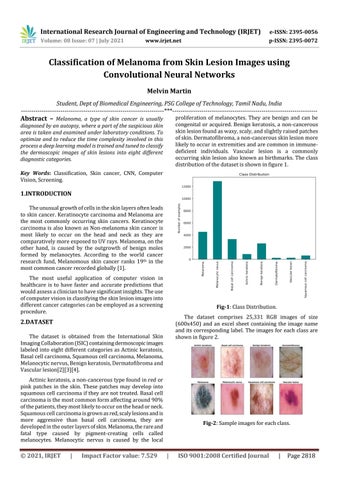
proliferation of melanocytes. They are benign and can be congenital or acquired. Benign keratosis, a non-cancerous skin lesion found as waxy, scaly, and slightly raised patches of skin. Dermatofibroma, a non-cancerous skin lesion more likely to occur in extremities and are common in immunedeficient individuals. Vascular lesion is a commonly occurring skin lesion also known as birthmarks. The class distribution of the dataset is shown in figure 1.
diagnosed by an autopsy, where a part of the suspicious skin area is taken and examined under laboratory conditions. To optimize and to reduce the time complexity involved in this process a deep learning model is trained and tuned to classify the dermoscopic images of skin lesions into eight different diagnostic categories. Key Words: Classification, Skin cancer, CNN, Computer Vision, Screening.
1.INTRODUCTION The unusual growth of cells in the skin layers often leads to skin cancer. Keratinocyte carcinoma and Melanoma are the most commonly occurring skin cancers. Keratinocyte carcinoma is also known as Non-melanoma skin cancer is most likely to occur on the head and neck as they are comparatively more exposed to UV rays. Melanoma, on the other hand, is caused by the outgrowth of benign moles formed by melanocytes. According to the world cancer research fund, Melanomous skin cancer ranks 19th in the most common cancer recorded globally [1]. The most useful application of computer vision in healthcare is to have faster and accurate predictions that would assess a clinician to have significant insights. The use of computer vision in classifying the skin lesion images into different cancer categories can be employed as a screening procedure.
Fig-1: Class Distribution. The dataset comprises 25,331 RGB images of size (600x450) and an excel sheet containing the image name and its corresponding label. The images for each class are shown in figure 2.
2.DATASET The dataset is obtained from the International Skin Imaging Collaboration (ISIC) containing dermoscopic images labeled into eight different categories as Actinic keratosis, Basal cell carcinoma, Squamous cell carcinoma, Melanoma, Melanocytic nervus, Benign keratosis, Dermatofibroma and Vascular lesion[2][3][4]. Actinic keratosis, a non-cancerous type found in red or pink patches in the skin. These patches may develop into squamous cell carcinoma if they are not treated. Basal cell carcinoma is the most common form affecting around 90% of the patients, they most likely to occur on the head or neck. Squamous cell carcinoma is grown as red, scaly lesions and is more aggressive than basal cell carcinoma, they are developed in the outer layers of skin. Melanoma, the rare and fatal type caused by pigment-creating cells called melanocytes. Melanocytic nervus is caused by the local
© 2021, IRJET
|
Impact Factor value: 7.529
Fig-2: Sample images for each class.
|
ISO 9001:2008 Certified Journal
|
Page 2818