International Research Journal of Engineering and Technology (IRJET)
e-ISSN: 2395-0056
Volume: 08 Issue: 06 | June 2021
p-ISSN: 2395-0072
www.irjet.net
AN AUTOMATED SYSTEM FOR CLASSIFICATION OF DIABETIC RETINOPATHY USING FASTER-RCNN K. Sakthiyavathi1, S. Amutha2, A. Vigneshwari3, T. Devadharshini4, R. Kiruthiga5 1Assistant
Professor, Department of Information Technology, Sri Manakula Vinayagar Engineering College, Puducherry 2Assistant Professor, Department of Information Technology, Sri Manakula Vinayagar Engineering College, Puducherry 3Department of Information Technology, Sri Manakula Vinayagar Engineering College, Puducherry 4Department of Information Technology, Sri Manakula Vinayagar Engineering College, Puducherry 5Department of Information Technology, Sri Manakula Vinayagar Engineering College, Puducherry ---------------------------------------------------------------------***---------------------------------------------------------------------and require trained ophthalmologists. The lack of skilled ABSTRACT: Diabetic Retinopathy is an eye disease caused as clinicians also leaves a large proportion of patients untreated and therefore receiving medical help too late, in part due to poor adherence and access to the retina screening process. However, early detection and prevention of DR progression are essential to reduce the rising threat of DR. Artificial intelligence offers a better solution to this problem. Deep learning is used for an end-to-end assessment of medical images to generate a predicted output. The diagnostic use of Fast RCNN algorithms is spreading in various medical healthcare areas like radiology and pathology. In ophthalmology, groundbreaking work has recently been conducted on the automation of DR detecting and prediction of various risk factors by Fast RCNN analysis of CFPs.
a result of semipermanent polygenic disease. Because the disorder progresses it results in distortion and blurred vision. The identification of DR stages color structure image needs dexterous clinicians to spot the presence of vital features that makes this a tough and time overwhelming task. As the DR accompanies numerous stages and differing challenges, it's tough to DR and it's tedious. Right now, build up a computerized division based mostly on order model for DR. At First, the original images are resized and green channels are extracted from the structure pictures. Then, the Adaptive Histogram Equalization (AHE) an image process technique is employed to enhance the distinction of the image and enhance the sides of the image. Later, The Faster R-CNN is utilized for classifying the structure into totally different grades of DR. This Faster R-CNN approach was found to be an efficient algorithm concerning speed and accuracy. The approximate accuracy of 89.5% was acquired from the Faster R-CNN.
Key Words: Fast Regional convolutional neural network (FRCNN), Diabetic retinopathy, deep learning, segmentation
I.
INTRODUCTION
Diabetic retinopathy (DR), a sight-threatening disease, happens because of diabetics that bring about the harm of cells in the retina when the glucose level of the patient was untypical. Around 29% of sugar patients with age above 42years possess Diabetic eye disease, and among them, 4.3% have an extreme level of DR that leads to vision loss. Therefore, patients with diabetes are exposed to the danger of this eye disease. If DR is untreated, blood & fluid leak from blood vessels of the retina leads to permanent vision loss. The underlying province of DR separating the clinical is finished by utilizing the procedure of fundus imaging the affected retinal structure of the eyes might be detected by focusing the eye by a retina specialist or a trained grader. But these methods are manual, which are time-consuming, © 2021, IRJET
|
Impact Factor value: 7.529
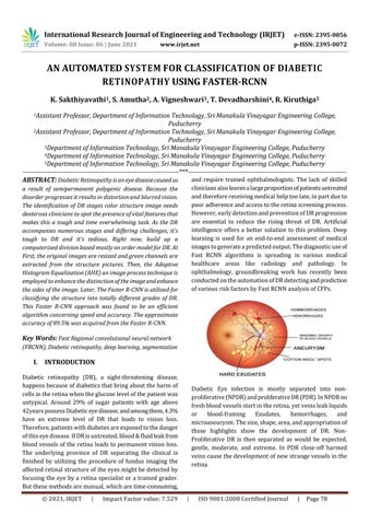
Diabetic Eye infection is mostly separated into nonproliferative (NPDR) and proliferative DR (PDR). In NPDR no fresh blood vessels start in the retina, yet veins leak liquids or blood-framing Exudates, hemorrhages, and microaneurysm. The size, shape, area, and appropriation of those highlights show the development of DR. NonProliferative DR is then separated as would be expected, gentle, moderate, and extreme. In PDR close-off harmed veins cause the development of new strange vessels in the retina.
|
ISO 9001:2008 Certified Journal
|
Page 78