International Research Journal of Engineering and Technology (IRJET) e-ISSN: 2395-0056 Volume: 08 Issue: 04 | Apr 2021
www.irjet.net
p-ISSN: 2395-0072
LUNG CANCER DETECTION USING CT SCAN IMAGES KHEVNA VASANI1, AYUSHI SHAH2 1 Student,
Dept. of Biomedical Engineering, L.D. College of Engineering, Ahmedabad, Gujarat, India Dept. of Biomedical Engineering, L.D. College of Engineering, Ahmedabad, Gujarat, India ---------------------------------------------------------------------***--------------------------------------------------------------------2 Student,
Abstract – Lung cancer is a malady caused by the
potential to diagnose this disease at an earlier stage. Cancer that begins from the lungs is termed as primary lung cancer. Cancer that spreads to the lungs from another place in the body is termed as secondary lung cancer. Human lung cancer is categorized into two main histopathological groups: non‐small cell lung cancer (NSCLC) and small‐cell lung cancer (SCLC). In the biomedical field, the examination and diagnosis of the lung CT image by field experts are a sensitive process that need time and high qualification. The subjective examination leads to variability among the observers. For these reasons, computer-based systems are required. The diagnosis process is supported by utilizing existing technological means and software. Thus, the cost and diagnosis effort can be notably reduced. Image processing techniques have been proven efficient to detect tumor cells. MATLAB is an optimum image processing software that can be used for the same. The first stage starts with collecting the CT scan images (normal and abnormal) from the open-source database. Several image enhancement techniques improve visual quality of an image. The third stage applies image segmentation algorithms which plays an effective role in image processing stages, and the fourth stage gives an indicator of normality or abnormality of images.
involuntary proliferation of cells within the lung tissue. Symptoms could be similar to those of respiratory problems or infections and typically there may be no symptoms at all. Early detection of cancerous cells within the lungs is indispensable as they provide oxygen to and excrete out carbon dioxide from the body. Fatality rate are often reduced by early detection and treatment of the disease. The method of early detection of cancer plays crucial role in preventing cancerous cells from multiplying and spreading. Lung cancer has been attracting the eye of medical and sciatic communities in the latest years as a result of its high prevalence allied with the challenging treatment. In recent years the image processing algorithms are widely used in various medical areas in order to enhancing the earlier detection and treatment stages, during which time factor is extremely important to diagnose the disease within the patient. However, CT scan imaging is best imaging technique in the medical field, it's difficult for doctors to interpret and diagnose the cancer from CT scan images. The main purpose of this project is to develop a CAD (Computeraided Diagnosis) system using MATLAB for detecting the initial carcinoma nodules using the lung CT scan images. Key Words: Lung Cancer, Cancerous Cells, CT Scan, CAD, MATLAB etc.
1.1 Aims and Objectives
1. INTRODUCTION
1) Performing Image segmentation following the Image Enhancement using various available techniques. 2) Detection of possible cancerous lung nodule locations, from enhanced segmented images. 3) Analysis and comparison of different algorithms to procure most suitable techniques.
CANCER is one among the most severe health issues within the world. Lung cancer is that the leading reason behind cancer deaths world. Carcinoma cancer cells have defects like autonomy of growth signals, insensitivity to growthinhibitory signals, limitless replicative potential, and tissue invasion and metastasis among the regulatory circuits that govern normal cell proliferation as well as homeostasis. It takes a series of mutations to create a cancer cell. Carcinoma arises from abnormal growth of epithelial cells in the lung. The clinical manifestations of lung cancer are usually varied. Patients are sometimes asymptomatic in the early stages of the disease. The lack of symptoms is especially true for lung cancers that originate in the periphery of the lungs. Approximately 5% to 10 % of patients with lung cancer does not show symptoms at the time of examination. These cancers are generally detected during evaluation for an unrelated health issues or on a chest radiograph performed for preoperative evaluation. More effective screening such as low-dose spiral computed tomography (CT) scanning that gives detailed information because of the sections of CT images structure, may allow for improvement in the
© 2021, IRJET
|
Impact Factor value: 7.529
Hence, reducing required time for analysis and improving the quality of diagnosis. Consequently, minimizing the frequency of biopsies for lung cancer detection.
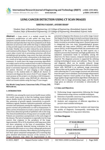
1.2 Flow Diagram CT SCAN IMAGES
IMAGE ENHANCEMENT
|
GABOR FILTER
ISO 9001:2008 Certified Journal
|
Page 3600