International Research Journal of Engineering and Technology (IRJET)
e-ISSN: 2395-0056
Volume: 08 Issue: 04 | Apr 2021
p-ISSN: 2395-0072
www.irjet.net
Detecting Covid-19 In Chest X-ray Images Using Neural Networks Saarthak Gupta1 1Student,
Shikshantar Senior Secondary School, Gurugram, Haryana, India ---------------------------------------------------------------------***---------------------------------------------------------------------
Abstract – In this paper we study the applications of
a system which can perform this task automatically greatly aids the medical fraternity.
Artificial Intelligence, particularly, neural networks in predicting whether a patient is inflicted with Covid-19, the illness caused by the SARS-CoV-2 virus. Convolutional Neural Networks (CNN), a subtype of deep neural networks are the most suitable for image analysis. In this study, the Convolutional neural network algorithm is applied to chest X-ray images of patients afflicted with various respiratory pathologies to determine if they are suffering from Covid-19. Another key aspect is differentiating between Covid-19 and other lung illnesses like viral pneumonia.
2. DATA COLLECTION AND ANALYSIS The dataset on Covid-19 radiography images was created by researchers from the Qatar University in Doha, Qatar and the University of Dhaka in Dhaka, Bangladesh [5,6] along with other collaborators. Total number of images is 21,165. An overview of the dataset is presented in Table-1. Table -1: Covid-19 Radiography Dataset Overview
Key Words: Neural Networks, COVID-19, Convolution, X-ray, Image Analysis
Covid-19 Radiography Data
1. INTRODUCTION With a shortage of testing kits and healthcare infrastructure in many countries overwhelmed, new, accurate and automated methods to detect Covid-19 infections are the need of the hour. Modern A.I. (Artificial Intelligence) techniques can be effective to detect COVID19 in medical images, particularly when radiologists are not available [1]. Covid-19 produces ground glass and consolidative opacities with a bilateral, peripheral, and lower lung distribution [2]. We aim to detect these lung opacities using Convolutional Neural Networks and help identify Covid-19 in the medical images of patients.
Image Class
Percent
Number of Images
Healthy
48.2%
10,192
Lung Opacity
28.4%
6,012
Covid-19
17.1%
3,616
Viral Pneumonia
6.4%
1,345
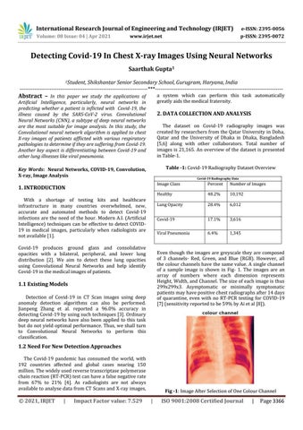
Even though the images are greyscale they are composed of 3 channels- Red, Green, and Blue (RGB). However, all the colour channels have the same value. A single channel of a sample image is shown in Fig- 1. The images are an array of numbers where each dimension represents Height, Width, and Channel. The size of each image is thus 299x299x3. Asymptomatic or minimally symptomatic patients may have positive chest radiographs after 14 days of quarantine, even with no RT-PCR testing for COVID-19 [7] (sensitivity reported to be 59% by Ai et al [8]).
1.1 Existing Models Detection of Covid-19 in CT Scan images using deep anomaly detection algorithms can also be performed. Jianpeng Zhang et al. reported a 96.0% accuracy in detecting Covid-19 by using such techniques [3]. Ordinary deep neural networks have also been applied to this task but do not yield optimal performance. Thus, we shall turn to Convolutional Neural Networks to perform this classification.
1.2 Need For New Detection Approaches The Covid-19 pandemic has consumed the world, with 192 countries affected and global cases nearing 150 million. The widely used reverse transcriptase polymerase chain reaction (RT-PCR) test can have a false negative rate from 67% to 21% [4]. As radiologists are not always available to analyse data from CT Scans and X-ray images,
© 2021, IRJET
|
Impact Factor value: 7.529
Fig -1: Image After Selection of One Colour Channel
|
ISO 9001:2008 Certified Journal
|
Page 3366