International Research Journal of Engineering and Technology (IRJET)
e-ISSN: 2395-0056
Volume: 08 Issue: 04 | Apr 2021
p-ISSN: 2395-0072
www.irjet.net
DETECTION OF TUMOUR FROM MRI SCAN IMAGES OF BRAIN USING IMAGE PROCESSING TECHNIQUES BY MATLAB Chaudhari Divyalata R #1, Himanshu A patel*2, Tejas V Bhatt#3 1-3Biomedical Engineering M.tech, (U. V. Patel College of Engineering, Ganpat University, Mehsana, Gujarat-(384012) --------------------------------------------------------------------------***--------------------------------------------------------------------
Abstract— Tumour detection is a one of the medical issue
areas of the brain and cause serious health issues. Benign tumors can be removed, and they are rarely grown back.
that is still challenging for medical field. A brain tumour is a mass of nerve tissues that have grown uncontrollably .There are two main types of tumour malignant and benign tumours. Medical image processing techniques plays a most significant role in the diagnosis of brain tumours. In this diagnosis the medical image data obtained from various biomedical devices. This data is acquired from various imaging techniques like X-ray, CT-scan and MRI. Radiologist perform tumour extraction on data acquire from magnetic resonance imaging (MRI) which is very time consuming. Brain tumour segmentation is a significant process to extract information from complex (MRI).Medical image processing is the most useful and developed field now a day. In this paper mentioned the strategy to detect and extraction of brain tumour from MRI scan images of the brain. This method incorporates with some noise removal filters, histogram equalization, segmentation unit and morphological operations and these all are the basic concept of image processing. Using MATLAB it takes less time to detect tumour.
1.2 Malignant Brain Tumour: Malignant brain tumours have a heterogeous structure of cells. They are likely to grow fast and raid into the nearby healthy brain tissue. MRI is an advanced medical imaging technique used to examine several regions of the human body. MRI imaging is used when treating brain tumours, ankle, and foot. From these highresolution images, we can conclude detailed anatomical information to examine human normal brain and detect abnormalities. Nowadays there are various techniques for classifying MR images, which are fuzzy methods, neural networks, atlas methods, knowledge based techniques, variation segmentation. MRI consists of T1 weighted, T2 weighted and PD (proton density) weighted images.
Key words— Brain tumour, MRI image, Histogram equalization, Watershed segmentation, MATLAB.
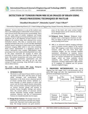
2. PROPOSED METHODOLOGY: This block diagram has two stages, first is pre-processing unit of given MRI image and after that segmentation and then performs morphological operations.
1. INTRODUCTION The brain is a soft mass of tissue which is cover by the bones of the skull, and fine layers of tissue called meninges and cerebrospinal fluid. Sometimes in the brain occurs abnormal growth of tissues causing brain tumour. Epilepsy is a brain disorder in which clusters of nerve cells occurs and the brain signals became abnormally. Here, neurons produce electrical impulses that act on various neurons, glands and muscles to produce human thoughts, feelings and actions. Here, the normal pattern of neuronal activity became disturbed causing strange sensations, emotions, behavior and loss of consciousness. Tumour are classify basically into two types:
Input MRI Image
|
Impact Factor value: 7.529
Threshold segmentatio n
Morphologi cal operation Tumour Detected (output)
1.1 Benign Brain Tumour: The benign tumour has uniformity in cell structure and does not contain cancer cells. They don't rise to other parts of the body. The benign tumours can grab sensitive
© 2021, IRJET
Preprocessing unit
Centre and area unit
Watershed segmentatio n
Fig. 1. Block Diagram for Brain Tumour Extraction
|
ISO 9001:2008 Certified Journal
|
Page 3013