International Research Journal of Engineering and Technology (IRJET) Volume: 08 Issue: 04 | Apr 2021
www.irjet.net
e-ISSN: 2395-0056 p-ISSN: 2395-0072
A Survey on Detection and Segmentation of Optic Disc in Retinal Images Prof. Nilima Kulkarni1, Ameya Kale2, Ishan Jawade3, Pratik Kakade4, Rushikesh Jadhav5 2, 3, 4,5
Student, Dept. of Computer Science and Engineering, MIT School of Engineering, Maharashtra, India Professor, Dept. of Computer Science and Engineering, MIT School of Engineering, Maharashtra, India ------------------------------------------------------------------------***------------------------------------------------------------------1
Abstract - Optic disc detection in retinal images is a trivial step in the process of diabetic retinopathy and glaucoma detection. Thus, it plays an important role in automatic retinal screening systems. Segmentation is also considered as one of the methods to locate the position of optic disc in optic images. Multiple methodologies have been developed for optic disc detection and disc diameter calculation, few of these literatures are discussed in this paper. These methods include conventional approaches using machine learning algorithms as well as deep learning-based object detection and segmentation approaches. Key Words: Optic Disc, Object Detection, Segmentation, Deep Learning, CNN.
1. INTRODUCTION Optic disc (OD), also known as the optic nerve head is a small blind spot in the eye which acts as an exit point for the ganglion cell axons leaving the eye. It plays a very important role in diabetic retinopathy and glaucoma detection. There are multiple methodologies which can help to find out the various anomalies in the optic disc size which can further help us in detection of the above- mentioned diseases.
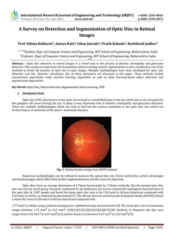
Fig -1: Retinal fundus image from DRIVE dataset Numerous methodologies can be utilized to measure the optical disc size. Every method has certain advantages and disadvantages which affect their further implementations and the research objectives. Optic discs have an average dimension of 1.76mm horizontally by 1.92mm vertically. But the normal optic disc size can vary by racial group. Research conducted by the Baltimore eye survey studied the topological characteristics of the optic disc in 3,387 people and found the mean optic disc area to be 2.94 mm2 in African Americans compared with 2.63 mm2 in whites, as obtained by planimetry[17]. The African Descent and Glaucoma Evaluation Study (ADAGES) found a mean disc area of 2.06 mm2 in African Americans compared with 1.77 mm2 in whites using confocal scanning laser ophthalmoscopy measurements [9]. The mean disc area in Caucasians ranges between 1.73 mm2 to 2.63 mm2, [10][11][12][13][14][15][16][17][18]. Similarly in Hispanics the disc area ranges from 2.46 mm 2 to 2.67 mm2,[16] and for Asians it is between 2.47 mm2 to 3.22 mm2[15].
© 2021, IRJET
|
Impact Factor value: 7.529
|
ISO 9001:2008 Certified Journal
|
Page 1545