International Research Journal of Engineering and Technology (IRJET)
e-ISSN: 2395-0056
Volume: 07 Issue: 09 | Sep 2020
p-ISSN: 2395-0072
www.irjet.net
Early Detection of Brain Stroke using MRI Images R Aishvarya1, RJ Anand2, Batchali Vasundhara3, Dr S Sudha4 1,2,3Electronics
and Communication Engineering, Easwari Engineering College, Chennai -78 Dept. of Electronics & Communication Engineering, Easwari Engineering College, Tamil Nadu, India ---------------------------------------------------------------------***---------------------------------------------------------------------4Professor,
Abstract - Medical imaging is a well developing and
demanding technology in today’s medical field. Early Brain stroke detection plays an important role as it can limit the brain damage and also reverse the long term repercussions caused by the complexity of the stroke .However, the location of ischemic stroke in the MRI image is not apparent and the physical identification of lesion area takes additional epoch. The purpose of this paper is to develop an automated stroke detection algorithm using machine learning. The proposed methodology describes a series of novel techniques to detect & categorize brain stroke. The entire process is divided into six distinctive stages. Subsequent to preprocessing the image to functional standards, Gabor Filters are utilized to improve the quality of the MRI image. Adaptive histogram equalization (AHE) is the technique adapted for enhancement. Finally Fuzzy C Means technique is used to fragment the image and GLCM is applied for extracting features. These features are trained and considered for classification process through Multiclass SVM. From the experimental results, the accuracy of the proposed module is found to be higher than 90%. The algorithm proposed is simple, efficient and less time consuming.
Key Words: MRI images, Preprocessing, Filtering,
Enhancement, Fuzzy c means, Gray Level Co-occurrence Matrix (GLCM), Multi SVM.
Being an imperative organ, human brain is dependent to a durable blood supply analogous to our heart for durability. The malfunctioning of the arteries that supply blood to the brain roots a brain stroke. This brain attack can be disengaged into two, the ischematic stroke and hemorrhagic stroke which disarray the brain function. The visual representation of the interior human body can be caused by bio medical imaging technique. Multifarious imaging techniques are used in medical monopoly such as x-ray, ultra sounds, Computed Tomography (CT) scan and Magnetic Resonance Imaging(MRI) scan. . On comparison with the other medical imaging, The MRI plays a vital role in dispensing analogous images of the brain with equivalent resolution in various projections. On obtaining images in multiple planes, augment the versatility in diagnosing utility. This gives a beneficial advantage for radiation or surgical treatment planning. The former part of the paper delineates the background of the brain stroke recognition using image processing techniques and posterior, its output is given admittance into the filtering part. Later, estimations of various features are ascertained. The system has undergone |
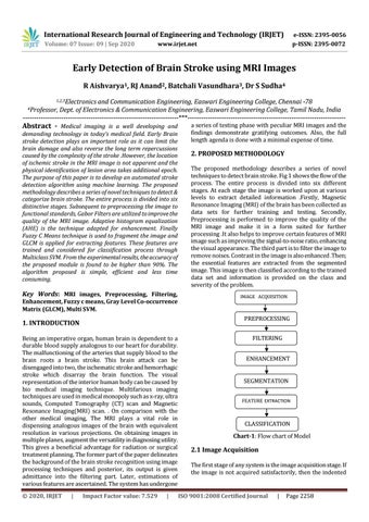
2. PROPOSED METHODOLOGY The proposed methodology describes a series of novel techniques to detect brain stroke. Fig 1 shows the flow of the process. The entire process is divided into six different stages. At each stage the image is worked upon at various levels to extract detailed information .Firstly, Magnetic Resonance Imaging (MRI) of the brain has been collected as data sets for further training and testing. Secondly, Preprocessing is performed to improve the quality of the MRI image and make it in a form suited for further processing .It also helps to improve certain features of MRI image such as improving the signal-to-noise ratio, enhancing the visual appearance. The third part is to filter the image to remove noises. Contrast in the image is also enhanced .Then; the essential features are extracted from the segmented image. This image is then classified according to the trained data set and information is provided on the class and severity of the problem. IMAGE ACQUISITION
PREPROCESSING
1. INTRODUCTION
Š 2020, IRJET
a series of testing phase with peculiar MRI images and the findings demonstrate gratifying outcomes. Also, the full length agenda is done with a minimal expense of time.
Impact Factor value: 7.529
|
FILTERING ENHANCEMENT SEGMENTATION FEATURE EXTRACTION
CLASSIFICATION Chart-1: Flow chart of Model
2.1 Image Acquisition The first stage of any system is the image acquisition stage. If the image is not acquired satisfactorily, then the indented
ISO 9001:2008 Certified Journal
|
Page 2258