International Research Journal of Engineering and Technology (IRJET)
e-ISSN: 2395-0056
Volume: 07 Issue: 09 | Sep 2020
p-ISSN: 2395-0072
www.irjet.net
Glaucoma Disease Detection by using Tree Bagging Algorithm in Fundus Images of Eye Payal N. Rabade1, Radhika Harne2 1M.Tech
Student, Department of Electronics Engineering, Government College of Engineering Amravati, Maharashtra, India. 2Assistant Professor, Department of Electronics Engineering, Government College of Engineering Amravati, Maharashtra, India. ---------------------------------------------------------------------***---------------------------------------------------------------------Abstract - Glaucoma is the second leading cause of blindness across the world. It is an eye disease in which Intra-ocular Pressure (IOP) in the eye increases continuously, that affects the optic nerve of eye which carries signals to the eye. This disease can leads to permanent blindness if not treated at earlier stage. In this paper, we present a novel method based on feature extraction and bootstrap aggregating, also called tree bagging to detect Glaucoma and classify normal eye and glaucomatous eye patient. Bagging is a machine learning ensemble meta-algorithm designed to improve the stability and accuracy, used in statistical classification. Wavelet based texture feature and statistical features are used for detection of glaucoma from fundus images. Tree Bagger classifiers is used for classification of normal eye patient and patient having Glaucoma. Classification accuracy of tree Bagger classifier is 96.1% for fundus image of different patient. Processing time of tree bagger classifier for testing dataset is 16.40s. Key Words: Glaucoma, Intra ocular pressure(IOP), Optic nerve, Fundus image, Tree bagger classifier. 1. INTRODUCTION Glaucoma comes second after diabetic retinopathy in eye diseases which causes blindness. Glaucoma is a eye disease that damages the optic nerve in the eye that carries the images that we see to our brain. In healthy eye, a transparent liquid called the aqueous humor circulates inside the front portion of the eye to maintain a constant healthy eye pressure. The eye continuously produces a small amount of aqueous humor, an equal amount of this liquid flows out of the eye through a microscopic drain which is known as a trabecular meshwork in the drainage angle. In case of glaucoma patients the aqueous humor does not flow through the drainage angle properly causing fluid pressure called as intraocular pressure (IOP) in the eye to increase, and this pressure causes damage to the optic nerve fibers. These damaged nerve fibers leads to patches of vision loss, and if not treated at early stage it may lead to total blindness. Symptoms also appear gradually, starting with preferable vision loss, which may go unnoticed until the central vision is affected. Fundus images are obtained from fundoscopy, which is one of the modern medical imaging techniques that helps ophthalmologists to identify structural changes in the Š 2020, IRJET
|
Impact Factor value: 7.529
|
optic disc to detect Glaucoma. In India, at least 12 million people affected by Glaucoma and nearly 1.2 million people blind from the disease. Glaucoma prevalence increases with age. Image processing based approach for analyzing fundus images for diagnosis of glaucoma is an emerging area of research and some work has been reported in this area.
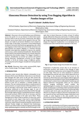
Fig -1: Input Fundus Image from Drishti-GS1 dataset. Progression of glaucoma occurs due to an increase in intraocular pressure and results in the damage of optic nerve. Progression of glaucoma can be stopped if detected at an early stage. There are no early symptoms in patients having glaucoma and the only way to detect glaucoma at an early stage is the structural change that occurs in the internal eye. Most Glaucoma cases happen without signs and symptoms because peripheral vision can be damaged before an individual’s central vision is affected [4]. Fundoscopy and Optical Coherence Tomography (OCT) are two modern medical imaging techniques which enables the ophthalmologists to analyze the internal structural retinal details. Fundoscopy is a technique which reveals the internal fundus details[11]. In glaucoma detection, Fundoscopy helps to examine optic nerve, optic cup and optic disc as it is cost effective as compare to Optical coherence tomography, So in our research we have used fundus images. Glaucoma is not curable, so early diagnosis is the only possible way to prevent loss of vision. Computer aided diagnosis system may help screening the disease [2]. 2. LITERATURE SURVEY Juan Carrillo, Lola Bautista et al.[1] gives computational tool for automatic glaucoma detection from fundus image of the eye. The disc segmentation was done by thresholding, the ISO 9001:2008 Certified Journal
|
Page 2105