International Research Journal of Engineering and Technology (IRJET)
e-ISSN: 2395-0056
Volume: 07 Issue: 08 | Aug 2020
p-ISSN: 2395-0072
www.irjet.net
CLASSIFICATION OF MELANOMA FROM DERMOSCOPIC IMAGES USING DEEP LEARNING Hima H A1, Celine Mary Stuart2 1M.
Tech Student, Dept. of Electronics and Communication, Government Engineering College, Kerala, India Professor, Dept. of Electronics and Communication, Government Engineering College, Kerala, India ---------------------------------------------------------------------***---------------------------------------------------------------------2Associate
Abstract - Malignant melanoma is one of the rapidly
itchiness, skin breakdown. It also rarely occur on normally appearing skin.
increasing and deadly diseases in the world. Early diagnosis is of great importance for treating the disease. Accurate observation of skin lesions is needed for melanoma detection. Dermoscopy is a non-invasive technique for observation of skin lesion. Manual observation of dermoscopic images for classification of lesion as benign or malignant can be inaccurate and subjective. Therefore computer aided diagnosis (CAD) plays a significant role for assisting in melanoma detection. Steps involved in traditional computer aided diagnosis of dermoscopic images involve a) Segmentation b) Feature extraction and c) Classification. Rather than using different tools for segmentation, feature extraction and classification, convolutional neural network (CNN) – a deep learning method can be used inorder to do automatic classification of skin lesion. In this work xception architecture of CNN is used as an end to end classification framework. This architecture is trained using three optimizers. Classification performance of the architecture with three optimizers is analysed using thresholding and ranking metrics. Using these metrics the optimal combination of xception architecture and optimizer for melanoma classification is identified.
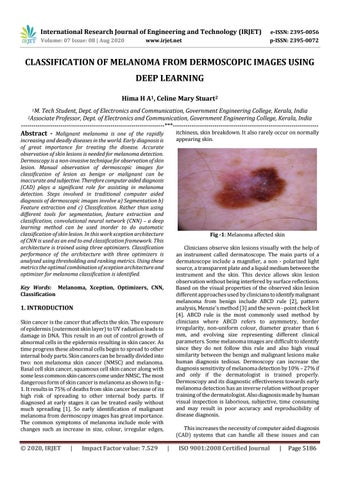
Fig -1: Melanoma affected skin Clinicians observe skin lesions visually with the help of an instrument called dermatoscope. The main parts of a dermatoscope include a magnifier, a non - polarized light source, a transparent plate and a liquid medium between the instrument and the skin. This device allows skin lesion observation without being interfered by surface reflections. Based on the visual properties of the observed skin lesion different approaches used by clinicians to identify malignant melanoma from benign include ABCD rule [2], pattern analysis, Menzie’s method [3] and the seven - point check list [4]. ABCD rule is the most commonly used method by clinicians where ABCD refers to asymmetry, border irregularity, non-uniform colour, diameter greater than 6 mm, and evolving size representing different clinical parameters. Some melanoma images are difficult to identify since they do not follow this rule and also high visual similarity between the benign and malignant lesions make human diagnosis tedious. Dermoscopy can increase the diagnosis sensitivity of melanoma detection by 10% – 27% if and only if the dermatologist is trained properly. Dermoscopy and its diagnostic effectiveness towards early melanoma detection has an inverse relation without proper training of the dermatologist. Also diagnosis made by human visual inspection is laborious, subjective, time consuming and may result in poor accuracy and reproducibility of disease diagnosis.
Key Words: Melanoma, Xception, Optimizers, CNN, Classification
1. INTRODUCTION Skin cancer is the cancer that affects the skin. The exposure of epidermis (outermost skin layer) to UV radiation leads to damage in DNA. This result in an out of control growth of abnormal cells in the epidermis resulting in skin cancer. As time progress these abnormal cells begin to spread to other internal body parts. Skin cancers can be broadly divided into two: non melanoma skin cancer (NMSC) and melanoma. Basal cell skin cancer, squamous cell skin cancer along with some less common skin cancers come under NMSC. The most dangerous form of skin cancer is melanoma as shown in fig 1. It results in 75% of deaths from skin cancer because of its high risk of spreading to other internal body parts. If diagnosed at early stages it can be treated easily without much spreading [1]. So early identification of malignant melanoma from dermoscopy images has great importance. The common symptoms of melanoma include mole with changes such as increase in size, colour, irregular edges,
© 2020, IRJET
|
Impact Factor value: 7.529
This increases the necessity of computer aided diagnosis (CAD) systems that can handle all these issues and can
|
ISO 9001:2008 Certified Journal
|
Page 5186