International Research Journal of Engineering and Technology (IRJET)
e-ISSN: 2395-0056
Volume: 06 Issue: 06 | June 2019
p-ISSN: 2395-0072
www.irjet.net
Survey Based on Detection of Optic Disc in Retinal Images Using Segmentation Based Techniques D Binny Jeba Durai1, Dr. T. jaya2 1Udhya
Polytechnic College, Vellamadam. Institute of Technology, Thovali. ----------------------------------------------------------------------***--------------------------------------------------------------------2CSI
Abstract— Automated localization and detection of the optic disc (OD) is an essential step in the analysis of digital diabetic retinopathy systems. Accurate localization and detection of optic disc boundary is very useful in proliferative diabetic retinopathy where fragile vessels develop in the retina. Detection of optic disc area is complex because it is located in an area that is considered as pathological blood vessels. The optic disc appears as a round region usually brighter than the surrounding. The results achieved by different algorithms can be compared when algorithms are applied on the same standard databases. In this paper we compare the eccentricity, accuracy and brightness using different segmentation techniques.
II. LITERATURE REVIEW
Keywords — optic disc, segmentation, fundus image, detection
Hough transform [4] is a technique capable of finding geometric shapes within an image and was employed to detect the OD. The Hough transform uses a couple of methods. In the first one, Hough transform was applied only to pixels on or close to the retinal vasculature in a binary image of the vasculature. The binary vasculature was dilated in order to increase the possible OD candidates. Alternatively, in the second method Hough transform was applied once again but only to the brightest 0.35% of the fuzzy convergence image obtained. Once more, dilation was applied to the convergence image to overcome the gaps created by small vessels. The center and boundary of the optic disc are found by applying the Hough transform to the gradient image. The Hough transform is a method for finding shapes in an image. The basic idea behind the Hough transform is to transform the image into a parameter space that is constructed specifically to describe the desired shape analytically. Maxima in this parameter space then correspond to the presence of the desired shape in image space.
Frank ter Haar [6] applied illumination equalization to the green-band of the image, and then a resolution pyramid using a simple Haar-based discrete wavelet transform was created. Finally, the brightest pixel at the fifth level of the resolution pyramid was chosen to correspond to the OD area. They also proposed an alternative to the later method based on the pyramidal decomposition of both the vasculature and the greenband, where the fifth level of the resolution pyramid for both the illumination equalized green-band and the binary vessel segmentation are summed, and the highest value corresponds to the OD center.
I. INTRODUCTION Diabetic Retinopathy (DR) is a result of long term diabetes mellitus and is a significant growing public health problem. It is one of the predominant causes of blindness. It causes pathological changes of the retina such as microaneurysms, intraretinal microvascular abnormalities, venous bleeding and neovascularities as well as haemorrhages, exudates and retinal oedema. Regular screening of Diabetic Retinopathy is indispensable so that appropriate and timely treatment can be given which thereby reduces the incidence of impaired vision and blindness from this condition. Current methods of detection and assessment of diabetic retinopathy are manual, expensive and require highly trained personnel to read large number of fundus images. The efficiency can be improved by automating the initial task of analyzing the huge amount of retinal fundus images. The optic disc is the brightest part in fundus images that can be seen as a pale, round or slightly oval disk. It is the entrance region of blood vessels and also acts as a landmark and reference for the other features in the fundus image. There are several methods for optic disc detection.
The circular hough transform (CHT) is used to detect the OD which has a roughly circular shape. The retinal vasculature in the green-band image was suppressed using the closing morphological operator. The Sobel operator and a simple threshold were then used to extract the edges in the image. CHT [8] was finally applied to the edge points, and the largest circle was found consistently to correspond to the OD. This is suitable for normal healthy fundus images where in optic disc is alone the brightest region of the image. But our
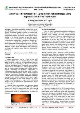
Fig 1. Original Retinal Fundus Image
© 2019, IRJET
|
Impact Factor value: 7.211
|
ISO 9001:2008 Certified Journal
|
Page 795