International Research Journal of Engineering and Technology (IRJET)
e-ISSN: 2395-0056
Volume: 06 Issue: 02 | Feb 2019
p-ISSN: 2395-0072
www.irjet.net
A Review Paper on Detection of Bone Tumor using Comparative Analysis of Segmentation Technique Prachi B.Tamgadge1, Dr. N.K Choudhari2, Dr. D.M. Kate3 1PG
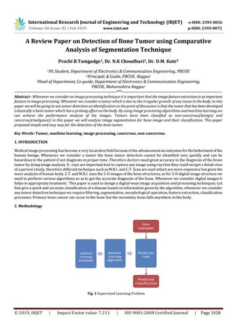
Student, Department of Electronics & Communication Engineering, PBCOE 2Principal, & Guide, PBCOE, Nagpur 3Head of Department, Co-guide, Department of Electronics & Communication Engineering, PBCOE, Maharashtra Nagpur ----------------------------------------------------------------------***--------------------------------------------------------------------Abstract - Whenever we consider an image processing technique it is important that the image feature extraction is an important feature in image processing. Whenever we consider a tumor which is due to the irregular growth of any tissue in the body. In this paper we will be going to use tumor detection an identification so the point of discussion is that the tumor that has been developed is basically a bone tumor which has a prolong effect on the body. By using image processing algorithms and machine learning we can achieve the performance analysis of the images. Tumors have been classified as non-cancerous(benign) and cancerous(malignant) in this paper we will analyze image segmentation for bone image and their classification. This paper proposed simple and easy way for the detection of the bone tumor. Key Words: Tumor, machine learning, image processing, cancerous, non-cancerous. 1. INTRODUCTION Medical image processing has become a very locarative field because of the advancement an outcomes for the betterment of the human beings. Whenever we consider a tumor the bone tumor detection cannot be identified very quickly and can be hazardous to the patient if not diagnosis in proper time. Therefore doctors need great accuracy in the diagnosis of the brain tumor by doing image analysis. X –rays are important tool to capture any image using rays but they could not get a detail view of a person’s body, therefore different technique such as M.R.I. and C.T. Scan are used which are more expensive but gives the more analysis of human body. C.T. and M.R.I. uses the 3-D images of the bone structures, so for 3-D digital image structure we need to perform various algorithms so as to get the accurate diagnosis of the bone. Whenever we consider digital images it helps in appropriate treatment. This paper is used to design a digital ways image acquisition and processing techniques. Let him give a quick and accurate classification of a disease based on information given by the algorithm, whenever we consider any tumor detection technique we require filtering, segmentation, morphological operation, feature extraction, classification processes. Primary bone cancer can occur in the bone but the secondary bone falls anywhere in the body. 2. Methodology
© 2019, IRJET
|
Impact Factor value: 7.211
|
ISO 9001:2008 Certified Journal
|
Page 1458