International Research Journal of Engineering and Technology (IRJET) e-ISSN: 2395-0056
Volume: 12 Issue: 05 | May 2025 www.irjet.net p-ISSN: 2395-0072


International Research Journal of Engineering and Technology (IRJET) e-ISSN: 2395-0056
Volume: 12 Issue: 05 | May 2025 www.irjet.net p-ISSN: 2395-0072
Shrikant
Bhaginath Bodkhe1, Dr. Sugandha Nandedkar2, Dr. Shaikh Shoaib3
1B. Tech in Computer Science and Engineering (Artificial Intelligence & Machine Learning), Deogiri Institute of Engineering and Management Studies, Aurangabad, Maharashtra
2,3 Assistant Professor, Deogiri Institute of Engineering and Management Studies, Aurangabad, Maharashtra ***
Abstract - Skin diseases are one of the most common problems with human health around the world, affecting millions of indicisualseachyear. Earlyandaccuratediagnosis is extremelyimportant intimelytreatment andmanagement. This project presents a deep learning-based system for automatic classification of skin diseases using convolutional neural networks (CNNs)thatfoldstheMobilenetV2asthebase model. The data record consists of eight different classes of skin infections, including bacteria, fungi, parasitic and viral diseases. Images are subject to preprocessing steps such as resizing, normalization, and labeling coding to ensure consistency and optimal performance. The proposed framework reaches 97% accuracy and demonstrates its potential as an efficient and reliable means of supporting dermatologists in the diagnosis of skin diseases.
Key Words: Skin Disease Detection, Convolutional Neural Network (CNN), MobileNet V2, Deep Learning, Image Classification, Medical Diagnosis, Artificial Intelligence, Accuracy97%.
Theskinisthelargestorganofthehumanbody,playinga crucialroleasaprotectivebarrieragainstbacteria,viruses, germs, and harmful ultraviolet (UV) radiation. Beyond protection, it performs several vital functions such as regulating body temperature, enabling the sensation of touch, and contributing to overall physical appearance. Healthy skin not only reflects good health but also significantly boosts self-confidence. Yet skin, while being important,isthesubjectofmanydiseases,mostofwhichare increasingly common in today's world. There are some highly contagious skin conditions which have a very big publichealthimpact.Onmanyoccasions,peoplemisstheir firstdoseordelaytreatmentsintheearlystagesofcancer development because they are unaware or cannot afford treatmentortheyare too busy.Ifthatis, negligence often leadsto moreserious complications,even lifethreatening ones, such as skin cancer. A lot of awareness needs to be raisedconcerningskinhealth,earlydiagnosis,andgetting timelytreatmentwithaviewtoavoidingsuchoutcomes.In thecaseofskincare,Iamhappytosaythat prioritizing it and acknowledging the importance of it as an important organcouldleadtohealthierlivesandlessenskindisease burdenonindividualaswellasthehealthcaresystem.
Eachyear,about900millionpeoplearoundtheworldhave skindiseases–fromsimpleconditionslikeacnetopsoriasis
and skin cancer, a WHO report says. In 2019, there were 4,859,267,654newcasesofskinandsubcutaneousdiseases (95%UI4,680,693,440–5,060,498,767cases)worldwide,of whichbacterialandfungalskindiseasescomprisedthebulk ofthenewcases.Afourthmakeup34%andfifthaccounted for23%ofthetotal[1].
Accordingtothereport,"4,859,267,654(95%uncertainty interval [UI], 4,680,693,440–5,060,498,767) new skin and subcutaneousdiseasecasesthatwereidentified,mostwere fungal(34.0%)andbacterial(23.0%)skindiseases,which accountedfor98,522(95%UI75,116–123,949)deaths.The burden of skin and subcutaneous diseases measured in DALYs was 42,883,695.48 (95% UI, 28,626,691.71–63,438,210.22)in2019,5.26%ofwhichwereyearsoflife lost,and94.74%ofwhichwereyearslivedwithdisability" (Yakupuetal.,2023)[1].
Wehaveseenthegrowthofmanytechnologiesasmedical sciencehasadvancedandmanyofthesehaveenabledusto better diagnose those skin diseases. Despite this, many derms still depend on manual inspection to spot skin conditions.Thoughthisarchetypicalapproachshallalways betheoptionofmanypractitioners,itisn’tflawless.Itisa timeconsuming,menialandpronetohumanerror,manual diagnosis. The outcome of which is that despite the complexity of certain dermatological conditions, it is not uncommonthatdifferentspecialistscometoverydifferent conclusionsaboutadiagnosisortreatmentplan.
Acombinationofclinicalexperiencewithunpredictableskin presentations may make the continued reliance on blind manual examination a reality. Skepticism towards fully automationhasalsobeenprovokedbythesefactors.Though the potential benefits for integration of computer based diagnostictoolsintodermatologymaynotbesoobvious,the potential benefits are worthwhile. They also available in automated systems, much faster and more accurate in diagnosis with artificial intelligence (AI) and machine learning(ML)involved.Withaccesstohugeamountsofdata, thesemodelsareabletoidentifydelicatepatternsthatmay be passed over for the human eye, helping to lower diagnosticmistakesandvariety.
Rather, the goal of AI/ML systems would be to act as a powerful decision support tool rather than replacing dermatologists. They might help clinicians make more consistent and precise diagnoses, ultimately helping patients. These technologies can become novel research

International Research Journal of Engineering and Technology (IRJET) e-ISSN: 2395-0056
Volume: 12 Issue: 05 | May 2025 www.irjet.net p-ISSN: 2395-0072
elements in dermatology when their components are integrated together properly. Automated diagnostic tools complement rather than replace traditional methods, bridgingbetweenhumanexpertiseandcapacitytoperform moreroutinetasks.
Anautomationintegrationintothediagnostic processhas the potential to transform future outcomes. In addition to theabilitytorevolutionizedetectionofskindiseases,ithas thepotentialtoimproveoverallpatientcarestandards,as wellasmeetinghealthcaredeliveryneeds.Steppingtoward these advancements better means a more speeds up and moredependabledermatologicalpractice,onethatexploits the strengths of each human in addition to computational exactness.
Skindiseasesirritatemillionsofpeoplearoundtheworld. Earlydetectionbenefitsthepatientoutcomesandrelieves health care systems of a burden. In recent years, more researchershavebeenusingdeeplearning asabranchof artificial intelligence tobuildautomatedsystemstospot and classify skin diseases in medical images. Designed to assistdermatologistsinimprovingaccuracyandefficiencyin dermatologicdiagnostic procedures,these systemsaim to reduce the delays caused by the time required for biopsy processingandreportingtoinitialdiagnosis.
Inthispaper,wereviewstateoftheartfordeeplearningfor skin disease prediction. It looks at the use of different methodologies in recent studies, convolutional neural networksinparticularaswellasotheradvancedAImodels. A review of distributed SML is provided along with the discussion of some ongoing challenges facing the technologies, including data quality, variability in skin presentation, and the requirement of large and diverse datasets for training robust models. Moreover, it explores whatfutureresearchshouldentail,includingembeddingthe AI tools in a clinical setting and how to implement these toolsinanethicalandtransparentway.Finally,overall,deep learningcouldenablea revolutioninthediagnosis ofskin disease,andsubsequentimprovementsinpatientcare.
1. Ki V., Rotstein C. proposed the work [2] on Bacterial skin and soft tissue infections in adults: The epidemiology,pathogenesis,diagnosis,andtreatment of infections are reviewed herein. Likely pathogens, routeofentry,diseaseseverity,andassociatedclinical complications will determine the selection of antimicrobial therapy. Knowledge of these factors is important for treatment and improved patient outcomes for infectious diseases, including proper therapeutictherapeuticstrategies.
2. Sae-limW.,WettayaprasitW.,AiyarakP[3]presenteda skin lesion classification approach based on the light weight deep Convolutional Neural Networks (CNNs),
called MobileNet. They employed MobileNet and proposed the modified MobileNet for skin lesion classification.
3. Bandyopadhyay et al. [4] proposed a model by combining deep learning (DL) and machine learning (ML). Deep Neural Networks (AlexNet, GoogleNet, ResNet50,andVGG16) wereused toperformfeature selection.ModelsofSupportVectorMachine,Decision Tree,andEnsembleBoosting(AdaBoost)wereusedfor classification.Forthisreason,a comparativeanalysis was carried out to compare and identify the most effective prediction model to predict the optimal combination of inputs for high classification performance.
4. Madurangaetal.[5]presentedanartificialintelligence (AI)basedmobileapplicationforthedetectionofskin diseases. To classify skin diseases, they used Convolutional Neural Networks (CNN) on the HAM10000dataset.Additionally,theyusedMobileNet withtransferlearningtocreateamobileapplicationfor fastandpreciserecognition,achievinganaccuracyof 85%.
5. Shanthi et al. [6] proposed a method to detect four types ofskin diseases using computer vision. For the proposed method, convolutional neural networks (CNNs) comprising about 11 layers – Convolution, Activation, Pooling, Fully Connected, and Softmax Classifier–areused.Wevalidatethisarchitecturewith care data from the DermNet database, recognizing samplesof30to60differentskinconditions.Though the focus here is in four core categories: eczema herpeticum,urticaria,keratosis,andacne.Withthis,we get an impressive accuracy between 98.6% and 99.04%.
A Python-based AI and computer vision system for diagnosing and identifying skin conditions. Oversaw the wholeprojectpipeline,includingthecollectionofdata,preprocessing,andmodeltraining.shownproficiencywithdeep learningandmachinelearningapproachesbyusingKeras, TensorFlow, OpenCV, and other libraries for CNN models andimageprocessing.
Inthisthesis,asystemforskindiseasediagnosisisproposed basedonadvanceddeeplearningmethodsandeffectivedata preprocessingtechniques.Thecoreofthesystemisthepre trained MobileNet V2 model, a lightweight architecture modelwithhighperformanceonimageclassificationtasks. In this work, the discriminative features of input medical images are extracted using MobileNet V2 and using the extractedfeatures,eightdifferenttypesofskindiseasesare classified accurately. For the robustness and versatility of the model across different datasets, a thorough

International Research Journal of Engineering and Technology (IRJET) e-ISSN: 2395-0056
Volume: 12 Issue: 05 | May 2025 www.irjet.net p-ISSN: 2395-0072
preprocessingpipelineisdeveloped.Theseincludethemost basic steps of the training which are image resizing and image normalization to standardize input and typically improveperformancebyreducingvariability.They'regoing to allow the model to generalize better across different image sources, different image quality levels and it also makessurethattheresultsallowthesameresulteverytime.
Figure 1 shows the system architecture, which brings togetherallcomponents dataacquisition,preprocessing, featureextraction,anddiseaseclassification onaunified platform. With this holistic design, efficient operation, scalability, and simple field deployment is enabled for clinicalenvironments.
The system combines a powerful, very lightweight deep learningmodelcombinedwithoptimizeddatahandlingto provide a practical and effective automated skin disease diagnosissolution.Usercanefficientlyreducetimeandeffort formedicalprofessionalstoreachadiagnosisandenhances diagnostic accuracy. Additionally, it may be used more broadlyinbothtelemedicineandremotehealthcarecontexts where specialized dermatological expertise may be unavailable. In short, the proposed system shows how (appropriately)combiningmodernAItoolswithgooddata processing can take the field of dermatology forward considerably.Notonlydoesithelpscliniciandiagnosefaster and more accurate, it also improves patient care and healthcareefficiency.

A. Deep Learning Model Construction:
A Convolutional Neural Network typically has three layers.
1) Input Layer: Supposetheimagehas32widthand32 heightandR,G,andBcolorchannels,itwillretain theimage'srawpixel([32x32x3])values.
2) Convolution Layer: Thislayercomputestheresults ofneuronsattachedtonearbyinputcomponents.A dotproductbetweenweightsandatinyregionin theinputvolumethattheyarereallyconnectedto is calculated by each neuron. For example consider,ifwedecidetouse12filters,thevolume willbe[32x32x12].
3) ReLU Layer: Thislayerisusedtoapplyapointwise activation function, for example, max (0, y) thresholding as 0. The result ([32x32x12]) correspondstovolumesofthesamesize.
4) Poolinglayer: Thislayerisusedtoperformadownsamplingoperationsalongthespatialdimensions, whichresultsinavolumeof[16x16x12]
5) Locally Connected Layer: After receiving an input from the previous layer, this typical neural network layer calculates the class scores and producesa1-Dimensionalarraythatisequaltothe number of classes. Next, we will add a Pooling layer to our Convolutional layer so that each feature map may be combined into a Pooled feature map. Ensuring spatial invariance in our imagesistheprimarygoalofthepoolinglayer.It also helps us reduce the size of our photos and preventoverfittingofourdata.Afterflatteningall ofourpooled photosinto a singlelongvectoror column,wewillnextfeedallofthesevaluesinto ourartificialneuralnetwork.
B. Dataset and Dataset Handling:
Certain pathogens, like fungi and bacteria, are normally found on the skin, but when they multiply too much, the immunesystemisunabletocontrolthem.Inthiscase,an infectioncanresult.Themicroorganismresponsiblefora skindiseasedeterminesitsrootcause.
Here I am using the dataset of skin diseases from Kaggle:
(https://www.kaggle.com/datasets/subirbiswas19/ski n-disease-dataset)
ThedatasetusedinthisstudywassourcedfromKaggleand containslabeledimagesrepresentingeightcategoriesofskin diseases. The dataset is organized in a structured folder format,whereeachfoldercorrespondstoaspecificdisease type.Thisstructurefacilitatesstraightforwardimage-label mapping for training purposes. The dataset includes infections caused by bacteria (cellulitis, impetigo), fungi (athlete’sfoot,nailfungus,ringworm),parasites(cutaneous larvamigrans),andviruses(chickenpox,shingles).
This dataset directory is arranged into a structured hierarchy, with each subdirectory representing a distinct diseasecategory.

International Research Journal of Engineering and Technology (IRJET) e-ISSN: 2395-0056
Volume: 12 Issue: 05 | May 2025 www.irjet.net p-ISSN: 2395-0072
Thedatasetcontainstotalof8classes,whichareasfollows
1.BacterialInfections-cellulitis
2.BacterialInfections-impetigo
3.FungalInfections-athlete-foot
4.FungalInfections-nail-fungus
5.FungalInfections-ringworm
6.ParasiticInfections-cutaneous-larva-migrans
7.Viralskininfections–chickenpox
8.Viralskininfections–shingle
C. Data Pre-processing:
1) Image Loading and Verification: Picturesareloaded into memory from the dataset directory. Image verificationisdonetofindanddealwithanycorrupted or unreadable images in order to guarantee data integrity.
2) ImageResizing: Everyimageisresizedinto224x224 pixels, which is a standard size. This size procedure aims to normalize image dimensions and promote consistencythroughdatasets.
3) Label Extraction: The subfolder structure in directory provides labels for each image combining each image with a specific disease group. Disease categoriescontainnumericalidentifiers.
4) Data Normalization: Thepixelvaluesofthecreated imagesarenormalizedtobeclassifiedintotherange01. This normalization procedure prevents any of the characteristics from controlling the model's learning process.
D. Deep Learning Model Construction:
1) Pre-Trained Model Utilization: The classification modelisentirelybasedonapre-traineddeeplearning version known as MobileNet V2. This pre-trained versionisinitializedwithweightslearnedfromlarge imagedatasetsandprovidesusefulfeatureextraction capabilities.
2) Input Layer: The model design has an entry layer with a fixed input shape of 224x224x3. This level guaranteescompatibilitywiththefeatureextractorand actsasanentrancefactorforimages.
3) Feature Extractor Layer: The pre-trained feature extractor layer, MobileNet V2, is integrated into the model architecture. This layer extracts relevant
features from the input image and captures
discriminatoryinformationfordiseaseclassification.
4) Output Layer: An output layer with SoftMax activationisaddedtothemodel.Thislayerconsistsof eightneuronscorrespondingtoeightdiseaseclasses. SoftMaxactivationcalculatesprobabilitydistributions through disease classes and promotes multi-class classification.
E. Model Training:
1) Compilation: The model is compiled with loss and optimizationfunctionsnamelysparsecategoricalcross entropylossandAdamoptimizer,respectivelyareused formulti-classclassification.
2) Training: Thecompiledmodelistrainediteratively onpre-processedtrainingdata.Duringtraining,model parameters are adjusted based on computed loss to minimizepredictionerrors.Theoptimizationprocess continues for multiple epochs until convergence is achieved.
A. Experimental Setup:
The experiment includes two deep learning models, MobileNetandInceptionV3onthedatarecordstotrain andevaluate,includingimagesofvariousskindiseases. Thedatasetconsistedofatotalof1159samplesfrom eight different classes, including cellulitis, impetigo, athlete'sfoot,nailfungus,ringworm,cutaneouslarva migrans, chickenpox, and shingles. I prepared the photosandchangedthemtomeettheinputformatof each model. The performance of the model was assessed using standard rating metrics, including accuracy,loss,precision,recall,andF1-score
B. Model Evaluation:
TheperformanceoftheMobileNetandInceptionV3models was evaluated using a held-out test set. Fig. 2 shows classificationreportsforbothmodels:

Figure-2: Classification Report for MobileNet and InceptionV3

International Research Journal of Engineering and Technology (IRJET) e-ISSN: 2395-0056
Volume: 12 Issue: 05 | May 2025 www.irjet.net p-ISSN: 2395-0072
C. Model Comparison:
BothMobileNetandInceptionV3showedexceptional overallaccuracyinthetestset.However,theaccuracy of the MobileNet (97% compared to96%) was somewhathigherthanthatoftheInceptionV3.Despite some differences in certain classes, overall accuracy, recall,andF1scoresforeachclassweresimilarinboth models.
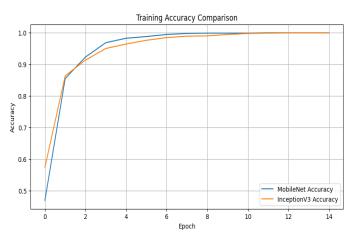
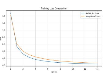
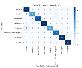
Graphsandplotsillustratingthetrainingaccuracy,training loss, and confusion matrices for both MobileNet and InceptionV3areprovidedbelowinfigure2:


Figure-3: Training Accuracy Comparison and Training Loss Comparison.


Figure-4: Confusion Matrix Comparison for MobileNet and InceptionV3.
D. Discussion:
TheresultsshowthatbothMobileNetandInceptionV3areeff ectivelyclassifiedintheclassificationofskindiseasesfrom images.Despiteslightvariationsinperformance,bothmodel sarehighlyaccurateandshowrobustnessindifferentdiseas eclasses.ThechoiceofMobileNetandInceptionV3dependso nfactorssuchascomputationalefficiency,modelcomplexity, andspecificapplicationrequirements.
1. Dataset Expansion: The models' ability for generalizationcanbeimprovedbygatheringlargerand morevarieddatasets.Tomakethemodelmorereliable and useful to a broader population, incorporate additionalvariationsinskinconditions,demographics, andimagecharacteristics

International Research Journal of Engineering and Technology (IRJET) e-ISSN: 2395-0056
Volume: 12 Issue: 05 | May 2025 www.irjet.net p-ISSN: 2395-0072
2. Fine-tuning and Transfer Learning: Use domainspecific data to investigate fine-tuning methods on previouslylearnedmodels.Thiscanassistinapplying the knowledge gained from big datasets (such as ImageNet) to the unique features of photos of skin diseases.
3. Ensemble Methods: Examine ensemble approaches, which aggregate predictions from several models to enhanceperformanceasa whole.Ensemble methods canproducemoreaccuratepredictionsandlessenthe drawbacksofindividualmodels.
4. Interpretability: To comprehend the model, techniqueslikeattentionprocessesandclassactivation cardscanbeused.Ifclinicsidentifythefeaturesofthe imagingthatthemodelvalueshighly,theycandiagnose patientsandscheduletreatmentsmoresuccessfully.
5. Real-time Deployment: Take into account that the modelwillbeusableinreal-timescenarios,suchmobile apps and healthcare platforms. This results in a high degreeofaccuracywhileloweringthemodel'ssizeand processingdemands.
6. Continued Evaluation: Ongoingassessmentbasedon freshdatatoguaranteethemodel'slong-termefficacy. It'scriticaltomaintainthemodelcurrentandrelevant in case skin conditions change and new situations emerge.
ThestudyconcludesthatMobileNetandInceptionV3models perform well in classifying skin diseases, as evidenced by theirabilitytocorrectlydetectarangeofskinailmentsfrom pictures. Both models demonstrated robustness across several illness classifications and attained a high overall accuracy.Eventhoughtherewereonlyslightperformance variations, factors including computing efficiency, model complexity, and application needs may influence which of MobileNetandInceptionV3isbest.
FuturedevelopmentsinCNN-basedskindiseasedetection appear promising. We can keep enhancing the precision, dependability, and application of these models in clinical practice and healthcare systems by growing datasets, improving model architectures, and investigating cuttingedgemethodslikeensemblelearningandinterpretability.
1. Yakupu,R.Aimaier,B.Yuan,B.Chen,J.Cheng,Y.Zhao, Y. Peng, J. Dong, and S. Lu, “The burden of skin and subcutaneous diseases: Findings from the Global Burden of Disease Study 2019,” Frontiers in Public Health, vol. 11, p. 1145513, Apr. 2023, doi: 10.3389/fpubh.2023.1145513.
2. V. Ki and C. Rotstein, “Bacterial skin and soft tissue infections in adults: A review of their epidemiology, pathogenesis, diagnosis, treatment and site of care,” Canadian Journal of Infectious Diseases and Medical Microbiology, vol. 20, no. 2, pp. 52–56, 2009, doi: 10.1155/2009/423868.
3. W. Sae-Lim, W. Wettayaprasit, and P. Aiyarak, “Convolutional Neural Networks using MobileNet for skin lesion classification,” in Proc. 12th Int. Conf. on Information Technology and Electrical Engineering (ICITEE),ChiangMai,Thailand,2020,pp.132–137,doi: 10.1109/ICITEE49829.2020.9271682.
4. S. K. Bandyopadhyay, P. Bose, A. Bhaumik, and S. Poddar, “Machine learning and deep learning integrationforskindiseasesprediction,”International Journal of Computer Science and Mobile Computing, vol.8,no.5,pp.88–93,May2019.
5. M.W.P.MadurangaandD.Nandasena,“Mobile-based skin disease diagnosis system using convolutional neuralnetworks(CNN),”inProc.Int.ResearchConf.on SmartComputingandSystemsEngineering(SCSE),Sri Lanka,2019,pp.50–55.
6. T. Shanthi, R. S. Sabeenian, and R. Anand, “Automatic diagnosis of skin diseases using convolution neural network,”MaterialsToday:Proceedings,vol.33,no.8, pp. 5602–5607, 2020, doi: 10.1016/j.matpr.2020.02.964.