International Research Journal of Engineering and Technology (IRJET)
e-ISSN: 2395-0056
Volume: 06 Issue: 01 | Jan 2019
p-ISSN: 2395-0072
www.irjet.net
Detecting Brain Tumor using K-Mean Clustering and Morphological Operations Shaheen M. Khan1, Radhika S. Kharade2, Vrushali S. Lavange3, Prof. D.B. Pohare4 1,2,3,4Department
of Electronics and Telecommunication Engineering, DES’s College of Engineering & Technology, Dhamangaon (Rly) ---------------------------------------------------------------------***--------------------------------------------------------------------Abstract:- Brain tumor is inherently serious and lifethreatening problem because of its character in the limited space of the intracranial cavity (space formed inside the skull). Tumor is the one of the most common brain disease and this is the reason for the diagnosis & treatment of the brain tumor has vital importance. CT scan is the technique used to produce computerized image of internal body tissues. Cells are growing in uncontrollable manner this leads to mass of unwanted tissue that is termed as neoplasm. Normally the anatomy of the Brain can be viewed by the CT scan. It is not affect the human body. Because it doesn’t use any radiation. In this paper we proposed segmentation of brain CT Scan Image using K-means clustering algorithm followed by morphological filtering which avoids the misclustered regions that can be formed after segmentation of the brain CT Scan Image for detection of tumor location.
procedure are introduced to estimate the world of the growth. 1.1 STRUCTURE OF BRAIN The human brain is that the central organ of the human system and with the medulla spinalis makes up the central system. The brain is protected by the os, suspended in liquid body substance, and isolated from the blood by the blood– brain barrier. However, the brain continues to be prone to injury, disease, and infection. Generally, human brain includes three major parts which controls different activities of human.
Key Words: CT scan, Brain Tumor, Benign, Malignant, Preprocessing, Feature extraction. 1. INTRODUCTION Image process could be a technique to convert a picture into digital kind and perform some operations on that, so as to induce AN increased image or to extract some helpful information from it. Image processing is widely used for diagnosis of diseases in agriculture as well as in biomedical. The main idea behind this paper is to study the design of a computer system able to detect the presence of a tumor in the digital images of the brain, and to accurately define its borderlines. Among different types of methods we focus on choosing appropriate method. Brain tumor is AN abnormal growth of cells within the bone. Normally the growth can grow from the cells of the brain, blood vessels, nerves that emerge from the brain. There are 2 kinds of growth that arebenign (non-cancerous) and malignant (cancerous) tumors. The former is delineate as slow growing tumors which will exert doubtless damaging pressure however it'll not unfold into close brain tissue. However, the latter is delineate as fast growing growth and it's ready to unfold into close brain. Tumors will injury the conventional brain cells by manufacturing inflammation, exerting pressure on parts of brain and increasing pressure within the skull. In this project, CT SCAN scan images are used for the analysis. CT SCAN is a very powerful tool to diagnose the brain tumors. It gives pictures of the brain and requires no radiation. The noninheritable image is analyzed mistreatment image process strategies. Image segmentation and bunch
© 2019, IRJET
|
Impact Factor value: 7.211
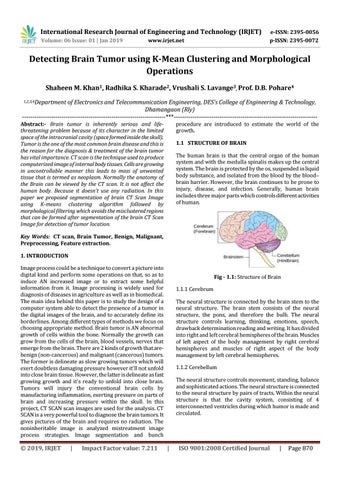
Fig - 1.1: Structure of Brain 1.1.1 Cerebrum The neural structure is connected by the brain stem to the neural structure. The brain stem consists of the neural structure, the pons, and therefore the bulb. The neural structure controls learning, thinking, emotions, speech, drawback determination reading and writing. It has divided into right and left cerebral hemispheres of the brain. Muscles of left aspect of the body management by right cerebral hemispheres and muscles of right aspect of the body management by left cerebral hemispheres. 1.1.2 Cerebellum The neural structure controls movement, standing, balance and sophisticated actions. The neural structure is connected to the neural structure by pairs of tracts. Within the neural structure is that the cavity system, consisting of 4 interconnected ventricles during which humor is made and circulated.
|
ISO 9001:2008 Certified Journal
|
Page 870