International Research Journal of Engineering and Technology (IRJET)
e-ISSN: 2395-0056
Volume: 06 Issue: 10 | Oct 2019
p-ISSN: 2395-0072
www.irjet.net
DETECTION OF WHITE BLOOD CELL IMAGE USING NUCLEUS SEGMENTATION Rufia farveen1, Ashwiniya2, Thanapriyanka3, Devasundharam4, Rajesh kumar5 1,2,3,4Dept.
of Bio Medical Engineering, Rajiv Gandhi College of Engineering and Technology, Puducherry, India. Professor, Dept. of Bio Medical Engineering, Rajiv Gandhi College of Engineering and Technology, Puducherry, India ---------------------------------------------------------------------------***-------------------------------------------------------------------------5Assistant
Abstract- Leukemia is the most critical blood disease, common in children and adults. Images processing steps like image enhancement, edgedetection, image segmentation and feature extraction are applied on microscopic images. Detection through images is fast and feasible method as they avoid special need of equipment for lab testing. This project aims to provide user friendly software based on MATLAB allowing for quick user interaction with a simple tool for the detection and segmentation of white blood cells from images of blood samples. The Sober method with FCM (Fuzzy Cmeans clustering) is used for detection and feature extraction of blood cells. KEY WORDS: White Blood Cell Segmentation, Sobel Method with FCM, Detection of Leukimia Types. 1. INTRODUCTION Blood disorder is considered among the most dangerous of diseases that can lead to death. Many of these blood diseases are defined to white blood cells such as Leukemia.Leukemia is an abnormal condition in the body because it produces excess white blood cells (leucocytes) in the bloodstream and spinal cord.Therefore, these abnormal conditions will disrupt the production of other blood cells and disrupt the flow of blood into the vital organs.The screening of prepared blood samples by pathologists for counting and classification of WBCs is tedious, slow, and time-consuming. Consequently, an automatic system based on image processing techniques is useful for aiding pathologists.Meanwhile, segmentation is one of the main steps in microscopic image analysis that the result of this step directly influences in the outcomes of the following steps such as feature extraction and classification.
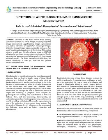
FIG.1 LEUKEMIA Leukemia is the most critical blood disease, common in children and adults. A majority cancer cell begins in body parts but leukemia is the type of cancer which begins and grows in blood cells. Blood is crucial content without which metabolic functions of body severely affects .Human system is like, cell grows and multiply into new cells. Old cells are destroyed and so that new cells can take their place. In cancer, an old cell does not die and remains in the blood so that new cells which are produced cannot get enough space to live. In this way, functioning of blood disturbs and white blood cells production is abnormal and uncontrolled. 1.1 COMPONENTS OF HUMAN BLOOD CELL: Blood cells are produced from the stem cells present in bone marrow. Blood consists of following components. • Red Blood Cells (Erythrocytes): RBCs has a capacity to carry oxygen to and take back CO2 away from tissues. • White Blood Cells (Leukocytes): WBCs are the cell which fights with the foreign bodies and prevents from infection. There are different types of WBCs like lymphocytes, monocytes, eosinophils, basophils and neutrophils.
© 2019, IRJET
|
Impact Factor value: 7.34
|
ISO 9001:2008 Certified Journal
|
Page 187