International Research Journal of Engineering and Technology (IRJET)
e-ISSN: 2395-0056
Volume: 04 Issue: 07 | July -2017
p-ISSN: 2395-0072
www.irjet.net
Diagnosis of Diabetic Retinopathy by Detection of Microneurysm And Exudates Gayatri M. Madale, Prof. Rahul Mulajkar Dept. of Electronics and telecommunication Jaihind college of Engineering,kuran, Pune,India ---------------------------------------------------------------------***---------------------------------------------------------------------
Abstract— Now a days diabetic retinopathy (DR) has turned out to be not kidding disease among diabetic patients . It would recognize at the early stage else it will causes to aggregate visual impairment .The paper proposes to programmed ID of exudates pathologies in retinopathy fundus pictures a novel strategy is computational insight method. To concentrate components of fundus picture like blood vessels, optical nerve, red sores and white sores together with surface element examination utilizes the remarkable execution of morphological administrators.
Over all approach
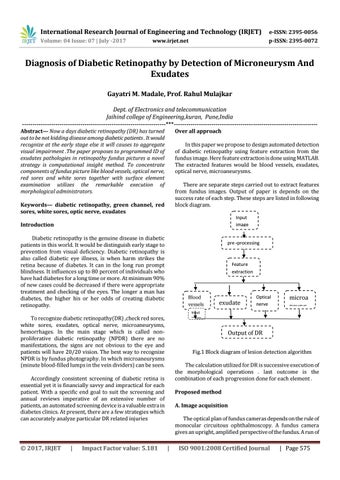
In this paper we propose to design automated detection of diabetic retinopathy using feature extraction from the fundus image. Here feature extraction is done using MATLAB. The extracted features would be blood vessels, exudates, optical nerve, microaneurysms. There are separate steps carried out to extract features from fundus images. Output of paper is depends on the success rate of each step. These steps are listed in following block diagram.
Keywords— diabetic retinopathy, green channel, red sores, white sores, optic nerve, exudates
Input image
Introduction Diabetic retinopathy is the genuine disease in diabetic patients in this world. It would be distinguish early stage to prevention from visual deficiency. Diabetic retinopathy is also called diabetic eye illness, is when harm strikes the retina because of diabetes. It can in the long run prompt blindness. It influences up to 80 percent of individuals who have had diabetes for a long time or more. At minimum 90% of new cases could be decreased if there were appropriate treatment and checking of the eyes. The longer a man has diabetes, the higher his or her odds of creating diabetic retinopathy.
pre -processing
Feature extraction
Blood vessels Input image
To recognize diabetic retinopathy(DR) ,check red sores, white sores, exudates, optical nerve, microaneurysms, hemorrhages. In the main stage which is called nonproliferative diabetic retinopathy (NPDR) there are no manifestations, the signs are not obvious to the eye and patients will have 20/20 vision. The best way to recognize NPDR is by fundus photography. In which microaneurysms (minute blood-filled lumps in the vein dividers) can be seen.
|
Impact Factor value: 5.181
Optical nerve
microa neurys ms
Output of DR Fig.1 Block diagram of lesion detection algorithm The calculation utilized for DR is successive execution of the morphological operations . last outcome is the combination of each progression done for each element .
Accordingly consistent screening of diabetic retina is essential yet it is financially savvy and impractical for each patient. With a specific end goal to suit the screening and annual reviews imperative of an extensive number of patients, an automated screening device is a valuable extra in diabetes clinics. At present, there are a few strategies which can accurately analyze particular DR related injuries
© 2017, IRJET
exudate s
Proposed method A. Image acquisition The optical plan of fundus cameras depends on the rule of monocular circuitous ophthalmoscopy. A fundus camera gives an upright, amplified perspective of the fundus. A run of
|
ISO 9001:2008 Certified Journal
| Page 575