International Research Journal of Engineering and Technology (IRJET)
e-ISSN: 2395 -0056
Volume: 03 Issue: 02 | Feb-2016
p-ISSN: 2395-0072
www.irjet.net
DESIGN OF ULTRAFAST IMAGING SYSTEM FOR THYROID NODULE DETECTION Aarthipoornima Elangovan1, Jeyaseelan.T2 PG Student, Department of Electronics and Communication Engineering Kings College of Engineering, Punalkulam, Tamilnadu, Affiliated to Anna University ,Chennai, India 2 Assistant Professor , Department of Electronics and Communication Engineering Kings College of Engineering, Punalkulam, Tamilnadu, Affiliated to Anna University ,Chennai, India 1
---------------------------------------------------------------------***--------------------------------------------------------------------1.1 Thyroid gland
Abstract - A complete solution to estimate the volume of the
thyroid gland directly from ultrasound (US) images is proposed in this paper. Physicians usually diagnose the pathology of the thyroid gland by its volume. However, even if the thyroid glands are found and the shapes are hand-marked from ultrasound images, most physicians still depend on computed tomography (CT) images, which are expensive to obtain, for precise measurements of the volume of the thyroid gland. This approach relies heavily on the experience of the physicians and is very time consuming. Patients are exposed to high radiation when obtaining CT images. In contrast, Ultrasound imaging does not require ionizing radiation and is relatively inexpensive. Ultrasound imaging is thus one of the most commonly used auxiliary tools in clinical diagnosis. The radial basis function neural network is used to classify blocks of the thyroid gland. The integral region is acquired by applying a specific-region-growing method to potential points of interest. The parameters for evaluating the thyroid volume are estimated using a particle swarm optimization algorithm. Simulation results of the thyroid show that the region segmentation can be automatically achieved and the volume of thyroid nodule can be precisely measured.
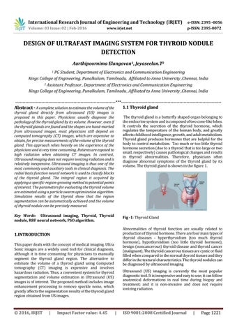
The thyroid gland is a butterfly shaped organ belonging to the endocrine system and is composed of two cone-like lobes. It controls the secretion of the thyroid hormone, which regulates the temperature of the human body, and greatly affects childhood intelligence, growth, and adult metabolism. Thyroid gland produces hormones that are helpful for the body to control metabolism. Too much or too little thyroid hormone secretion (due to a thyroid that is too large or two small, respectively) causes pathological changes and results in thyroid abnormalities. Therefore, physicians often diagnose abnormal symptoms of the thyroid gland by its volume. The thyroid gland is shown in the figure 1.
Key Words: Ultrasound imaging, Thyroid, Thyroid nodule, RBF neural network, PSO algorithm.
Fig -1: Thyroid Gland Abnormalities of thyroid function are usually related to production of thyroid hormone. There are four main types of thyroid diseases - hyperthyroidism (too much thyroid hormone), hypothyroidism (too little thyroid hormone), benign (noncancerous) thyroid disease and thyroid cancer (malignant). The thyroid cancerous tissues are cystic or fluid filled when compared to the normal thyroid tissues and they differ in the textural characteristics. The thyroid nodules can be diagnosed by ultrasound imaging
1.INTRODUCTION This paper deals with the concept of medical imaging. Ultra Sonic images are a widely used tool for clinical diagnosis, although it is time consuming for physicians to manually segment the thyroid gland region. The alternative to estimate the volume of a thyroid gland using Computed tomography (CT) imaging is expensive and involves hazardous radiation. Thus, a convenient system for thyroid segmentation and volume estimation in Ultrasound (US) images is of interest. The proposed method includes image enhancement processing to remove speckle noise, which greatly affects the segmentation results of the thyroid gland region obtained from US images.
Š 2016, IRJET
|
Impact Factor value: 4.45
Ultrasound (US) imaging is currently the most popular diagnostic tool. It is inexpensive and easy to use; it can follow anatomical deformations in real time during biopsy and treatment; and it is non-invasive and does not require ionizing radiation.
|
ISO 9001:2008 Certified Journal
|
Page 1221