International Research Journal of Engineering and Technology (IRJET)
e-ISSN: 2395 -0056
Volume: 03 Issue: 10 | Oct -2016
p-ISSN: 2395-0072
www.irjet.net
Diabetic Retinopathy Diagnosis Using Image Mining Abhilash Bhaisare1, Sagar Lachure2, Amol Bhagat3, Jaykumar Lachure4 1Student,
CSE Department, YCCE, Nagpur Professor, CT Department, YCCE, Nagpur 3Assistant Professor, CSE Department, PRMCEM, Badnera 2Assistant
---------------------------------------------------------------------***---------------------------------------------------------------------
Abstract - Diabetic Retinopathy is an eye problem that can cause blindness. It occurs when sugar level in blood increases, blood vessels in the back of the eye becomes weak and because of this vessel leaks the blood and lipoproteins fluid. Signs of Diabetic Retinopathy are floating spot in vision and blocked vision. If we detected early the sign of Diabetic Retinopathy then may be able to prevent additional vision loss. The main motivation is to identify the input image is normal or abnormal. When the input image is found abnormal then analysis for further DR stages is done. To identify abnormal image there are various techniques and methodology used in image mining. Image mining is an extension of data mining technique. Most of the image processing algorithms are used in image mining for preprocessing. Preprocessing Stage is one of the most commonly used in image processing for image enhancement. Features are extracted when detecting exudates. After extracting the features, classification algorithms are used. After that result is obtained and result will display that image how much normal or abnormal. Now a days web based systems are mostly useful as compare to stand alone system. If we implement web based system for detection of diabetic retinopathy, it is beneficial for rural patients and it is also save the time and money of diabetic patients.
Four stages of Diabetic Retinopathy are as follows: First stage is known as Mild Non-Proliferative Diabetic Retinopathy (Mild NPDR). In this stage, there will be balloon like swelling in the blood vessels in the retina and small balloon like swelling in the blood vessels known as Microaneurysms. Second stage is known as Moderate Non-Proliferative Diabetic Retinopathy (Moderate NPDR). In this stage, some of the blood vessels in the retina will become blocked.
Third stage is known as Severe Non-Proliferative Diabetic Retinopathy (Severe NPDR). In this stage, more blood vessels are blocked that’s why the areas of the retina will not receiving enough blood. Without proper flow of blood, the retina will not grow new blood vessels and to replace the damaged blood vessels. Fourth stage is known as Proliferative Diabetic Retinopathy (PDR). This is advanced stage. New blood vessels will begin to grow in the retina, but they will be weak blood vessels. So weak blood vessels can leaks blood and lipoproteins fluid. In this stage chances of completely blindness increases.
Key Words: Diabetic Retinopathy, Lipoproteins, Image Mining, Pre-processing, Exudates.
1. INTRODUCTION Diabetic Retinopathy is an eye problem that can cause blindness. Small blood vessels in the back of the eye called as retinal blood vessels. Signs of Diabetic Retinopathy are floating spot in vision, blurred vision and blocked vision. When sugar level in blood increases, blood vessels in the back of the eye becomes weak and because of this vessel leaks the blood and lipoproteins fluid. After that fluid become floating spot in vision so that Diabetic patient can not see anything completely through the vision. If we do not do the treatment of this disease on the time then it may be possible of complete vision loss or blindness. If we detected early the sign of Diabetic Retinopathy, it is possible to prevent additional loss of vision.
© 2016, IRJET
|
Impact Factor value: 4.45
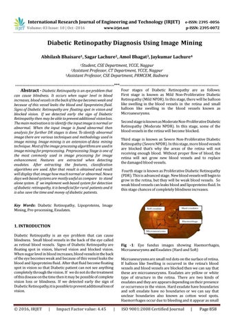
Fig -1: Eye fundus images showing Haemorrhages, Microaneurysms and Exudates (Hard and Soft) Microaneurysms are small red dots on the surface of retina. If balloon like Swelling is occurred in the retina’s blood vessels and blood vessels are blocked then we can say that these are microaneurysms. Exudates are yellow or white type of structure in the retina. There are two kinds of exudates and they are appears depending on their presence or occurrence in the vision. Hard exudate have boundaries and soft exudate have no boundaries or we can say that unclear boundaries also known as cotton wool spots. Haemorrhages occur due to bleeding and it appear as small
|
ISO 9001:2008 Certified Journal
|
Page 858