
International Research Journal of Engineering and Technology (IRJET) e-ISSN:2395-0056
Volume: 11 Issue: 05 | May 2024 www.irjet.net p-ISSN:2395-0072

International Research Journal of Engineering and Technology (IRJET) e-ISSN:2395-0056
Volume: 11 Issue: 05 | May 2024 www.irjet.net p-ISSN:2395-0072
Sahil Sawant1, Prof. Mario Pinto2
1Student, Department of Information Technology and Engineering, Goa College of Engineering, Farmagudi, Goa, India
2Assistant Professor, Department of Information Technology and Engineering, Goa College of Engineering, Farmagudi, Goa, India ***
Abstract- Pneumonia, a prevalent and potentially lifethreateningrespiratoryinfection,posessignificantchallenges intimelyandaccuratediagnosis.Leveragingadvancementsin machine learning (ML) techniques, this research aims to developanefficientandreliablepneumoniadetectionsystem.
Through the analysis of chest X-ray images, a convolutional neural network (CNN) model is trained to differentiate between pneumonia-infected and healthy lung images. The dataset used for training and validation comprises a diverse set of chest X-ray images collected from various sources. The proposed model demonstrates promising results, achieving high accuracy and sensitivity in pneumonia detection. Moreover, interpretability techniques are employed to elucidate the decision-making process of the CNN model, enhancing its clinical relevance and trustworthiness. The developed system holds considerable potential for aiding healthcare professionals in prompt and accurate pneumonia diagnosis, thereby facilitating timely intervention and improving patient outcomes.
Key Words: Pneumonia detection, Convolutional Neural Networks, Deep Learning, Chest X-ray images
Pneumoniaisaleadingcauseofmorbidityand mortality worldwide, particularly among children and the elderly. Early detection and prompt treatment of pneumonia are crucialforpreventingcomplicationsandimprovingpatient outcomes. Chest X-ray imaging is commonly used for the diagnosis of pneumonia due to its accessibility and effectiveness in detecting abnormalities in lung tissue. However, the interpretation of chest X-ray images can be challengingandtime-consuming,requiringexpertisefrom radiologists. Automated methods based on deep learning techniques offer a promising solution to streamline the diagnosisprocessandimprovetheefficiencyandaccuracyof pneumoniadetection.
The project focuses on developing a machine learningbased system for the early and accurate detection of pneumonia from chest X-ray images. Pneumonia, a prevalentrespiratoryinfection,requirestimelydiagnosis for effective medical intervention. The study utilizes a diverse dataset containing both normal and pneumonia-
affectedcases,ensuringcomprehensivemodeltrainingand evaluation.
The methodology involves extracting relevant image featuresfromchestX-rays,encompassingtexture,intensity, and shape descriptors. To address challenges related to dataset variability and generalization, feature engineeringandselectiontechniquesareemployed.
Theresearchaimstoshowcasethepotentialof machine learning models in pneumonia detection, with a focus on practicalapplicabilityinclinicalsettings.Theultimategoal istoprovidehealthcareprofessionalswitha reliabletool that aids in the early identification of pneumonia cases, contributingtoimprovedpatientoutcomes.
This project contributes to the broader field of medical diagnostics, highlighting the promising role of machine learninginenhancingpneumoniadetectioncapabilities.As technology continues to intersect with healthcare, the findings from this study aim to pave the way for the integration of machine learning models into real-world clinical workflows, supporting healthcare professionals in making timely and accurate diagnostic decisions for pneumonia.
TheauthorproducedandshowcaseamergedDLmodelfor identifyingPneumoniapatientsfromCXR.Intheproposed model,threedistinctmodelsaretrainedontheCXRdataset. The first of them is a bespoke CNN model. Xception and EfficientNetB4arethetwoothermodels.
Severaldataaugmentationandpreprocessingstrategiesare utilized, along with hyperparameter tuning. A composite model is generated by giving different trained models weights based on their accuracy and recall rates.Several performance metrics are improved compared to the prior art,thankstothesuggestedapproach.[1]
In this paper ensemble of 3 CNN model was used Deep transfer learning is used to deal with the data shortage Dataset is collected which is than pre-processed Image enhancementisdoneutilizingthresholdLBP(Localbinary pattern)featureextractiondoneDataaugmentationtoform anewdatapointandsplitintotwodatasets.[2]
International Research Journal of Engineering and Technology (IRJET) e-ISSN:2395-0056
Volume: 11 Issue: 05 | May 2024 www.irjet.net p-ISSN:2395-0072
The author in this paper focuses on early detection of pneumonia. From X-ray images using a pre-trained CNN model Resnet50 Comparison is made between the two models.ThemodelsusedareCNNandResNet50.[3]
InthistheauthorDensenet121modelisused.Twoclassifier areusedonefortheidentificationofpneumonia.Theother fortheidentificationoftype. CausedbybacteriaorVirus. Here transfer learning is used in which the pre-trained modelsareused.[4]
Alexnetmodelisusedforthedetectionofpneumonia.Itis capableofidentifyingpneumoniaatearlystagesComparison is made between various other models like VGG16, CNN, RESNET.[5]
Model is created using Tensorflow. They created a CNN modelwithtwoconvolutionallayersandonefullyconnected layer, followed by flattening to generate a fully connected layer They applied sigmoidal activation function on fully connectedlayerThisresultintheincreaseinaccuracy.[6]
Machinelearning(ML)algorithmshavesteadilygained the interestofresearchersoverthelastfewyears.Thistypeof method can make full use of the massive ability to create computer calculators in image processing with predeterminedalgorithmstages.Traditionalmachinelearning methodsfordividingjobs,ontheotherhand,necessitatethe use of manual design algorithms or the manual setting of output layers to separate images. In response to the aforementioned situation, LeCun et al. offered a CNN approach,whichcanautomaticallyextractfeatureswiththe useofconstantlystackingfeaturesandexitthattheincluded photosmaynotbeinanyclass.
Theshallownetworksareverydeepandconcentrateonthe image's low-level features. CNN the model increasingly exposesadvancedfeaturesasthenumberofnetworklayers increases. CNN learns the distinctions between different
imagesbycombiningandevaluatingthesepriorityfeatures, and it uses a back-propagation technique to update and recordlearnedparameters.CNN'sconceptistouseaspecific convolution kernel to filter a prior picture or map componenttobuildthenextlayermapelement,aswellas mergefunctionslikemergingfunctionstominimisefeature mapscaleandmitigationtocount.Thecreatedcomponentis thengiventothenon-linearactivationfunctionmappingto improve the model's simulation capabilities The most commonintegrationtasksincludemidandhighintegration.
Thepluralofintegrationdenotesthattheelementsentto theintegrationlayerissplitintomanyregions,witheach sub-regionhavingadifferentsizeintermsofhorizontal andverticalsteps.Thesoledistinctionbetweenhighand medium integration is a lower region where the aggregation rate yields the average of each sub-region. ReLU (Rectified Linear Units) and Sigmoid are two commonactivationevents.
Image elements are automatically extracted using segmentation and a continual accumulation of convolutional processes, integration functions, indirect openingfunctions,andothercompletelyintegratedlayers. Then, by evaluating these derived characteristics, it is possibletoextractpneumoniafromthephotosprocessed bythemodel.Themodel'sgeneralcapacityisincreasedby fully utilising pixel-level image information. The most prominentneuralframeworkhasbeenproposedinpast fewdecadesforin-depthlearningdevelopment.,suchas AlexNetandVGGNet.
However, when the number of layers in the network increase, Instead of learning the numerous productive features,theneuralnetworkwillbemodifiedtoparticular partsofthetrainingimage,whichmakesthemodelsimilar to the capacity declines and creates congestion. The remaining communication framework was proposed to overcometheproblemofnetworkdepth.Sincethen,neural networks have advanced, garnered a lot of attention and research, and have formed the foundation for a lot of occupations. We also looked at the efficiency of residual connectionsinourreducedCNNarchitecturewithonlyafew layersinthisstudy.
4.1
The proposed database, which will be used to test the model's performance, comprises a total of 5863 X-ray pictures via Kaggle.Dr. Paul Mooney created a Kaggle contestin2017toclassifyviralandbacterialpneumonia. Itdiffersfromtheotherdatasetssinceitcontains5,863 paediatric photos. We're talking about the updated versionofthisdataset.
International Research Journal of Engineering and Technology (IRJET) e-ISSN:2395-0056
Volume: 11 Issue: 05 | May 2024 www.irjet.net p-ISSN:2395-0072
Thedatabaseisfurtherdividedintothreefolders(train, test,andval)withsubfoldersforeachimagecategory (Pneumonia/General).Figure1showsafewinstances of common and pneumonia photos that have been scaled to a static size. Due to the low amount of exposureinpatients,chestX-rayimagesalwaysshow symptomsoflimitedbrightness,andchestX-rayimages alwayshaveblack,white,andgreypants.
Thelungsareonbothsidesofthethoraciccavity,and the lung area is plainly visible on an X-ray since it is virtuallyblack.Theheart,whichissituatedbetweenthe lungs, appears practically as white as X-rays can go through it entirely. Because bones are comprised of proteinandareexceedinglydense,X-rayscannotpass through them, leaving the bones virtually white. Furthermore,theboneshavedistinctedges.
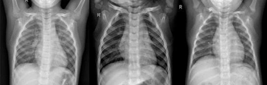
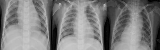
-1:Examplesfromthedataset.(a)normal cases, (b) pneumoniacases
Table2liststhetacticsemployedthroughoutthisarticle. Rescaleisavaluethatwewillmultiplythedatabybefore any other processing in our investigation. Our original photos had RGB coefficients ranging from 0 to 255, but valueslikethiswouldbetoohighforourmodelstohandle (givenatypicallearningrate),sowescalethemdownbya factor of 1./255. shear range is used to apply shearing transformations at random. When there are no assumptionsofhorizontalasymmetry,zoomrangeisused
torandomlyzoominsidephotographs,andhorizontalflip is used to randomly flip half of the images horizontally (e.g.real-worldpictures)
Datapre-processingtechniquesusedinthisstudy
Rescale 1./255
ZoomRange 0.2
ShearRange 0.2
Horizontal_Flip True
Table 2:- DataPreprocessingtechniques
Inthisstudy, wedesigneda CNN model withfive convolutionallayerstoextractthefeaturesofchestXrayimagesandusethosefeaturestodetectifapatient suffersfrompneumonia.InOurCNNArchitecture,We startedwithalowerfiltersettingof32andworkedour wayuplayerbylayer.AlayerofConv2Dwasusedto buildthemodel,followedbyalayerofMaxPooling.An oddnumber,suchas3x3,isdesirableforkernelsize.
TheactivationfunctionsTanh,ReLU,andotherscan beemployed,butReLUisthemostpopular.inputshape acceptsthewidthandheightofanimage,withthelast dimension serving as a colour channel. After that, we flattenedtheinputandaddedANNlayers.
f(x)=max(0,x)
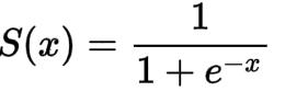
S(x) = Sigmoid
f(x) = ReLU
Volume: 11 Issue: 05 | May 2024 www.irjet.net p-ISSN:2395-0072
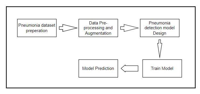
Fig -2: UMLDiagram
Table 3:ComparisonofvariousPneumonia&otherlungdiseasesdetectiontechniques
Ref.no.
Publisher / Year Proble m definitio n
[1] ICACI/ 2018 Detection of lung cancer
[2] IEEE/ 2016
[3] arXiv 2018
[4] Stamford University / 2017
Detection of lung diseases likeLung Cancer,TB , Pneumonia
Detection of thorax diseases
Detection of Pneumon ia
Detectionof Tuberculosis
[5]RSNA/2017
Boneshadow exclusion(#2)
Segmentation(#3)
Segmentationafter bone shadow exclusion(#4)
Exclusionofoutliers by t-SNEmethod(#5)
Imagepreprocessing Lung segmentation
Featureextraction Image classification
Globalbranchtakes input Localbranchis trainedafter discoveringlocallesion regionandcropping Finally globalandlocal branches arecombinedtofine tune
Imagedownscaling to 224*224
Normalizebased on standard deviationand mean
Randomhorizontal flipping
Imagesareresized to256x256
Imagesare augmentedusing 1..Random cropping(227x227 pixel)
CNN
Tensorflow GPUused NVIDIA Tesla K40c card 71% JSRT Highly accurate
ANN(Feed forward neural network) with sigmoid activation function
Attention guided CNN (sigmo id functio n) NA
DCNN (DenseNet)
AlexNet GoogleNet 1.LinuxOS 2.Caffe framework
Sasoo hospital, Pune (Datasetof 80patients)
AUC(0.871) ChestX-ray 14
Itcan detect multiple lung diseases
Ituses small dataset which mightnot containall the cases
Itisnot robust whenthere are changes inthe size and positionof CXRimages
Ityields better accurac y compar edto other methods Relatively insensitive to parameter changes
AUC(0.76) ChestXray14 indicating absence or presence of14 different pathology classes Onlyfrontal radiograph were present
AUC(0.99) 1007chest radiograph
ImageNet performe dbetter thanthe untrained networks Thisalgorithm canonlyusefor TBdetection
International Research Journal of Engineering and Technology (IRJET) e-ISSN:2395-0056
Volume: 11 Issue: 05 | May 2024 www.irjet.net p-ISSN:2395-0072
[6]IEEE/2017
Detectionof Pneumonia
[7] Springer/2018
[8] Isabel BushStanford Computer Science353 SerraMall, Stanford,CA 94305
Meltilabel classificationof thoracic diseasesin chest radiographs
Distinguish between benignand malignant nodulesto detectlung cancer
[9]IEEE/2017 classificationof eightcommon thoracic diseases
Detectionof thoraxdiseases
[10]HIKARI ltd/2015
[11] Springerlink February2017
[12] Appliedscience 2018
Dominant technologyfor tacklingCADin thelungs
2.meansubtraction 3.mirrorimages
Signal segmentation Wavelet decomposition Powerspectral densityStatistical parameter Fourier transform Continuous wavelet Transform
Binary relevence(BR) PairWise Error(PWE) Softmaxactivation weightedcross entropyloss calculated
Localisationand classification
Baseline: DensNet161 Boostedcascade network
Weakly-supervised pathology localization Multi-labeldisease classification
Imagepreprocessing,lung fields segmentation, features calculation, classification CADSystem
Pulmonaryimage analysisComputeraideddetection Computer-aided diagnosisImage processing rule-basedstudy
Detectionof pneumonia DataCollectionand Preprocessing VGG16 CAMandgradCAMvisualizati ontools
detecting diseases
chestX-Ray14
Thisislow cost,noncontact, and noninvasi ve Usedveryless input/refers(22 signals)
Boosted cascade approach give increased performa nce BRapproachit doesnotmodel theinterclass relationwith example
[13]IEEE/2013
Detectionof Tuberculosis
Pre-processing FeaturesImages ExtractionImages Identification
StatisticalImage Feature PCAforFeature VectorDimension Reduction Minimum Distance Classifier
dataset used
Higher accurate ResNetmodelis unableto determineits preciselocation
Effective methods ofimage preproces sing, features calculatio n Automating thoraxdiseases detectionstill remains unsolveddueto itscomplexity
ConvNets arebetter feature extractor Computed tomography (CT)
Ray14 Highly Accurate
Preprocessed images used NA
International Research Journal of Engineering and Technology (IRJET) e-ISSN:2395-0056
Volume: 11 Issue: 05 | May 2024 www.irjet.net p-ISSN:2395-0072
5. Results
In this study, we explored the effectiveness of utilizing a deeplearningarchitecturewithfiveconvolutionallayersfor pneumonia detection. Through rigorous experimentation andevaluation,wehavedemonstratedthecapabilityofthe model to accurately classify chest X-ray images into pneumonia-positiveandpneumonia-negativecases. The utilization of convolutional layers allows the model to effectivelycaptureintricatepatternsandfeaturesindicative ofpneumonia,leadingtorobustperformance.
6. Conclusion
Inconclusion,oursurveyonpneumoniadetectionutilizinga deep learning model with five convolutional layers has providedvaluableinsightsintothecurrentstateofthefield. Throughacomprehensivereviewofexistingliteratureand methodologies,wehavegainedadeeperunderstandingof thechallengesandopportunitiesinemployingdeeplearning techniquesforpneumoniadiagnosis.
Overall, our survey contributes to the ongoing dialogue surrounding the application of deep learning in medical imaging and underscores the importance of collaborative efforts between researchers, clinicians, and industry stakeholders to further advance the field of pneumonia diagnosis. By addressing the challenges identified in this surveyandbuildingupontheexisting bodyofknowledge, we can continue to improve the accuracy, efficiency, and accessibility of pneumonia detection methods, ultimately leading to better healthcare outcomes for patients worldwide.
1. Gang,Peng,WangZhen,Wei Zeng,YuriGordienko, Yuriy Kochura, Oleg Alienin, Oleksandr Rokovyi, and Sergii Stirenko. "Dimensionality reduction in deep learning for chest x-ray analysis of lung cancer." In 2018 Tenth International Conference on Advanced ComputationalIntelligence(ICACI),pp.878-883.IEEE, 2018.
2. Khobragade,Shubhangi,AdityaTiwari,C.Y.Patil, andVikram Narke."Automaticdetectionofmajorlung diseasesusingChest Radiographsandclassificationby feed-forwardartificialneural network."In2016IEEE 1st International Conference on Power Electronics, IntelligentControlandEnergySystems(ICPEICES),pp. 1-5.IEEE,2016.
3. Guan,Qingji,YapingHuang,ZhunZhong,Zhedong Zheng, Liang Zheng, and Yi Yang. "Diagnose like a radiologist: Attention guided convolutional neural network for thorax disease classification." arXiv preprintarXiv:1801.09927(2018).
4. Rajpurkar, Pranav, Jeremy Irvin, Kaylie Zhu, BrandonYang,Hershel Mehta,TonyDuan,DaisyDing etal."Chexnet:Radiologist-level pneumoniadetection on chest x-rays with deep learning." arXiv preprint arXiv:1711.05225(2017).
5. Lakhani, Paras, and Baskaran Sundaram. "Deep learning at chest radiography: automated classification of pulmonary tuberculosis by using convolutionalneuralnetworks."Radiology284,no.2 (2017):574-582.
6. Pingale,TejashreeH.,andH.T.Patil."Analysisof CoughSoundfor PneumoniaDetectionUsingWavelet Transform and Statistical Parameters." In 2017 International Conference on Computing, Communication,ControlandAutomation(ICCUBEA), pp.1-6. IEEE,2017.
7. Kumar, Pulkit, Monika Grewal, and Muktabh Mayank Srivastava. "Boosted cascaded convnets for multilabel classification of thoracic diseases in chest radiographs." In International Conference Image AnalysisandRecognition,pp.546-552.Springer,Cham, 2018.
8. Chan, Heang-Ping, Berkman Sahiner, Lubomir Hadjiyski, Chuan Zhou, and Nicholas Petrick. "Lung nodule detection and classification." U.S. Patent Application10/504,197,filedSeptember22,2005.
9. Wang, Xiaosong, Yifan Peng, Le Lu, Zhiyong Lu, Mohammadhadi Bagheri, and Ronald M. Summers. "Chestx-ray8:Hospital-scalechest x-raydatabaseand benchmarksonweakly-supervisedclassification and localization of common thorax diseases." In Proceedings of the IEEE conference on computer visionandpatternrecognition,pp. 2097-2106.2017.
10. Zakirov,A.N.,R.F.Kuleev,A.S.Timoshenko,andA.V. Vladimirov."Advancedapproachestocomputer-aided detection of thoracic diseases on chest X-rays." Appl MathSci9,no.88(2015): 4361-4369.
11. vanGinneken,Bram."Fiftyyearsofcomputeranalysis inchest imaging: rule-based, machine learning, Deep learning." Radiologicalphysicsandtechnology10,no.1 (2017):23-32.
12. Rajaraman,Sivaramakrishnan,SemaCandemir,Incheol Kim,George Thoma,andSameerAntani."Visualization and interpretation of convolutional neural network predictionsindetectingpneumoniain pediatricchest radiographs."AppliedSciences8,no.10(2018):1715.
13. DivyeshRanpariya;ParinParikh;ManishI.Patel;Ruchi Gajjar, “ A CNN based Hybrid Model for Pneumonia ClassificationUsingChestX-rayImages,”2022
International Research Journal of Engineering and Technology (IRJET) e-ISSN:2395-0056
Volume: 11 Issue: 05 | May 2024 www.irjet.net p-ISSN:2395-0072
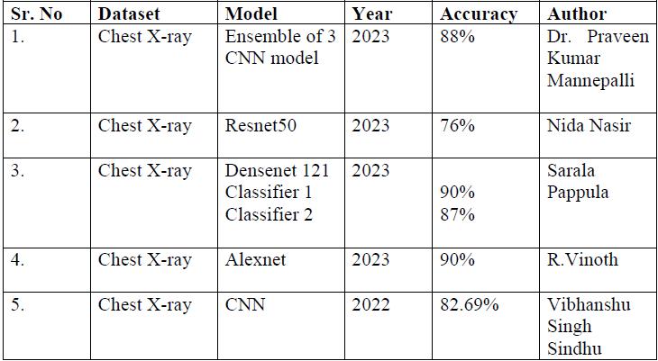
14. Dr.PraveenKumarMannepalli,“AnEarlyDetectionof Pneumonia in CXR Images using Deep Learning Techniques”,2023 International Conference on Innovative Data Communication Technologies and Application.
15. NidaNasiretal.”PneumoniaDetectioninChestX-ray Images using ResNet50 Model”, 2023 Advances in Science and Engineering Technology International Conferences(ASET).
16. Sarala Pappula et al.“Detection and Classification of PneumoniaUsingDeepLearningbytheDenseNet-121 Model”, 2023 Advances in Science and Engineering TechnologyInternationalConferences(ASET)
17. R.Vinothetal.“PneumoniaDetectionfromChestX-Ray using AlexNet Image Classification Technique”, 2023 7thInternationalConferenceonIntelligentComputing and Control Systems (ICICCS) | 979-8-3503-97253/23/$31.00 ©2023 IEEE | DOI: 10.1109/ICICCS56967.2023.10142405
18. Vibhanshu Singh Sindhu et al. “Pneumonia Detection UsingImproved ConvolutionalNeuralNetwork”, 2022 8th International Conference on Advanced Computing and Communication Systems (ICACCS) | 978-1-6654-0816-5/22/$31.00©2022IEEE
19. K.Weiss,T.M.Khoshgoftaar,andD.Wang,“Asurveyof transfer learning.” Journal of Big Data, vol. 3, no. 1, 2016,doi:10.1186/s40537-016-0043-6.
20. M.F.Hashmi,S.Katiyar,A.G.Keskar,N.D.Bokde,andZ. W.Geem,“EfficientPneumoniaDetectioninChestXray ImagesUsingDeepTransferLearning,”Diagnostics,vol. 10, no. 6, p. 417, Jun. 2020, doi: 10.3390/diagnostics10060417.
21. V. Chouhan, S. K. Singh, A. Khamparia, D.Gupta, P. Tiwari, C. Moreira, R. Damaˇseviˇcius, and V. H. C. de Albuquerque, “A Novel Transfer Learning Based Approach for Pneumonia Detection in Chest X-ray Images,” Applied Sciences, vol. 10, no. 2, p. 559, Jan. 2020,doi:10.3390/app10020559.
22. T. Rahman, M. E. H. Chowdhury, A. Khandakar, K. R. Islam, K. F. Islam, Z. B. Mahbub, M. A. Kadir, and S. Kashem,“TransferLearningwithDeepConvolutional NeuralNetwork(CNN)forPneumoniaDetectionUsing ChestX-ray,”AppliedSciences,vol.10,no.9,p.3233, May2020,doi:10.3390/app10093233.