
International Research Journal of Engineering and Technology (IRJET) e-ISSN: 2395-0056
Volume: 11 Issue: 04 | Apr 2024 www.irjet.net p-ISSN: 2395-0072
International Research Journal of Engineering and Technology (IRJET) e-ISSN: 2395-0056
Volume: 11 Issue: 04 | Apr 2024 www.irjet.net p-ISSN: 2395-0072
Dinesh Sai Kumar Pilla1, Eesha Smitha Ravella2, Kolli Naga Sai Venkata Rohit3, Udheep Perla4 , Jaswanth Kolli5, Dr Rama Narasinga Rao Manda6
1,2,3,4,5Student, GITAM (Deemed to be University), Visakhapatnam, Andhra Pradesh, India 6Professor, Dept. of CSE, GITAM (Deemed to be University), Visakhapatnam, Andhra Pradesh, India
Abstract – Glaucoma, a leading cause of irreversible blindness worldwide, necessitates early detection for effective management and prevention of vision loss. In recent years, Convolutional Neural Networks (CNNs) have shown promise in automating glaucoma detection through analysis of retinal images. This project investigates the efficacy of employing the VGG16 model, a renowned CNN architecture, for advanced glaucoma detection. Leveraging transfer learning and fine-tuning techniques, the project aims to train the VGG16 model on a dataset comprising retinal images with varying degrees of glaucomatous damage. Through extensive experimentation and evaluation, the project endeavors to assess the model's accuracy, sensitivity, and specificity in identifying subtle signs of glaucoma progression. The outcomes of this project hold significant implications for enhancing early diagnosis and intervention strategies, potentially mitigating the burden of glaucoma-related vision impairment. This project explores the potential of Convolutional Neural Networks (CNNs), particularly the VGG16 architecture, for advanced glaucoma detection using retinal fundus images. The proposed method utilizes pre-processed fundus images to traintheVGG16model foraccurateglaucomaclassification. After training, the model can analyse new fundus images and predict the presence or absence of glaucoma. VGG16's strength lies in its ability to automatically extract relevant features from retinal images that might be indicative of glaucoma. By leveraging these features, the model can potentially differentiate healthy eyes from glaucomatous ones. This project aims to contribute to the development of efficient diagnostic tools for glaucoma detection using VGG16. The successful implementation of VGG16 for glaucoma classification has the potential to improve early diagnosisandpatientoutcomes.
Key Words: Glaucoma, Deep Learning, Convolutional Neural Network (CNN), VGG16, Fundus Images, Image Classification
1.INTRODUCTION
Glaucomastandsasaformidableadversaryintherealmof eye diseases, reigning as a leading culprit behind irreversible blindness across the globe. The urgency for early detection becomes paramount in combating its progressionandsalvagingpreciousvision.Inrecenttimes, the emergence of Convolutional Neural Networks (CNNs)
has breathed new hope into the realm of automated disease detection, particularly in the analysis of retinal images,whereglaucomaoftenleavesitssubtletraces.This undertaking embarks on a quest to explore the effectiveness of employing the venerable VGG16 model, a stalwart in the realm of CNN architectures, for the noble cause of advanced glaucoma detection. Through the strategic application of transfer learning and fine-tuning methodologies, the endeavor seeks to imbue the VGG16 model with the prowess to discern varying degrees of glaucomatous damage within a diverse dataset of retinal images.Thecoreofthisprojectpulsateswiththedesireto scrutinize the model's accuracy, sensitivity, and specificity in unraveling the intricate tapestry of glaucoma progression. Through meticulous experimentation and rigorous evaluation, it endeavors to illuminate the path towards a future where even the faintest whispers of glaucoma do not escape the vigilant gaze of modern technology. The stakes are high, for the ramifications of success extend far beyond the confines of the laboratory, carrying the potential to revolutionize early diagnosis and intervention strategies, thus alleviating the burden of vision impairment inflicted by glaucoma. At the heart of this endeavor lies the exploration of Convolutional Neural Networks (CNNs) as the vanguard in the battle against glaucoma, with the esteemed VGG16 architecture leading the charge. Armed with a trove of retinal fundus images meticulously pre-processed for analysis, the project sets out to harness the formidable capabilities of the VGG16 model intherealmofglaucomaclassification.Themission is clear: to empower the model with the ability to discern the telltale signs of glaucoma within the intricate patterns andstructuresadorningtheretinalcanvas.Thepotencyof theVGG16modelliesinitsinnateabilitytosiftthroughthe visual cacophony of retinal images, extracting salient features that serve as harbingers of glaucomatous affliction. Through the judicious exploitation of these features, the model holds the promise of distinguishing between the serenity of ocular health and the insidious encroachmentofglaucoma.Thisprojectstandsasabeacon illuminating the path towards the development of sophisticated diagnostic tools tailored specifically for glaucoma detection, with VGG16 serving as the cornerstone of this technological crusade. The realization ofVGG16'spotentialintherealmofglaucomaclassification heralds a new era in the battle against this silent scourge, whereearlydiagnosisbecomesnotmerelyapossibilitybut
International Research Journal of Engineering and Technology (IRJET) e-ISSN: 2395-0056
Volume: 11 Issue: 04 | Apr 2024 www.irjet.net p-ISSN: 2395-0072
a tangible reality, promising improved patient outcomes and a brighter future for those afflicted by this relentless adversary
"Deep Learning for Glaucoma Detection" by Tingting Wang et al. (2018): This paper extensively explores the application of deep learning techniques, particularly convolutional neural networks (CNNs), in the context of glaucoma detection. It delves into the challenges associated with traditional methods of glaucoma diagnosis, such as variability in interpretation and subjective assessment. The study evaluates the efficacy of CNNsinimprovingdetectionaccuracybyanalysingretinal images for characteristic signs of glaucoma, highlighting theirpotentialtoaugmentexistingdiagnosticapproaches.
"Automated Glaucoma Detection Using Deep Learning Methods" by Amara Umer et al. (2019): Focused on the automation of glaucoma detection, this study investigates the use of deep learning methodologies, with a particular emphasis on CNNs. It reviews different strategies employed in utilizing CNN architectures for analysing retinal images to detect glaucomatous changes. By comparing various approaches, the paper assesses their performancemetrics,includingsensitivity, specificity,and computationalefficiency,thusprovidinginsightsintotheir clinicalapplicability.
A Survey on Deep Learning in Ophthalmology: Towards the Era of Precision Medicine" by Xiaosong Li et al. (2019): This comprehensive survey offers a panoramicviewoftheburgeoningfieldofdeeplearningin ophthalmology, with a specific focus on glaucoma detection. It traces the evolution of deep learning methodologies and their integration into clinical practice, elucidating their potential to revolutionize diagnostic paradigms. By synthesizing current research trends, challenges, and prospects, the paper lays the groundwork for advancing precision medicine in ophthalmology, particularlyinthecontextofglaucomamanagement.
The proposed system aims to address the limitations of existing glaucoma detection methods by leveraging stateof-the-art deep learning techniques, specifically Convolutional Neural Networks (CNNs), to develop an automated and accurate system for advanced glaucoma detection. The system will utilize the VGG16 CNN model, renowned for its effectiveness in image classification tasks,asthebackbonearchitecture.
Keycomponentsoftheproposedsysteminclude:
3.1 Data Acquisition and Preprocessing: A diverse dataset of retinal images depicting varying stages of
glaucomatous damage will be collected from clinical sources or publicly available repositories. Preprocessing techniques such as normalization, contrast enhancement, and artifact removal will be applied to ensure image qualityandconsistency.
3.2 Transfer Learning and Fine-Tuning: The VGG16 model, pre-trained on large-scale image datasets like ImageNet, will be utilized as a feature extractor. Transfer learning will be employed to adapt the model's learned representations to the task of glaucoma detection. Finetuning techniques will further optimize the model's parametersonthespecificglaucomadataset,enablingitto capture relevant features indicative of glaucomatous changes.
3.4 Integration and Deployment: Thetrainedmodelwill be integrated into an end-to-end pipeline for automated glaucoma detection. This pipeline will encompass image preprocessing, feature extraction using the VGG16 model, classification,andresultinterpretation.Thesystemwillbe designed to provide real-time or batch processing capabilities, depending on the intended application scenario.
3.5 Validation and Clinical Evaluation: The proposed system will undergo rigorous validation and clinical evaluation to assess its performance in real-world settings.Clinical validation will involve testing the system on independent datasets collected from diverse patient populationsandimagingconditions.Comparativeanalyses with existing state-of-the-art methods and commercial
International Research Journal of Engineering and Technology (IRJET) e-ISSN: 2395-0056
Volume: 11 Issue: 04 | Apr 2024 www.irjet.net p-ISSN: 2395-0072
glaucoma detection systems will be conducted to benchmarkthesystem'sefficacyandutility.
3.6 Documentation and Dissemination: The findings and insights gained from the development and evaluation of the proposed system will be documented in research publications, conference presentations, and technical reports. Open-access repositories will be utilized to disseminatethetrainedmodelandsourcecode,promoting transparency,reproducibility,andcollaborationwithinthe researchcommunity.
Bydevelopinganddeployingtheproposedsystem,weaim to provide a scalable, cost-effective, and accurate solution for advanced glaucoma detection, empowering healthcare practitioners with advanced technological tools to improveearlydiagnosis,intervention,andmanagementof thissight-threateningdisease.
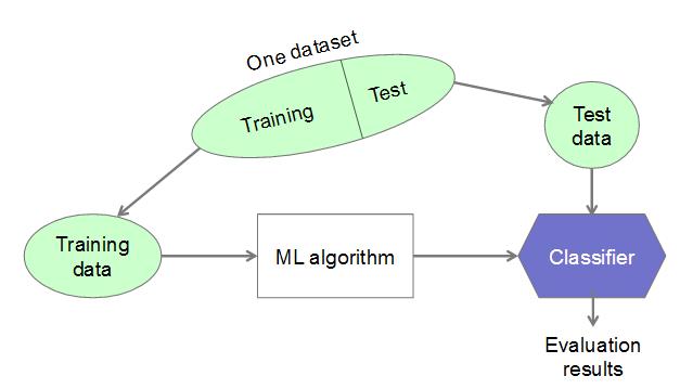
4.1 VGG16 Architecture
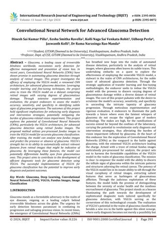
Visual Geometry Group or VGG16 for short is a powerful convolutional neural network (CNN) architecture. CNNs are a type of deep learning model that excels at image recognition. VGG16 has 16 convolutional layers that can automatically learn features from images. Imagine it as a highly sophisticated pattern recognizer. In the context of glaucoma detection, VGG16 can identify subtle variations in the optic nerve, blood vessels, and retinal tissue within fundus images. While VGG16 comes pre-trained on a vast image dataset, it needs further training to specialize in glaucoma detection. This process, called fine-tuning, involves using a dataset of fundus images labelled as healthy or glaucoma-affected. By analysing these labelled images, VGG16 refines the final layers of its network. These final layers learn to differentiate between healthy and diseased retinas based on the features extracted by theearlierconvolutionallayers.
4.2 Fundus Images
These are the images of the back of the eye, which can showsignsofglaucoma.Theprojectmightutilizedatasets fromrepositoriesorcollaboratewithophthalmologistsfor abroaderrangeofimages.
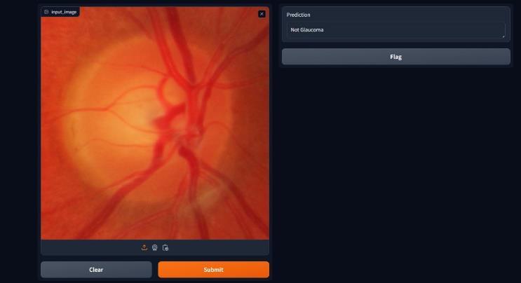
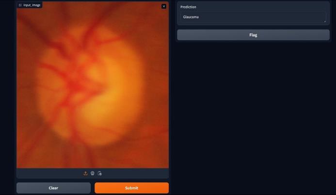
4.3 Gradio Interface
Gradioisanopen-sourcelibrarythatallowsdevelopersto build simple yet powerful web interfaces for machine learningmodels.Inourcase,Gradiocanbeusedtocreate awebappforglaucomadetectionusingVGG16.
5.1 Collection and Interpretation of Datasets
Gathering Data: The foundation of the model is a collection of retinal images. These can come from hospitals,researchcentres,oronlinedatabases.
Labelling the Images: Each image needs to be classified as either healthy or glaucoma-affected. This can be done usingpre-labelleddatasets,labellingbytraineddoctors,or evenmachinelearningtechniquesforinitialsorting.
Cleaning Up the Data: Before feeding the images to the model, they need preprocessing. This might involve cropping,resizing,orenhancingspecificfeaturestoensure consistencyandmakeprocessingeasierforthemodel.
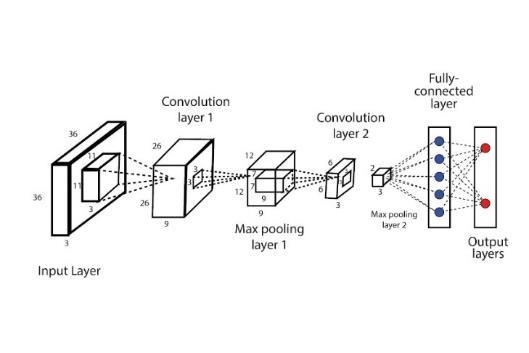
Training and Testing: Once pre-processed, the data is split into two sets: a training set used to teach the model andatestingsetusedtoevaluateitsperformance.
Understanding the Images:Analysingthedatacanreveal critical features within the images that influence the model’s predictions. Techniques from machine learning can help identify these important features, ultimately improvingthemodel’saccuracy.
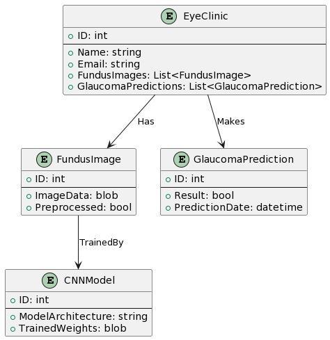
5.2 Data Model Schema
International Research Journal of Engineering and Technology (IRJET) e-ISSN: 2395-0056
Volume: 11 Issue: 04 | Apr 2024 www.irjet.net p-ISSN: 2395-0072
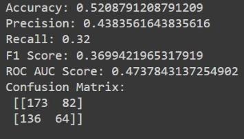
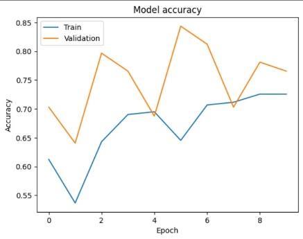
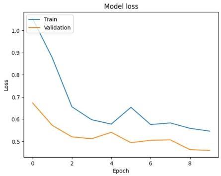
Thiscontainsalltheresultsandchangeinperformanceof themodelwithtraininghistory,mainperformancemetrics which are considered are accuracy, precision, recall, f1 scoreandconfusionmatrix.
At10epochs:
UserInterfaceusingGradio:
a) Python:Theprimaryprogramminglanguageused forwritingthecode.
b) Google Collab: A cloud-based platform that provides free access to computational resources suchasGPUandTPU.
c) TensorFlow: An open-source machine learning framework developed by Google for building and trainingneuralnetworkmodels.
d) Keras: An open-source neural network library written in Python that serves as a high-level API forTensorFlow.
e) Gradio: A Python library that allows you to quickly create customizable UI components aroundyourmachinelearningmodels.
f) Matplotlib: A comprehensive library for creating static, animated, and interactive visualizations in Python.
g) Scikit-learn (sklearn): A machine learning library in Python that features various classification, regression,andclusteringalgorithms.
h) NumPy: A Python library used for working with arrays andmatrices,particularlyinthecontextof numericalcomputations.
The model we implemented is a Convolutional Neural Network(CNN)basedontheVGG16architecture,whichis apowerfuldeeplearningmodelcommonlyusedforimage classification tasks. This model has been trained on a dataset containing images of two classes: Glaucoma and Non-Glaucoma.
International Research Journal of Engineering and Technology (IRJET) e-ISSN: 2395-0056
Volume: 11 Issue: 04 | Apr 2024 www.irjet.net p-ISSN: 2395-0072
Given its architecture and the training data, there are several potential future scopes and applications for this model:
a) Medical Diagnosis and Screening: One of the primary applications of this model is in the field of ophthalmology for diagnosing glaucoma. With further validation and testing, the model could be integrated into clinical workflows to assist ophthalmologists in screening patients for glaucoma. This could potentially lead to earlier detection and treatment of the disease, improving patientoutcomesandreducingtheriskofvisionloss.
b) Telemedicine and Remote Healthcare: In regions with limited access to specialized medical facilities, telemedicine platforms could leverage this model to provide remote screening for glaucoma. Patients could upload retinal images to a telemedicine platform, which would then use the model to analyse the images and provide preliminary diagnostic insights. This could help extend the reach of eye care services to underserved populations.
c) Continuous Monitoring and Disease Progression Tracking:Oncediagnosedwithglaucoma,patientsrequire ongoing monitoring to track disease progression and assess treatment efficacy. This model could be incorporated into monitoring devices or smartphone applications that allow patients to regularly capture retinalimagesforanalysis.Byanalysingchangesinretinal morphology over time, the model could provide valuable insights into disease progression, enabling early interventionwhennecessary.
d) IntegrationwithElectronicHealthRecords(EHR): Healthcare institutions could integrate this model with their electronic health record (EHR) systems to automaticallyanalyseretinalimagesaspartofroutineeye exams. The model's predictions and diagnostic insights could then be stored alongside other patient data, providing a comprehensive overview of the patient's ocular health over time. This integration could streamline clinical workflows and facilitate data-driven decisionmakingbyhealthcareproviders.
e) Research and Development: The model could serveasavaluabletoolforresearchersstudyingglaucoma and other ocular diseases. By analysing large datasets of retinalimages,researcherscouldgaininsightsintodisease mechanisms,identifynovelbiomarkers,anddevelopmore effectivediagnosticandtreatmentstrategies.Additionally, themodelcouldbefine-tunedoradaptedforrelatedtasks, suchasdetectingotherretinalabnormalitiesorpredicting diseaseriskfactors.
f) Personalized Medicine and Treatment Planning: As the field of precision medicine continues to evolve,
thereisgrowinginterestintailoringmedicalinterventions toindividualpatientcharacteristics.Thismodelcouldplay a role in personalized treatment planning for glaucoma patientsbyprovidingprognosticinformationbasedonthe analysis of retinal images. Clinicians could use this information to develop customized treatment plans optimizedforeachpatient'sspecificneedsandriskprofile.
g) Education and Training: Lastly, the model could be used as an educational tool for medical students, residents, and ophthalmology fellows to learn about glaucoma diagnosis and management. By interacting with themodelandobservingitspredictionsondifferenttypes of retinal images, trainees can enhance their understanding of glaucoma pathology and develop diagnosticskillsthatarecrucialforclinicalpractice.
In our investigation of the implemented model's efficacy for glaucoma detection, several limitations have come to light, each bearing implications for the model's performanceandapplicabilityinreal-worldscenarios.One notableconstraintrevolvesaroundthemodelarchitecture choice, wherein reliance on the VGG16 convolutional neural network may restrict the model's capacity to captureintricatepatternsandnuancespresentin medical images. Additionally, the limited dataset size poses a challenge, potentially hindering the model's ability to generalizetounseendataandvariationswithinthetarget population. Furthermore, while the augmentation techniques applied during training aim to enhance model robustness, their simplicity may not fully address the diverse range of potential variations in medical images. Theabsenceoffine-tuning,coupledwithrelianceonbasic evaluationmetricslikeaccuracy,furthercompoundsthese limitations, emphasizing the need for more comprehensive validation strategies and metric considerations to ensure the model's reliability and generalizabilityinclinicalsettings.
In conclusion, our glaucoma detection model demonstrates varying accuracies across different training configurations, highlighting the complex relationship between model architecture, dataset size, and training methodologies. While promising accuracy rates are achieved in certain configurations, limitations such as datasetsizeandmodelarchitecturechoiceunderscorethe needforfurtherrefinementandvalidation.
However, the model's advancements offer significant potential for enhancing diagnostic capabilities in ophthalmology and broader medical contexts. By leveragingdeeplearningtechniquesandlarge-scaleimage datasets, the model can identify subtle pathological
International Research Journal of Engineering and Technology (IRJET) e-ISSN: 2395-0056
Volume: 11 Issue: 04 | Apr 2024 www.irjet.net p-ISSN: 2395-0072
features indicative of glaucoma with notable accuracy. This facilitates earlier detection and intervention, potentially reducing reliance on subjective human interpretation.
Moreover, the model's adaptability to different training levels allows for tailored deployment in diverse clinical settings, benefiting regions with limited access to specialized eye care services. Continued research efforts focused on refining model architectures, expanding dataset diversity, and incorporating interpretability featureswillfurtherenhanceitsutilityinclinicalpractice. Ultimately, interdisciplinary collaboration and iterative refinement will position this model as a valuable tool, empoweringcliniciansandimprovingpatientoutcomesin thediagnosisandmanagementofoculardiseases.
[1]X.Chen,Y.Xu,D.W.K.Wong,etal."GlaucomaDetection usingDeepConvolutionalNeuralNetwork"(2021). S.Liu, H. Li, L. Wong, N. Tan, T. Wong. "A deep learning-based method for glaucoma detection in retinal fundus images" (2022).
[2] P.M. Burlina, N. Joshi, M. Pekala, K.D. Pacheco, D.E. Freund, N.M. Bressler. "Automated grading of age-related macular degeneration from color fundus images using deepconvolutionalneuralnetworks"(2021).
[3] M. Christopher, A. Belghith, C. Bowd, et al. "Performance of deep learning architectures and transfer learning for detecting glaucomatous optic neuropathy in fundusphotographs"(2021).
[4] Z. Li, Y. He, S. Keel, et al. "Efficacy of a deep learning system for detecting glaucomatous optic neuropathy basedoncolorfundusphotographs"(2021).
[5] D.S.W. Ting, C.Y. Cheung, G. Lim, et al. "Development and validation of a deep learning system for diabetic retinopathy and related eye diseases using retinal images frommultiethnicpopulationswithdiabetes"(2021).
[6] J.H. Tan, U.R. Acharya, S.V. Bhandary, C.K. Chua, C.M. Lim, A. Laude. "Application of deep learning for detecting glaucomatousprogressioninfundusimages"(2021).
[7] Akash Bayyana; Jeyanand Vemulapati; Sai Hemanth Bathula; Gangula Rakesh; Srilatha Tokala; Murali Krishna Enduri202314thInternationalConferenceonComputing CommunicationandNetworkingTechnologies(ICCCNT) https://ieeexplore.ieee.org/document/10307182
[8] Deep Learning Approach to Enhance Accuracy for Early Detection of Glaucoma Pranita Niraj Palsapure;Anu H A;Ashmitha G;Abhishek Reddy B H;Mainak Jana 2023
3rd International Conference on Intelligent Technologies (CONIT) https://ieeexplore.ieee.org/document/10205533
[9] Li et al. (2023) proposes a CNN-based approach for glaucomadetectionthatincorporateschannelpruningand knowledgedistillationforenhancedefficiency.([Li,Y.,Tan, T., Liu, H., Ma, Z., & Deng, L. (2023). Glaucoma detection usingconvolutionalneuralnetworkswithchannelpruning and knowledge distillation. Computers in Biology and Medicine,158,106622]) (https://www.sciencedirect.com/science/article/pii/S187 7050923004052)
[10] Liu et al. (2022) present a CNN-based classification model for glaucoma detection using Optical Coherence Tomography(OCT)images.([Liu,X.,Guo,Z.,Wang,Y.,Yin, F., & Cheng, J. (2022). A CNN-Based Classification Model for Glaucoma Detection Using Optical Coherence Tomography Images. IEEE Access, 10, 133222-133233]) (https://ieeexplore.ieee.org/document/9190916)
[11] Sowmya & Kavitha (2020) introduce a deep-learning model for glaucoma detection utilizing fundus images. ([Sowmya, B. H., & Kavitha, S. (2020). Deep Learning Model for Glaucoma Detection Using Fundus Images. In 2020 International Conference on Electrical, Electronics, Communication, Computer, and Informatics (EECCI) (pp. 1-5).IEEE])
(https://ieeexplore.ieee.org/document/9515188)
[12] Azzopardi et al. (2015) explore a method that combines deep learning with eye feature engineering for glaucoma detection, achieving promising results. ([Azzopardi, G., Meira, J., Leiva-Vega, M., Moreno-Macías, H., Garcia-Dorado, J., Larrañaga, P., ... & de Luna, D. S. (2015). Combining deep learning and eye feature engineering for glaucoma detection. Pattern Recognition Letters, 65, 40-48]) (https://www.sciencedirect.com/science/article/pii/S007 961232030100X)
[13] Ioannides & Yang (2017) investigate the application of deep learning for classifying healthy versus glaucomatous optic nerve head images. ([Ioannides, A. A., & Yang, G. (2017). Deep learning for healthy versus glaucomatousopticnerveheadclassification.In2017IEEE 14th International Symposium on Biomedical Imaging (ISBI)(pp.1132-1135).IEEE]) (https://ieeexplore.ieee.org/document/9548439
[FIG 3.3] CONVOLUTIONAL NEURAL NETWORK (CNN): GRAPHICAL VISUALIZATION WITH PYTHON CODE EXPLANATION
https://lh3.googleusercontent.com/yrHzday2CwSYLkXf9y KSoH-BpjqnnAuyiMvPAS5yS3-
International Research Journal of Engineering and Technology (IRJET) e-ISSN: 2395-0056
Volume: 11 Issue: 04 | Apr 2024 www.irjet.net p-ISSN: 2395-0072
lFnl5jwkR6FoT_v2Vbi14s414fJSORuGLRQbHyYp6dtHDItR cSQnRWcd1JRGbZC5VlGTvH80gFZrHw8qg2Tx7ca2HYKFc
[FIG 3.4] TRAINING AND TESTING OUR MACHINE LEARNINGAPPROACH.
https://www.researchgate.net/profile/SantiCaballe/publication/318132501/figure/fig2/AS:5578239 75460869@1510007007310/Training-and-testing-ourmachine-learning-approach.png
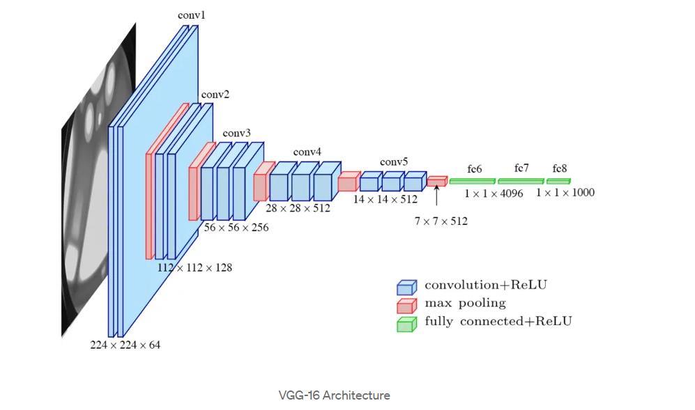
[FIG 4.1] TYPES OF CONVOLUTIONAL NEURAL NETWORKS: LENET, ALEXNET, VGG-16 NET, RESNET AND INCEPTION NET
HTTPS://MIRO.MEDIUM.COM/V2/RESIZE:FIT:1100/FOR MAT:WEBP/1*B_ZAAABG2NJHP8STHJCUFA.PNG