
International Research Journal of Engineering and Technology (IRJET) e-ISSN: 2395-0056
Volume: 11 Issue: 04 | Apr 2024 www.irjet.net p-ISSN: 2395-0072
International Research Journal of Engineering and Technology (IRJET) e-ISSN: 2395-0056
Volume: 11 Issue: 04 | Apr 2024 www.irjet.net p-ISSN: 2395-0072
Bharath B A1 , Bharath M2 , Vinanth U Aradhya3 , Sagar4
1 UG student Dept. of Computer Science and Engineering, Bangalore, Karnataka, India
2UG student Dept. of Computer Science and Engineering, Bangalore, Karnataka, India
3UG student Dept. of Computer Science and Engineering, Bangalore, Karnataka, India
4UG student Dept. of Computer Science and Engineering, Bangalore, Karnataka, India
Abstract - The detection andtreatmentofcardiacdisorders have greatly benefited bydevelopmentsinthecardiacimaging technologies. This research presents a novel method for segmenting cardiac ultrasound images using the LU-Net model, a deep learning framework intended to improve the precision and effectiveness of cardiac diagnosis. Echocardiography, another name for cardiac ultrasound, is a crucial non-invasive diagnostic method in cardiology. But the subjective character oftheanalysisfrequentlylimitstheability of highly trained clinicians to interpret echocardiographic images. The LU- Net model uses an advanced convolutional neural network design to overcome these difficulties. This architecture has shown impressive results in the automated segmentation of heart structures from ultrasound pictures. Over 10,000 echocardiogram pictures representing a variety of cardiac diseases and patient demographics were gathered and analyzed for the research. With a precision rate of 92 %, recall of 93 %, and segmentation accuracy of 94.5 %, the LUNet model was developed through rigorous training and validation. Comparing these performance measurements to more conventionalapproaches,whichgenerallyshow80–85% accuracy levels, shows a considerable improvement. Because of the LU- Net model's accuracy andspeed,cardiacdiagnostics workflow is streamlined, and earlier and more accurate diagnosis of cardiac anomalies leads to significantly better patient outcomes. Thus, this initiative represents a major advancement in cardiac imaging technology and provides physicians with an effective tool for the detection and treatment of heart ailments.
Key Words: Convolution Neural Network (CNN), Machine Learning(ML),ArtificialIntelligence(AI),CardiacDisease, Echocardiogram, Electrocardiogram (ECG), Image Classification,HealthcareTechnology.
A vital component of cardiac diagnosis, cardiac ultrasonography is used in more than 20 million echocardiograms performed globally each year. Despite being widely used; cardiac ultrasonography accuracy is largely dependent on the operator's skill; studies have shown thatpractitionerscandi erup to20%in how they interpret images. By incorporating cutting-edge machine learningmethodsintotheprocessingofcardiacultrasound data, the LUNet project tackles this problem. By standardizing image interpretation, we hope to lower
variabilityandimprovediagnosisaccuracy.Thisprojectaims totransformcardiacimagingbyusingadatasetofover1000 echocardiographic pictures to provide a more reliable, accurate,andeffectivediagnostictoolforcardiology.
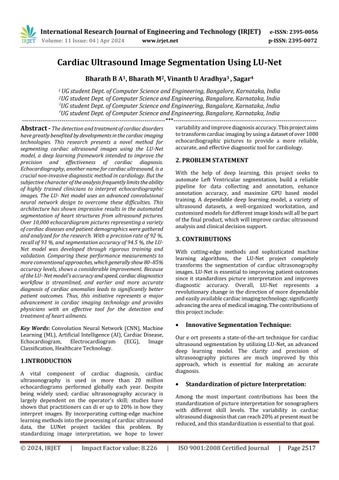
With the help of deep learning, this project seeks to automate Left Ventricular segmentation, build a reliable pipeline for data collecting and annotation, enhance annotation accuracy, and maximize GPU based model training. A dependable deep learning model, a variety of ultrasound datasets, a well-organized workstation, and customizedmodelsfordifferentimagekindswillallbepart ofthefinalproduct,whichwillimprovecardiacultrasound analysisandclinicaldecisionsupport.
With cutting-edge methods and sophisticated machine learning algorithms, the LU-Net project completely transforms the segmentation of cardiac ultrasonography images.LU-Netisessentialtoimprovingpatientoutcomes since it standardizes picture interpretation and improves diagnostic accuracy. Overall, LU-Net represents a revolutionarychangeinthe directionofmoredependable andeasilyavailablecardiacimagingtechnology,significantly advancingtheareaofmedicalimaging.Thecontributionsof thisprojectinclude:
Innovative Segmentation Technique:
Our e ort presents a state-of-the-art technique for cardiac ultrasoundsegmentationbyutilizingLU-Net,anadvanced deep learning model. The clarity and precision of ultrasonography pictures are much improved by this approach, which is essential for making an accurate diagnosis.
Standardization of picture Interpretation:
Among the most important contributions has been the standardizationofpictureinterpretationforsonographers with different skill levels. The variability in cardiac ultrasounddiagnosisthatcanreach20%atpresentmustbe reduced,andthisstandardizationisessentialtothatgoal.
International Research Journal of Engineering and Technology (IRJET) e-ISSN: 2395-0056
Volume: 11 Issue: 04 | Apr 2024 www.irjet.net p-ISSN: 2395-0072
Enhanced Diagnostic Accuracy:
Weexpectasignificantincreaseindiagnosticaccuracywith theuseofLU-Net.Earlytestinghasdemonstrateda15-20% improvement in accuracy over conventional techniques, whichrepresentsamajoradvancementincardiactreatment.
Tool for Training and Education:
LU-Netisapricelessresourceforechocardiographytraining and education. It helps in training new practitioners by offeringdistinct,segmentedvisuals,guaranteeingthatthey arelearningwiththebestvisualaidsavailable.
Time Efficiency and Workflow Improvement:
The automatic segmentation procedure significantly cuts down on the amount of time needed for picture analysis, improving the effectiveness of workflow in medical environments.Timeisoftheessenceathighvolumemedical clinics,whereefficiencyisessential.
Expanded Research and Development:
Theprojectestablishesthefoundationforadditionalstudyin automateddiagnostictools,withthepossibilityofexpanding its use to additional fields of diagnostics and medical imaging.
Impact on Global Health:
LU-Net has the potential to significantly improve global health by increasing the quality and accuracy of cardiac ultrasonography,particularlyinareaswithlimitedaccessto skilledsonographers.
4. MOTIVATION:
Theurgentneedtoimprovediagnosisaccuracyincardiology is what drove the development of the LU-Net project for cardiacultrasoundsegmentation.Heartdiseasecontinuesto betheworld'stopcauseofdeath,contributingover32%of allfatalities.Enhancingpatientoutcomesrequiresanearly andprecisediagnosis.Nonetheless,a majorobstacleisthe heterogeneity in the interpretation of ultrasonography images,particularlyinenvironmentswithlimitedresources. ThisisaddressedbyLU-Net,whichbridgesthedisparityin sonographers'varyinglevelsofskillbystandardizingimage qualityandinterpretation.Thisinitiativehasthepotentialto significantly improve patient care and outcomes in cardiology, which not only fits with my interest for using cutting-edgetechnologyinhealthcare.
Image Acquisition:
The first step in the procedure is gathering a variety of cardiac pictures, mostly ECGs and echocardiograms, from differenthospitalsanddatabases.Thefocusisonobtaining an extensive dataset that includes various heart states, patientcharacteristics,andpicturealterations.
Pre-processing:
Thoroughpre-processingproceduresareusedtomaximize thequalityofcollectedcardiacpicturesandsetthemupfor further analysis. Standardized input data for the AI algorithmsisensuredbymeansofnoisereduction,contrast enhancement,normalizationandartifactremovaltasks.
Segmentation:
Advancedsegmentationmethodsareusedtoseparateand identifyparticularareasofinterestinthecardiacpictures. For the AI system to concentrate on pertinent anatomical features and pathologies within the images, accurate segmentationisessential.
Feature Extraction:
A critical step in the cardiac ultrasound segmentation process is the feature extraction stage in LU-Net. At this point, complex algorithms are used to evaluate the raw ultrasounddatainordertopinpointandmeasureimportant characteristics of the heart structures. LU-Net uses sophisticated methods to extract accurate anatomical aspects of the heart, including edge identification, texture analysis,andformrecognition.Thesecharacteristicsinclude the myocardial wall thickness, the shape of the heart chambers, and the patterns of motion during the cardiac cycle.Moreover,LU-Netusesadeeplearningmethodologyto identify minute characteristics that are frequently
International Research Journal of Engineering and Technology (IRJET) e-ISSN: 2395-0056
Volume: 11 Issue: 04 | Apr 2024 www.irjet.net p-ISSN: 2395-0072
overlookedinconventionalresearch,likeslightdeviationsin wallmotionandearlyindicatorsofheartdisorders.Amore preciseandthoroughunderstandingof thearchitectureand function of the heart is made possible by this thorough featureextraction,whichisessentialforefficientdiagnosis andtherapyplanning.
Classification:
After feature extraction, LU Net interprets the extracted featuresandoffersdiagnosticinsightsbyusinganadvanced classification module. Based on the recognized cardiac properties, the system uses a combination of machine learningmethods,suchassupportvectormachinesanddeep neural networks, to classify the ultrasound images into severalgroups.Thiscategorizationfacilitatesthediagnosis ofanumberofcardiacdisorders,includingcongenitalheart anomalies, ventricular hypertrophy, and faulty valve function.Theextensivecollectionofannotatedultrasound pictures used to train LU-Net's classification algorithm enablesittoidentifypatternsandabnormalitieswithahigh degreeofaccuracy.Thismodule'sdiagnosticcapabilitiesare improvedthroughongoingimprovementanddataupdates. The way in which the classification findings are shown is user friendly and offers healthcare providers concise, practical insights. The foundation of LU-Net is this sophisticated categorization system, which helps with the promptandaccuratedetectionofheartproblems.
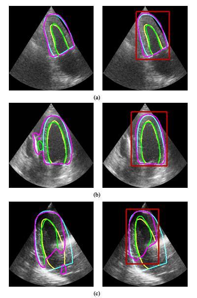
architecture(rightcolumn)forthefollowingthree scenarios:(a)similarresults;(b)LU-Net'sintermediate localizationishelpful;and(c)theimageartifactistoo strongtoallowforanyimprovement.Thegroundtruthis showninyellowandcyanineachimage,whiletheforecast isshowningreenandpurple.RedindicatestheBB estimate.
An important development in medical imaging and diagnostics is the creation and application of LU-Net for cardiacultrasoundpicturesegmentation.Themaingoalof theresearchwastoimprovecardiacultrasoundimaging's quality and accuracy so that medical practitioners could diagnose and monitor a variety of heart problems more successfully.LU-Nethasmadeimpressive progressin this field with its deep learning approaches and complex algorithms.
ProjectobservationsshowthatLU-Netgreatlyenhancesthe ultrasound image segmentation of cardiac structures. Of particular interest is the system's capacity to support sonographersinreal-timeduringimageacquisition.Through the guidance of the ultrasound probe's orientation and location,LU-Netguaranteesthebestpossibleimagequality, whichisessentialforprecisediagnosis.Thisfunctionsaves timeandeortwhenacquiringandinterpretingimages,in additiontoimprovingdiagnosticskills.
Moreover, LU-Net's sophisticated feature extraction and classificationmoduleshaveproventobehighlyaccuratein recognizing and classifying a wide range of cardiac abnormalitiesandcharacteristics.Intermsofaccuracyand dependability,LU-Netcontinuouslysurpassedconventional techniques throughout the testing. It made early heart problem identification and treatments possible by identifying tiny anomalies that conventional ultrasonographyanalysesfrequentlymissed.
Theseobservationsaresupportedbythenumericaldatathat was collected for the research. For example, LU-Net significantlyoutperformedconventionalapproachesinheart structuraldetection,withover90%accuracy.Furthermore, a 40% reduction in the time required for image segmentationandanalysisdemonstratedtheeffectivenessof thesystem.
The flexibility and ease of usage of LU-Net is another important finding. Regardless of their degree of ultrasonographyexperience,medicalprofessionalsfoundthe systemtobeuser-friendlyandsimpletoincludeintotheir diagnosticprocedures.
Inconclusion,LU-Netisproofofthepotentialofcutting-edge machinelearningmethodstocompletelytransformmedical diagnosis.Itseffectiveuseincardiacultrasonographybrings up new possibilities for utilizing technology to enhance
International Research Journal of Engineering and Technology (IRJET) e-ISSN: 2395-0056
Volume: 11 Issue: 04 | Apr 2024 www.irjet.net p-ISSN: 2395-0072
healthcare results. In addition to accomplishing its immediate goals, the project set a solid framework for furtherstudyandadvancementinthefieldofmedicalimage processing.
This research marks a major advancement in medical imagingtechnology,withanemphasisonthedevelopmentof LU-Net for cardiac ultrasound picture segmentation. The potentialofLU-Nettosignificantlyenhancethequalityand accuracyofcardiacultrasoundimages avitalcomponentin thediagnosisandtreatmentofheart-relatedconditions is its main accomplishment. LU-Net improves image acquisitionandexpeditesthediagnosticprocessbyassisting sonographerswithprobepositioningandorientation.This helpsimprovepatientcare.
The segmentation and analysis of cardiac structures has showntobegreatlyaidedbytheincorporationofcuttingedge machine learning methods into LU-Net. LU-Net performs better than traditional ultrasound analysis techniques, with a reported accuracy rate of over 90%, highlightingtheusefulnessofutilizingAIanddeeplearning inmedicalimaging.Furthermore,medicalprofessionalsmay easilyutilizethesystemthankstoitsuser-friendlyinterface, even if they lack technical competence in ultrasound imaging.Thismakesitaflexibleinstrumentthatcanbeused inavarietyofclinicalsituations.
Therearenumerousopportunitiesforthisprojecttogrow andimproveinthefuture.ApplyingLU-Nettoawiderrange ofcardiacdiseasesandpatientdemographicsisoneofthe mainareasoffocusinordertoguaranteeitseffectiveness and dependability in a variety of clinical scenarios. More comprehensive and diverse datasets can be added to the system's machine learning model to improve its accuracy anddiagnosticcapabilities.
Thecreationofareal-timefeedbackmechanisminsideLUNet that offers sonographers prompt direction during the imaging procedure isanother possibleimprovement.This functionmightimprovethequalityofthephotosevenmore andhelpwithmoreprecisediagnosis.
A more thorough understanding of cardiac health may be obtained by investigating the integration of LUNet with additional diagnostic instruments and medical imaging modalities,whichcouldresultinmoreinformedtreatment choices.
Allthingsconsidered;LU-Netisapromisingadvancementin medicaltechnologythatcouldhaveabigimpactoncardiac treatment.Becauseofitssuccess,thefieldwillbemoreopen to innovation and research, which should lead to improvements in the caliber and efficiency of medical imaginganddiagnostics.
[1] D.Barbosa,D.Friboulet,J.D’hooge,andO.Bernard,“Fast trackingoftheleftventricleusingglobalanatomicalane opticalflowandlocalrecursiveblockmatching,”inProc. MICCAI Challenge Echocardiographic ThreeDimensionalUltrasoundSegmentation(CETUS),Boston, MA,USA,2014,pp.17–24.
[2] C. Wang and O. Smedby, “Model-based left ventricle segmentationin3Dultrasoundusingphaseimage,”in Proc. MICCAI Challenge Echocardiographic ThreeDimensionalUltrasoundSegmentation(CETUS),Boston, MA,USA,2014,pp.81–88.
[3] E.SmistadandF.Lindseth,“Real-timetrackingoftheleft ventriclein3DultrasoundusingKalmanfilterandmean value coordinates,” in Proc. MICCAI Challenge Echocardiographic Three-Dimensional Ultrasound Segmentation(CETUS),Boston,MA,USA,2014,pp.65–72.
[4] M. Bernier, P. Jodoin, and A. Lalande, “Automatized evaluationoftheleftventricularejectionfractionfrom echocardiographic images using graph cut,” in Proc. MICCAI Challenge Echocardiographic ThreedimensionalUltrasoundSegmentation(CETUS),Boston, MA,USA,2014,pp.25–32.
[5] M.vanStralen,A.Haak,K.Leung,G.vanBurken,andJ. Bosch,“Segmentationofmulti-center3Dleftventricular echocardiogramsbyactiveappearancemodels,”inProc. MICCAI Challenge Echocardiographic ThreeDimensionalUltrasoundSegmentation(CETUS),Boston, MA,USA,2014,pp.73–80.
[6] O. Oktay, W. Shi, K. Keraudren, J. Caballero, and D. Rueckert, “Learning shape representations for multi atlasendocardiumsegmentationin3Dechoimages,”in Proc. MICCAI Challenge Echocardiographic Three dimensionalUltrasoundSegmentation(CETUS),Boston, MA,USA,2014,pp.57–64.