
International Research Journal of Engineering and Technology (IRJET) e-ISSN: 2395-0056
Volume: 11 Issue: 04 | Apr 2024 www.irjet.net p-ISSN: 2395-0072

International Research Journal of Engineering and Technology (IRJET) e-ISSN: 2395-0056
Volume: 11 Issue: 04 | Apr 2024 www.irjet.net p-ISSN: 2395-0072
Ramnesh Kumar1 , Sankalp Rajpoot2 , Prateek Kumar Verma3, Mr. Suresh Kumar4
1B.Tech student, Information Technology, Galgotias College Of Engineering & Technology, Uttar Pradesh, India
2B.Tech student, Information Technology, Galgotias College Of Engineering & Technology, Uttar Pradesh, India
3B.Tech student, Information Technology, Galgotias College Of Engineering & Technology, Uttar Pradesh, India
4B.Tech faculty, Information Technology, Galgotias College Of Engineering & Technology, Uttar Pradesh, India
Abstract - Detecting brain tumors via Magnetic Resonance Imaging (MRI) is crucial but challenging due to the intricate nature of these abnormalities. A proposed method involves several steps, including sigma filtering, adaptive thresholding, and region detection, to analyze MR images. Shape features such as Major Axis Length, Euler Number, Minor Axis Length, Solidity, Area, and Circularity are extracted to characterize the tumors. This method employs two supervised classifiers: a C4.5 decision tree algorithm and a Multi-Layer Perceptron (MLP) algorithm. These classifiers distinguish between normal and abnormal brain cases, with abnormalities further classified into benign or malignant tumors. With a dataset of 250 brain MR images, the MLP algorithm achieves a notable precision of approximately 80%.
Key Words: Magnetic Resonance Imaging (MRI), Image Acquisition, Detection Region, Image preprocessing, Image Segmentation, Feature Extraction
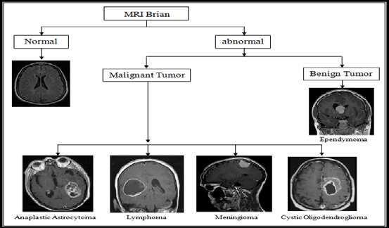
Brain tumors are solid neoplasms found within the skull, arisingfromuncontrolledandabnormalcelldivision.They typicallydevelopinthebrainitself,butcanalsomanifestin other locations such as lymphatic tissue, blood vessels, cranial nerves, and brain envelopes. Additionally, brain tumorscanresultfromthemetastasisofcancersoriginating elsewhere in the body. The classification of brain tumors hinges on factors like their location, the tissue type from whichtheyoriginate,theirmalignantorbenignnature,and otherconsiderations.
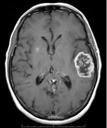
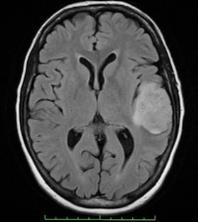
Primary brain tumors originate within the brain and are named based on the cell types from which they originate. They may be benign, such as Meningioma, which cannot metastasize.Conversely,theycanbemalignantandinvasive, exemplified by Lymphoma (characterized by a ring-like appearance),cysticoligodendroglioma(displayingrounded cellswithdistinctbordersandacentralnucleusresembling a"friedegg"),Ependymoma(arisingfromependymalcells andexhibitingmalignantbehaviordespitebenignhistology), and Anaplastic astrocytoma (a common high-grade astrocytoma).
Secondary brain tumors, also known as metastatic brain tumors,developfromcancercellsthathavemigratedtothe brainfromotherpartsofthebody.Typically,thesecancers
originate from primary tumors in organs such as the kidneys, lungs, breasts, or from melanomas on the skin. A brain scan offers a detailed visualization of the brain's internal structure. Among the most frequently utilized methods for brain imaging is MRI (Magnetic Resonance Imaging), renowned for its ability to provide exceptional insightsintothehumanbody.TocategorizeMRImages,two primary methodologies are employed: supervised techniques like support vector machines, k-nearest neighbors,andartificialneuralnetworks,andunsupervised techniquessuchasfuzzyc-meansandself-organizingmaps (SOM). Many studies have utilized a combination of both supervisedandunsupervisedtechniquestodistinguishMR Imagesas either normal or abnormal.Thisstudy employs supervisedmachinelearningtechniquestocategorizefive distinct types of abnormal brain MR Images, including Ependymoma, Lymphoma, Cystic Oligodendroglioma, Meningioma,andAnaplasticAstrocytoma,alongsidenormal images
1.SurajGroverandfellow writers unveiledanunfamiliar method for segmenting brain tumors in 3D MR pictures. Initially,segmentationofbrainMRpictureswascarriedout utilizing an inventive technique for tumor detection. Afterward, tumor detection leaned on selecting uneven regions.Thistechniqueconsidersthebrain'sasymmetrical planeandutilizesblurryclassification.Theresultsactasthe
International Research Journal of Engineering and Technology (IRJET) e-ISSN: 2395-0056
Volume: 11 Issue: 04 | Apr 2024 www.irjet.net p-ISSN: 2395-0072
foundationforsettinginmotionasegmentationprocessthat integratesspatialrelationsanddeformablemodels,leading toprecisesegmentationofbraintumors.
2.MaheshS.andfriendsputforthamethodologyfocusedon texture characteristics, particularly the Gray Level CooccurrenceArray(GLCM)derivedfromMRpictures.They employed a Sequential Forward Selection algorithm to pinpoint discriminative characteristics. Afterward, the method categorized MR images into usual and unusual categories by applying an advanced kernel-centered technique,justliketheAssistanceVectorMachine(SVM).
3.M.Jayanandfriends putintopracticeahybridalgorithm devised for brain tumor detection, leveraging statistical characteristics and a Vague Assistance Vector Machine (SVM) classifier. Their technique includes a four-step process.Initially,theyimplementedananisotropicfilterto diminishnoiseinthefirststep.Next,texturecharacteristics were taken out from MR pictures in the second step. Afterward,PrincipleConstituentAnalysis(PCA)wasutilized to reduce the characteristics of MR pictures to the most necessary ones in the third step. Eventually, tumor classificationintousualandunusualcategorieswasexecuted employingaManagerialclassifierbasedonVagueAssistance VectorMachineinthefinalstep.Theclassificationprecision accomplishedwas95.80%.
4.TanmayKapurandfriendstookadvantageofdatafrom both magnetized reverberation (MR) imaging and magnetized resonation spectroscopy (MRS) to help in clinical diagnosis. Their proposed technique encompasses numerousstages,encompassingsegmentation,characteristic extraction, and characteristic selection. A classification model was then built to segregate between usual and unusual brain cases. They employed a segmentation techniquefoundedonblurryconnectednesstooutlinetumor mass fences in MR pictures. Besides, they utilized the concentric circle technique to extract characteristics from regionsofinterest.Characteristicselectionwasimplemented to remove repetitious characteristics. Experimental discoveries highlight the efficiency of their approach in faithfullycategorizingbraintumorsinMRpictures.
5. Prachi Gadpayle and friends contrived a system specializedindetectingandcategorizingbraintumors.They exploited a spectrum of picture processing techniques, encompassing preprocessing, picture enrichment, segmentation, morphological actions, and characteristic extraction, custom-made for pinpointing brain tumors in MRI pictures. Noticeably, they included texture characteristics like the Gray Level Co-occurrence Array (GLCM) in tumor detection. Using classifiers just like the Backing Neural Network (BPNN) and the K-Nearest Companions(K-NN)algorithm,theyefficientlyclassifiedMRI brainpicturesintounusualandwholesomeones.
6. Ramteke and Monali recommended an automatic classificationofbrainMRpicturesintotwosectionsUsual andUnusualbasedonpicturecharacteristicsandautomatic mistakedetection.TheStatisticaltexturecharacteristicsetis picked up from usual and unusual pictures and then KNN classifierisutilizedforclassifyingpicture.TheKNNattains 80% classification rate. Xuan and Liao recommended statistical structure examination founded tumor segmentation technique. The intensity-based, symmetrybased,andtexture-basedcharacteristicsareextractedfrom MR picture. Then, classification technique employing AdaBoost is utilized to classify the MR picture into usual tissuesandunusualpictures.Thenormalaccuracyofabout 96.82% is attained. Othman et al. in recommended Probabilistic cerebral network technique for brain tumor classification. Primarily, the characteristics are extracted utilizingthepredominantcomponentanalysis(PCA)andthe classification is executed utilizing Probabilistic Neural Network (PNN). Ibrahim et al is recommended Neural Networktechniquefortheclassificationofthemagnetized reverberationhumanbrainpictures.Thecharacteristicsare extractedutilizingprincipalConstituentAnalysis(PCA)and then Back-WebDriver Neural Network is utilized as a classifiertoclassifyMRIbrainpicturesasusualorunusual.
Developing an Machine Learning Based Brain Tumor Detection Model:Methodology
3.1.
Inoursystem,weleveragereal-timedatacomprisingMRI images sourced from various hospitals and online repositories
Toensureconsistency,westandardizethedimensionsofthe images to 224x224 pixels. Upon acquisition, each image undergoesathoroughpreprocessingstagetoprepareitfor analysis.
Inimageprocessing,imageaccessionisdoneby reacquiring animagefromdatasetforprocessing.It'sthefirststepinthe workflowsequencebecause,withoutanimagenoprocessing is possible the image that's acquired is fully undressed. Then we reuse the image using the train path from the originaldevice
3.2.
The MRI dataset utilized in our study encompasses approximately2100images,representingbothnormaland abnormal brain scans. To enhance the quality of these images,weemployatechniqueknownassigmafilteringto reducenoiseinterference.
This process involves analyzing the pixels within a designatedareaandsmoothingoutvariationsthatexceeda
International Research Journal of Engineering and Technology (IRJET) e-ISSN: 2395-0056
Volume: 11 Issue: 04 | Apr 2024 www.irjet.net p-ISSN: 2395-0072
certain threshold. By applying sigma filtering, we aim to improve the clarity and accuracy of the images, thus facilitatingmoreprecisetumordetection.
After processing the images, the subsequent step is segmented.Hither,segmentationisbeingdoneemploying thresholding.Theunderlyingconceptofthresholdingisto simplify the visual data analysis. Thresholding bethinks a vastly popular segmentation technique utilized to differentiate the object pondered as a forefront from its surrounding. In this scenario, we are utlilizing binary thresholdingforsegmentation.Inbinarythresholding,each pixel enduresthesamethresholdvalue.Supposethepixel intensityvalueislesserthanthethreshold,itgetssetto0 (black);ifnot,itgetssetto255(white).
Segmentationstandsastheprocessofcarvinganimageinto myriad segments and isolating the tumor from regular tissues. Segmentation method owns the capacity to recognizeordeterminetheaberrantportionfromtheimage, cateringtotheanalysisofsize,volume,location,texture,and shapeofextractedimage.
Feature extraction is a pivotal step in harnessing the potentialofbigdatasets,particularlyintherealmofmedical imaginganalysissuchasbrainMRIscans.Byselectingand combining variables into meaningful features, we can significantly reduce the redundancy inherent in these datasets. This reduction not only streamlines the data processingpipelinebutalsoenhancesthelearningspeedof machinelearningalgorithms.
In our approach, we utilize morphological operations to extractfeaturesfromtheacquiredbrainMRIscans.These operations allow us to highlight distinct patterns and structures within the images, providing valuable insights into the underlying characteristics of the brain tissue.The transformed data, now represented by a reduced set of informativefeatures,isreferredtoasafeaturevector.This vectorencapsulatestheessentialinformationnecessaryfor subsequent analysis and model building. In our case, we
focus on extracting features that capture the textural propertiesofthesegmentedbrainMRIimages.
To achieve this, we employ the Gray Level Co-occurrence Matrix (GLCM) method, renowned for its robustness and highperformanceintextureanalysis.GLCMquantifiesthe spatialrelationshipsbetweenpixelintensityvalues,thereby encoding textural information that is instrumental in differentiatingbetweenvarioustissuetypesandpathological conditionsinbrainscans.ByleveragingGLCM-basedfeature extraction,weaimtoenhancethediscriminativepowerof our model and facilitate accurate tumor detection and classification.
Classificationcanbedefinedastheprocessofpredictinga class or category from observation values or given data points. The bracket of a biomedical image is a veritably importantstepforanautomatedComputerbackedDesign( CAD) system. At the end of these segmentation and discoveryprocess,decisionhasbeentakenrainfallthatMRI imageconsistsofanyexcrescenceornotandthenormalor theabnormalstatehasbeenchecked.
4.1.
The algorithm's efficacy has been rigorously evaluated across diverse performance metrics, encompassing True Positives (TP) and True Negatives (TN). TP quantifies the algorithm'scapacitytoaccuratelydiscerndamagedregions, while TN reflects its precision in identifying non-damaged areas. Conversely, False Positives (FP) denote instances where the algorithm incorrectly identifies non-damaged regionsasdamaged,whileFalseNegatives(FN)indicateits failuretorecognizedamagedregions.LeveragingTP,TN,FP, andFNvalues,keymetricssuchasAccuracy,Specificity,and Sensitivity are derived to comprehensively assess the algorithm's performance. This multifaceted evaluation underscoresthealgorithm'sdistinguishingbetweendamaged andnon-damagedregions,thuscontributingtoitsefficacyin clinicalapplications.


The experiment was carried out on 250 brain MR images. From eachimage,the texture basedfeaturesare extracted andwekatoolisusedforclassification.Thetexturebased featuressuchasenergy,contrast,correlation,homogeneity
International Research Journal of Engineering and Technology (IRJET) e-ISSN: 2395-0056
Volume: 11 Issue: 04 | Apr 2024 www.irjet.net p-ISSN: 2395-0072
areextractedusingGLCM.TheMulti-LayerPerceptron(MLP) and Naïve bayes with 66% percentage split is used for classification.In66%percentagesplit,66%oftheinstances areused for training and remaining instances are used for testing.
Table -1: ResultofNLPalgorithm
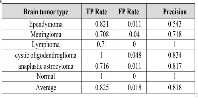
Table -2: Experimentalresultanalysis

Chart -2:Timetakenrepresentation
In the realm of brain MR image classification, the path to achieving accuracy is often a balancing act between time investmentandprecision.Here,theMulti-LayerPerceptron (MLP)emergesasthefrontrunner,boastingcommendable accuracy at approximately 88.2%. However, its triumph comeswithacaveat-theconstructionofitsmodeldemands aconsiderableamountoftime
FromtheTable1,wecanfindtheclassificationrateofbrain MRimagesusingMLPandNaivebayes.Theaccuracyofabout 88.2% and80.7%isobtainedrespectively
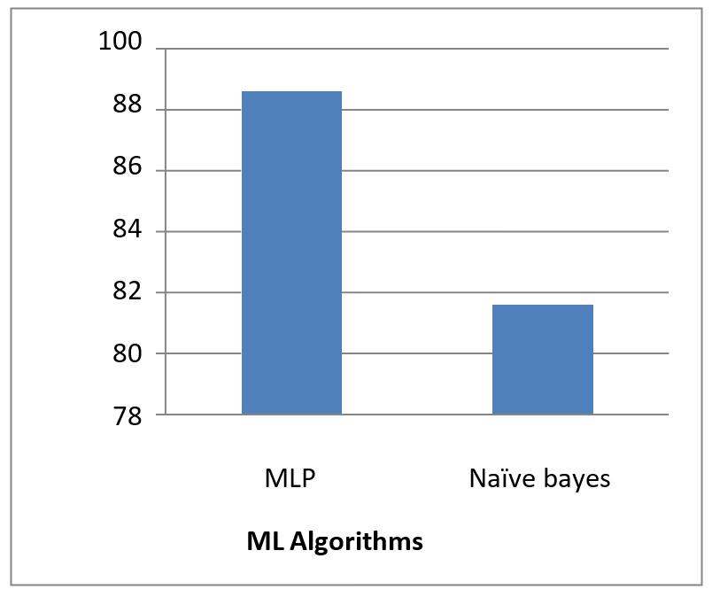
Chart -1:Accuracyrepresentation
Theaccuratebrainexcrescencediscoveryisstill veritably demanding because of tumor appearance, variable size, shape, and structure. Although excrescence segmentation styles have shown high eventuality in assaying and detecting the tumor in MR images, still numerous advancementsare neededtodirectlymemberandclassify the excrescence region. Being work has limitations and challengesfor relatingsubstructuresofexcrescenceregion andbracketofhealthyandunhealthyimages.Inshort,this checkcoversallimportantaspectsand rearmostworkdone sofarwiththeirlimitationsandchallenges.It'llbehelpfulfor theexperimenterstodevelopanunderstandingofdoingnew explorationinashorttimeandcorrectdirection.Thedeep literacy styleshavecontributedsignificantlybutstillbeara general fashion.These stylesprovidedbetterresultswhen trainingandtestingareperformedon analogousaccession characteristics(intensityrangeandresolution); still,aslight variationinthetrainingandtestingimagesdirectlyaffects therobustnessofthe styles.In unbornwork, exploration can be conducted to descry brain excrescences more directly, using real case data from any medium( different imageaccession(scanners).Handcraftedanddeepfeatures can be fused to ameliorate the classification results. also, lightweight styles similarasamountmachine literacyplay significant part to ameliorate the delicacy and efficacity thatsavethetimeofradiologistsandincreasethesurvival rateofcases.
International Research Journal of Engineering and Technology (IRJET) e-ISSN: 2395-0056
Volume: 11 Issue: 04 | Apr 2024 www.irjet.net p-ISSN: 2395-0072
[1] JPriyankaandBalwinderSinghconductedastudytitled "A Review On Brain Tumor Detection Using Segmentation"whichwaspublishedintheInternational JournalOfComputerScienceAndMobileComputingin July 2013. The study can be found on pages 48-54 of Volume2,Issue7.
[2] P. Narendran M, V.K. Narendira Kumar, and K. Somasundaram published a paper titled "3D Brain TumorsandInternalBrainStructuresSegmentationIn MR Images" in the I.J. Image, Graphics and Signal Processingin2012.ThepapercanbefoundinVolume1, pages35-43
[3] R. Siva Kumar and M. Karnan conducted a review on MRI image classification techniques, which was published in the International Journal of Research Studies In Computer Science and Engineering in May 2014.ThereviewcanbefoundinVolume1,Issue1,on pages21-28
[4] Komal Sharma, Akwinder Kaur, and Shruti Gujral publishedapapertitled"BrainTumorDetectionBased OnMachineLearningAlgorithms"intheInternational JournalofComputerApplicationsinOctober2014.The papercanbefoundinVolume103,No.1
[5] Mehdi Jafari and Shohreh Kasaei conducted a study titled"AutomaticBrainTissueDetectionInMRIImages UsingSeededRegionGrowingSegmentationandNeural Network Classification" which was published in the Australian Journal of Basic and Applied Sciences in 2011.ThestudycanbefoundinVolume5,Issue8,on pages1066-1079
[6] Hassan Khotanloua, Oliviercolliotb, Jamalatifc, and Isabelleblochconductedastudytitled"3DBrainTumor Segmentation In MRI Using Fuzzy Classification, Symmetry Analysis and Spatially Constrained Deformable Models" which was published online at www.sciencedirect.comin2009.Thestudycanbefound inFuzzySetsAndSystems160,onpages1457-1473
[7] Qiang Wang, Eirini Karamani, and Erickson Mir conductedastudyontheclassificationofbraintumors inMRimages.ThestudycanbefoundonSiteSeerx5m in2010.
[8] SupportvectormachineclassificationforMRIimages, Rajeswari S, and Theiva Jeyaselvi K, International Journal of Electronics and Computer Science Engineering,V1N3-1534-1539,2013.
[9] "AHybridMethodForMRIBrainImageClassification," Yudong Zhanga, Zhengchao Dongb, and Lenan Wua, Volume38,Issue8,Pages10049–10053,August2011.
[10] A.JayachandranandR.Dhanasekaran,"UsingTexture Features and Fuzzy SVM Classifier for Brain Tumor Detection and Classification of MR Images," Research JournalofAppliedSciences,EngineeringandTechnology 6(12),2264-2269,2013.
[11] Tae-Cheon Yang and Ibrahim Furkan Ince, "A Novel Low-Cost Eye Tracking and Blink Detection Method: Determining Eye Features Using Blob Extrac tion," SpringerBerlinHeidelberg,PP526–533,2009.
[12] The article "Brain Tumor Detection Using Neural Network" was published in August 2013 in the International Journal of Science and Modern Engineering(IJISME),Volume1,Issue9.PankajSapra, Rupinderpal Singh, and Shivani Khurana wrote the paper.
[13] IntheIEEEInternationalConferenceonInformaticsand Systems, INFOS 2012, Sabaa E.Amin and M.A. Mageed presented their work on "Brain Tumor Diagnosis Systems Based on Artificial Neural Networks and SegmentationUsingMRI".
[14] Dipali M. Joshi, N. K. Rana, and V. M. Misra (2010) presented their findings on "Classification of Brain Cancer Using Artificial Neural Network" at the IEEE International Conference on Electronic Computer Technology(ICECT).