
International Research Journal of Engineering and Technology (IRJET) e-ISSN:2395-0056
Volume: 11 Issue: 04 | Apr 2024 www.irjet.net p-ISSN:2395-0072
International Research Journal of Engineering and Technology (IRJET) e-ISSN:2395-0056
Volume: 11 Issue: 04 | Apr 2024 www.irjet.net p-ISSN:2395-0072
B.Harshitha
Department of Computer science and Engineering Institute of Aeronautical Engineering,Hyderabad
Department of Computer science and Engineering Institute of Aeronautical Engineering,Hyderabad
Telangana Telangana
B.Padmaja
Department of Computer science and Engineering Institute of Aeronautical Engineering,Hyderabad Telangana
Department of Computer science and Engineering Institute of Aeronautical Engineering,Hyderabad Telangana
ABSTRACT In this work, we propose a groundbreaking approach to address the challenges posed by conventional methods in diagnosing skin issues, particularly in dermoscopic image analysis. Skin problems affect millions worldwide, impacting their well-being and incurring significant medical expenses. Timely diagnosis is essential for effective treatment, yet existing techniques are often limited due to the diverse visual characteristics and overlapping symptoms of various skin conditions. To tackle this issue, we introduce a novel methodology that combines dermoscopic images with artificial intelligence to develop an autonomous diagnosis system capable of identifying multiple types of skin lesions. Early detection is key to preventing the progression of skin conditions, and our approach aims to provide a more accurate and efficient means of diagnosis.Utilizing deep learning techniques integrated with a computer-aided diagnosis system, our research seeks to revolutionize the recognition of skin problems. Actinic keratoses, Benign keratosis, Melanocytic nevi, Basal cell carcinoma, Dermatofibroma, Melanoma, and Vascular skin lesions are among the targeted illnesses. By leveraging advanced technology, such as imaging devices and routine skin examinations, we aim to enhance early detection efforts and improve overall health outcomes.The proposed methodology addresses the shortcomings of traditional diagnostic procedures, offering a promising solution for early diagnosis and intervention. With an impressive accuracy rate of 89%, our research demonstrates the effectiveness of our approach in accurately identifying and classifying various skin lesions.
E.Amrutha Varshini
Department of Computer science and Engineering Institute of Aeronautical Engineering,Hyderabad Telangana
This breakthrough has the potential to significantly impact dermatological diagnostics, paving the way for improved patientcareandoutcomes in dermatology.
Keywords Dermoscopic, skin issues, timely diagnosis, autonomous diagnosis system, deep learning techniques, computer-aided diagnosis, actinic keratoses, benign keratosis, melanocytic nevi, basal cell carcinoma, dermatofibroma, melanoma, vascular skin lesions, early detection, improved patient care
Skin disorders affect millions of people globally, which emphasizes the critical requirement of an accurate and timely diagnosis to enable effective treatment and preventfutureissues.Dermatologistsoftenfinditdifficultto identifyskinconditionsaccuratelyevenwiththeirextensive trainingbecausemanyailmentshavesimilarsymptoms and features.Combiningdeeplearningwithcomputervisionhas shown promise in the past several years for automating the detection of skin problems. In particular, two methods utilized in hybrid deep learning models recurrent neural networks (RNNs) and convolutional neural networks (CNNs) haveprovenremarkablysuccessfulatcategorizing skinimagesintodifferentdiseaseclasses.Theintersectionof deep learning and computer vision has revolutionized the diagnosis of skin diseases, especially in the field of dermatology. Convolutional neural networks (CNNs) and recurrent neural networks (RNNs) are two examples of hybriddeeplearningmodels,whicharearelativelynewidea
International Research Journal of Engineering and Technology (IRJET) e-ISSN:2395-0056
Volume: 11 Issue: 04 | Apr 2024 www.irjet.net
withgreatpotentialtoincreasetheprecisionandefficacy of skin disease diagnosis. This finding is particularly noteworthy given the global frequency of skin illnesses and the difficulties associated with overlapping symptoms between various dermatological conditions. Deep learning algorithms are employed to facilitate the automatic analysis of skin photographs, enabling the classification of various dermatological disorders into distinct categories. This reduces the possibility of problems resulting from a variety of skin disorders by speedingupthediagnosticprocessandensuringprompt intervention and treatment. Thanks to this cutting-edge technology, dermatologists may concentrate their skills on customizing care and enhancing treatment regimens The automated diagnostic architecture has been improved by the astute addition of feature fusion. Combining various elements from skin pictures, such as color, texture, and shape, is known as feature fusion. Above and beyond the constraints of conventional diagnostic methods, this technology can extract a wider range of data from the intricate pictures of skin. Combining these divergent features yields a comprehensive data representation that provides a close-uplookatallthemanyfacetsofskindisorders.
Effective management of skin diseases depends onearlydiagnosis andtreatment.Numerous approaches have surfaced, each providing distinct benefits throughout the diagnosis procedure. Dermoscopy is a technique for examining skin lesions that makes use of portable light and magnification-equipped equipment. Thismethodhelpsidentifytinysignalsthatmayindicate different skin problems by giving a detailed image of structures invisible to the unaided eye. Total body photography is a useful technique for tracking the evolution of particular lesions over time. It captures high-definition photographs of the entire skin surface. This approach is useful for monitoring the emergence of skin disorders and seeing any changes that could raise redflags.Artificialintelligence(AI)andmachinelearning havehadasignificantimpactondermatologybecauseof their capacity to evaluate vast volumes of skin imaging data. These technologies identify patterns and traits associated with various skin conditions, offering a rapid and automated screening method. Patients in teledermatology can share images and have virtual consultations with dermatologists thanks to the use of telecommunicationstechnology.Thisapproachimproves access to specialized care, particularly in impoverished areas. Reflectance confocal microscopy provides highresolution, real-time cellular pictures without the need for invasive treatments. This technique aids in the noninvasive evaluation of skin lesions, offering crucial insights into cellular architecture and aiding in the differentiationofbenignfrommalignantconditions.Mole mappingsystematicallytrackseachmoleanditschanges throughouttime,makingiteasiertospotanomaliesearly
p-ISSN:2395-0072
on. Skin biopsy remains the gold standard for a definitive diagnosis. To precisely identify the type of skin lesion and study it under a microscope, a small sample of tissue must be taken out. Mobile apps driven by artificial intelligence (AI) offer an additional approach to skin lesion analysis, allowing users to make educated guesses and seek professionaladvice.Thesemethodsarecombinedtocreatea comprehensiveplanforearlyskindiseasedetection.Seeking the assistance of medical specialists is still important for a clear diagnosis, treatment planning, and ongoing care, even thoughtechnologyhasmadehealthcaremoreaccessibleand efficient. When these many strategies are combined, a thorough plan for early intervention and improved skin conditionmanagementisproduced.
In recent years, Chen's Closed-Loop Method for Determining Skin Conditions Chen's groundbreaking work on the diagnosis of skin conditions employs a closed-loop methodologythatblendsself-learningwithavastamountof data. This innovative approach uses artificial intelligence techniques, specifically examining the architectures of LeNet-5, AlexNet, and VGG16. This research stands out becauseofChen'semphasisoncontinuousself-improvement and adaptability, which is crucial in the dynamic field of dermatology. Experimental validation demonstrates the usefulness of the closed-loop concept and provides a potential avenue for more accurate and responsive diagnosticdevices.Chen'sworkisanimportantadditionthat couldadvancedermatologicaldiagnosisandenhancepatient outcomes[2].Kawahara's Improved Multi-Resolution-Tract CNNforSkinLesionClassificationcombinespre-trainedand lesion-trained layers, demonstrating a major advancement in skin lesion classification. The hybrid approach enhances model understanding, promising improved diagnostic accuracyandadvancementsinmedicalimageprocessing[3]. Shanthi et al. leveraged Convolutional Neural Networks (CNNs), particularly AlexNet, to achieve remarkable accuracy (85.7%-93.3%) in identifying four skin diseases. Thisworkaddresseschallengesindermatologicaldiagnosis, emphasizing the potential for error in traditional methods and highlighting the complexity of distinguishing skin lesions. The study contributes to improved accuracy using CNN technology, marking a significant advancement in automated dermatological disease classification [4]. Wei et al. laid a robust foundation for multi-class skin disease identification through image processing and machine learning. Their intentional use of a median filter eliminated noise, and GLCM-based segmentation enhanced understanding. Employing SVM, they achieved notable accuracy (90%-95%) in classifying dermatitis, herpes, and psoriasis, showcasing the effectiveness of their system. image processing, making a substantial contribution to introduced Thisstudy illustratesthepotential enhancement of dermatological diagnosis through a combination of machine an innovative Computer-Aided Diagnosis (CAD) system, integrating various deep learning networks
International Research Journal of Engineering and Technology (IRJET) e-ISSN:2395-0056
Volume: 11 Issue: 04 | Apr 2024 www.irjet.net p-ISSN:2395-0072
(DenseNet-161,learning and advancing precision in skin disease diagnosis[5].Bajwa et al. ResNet-152, NASNet, and SE-ResNeXt-101) to advance dermatological diagnosis. Trained on DermNet and ISIC datasets, their CAD achieved average accuracies of 92.4% and 93%, respectively.
This work significantly enhances clinical decision support systems in dermatology by automating thereliabledetectionofskindisorders[6].Furthermore, Kousisetal.conductedathoroughinvestigationintoskin lesion classification, training and evaluating 11 CNN architectures. DenseNet169 emerged as the top performer with a remarkable accuracy of 92.25%, showcasing its superior capabilities for accurate and diverse skin lesion classification. This study not only offers insightful comparisons between various CNN designsbutalsounderscoresDenseNet169'spotentialas a leading contender for advancing skin lesion categorization[7].
Gouda et al. enhanced skin lesion classification usingstate-of-the-artdeeplearningmodelswithORGANbased preprocessing. Their novel method, incorporating ESRGAN and deep learning models like ResNet50 and InceptionV3, demonstrated improved accuracy in skin disease categorization. This work underscores the importance of preprocessing methods and diverse deep learning architectures for achieving accurate skin lesion classification, making a significant contribution to dermatological diagnostics[8]. Rajput et al. [9] modified the activation function in the AlexNet model to detect skin cancer diseases within the HAM10000 dataset. This adaptation resulted in increased accuracy, recall, and Fscore scores, reaching 98.20%. Meanwhile, Raza et al. [10] introduced an ensemble model for skin lesion classification, stacking Xception, Inceptionv3, InceptionResNet-V2, DenseNet121, and DenseNet201. Leveraging transfer learning and fine-tuning principles, their proposed model surpassed state-of-the-art procedures,achievinganaccuracyof97.93%.
IV. PROBLEM IDENTIFICATION
The current approach to early skin disease identification offers a wide range of diagnostic instruments; nevertheless, for best results, a few factors need to be taken into account. First, a more detailed assessment of each technology's applicability for particular skin disorders is needed to help medical professionals choose the best course of action for an accurate diagnosis. Patient acceptance and accessibility are important considerations because not everyone can easilyuseorbeateasewithcertaintechnologies,suchas mobileappswithAIcapabilitiesordermoscopy.Limited Focus on Specific Technologies: While the technique enumerates various technologies, it doesn't provide a
clear hierarchy or emphasize which approaches are more appropriateinwhichcircumstances.
A more concentrated discussion about the advantages and disadvantages of each technology would be beneficial because different skin conditions might require different diagnostic approaches. Patient Acceptance and Accessibility: In order for these strategies to be properly applied, patients must accept them. For example, not everyone may have easy access to dermoscopy equipment, full body photography equipment, or sophisticated smartphoneappswithAIcapabilities.Forthesetechnologies to be useful, patients must find them to be both widely accessible and comfortable. Difficulties with Integration: It could be challenging to combine several methods into an efficient diagnostic procedure. It's crucial to consider how different technologies might work in concert and support oneanotherinordertoprovideacomprehensiveevaluation. System compatibility, established protocols, and data sharing need to be taken into consideration. Training and Expertise: Specialized training is necessary for the accurate interpretation of sophisticated methods like reflectance confocalmicroscopyanddermoscopy.
Ensuring healthcare staff have the necessary knowledge and skills to use these tools is imperative. The system should also address the need for continuing education to keep healthcare personnel up to date on emerging technologies. Privacy and Ethical Concerns: Concerns about patient privacy, data security, and potential algorithmic biases arise when AI and machine learning are used to diagnose skin conditions. Addressing these issues willensurethatpatientdataisprotectedandthatAImodels aredevelopedandvalidatedinanethicalmanner.
Cost Implications: A number of the cutting edge technologies that are being proposed, such as AI-capable smartphone apps and reflectance confocal microscopy, may be expensive. Assessing the implementation's financial feasibility and other challenges is essential for widespread acceptance, especially in settings with constrained resources. Evidence-Based Validation: The methodology should highlight how important it is to use evidence to validate each technology. In order to validate the diagnostic precision of these methods, robust clinical studies and investigations are needed to support their reliability and efficacy. Holistic Patient Care: Although technology plays a significant role, the methodology should highlight how crucial it is to integrate technical techniques with holistic patient care. The interaction between patients and physicians as well as the experience of healthcare professionals remain critical to a comprehensive diagnosis andtreatmentplan.
© 2024, IRJET | Impact Factor value: 8.226 | ISO 9001:2008
International Research Journal of Engineering and Technology (IRJET) e-ISSN:2395-0056
Volume: 11 Issue: 04 | Apr 2024 www.irjet.net p-ISSN:2395-0072
The suggested plan of action is to submit an application. The user can identify the type of skin condition they have by taking pictures of their skin, entering their age, choosing anatomical areas, selecting gender, and selecting symptoms using the specially designed application. The user can click the "Detect" button to determine their skin condition once the page has loaded. You can determine whether the skin condition is abnormal or healthy in the first window. If the result is abnormal, pressing the continue button brings up a new window with the diagnosis of the fiveskin condition. The model returns Unknown if the diseasedoesnotfallintooneofthefivecategories.
After launching the app, users can use their phone's camera to snap direct photos of their skin. In additiontothevisualinput,usersarerequiredtosubmit criticalpersonaldatalikeageandgender.Userscanalso describe any relevant anatomical locations and symptoms, creating a comprehensive profile for a thorough evaluation. The main function of the application is its one-click ability to rapidly and simply identify skin concerns. After the gathered data has been loaded, users see the first diagnostic window. This window allows you to distinguish between skin disordersthatareproblematicandthosethatarenormal. If there is an abnormality found in the result, users can select "continue" to access a more detailed diagnostic box.
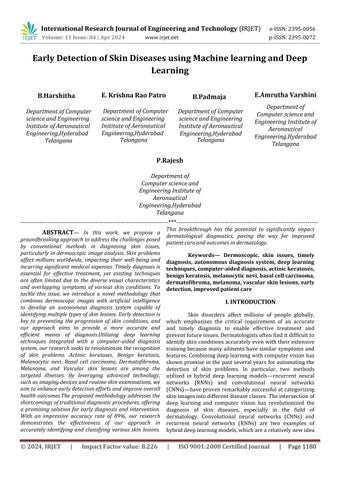
Using classification approaches, the bottleneck attributes of six previously trained models were retrieved and stored, enabling the differentiation of skin lesions. Block diagram of working model shows the pipeline of the proposed ensemble feature fusion approachforskinconditiondiagnosis.
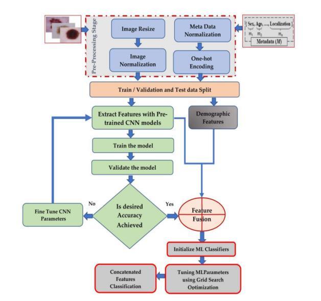
Fig1:Methodologyofthemodel
Step 1 (image pre-processing): In order to improve speed and model generalizability, we concentrated onsimplifyingthepreprocessingprocessesinStage1ofour work on employing CNNs to classify skin diseases. There were two main methods used: image scaling and normalization.Imagescalingcompensatedfordifferencesin dataset image sizes, while normalization ensured uniform pixel intensity by accounting for a variety of acquisition sources.Tomitigatepotentialfluctuationsinimagecontrast, a normalization method during training was introduced, scalingpixelvaluestoarangebetween -1and1.Inorderto optimize the CNN model's training procedure for enhanced performance ona variety of skinlesion images,thismethod simplified the preprocessing step. FIGURE-1 BLOCK DIAGRAMOFWORKINGMODEL
Stage 2 (feature extraction): Six CNN models were used for feature extraction in Stage 2: Xception, ResNet50, DenseNet201, InceptionV3, VGG19, and InceptionResnet. Low-dimensional vectors of extracted features were used in place of retraining the models, which resulted in a considerable reduction in training time. With its numerous layers, deep learning models are excellent at extracting features. By using convolutional and pooling layersafteroneanother,theyareabletocapturegeometric, edge, color, and texture features. While pooling layers performthresholdinganddimension reduction,convolution layers use digital filters. In order to guarantee thorough feature representation for ensuing diagnostic assessments, eachmodelwasextractedwith2048features,eachofwhich wasrepresentedbyavectoroflengthN2048,whereNisthe numberoftrainingphotos.
International Research Journal of Engineering and Technology (IRJET) e-ISSN:2395-0056
Volume: 11 Issue: 04 | Apr 2024 www.irjet.net
Stage 3 (metadata pre-processing): This removes missing data from the clinical data. Mostly the demographic features are finitely created categorical variables, represented as "strings" or "categories." The characteristics of these categories are converted to the categorical data format via one-hot encoding. For every floorwithinaItwasdecidedtointroduceanewvariable named category feature. A binary variable with the values 0 or 1 was allocated to each category. For instance,thereweretwonewclassificationsforsex:male and female. In this case, 0 denotes the absence of the category while 1 denotes its presence. Additionally, the demographic Age and other numerical data were normalized.
Stage 4 (feature concatenation): In thisstage, the information and picture features are combined to create a single feature vector. Initially, we fed the previously edited images of skin conditions into CNN's models. Convolutional, pooling, and auxiliary layers are usedbyCNNmodelstoextractdeepfeatures.2048deep features were produced as a result, and they were recorded as 6509 × 64 feature vectors. 6509 × 5 demographic features are present. The concatenated feature vector is 6509 × 65. After the category characteristicsofthedemographicdataareencodedonehot,theconcatenatedvectorwillhavealengthof6509× 85features.
Step 5 (skin lesion classification): The produced concatenated characteristics are given into a variety of machine learning classifiers. All of the skin lesion photographs were eventually divided into seven classes.
Ten thousand photos made up the study's input dataset. Without Metadata, the HAM10000 Dataset In the first experiment, pictures of skin lesions were classified using six pre-trained models. The collected characteristics from the previously trained CNN models were classified using three machine-learning classificationalgorithmsin order todistinguishbetween different types of skin lesions. To further improve the generalization ability and accuracy of the deep models, we used a combination of machine learning classifiers and pre-trained deep learning classifiers to diagnoseskinlesionsautonomously.
SAMPLE INPUT DATA:
p-ISSN:2395-0072
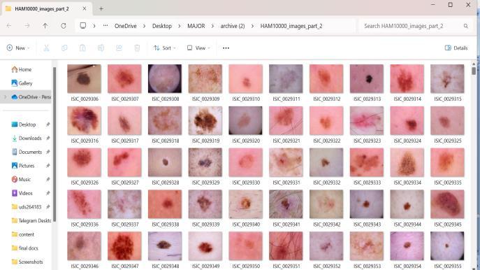
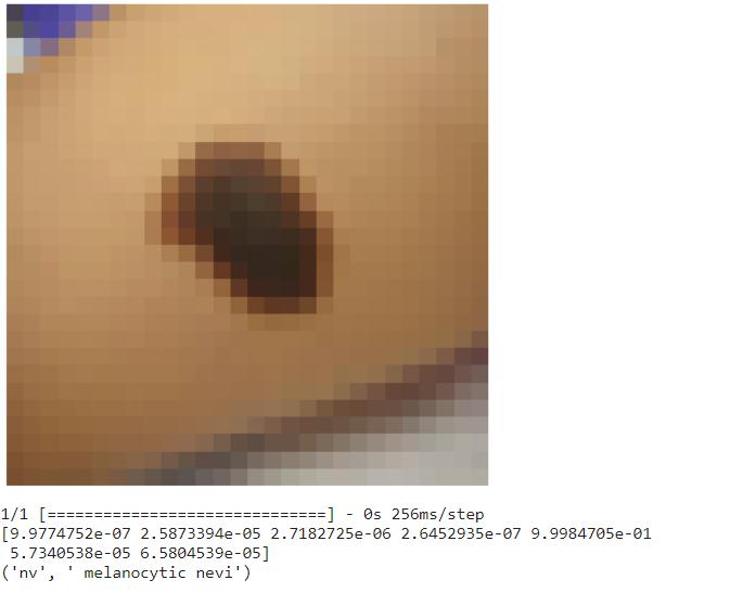
SAMPLE OUTPUT DATA:
Ten thousand photos made up the study's input dataset. Without Metadata, the HAM10000 Dataset In the first experiment, pictures of skin lesions were classified using six pre-trained models. The collected characteristics from the previously trained CNN models were classified using three machine-learning classification algorithms in order to distinguish between different types of skin lesions. To further improve the generalization ability and accuracy of the deep models, we used a combination of machine learning classifiers andpre-traineddeeplearningclassifierstodiagnoseskin lesionsautonomously.
International Research Journal of Engineering and Technology (IRJET) e-ISSN:2395-0056
Volume: 11 Issue: 04 | Apr 2024 www.irjet.net p-ISSN:2395-0072
In conclusion, the global prevalence of skin diseases is linked to major health and economical concerns. Our researchproposesadeeplearningandmachinelearning based computer-aided diagnosis system specifically designedfordermoscopicpictureanalysistoaddressthis problem. The acquired results are promising and warrantfurtherinvestigationintoabroaderspectrumof skindiseasesandclassifications.Anotherdisadvantageis the absence of dimensionality reduction strategies to improvefeatureselection.Furtherresearchwillfocuson exploring additional deep-learning techniques to improveclassificationaccuracyandtestingtheproposed system on benchmark datasets with a wider variety of skin conditions. The development of more dependable and effective diagnostic tools is the aim of this initiative inordertoimprovepatientoutcomes.
Thereissignificantandbroadpotentialfora deep learning-based skin disease detection programme. First, by investigation and application of cutting-edge deep learning architectures, such as transformer-based models and attention processes, the system can recognise complex patterns and diagnose patients with more accuracy. Second, integrating different data modalities such as genetics and patient history or imaging may lead to a deeper comprehension of skin conditions. To further evaluate and modify the system for worldwide use, it will be tested on a range of demographic groups and implemented in actual healthcare settings. By working with dermatologists and providing regular updates, the project will be further validated as a cutting-edge and essential diagnostic tool in dermatology through continued study into the interpretability and explainability of deep learning models.
[1] American Cancer Society. Important Data on Skin Cancer from Melanoma, 2022. The following URL is accessible online:https://www.cancer.org/cancer/melanoma-skinca ncer/about/key-statistics.html (retrieved October 27, 2022).
[2] Skin Disease Recognition Method Based on ImageColourandTextureFeatures,Wei,L.-S.;Gan,Q.;Ji, T.2018;8145713,Comput.Math.MethodsMed.(2018).
[3] Amarathunga, A.A.L.C., Ellawala, E.P.W.C.,Abeysekara, G.N., and Amalraj, C.R.J. (2015) Int. J.Sci. Technol. Res. 4, 174–178. Developed an expertsystem foridentifyingskinillnesses.
[4] Computer-Aided Diagnosis of Skin Diseases Using Deep Neural Networks Bajwa, M.N., Muta, K., Malik, M.I., Siddiqui, S.A., Braun, S.A., Homey, B., Dengel, A., Ahmed, S. Appl.Sci.2020,10,2488.
[5] Artificial Intelligence-Based Skin Classification UsingGMM:Monisha,M.,Suresh,A.,andRashmi,M.R.J.Med. Syst.2019,43,3.7.M.A.Kassem
[6]
Eltoukhy,M.M.Diagnostics11,1390(2021).
[7] Nature 2015, 521, 444; LeCun, Y.; Bengio, Y.; Hinton,G.Deeplearning.
[8] Classification of Skin Lesions into Seven Classes Using Transfer Learning with AlexNet Hosny, K.M.; Kassem, M.A.;Fouad,M.M.2020;J.DigitImaging33:1325–1334.
[9] Long, H.; Zhang, B.; Zhang, J.; et al. Wu, H.; Yin, H.; Chen, H.; Sun, M.; Liu, X.; Yu, Y.; Tang, Y. A Deep Learning, Image-Based Approach for Automated Diagnosis for InflammatorySkinDiseases.2020Ann.Transl.Med.8,581.
[10] Skin cancer localization and classification using a computerized decision support system Khan, M.A.; Akram, T.; Sharif, M.; Kadry, S.; Nam, Y. Computing Materials and Contin.2021,68,1041–1064.
[11] Using artificial intelligence algorithms, Alsaade, F.W., Aldhyani, T.H.H., and Al-Adhaileh, M.H. developed a recognition system for the diagnosis of melanoma skin lesions.MethodsMed.Comput.Math.2021,2021,9998379.
[12] Ali, S., Islam, K.; Rahman, M.; Haque, J.; Miah, S. an improved method of classifying skin cancer that combines transfer learning models and deep convolutional neural networks.MachineLearningApplications,5,100036,2021.
[13] Ameri, A. A deep learning method for identifying skin cancer in dermoscopy pictures. 2020; J Biomed Phys. Eng.10,801–806.
[14] Shanthi, T., Sabeenian, R.S., and Anand, R. Convolution Neural Networks for Automatic Skin Disease Diagnosis. Microsyst. 2020, 76, 103074; microprocessor.