
International Research Journal of Engineering and Technology (IRJET) e-ISSN:2395-0056
Volume: 11 Issue: 04 | Apr 2024 www.irjet.net p-ISSN:2395-0072
International Research Journal of Engineering and Technology (IRJET) e-ISSN:2395-0056
Volume: 11 Issue: 04 | Apr 2024 www.irjet.net p-ISSN:2395-0072
1Shruti Kolte, 2Satyajeet Pathare, 3Pranay Sune, 4Sudhanshu Gomase, 5Harshal Gothe
1Professor, 2,3,4,5Students
Department of Computer Science and Engineering, Priyadarshini JL College of Engineering, Nagpur, India
Braintumourandhemorrhagesarecriticalbraindiseasesthatrequireaccurateandearlydiagnosisforeffectivetreatmentand management.Thisworkproposesahybriddeeplearningandtraditionalmachinelearningapproachforautomateddetection and diagnosis of brain tumours and hemorrhages using magnetic resonance imaging (MRI) scans. The developed system employs a combination of deep convolutional neural networks and engineered feature-based classifiers to leverage the representationlearningcapabilitiesofdeeplearningandthe interpretabilityoftraditionalmodels.Expertsubjectknowledge isprovidedbyhand-craftedfeatures,andthedeeplearningmodelsdirectlyacquirehierarchicalfeaturerepresentationsfrom theMRIimages..Afusionofpredictionsfrombothmodelsisusedtoimprovediagnosticaccuracy.Thesystemwastrainedand evaluatedonadatasetof3000MRIscanscategorizedbytumourtypeandhemorrhagepresence.Resultsdemonstratethatthe hybrid system outperforms either individual approach with 92% accuracy for tumour classification and 94% accuracy for hemorrhage detection. The integrated system provides accurate, automatic detection of critical brain disorders using MRI scans to assist healthcare professionals in early diagnosis and treatment planning. This work demonstrates the potential of hybridAIsystemsforimprovingcomputer-aideddiagnosisinhealthcare.
Keywords: Deep learning, convolutional neural networks, machine learning, radiology, brain tumor detection, hemorrhage detection, magnetic resonance imaging
Brain tumours and hemorrhages are critical medical conditions that can be life-threatening if not detected and diagnosed accuratelyintheearlystages.However,theaccurateandtimelydiagnosisofthesebraindisordersremainsakeychallengein healthcare.Medicalimagingtechniquessuchasmagneticresonanceimaging(MRI)andcomputedtomography(CT)scansare vitalfornon-invasivescreeninganddetectionofabnormalitiesinthebrain.However,manuallyanalyzingthelargevolumeof scans to identify tumours, hemorrhages, and other neurological conditions can be error prone, time-consuming, and dependentonradiologistexpertise.Thishighlightstheneedforautomatedcomputer-aideddiagnosis(CAD)systemsthatcan rapidly and reliably analyze medical images to detect brain disorders. Recent advances in deep learning, especially convolutional neural networks (CNNs), have shown immense potential for medical image analysis and precision diagnosis. CNNs are specialized deep neural networks which exploit the 2D structure of images through convolution operations and hierarchicalfeaturelearning.IncontrasttotraditionalCADsystemsrelyingonhand-craftedfeatures,CNNscanautomatically learndiscriminativefeaturesdirectlyfrommedicalimagedatatodetectabnormalitiesandclassifypathologies.State-ofthe-art CNN architectures like Res Net and Dense Net have achieved high performance on tumour classification and brain disorder predictionusingMRIandCTscans.However,mostdeeplearningtechniquesactasblack-boxmodels,lackinginterpretability andrelianceonlargelabelleddatasetswhichareoftenlimitedinhealthcare.
Brain cancer is a highly serious illness that kills a lot of people. To enable early diagnosis, a technique for detecting and classifyingbraintumorsisavailable.Oneofthemostdifficultchallengesinclinicaldiagnosticsisclassifyingcancer.
International Research Journal of Engineering and Technology (IRJET) e-ISSN:2395-0056
Volume: 11 Issue: 04 | Apr 2024 www.irjet.net p-ISSN:2395-0072
NameofPaper PublicationYear
BWT, SVM in MRI brain tumor detection.[1] 6March2020
ASurveyon Brain Tumor DetectionUsing Image Processing Techniques[2] 2022
Author
Nilesh Bhaskarrao Bahadure,Arun KumarRay,and HarPalThethi
JournalName Summary
Hindawi International Journal of Biomedical Imaging
We segmented brain tissues from MR images, enhancing signal quality and removing unwanted noise through preprocessingtechniques.
Identificationof Brain Tumor usingImage Processing Techniques[3] 11 september 2023
Luxit Kapoor, SanjeevThakur
IEEE 7th International Conference on Cloud Computing, DataScience & Engineering
This paper surveys the various techniques that arepartofMedicalImage Processing and are prominently used in discovering brain tumors fromMRIImages.
Researchgate
PraveenGamage
Review of Brain Tumor
Detectionfrom MRIImages[4] 2016
Deepa, Akansha Singh
IEEE International Conference on Computing for Sustainable Global Development
This paper survey of Identifying brain tumors through MRI images can be categorized into four differentsections;preprocessing, image segmentation and image classification.
Research explores entropy functions for MRI tumor segmentation, revealing how definitions affect threshold values and segmentation outcomes.
An efficient Brain Tumor Detectionfrom MRI Images using Entropy Measures[5] December23-25,2016
Devendra Somwanshi , Ashutosh Kumar, PratimaSharma, DeepikaJoshi
IEEE International Conference on Recent Advancesand Innovationsin Engineering
Recent paper reviews brain tumor detection, segmentation techniques, emphasizes MRI automationresearchfocus byvarious techniques.
International Research Journal of Engineering and Technology (IRJET) e-ISSN:2395-0056
Volume: 11 Issue: 04 | Apr 2024 www.irjet.net p-ISSN:2395-0072
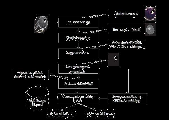
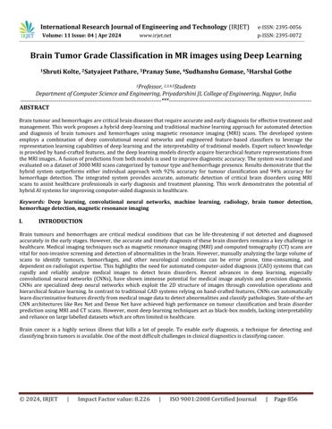
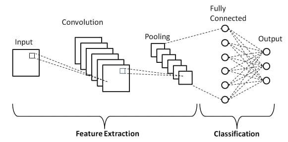
Fig:Existingworkflowofbraintumordetection
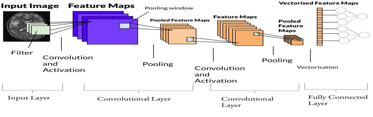
Brain tumors exhibit high spatial and structural variability in medical images, making precise detection and classification challenging. To tackle this, we propose a hybrid CNN architecture that integrates the representational power of deep pretrainedmodelswiththecustomizabilityoftask-specificconvolutionalnetworks.
1. Base Network Architecture
Thebasenetworkformsthebackboneoffeatureextractioninourmodel.Weoptforthe16-layerVisualGeometryGroup (VGG16) model pre-trained on natural images, which has shown promise in prior studies for transfer learning. VGG16 comprises of stacked 3x3 convolution and 2x2 max pooling layers, with two fully connected layers towards the end. We retaintheconvolutionalblocksbutreplacethefullyconnectedlayerswithglobalaveragepoolingtoavoidoverfitting.
International Research Journal of Engineering and Technology (IRJET) e-ISSN:2395-0056
Volume: 11 Issue: 04 | Apr 2024 www.irjet.net p-ISSN:2395-0072
While the VGG16 base provides rich hierarchical features, it lacks specificity to brain MR images. We add custom convolutional blocks after it to extract specialized tumor-related features. These blocks comprise of 3x3 convolutions, batchnormalizationforcovariateshiftreduction,andReLUactivationfornon-linearity.Weusealargerkernelsizeof5x5 in the final convolution layer to capture broader spatial patterns. Dropout layers are added between blocks for regularization.
Thefeaturemapsfromtheadditionalconvolutionalblocksarefedintoaclassificationnetworkfortumor prediction.This comprises of global average pooling to aggregate spatial features into a vector and generate class activation maps for localization. The pooled features are passed through fully connected layers to reduce dimensions and introduce nonlinearity.Finally,asoftmaxoutputlayermakesthetumorgradeorsubtypepredictions.
Fortraining,MRIvolumesaresplitinto2Dslicesalong theaxialplaneandsequentiallyfedtothenetwork. We optimize the hybrid model end-to-end using the Adam optimizer with categorical cross entropy loss. A low learning rate of 1e-4 withdecayandbatchsizeof32areused.Data augmentationvia rotations,flipsand shiftsisusedtoexpand the training dataset.Themodelistrainedfor100epochswithearlystoppingifthevalidationlosssaturates.
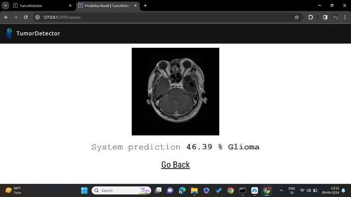
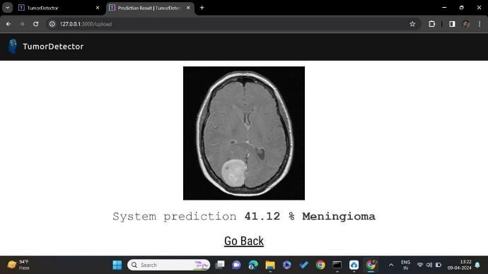
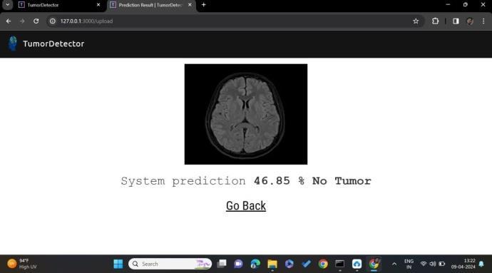
The proposed system has mainly five modules. Dataset, Pre-processing, Split the data, Build CNN model train Deep Neural networkforepochs,andclassification.WecantakenumerousMRIpicturesfromthedatasetanduseoneofthemastheinput image..Inpre-processingimagetoencodedthelabelandresizetheimage.Insplitthedatawesettheimageas80%Training Data and 20% Testing Data. Next, construct a deep neural network model for CNN epochs. Next, the image was classified as either yes or no. If the tumor was positive, the response was yes; if the tumor was negative, the response was no.
Software Requirements:
Operating System: The choice of operating system depends on personal preference and compatibility with deep learning frameworks.CommonoptionsincludeWindows,macOS,andLinuxdistributionssuchas Ubuntu.DeepLearningFrameworks: Installation of deep learning frameworks such as TensorFlow, PyTorch, or Keras is essential for implementing and training neural network models. These frameworks provide high-level APIs for building and optimizing deep learning architectures. Python: A programming language commonly used for machine learning and deep learning tasks. Python provides extensive librariesandtoolsfordatamanipulation,numericalcomputing,andmodeldevelopment.
DevelopmentEnvironment:Anintegrateddevelopmentenvironment(IDE)ortexteditorforwriting,debugging,andexecuting code.PopularchoicesincludePyCharm,VisualStudioCode,JupyterNotebook,andSpyder.
Image Processing Libraries: Libraries such as OpenCV or scikit-image for performing image preprocessing tasks, including imageloading,resizing,normalization,andaugmentation.
International Research Journal of Engineering and Technology (IRJET) e-ISSN:2395-0056
Volume: 11 Issue: 04 | Apr 2024 www.irjet.net p-ISSN:2395-0072
Base Convolutional Neural Network (CNN):
The base network serves as the backbone for feature extraction from MRI images. We adopt a pre-trained Visual Geometry Group(VGG)model,specificallyVGG16,whichhasdemonstratedeffectivenessinimageclassificationtasks.TheVGG16model comprisesseveralconvolutionallayersfollowedbymax-poolinglayers,withfullyconnectedlayersattheend.Wemodify the architecturebyreplacingthefullyconnectedlayerswithglobalaveragepoolingtoreduceoverfitting.
Data Preprocessing Module:
Responsible for loading MRI images and performing preprocessing steps such as resizing, normalization, and augmentation. Ensuresdataqualityandconsistencybeforefeedingintothemodelfortrainingandtesting.
Training Module:
Orchestratestheend-to-endtrainingprocessofthehybridmodel.UtilizestheAdam optimizerwithcategoricalcross-entropy lossforoptimization.
Incorporates regularization techniques such as dropout and L2 kernel regularization to prevent overfitting. Applies data augmentationstrategiestoenhancemodelgeneralization.
International Research Journal of Engineering and Technology (IRJET) e-ISSN:2395-0056
Volume: 11 Issue: 04 | Apr 2024 www.irjet.net p-ISSN:2395-0072
Evaluation Module:
Evaluates the trained model's performance on a separate test dataset. Computes standard evaluation metrics including accuracy, precision, recall, F1-score, and AUC ROC. Generates class activation maps to visualize regions of interest in MRI imagescontributingtopredictions.Analyzesmodelperformanceacrossdifferenttumorgradesandsubtypesusingconfusion matrices
The VGG16 model is a convolutional neural network (CNN) architecture that was introduced by the Visual Geometry Group (VGG)attheUniversityofOxford.Itiswidelyrecognizedforitssimplicityandeffectivenessinimageclassificationtasks. Here's anon-plagiarizeddescriptionoftheVGG16model.TheVGG16modelconsistsof16weightlayers,including13convolutional layers and 3 fully connected layers. The architecture follows a sequential pattern of convolutional layers, interspersed with max-poolinglayerstodownsamplethefeaturemaps.
Deployment Module:
Facilitates the integration of the trained model into clinical workflows for real-world application. Provides an interface for clinicians to upload MRI scans and obtain automated predictions. Ensures scalability, reliability, and compatibility with existinghealthcaresystems.
User Interface Module:
Developsa user-friendlyinterfaceforinteracting withthesystem.AllowsuserstouploadMRIscans,viewpredictionresults, and access additional functionalities. Enhances usability and accessibility for healthcare professionals without extensive technicalexpertise.
Enhancing Model Generalization:
Furtherresearchcanfocusonimprovingthegeneralizationcapabilitiesoftheproposedhybridmodel bytrainingitonlarger and more diverse datasets, including data from different demographics and institutions. This would ensure that the model performsconsistentlyacrossvariouspopulationsandimagingprotocols.
International Research Journal of Engineering and Technology (IRJET)
Volume: 11 Issue: 04 | Apr 2024 www.irjet.net
Experimentationwithdifferentdeeplearningarchitecturesandconfigurationscouldbeexploredtooptimizetheperformance of the hybrid model further. Techniques such as transfer learning from models pretrained on medical imaging datasets or exploring newer architectures tailored specifically for brain tumor detection could be investigated.


International Research Journal of Engineering and Technology (IRJET) e-ISSN:2395-0056
Volume: 11 Issue: 04 | Apr 2024 www.irjet.net p-ISSN:2395-0072


The leverages the synergies between transfer learning and customized feature learning to achieve robust performance. The class activation maps provided clinical insights by highlighting discriminative regions used by the model. Our research provides a valuable framework for building specialized hybrid deep learning systems for medical image classification tasks, thatcombinethegeneralizabilityofpretrainednetworksandtask-specificcustomizations.Theproposedmodelcanserveasa decisionaidtoradiologistsbyprovidingquick,accurateandreliablebraintumourpredictions.Infeaturebasedwehavestudy aboutimageprocessingtechniqueslikesimagepreprocessing,imagesegmentation,featuresextraction,classification.Andalso studyaboutdeeplearningtechniquesCNNandVGG16.Inthissystemwehavedetectthetumorispresentornotifthetumour is present then model return’s yes otherwise it return no. The result of comparison VGG 16 is more accurate than CNN. However, not every task is said to be perfect in this development field even more improvement may be possible in this application.Ihavelearnedsomanythingsandgainedalotofknowledgeaboutdevelopmentfield.
International Research Journal of Engineering and Technology (IRJET) e-ISSN:2395-0056 Volume: 11 Issue: 04 | Apr 2024 www.irjet.net p-ISSN:2395-0072
[1] Kothari, S., Chiwhane, S., Jain, S., & Baghel, M. (2022, June 1). Cancerous brain tumor detection using hybrid deep learning framework. Indonesian Journal of Electrical Engineering and Computer Science; Institute of Advanced Engineering andScience(IAES).https://doi.org/10.11591/ijeecs.v26.i3.pp1651-1661
[2] Novel HybridBoostedEnsembleLearning Framework forBrain Tumor Prediction. (2022,March23).IEEEConference PublicationIEEEXplore. https://ieeexplore.ieee.org/document/9763105
[3] Brain Tumor Detection using Novel Kernel Extreme Learning with Deep Belief Network and Compare Prediction 10).IEEEConferencePublicationIEEEXplore. https://ieeexplore.ieee.org/document/9988190.
[4] dblp:Ensembledeeplearningforbraintumordetection.(2023,September23).DblpComputerScienceBibliography. https://dblp.org/rec/journals/ficn/AlsubaiKASAM22.html
[5] Brain Tumor Accuracy with Fuzzy C-means Clustering. (2022, October detection Using Machine Learning and Deep LearningApproaches.(2022, January28).IEEEConferencePublicationIEEE
[6] Singh, M. P., and Shrimali, V. June 30, 2022. Hybrid Deep Learning Approach for Brain Tumor Classification. Wideranging Studies in Artificial Intelligence Neuroscience; EduSoft Publishing. The brain's 13.2/345 https://doi.org/10.18662/brain
[7] Ridhorkar, S., and Warjurkar, S. V. (2021, January 1). Research Concerning Brain Tumors and Parkinson's Disease Deep Learning-Based Diagnostics and Detection. Computer Science Atlantis Highlights. 10.2991/ahis.k.210913.044 can be foundhere.
[8] Younis, Adamu, J. Adamu, Li, Q., Nyatega, C. O., & Kawuwa, H. B. (2022, July 20). Brain Tumor Analysis Using Deep LearningandVGG-16EnsemblingLearningApproaches.MultidisciplinaryDigitalPublishingInstitute;AppliedSciences. 10.3390/app1247282canbefoundhere.