
International Research Journal of Engineering and Technology (IRJET) e-ISSN: 2395-0056
Volume: 11 Issue: 04 | Apr 2024 www.irjet.net p-ISSN: 2395-0072
International Research Journal of Engineering and Technology (IRJET) e-ISSN: 2395-0056
Volume: 11 Issue: 04 | Apr 2024 www.irjet.net p-ISSN: 2395-0072
Sri P. Anil Kumar1 , Thanuri Bhanu Harshitha2 , Yarlagadda Rajeswari3, Sanaka Venkata Karthikeya4, Ramireddy Usha Kumari5
1 Assistant professor, Department of Electronics and Communication Engineering, Seshadri Rao Gudlavalleru Engineering college, Andhra Pradesh, India 2,3,4,5 U.G Student, Department of Electronics and Communication Engineering, Seshadri Rao Gudlavalleru Engineering college, Andhra Pradesh, India
Abstract - Accurately diagnosing bone tumors is crucial for patient management and creating effective treatment plans. In this study, we propose a unique approach that uses deep learning algorithms to diagnose bone tumors using medical imaging data.Ourmethodcombinespicturesegmentationand the EfficientNetB0 architecture to enable high-performance tumorclassification.First,pre-processingandsegmentationof the input medical images are done to identify regions of interestthatmightbemalignancies.Theconvolutionalneural network EfficientNetB0 is then fed the segmented areas to carryoutfeatureextractionandclassification.EfficientNetB0 is known for its exceptional performance and computational efficiency, which enables strong learning from the separated tumor zones. We train and validate our model on a large dataset of annotated bone tumor images to guarantee generalizability and reliability. Our method's exceptional levels of sensitivity, specificity, and accuracy in diagnosing bone tumors are demonstrated by the results of our experiments.Thismethodoffersascalableandefficientwayto automatically identify tumors from medical images, which presents a potential way to improve clinical judgment in the diagnosis and treatment of bone malignancies.
Key Words: Deep Learning, Bone Tumors, Image Segmentation, EfficientNetB0, Classification, Radiology.
1.INTRODUCTION
Bonetumorsarecommongrowthsormassesoftissuethat develop inside the bones. These tumors can be benign or malignant; malignant tumors pose a major risk to health, includingthepossibilityofspreadanddeath,iftreatmentis not received. For patients with bone malignancies, timely planning of their treatment and an accurate diagnosis are crucial.Bonemalignanciesareusuallydiagnosedbyaclinical examination,imagingtechniquessuchasCT,MRI,andX-rays, andhistologicalanalysisoftissuesamplesobtainedthrough biopsy.
Inrecentyears,deeplearningmethodshavebecomemore and more popular for use in medical image analysis, particularly when dealing with issues like tumor segmentation and classification. The artificial intelligence field of deep learning has demonstrated remarkable
outcomesincomputervision,naturallanguageprocessing, andhealthcare,amongotherdomains.Deeplearningmodels, andinparticularconvolutionalneuralnetworks(CNNs),have shown promising results in automated medical image interpretationapplications.Comparedtotraditionalmethods, thesemodelsmayofferbenefitsincludingimprovedaccuracy, consistency,andefficiency.[2]
Imagesegmentationiscrucialformedicalimageanalysis becauseitmakesiteasiertoidentifyandseparateareasof interest like cancers from the surrounding anatomical structures.Byaccuratelysegmentingbonetumorlocations, clinicianscanobtainvaluableinformationontumorfeatures, such as size, shape, and texture, which are useful for diagnosisandtherapeuticplanning.Moreover,segmentation facilitatestheextractionofquantitativeelementsfromimage data that serve as algorithmic input for classification, enablingautomatictumorcategorization.[3]
[1]Severalscientistshaveseentrendsintheinformationand data that they have obtained from large databases and pertinent websites. Learning vector quantization, fuzzy theory, probabilistic neural networks, association rule mining,andsupportingvectormachinesarethemostwidely usedmethodsfordiagnosingandclassifyingbonecancer.In order to segment bone pictures, the k means clustering algorithmwasusedinthiswork.Toprocessthesegmented image further for the aim of identifying bone cancer, the mean intensity of the detected area is evaluated. It is recommended to classify medical images based on the presenceorabsenceofbonecancerusingthresholdvalues.
[2]Thebasisofthisworkisthefusionofcomputerscience with the biomedical field. Numerous image segmentation methods, including Sobel, Prewitt, Canny, K-means, and RegionGrowing,aredescribedinthispaper.Thesemethods canbehelpfulinunderstandingMRIandX-rayimagesaswell as in predicting the type of bone cancer. In order to use MATLABtodetectosteosarcomacancerpresentonbone,the studyalsodisplaystheoutcomesofedge-basedandregionbased image segmentation techniques applied to X-ray pictures.
[3Traditional methods that used very small images were time-consuming and noisy. The research offers automated imageprocessingtechniqueslikeWaveletdenoising,which improveimagequalityandremovenoisewhilepreserving diagnostic information, as a solution to this problem. By utilizing pre-processing methods and algorithms like Kmeansandedgesegmentation,thestudysuccessfullydetects bonecancerandestablishesitsstage.Furthermore,genetic algorithms are used to distinguish benign from malignant tumors.
[4]Anovelapproachfordeterminingthegradeandstageof cancer in long bones is presented in this paper, which is based on X-ray image processing. According to the recommendedapproach,supportvectormachines(SVM)are trained to identify healthy and cancerous bones based on certain features extracted from X-ray images of the bones. Thesiteswherecancerispresentareidentifiedusingadigital geometry-basedtechnique.Thepresentstageandgradeof the disease, as well as the underlying pattern of bone degeneration,arealldescribedbymeansofadecisiontree classifier. More importantly, the method produces a computer-aideddiagnostictoolthatdoctorsandparamedics canusewithease.
[5] This study uses the adaptive neuro-fuzzy inference system (ANFIS) in conjunction with feature selection to provide a unique method of early breast cancer diagnosis. ThismethodemploysANFISasanintelligentclassifierand the association rules (AR) technique as a featureselection algorithm. The value of radius has a big influence on how accuratetheANFISsystemis.Asaresult,inordertoobtain theoptimalradiusvalue,thecuckoooptimizationalgorithm (COA) was utilized in the recommended technique. The resultsshowthatthesuggestedtechniquehasgreatdetection accuracy when applied to the Wisconsin Breast Cancer Database(WBCD).
[6]Theboneisavitalcomponentofthehumanbody.Bones provide the body its ability to move. Bone fractures are commonin thehumananatomy. The doctors diagnose the fractured bone based on the X-ray image. The manual fracturediagnosisapproachislaboriousandpronetolarge errors. Therefore, it is necessary to develop an automated method for recognizing fractured bones. Deep Neural Networks(DNNs)areapopularmodelforpowerelectrical systems. This study has developed a deep neural network model to discriminate between bone that is fractured and healthy.Thesmalldatasetcausesthedeeplearningmodelto overfit.Assuch,strategiesfordataaugmentationhavebeen usedtoincreasetheamountofthedatacollection.Threetests with thesoftmaxandAdamoptimizers were conductedto evaluatethemodel'sperformance.
Volume: 11 Issue: 04 | Apr 20 www.irjet.net p-ISSN: 2395-0072
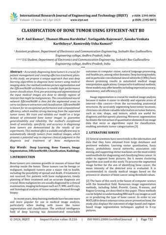
PicturesegmentationandtheEfficientNetB0architectureare proposedtobeusedinadeeplearning-basedbonecancer classificationsystem.Wepreprocessthebonetumorimages firstinordertoenhancetheirqualityandremovenoise.Next, we apply a state-of-the-art image segmentation technique such as U-Net to accurately distinguish the tumor regions. Afterwards,weutilizethesegmentedtumorregionsasinput for our EfficientNetB0 architecture, which serves as our classificationbackbone.EfficientNetB0isasuitablechoicefor thistasksinceitachievesabalancebetweenmodelsizeand accuracy.Werefinethepre-trainedEfficientNetB0modelto makeitappropriateforbonetumorclassificationusingour segmented tumor dataset. To improve our models' performanceandgeneralization,weusetechniqueslikedata augmentation and transfer learning. We use industrystandardcriteriaincludingaccuracy,precision,recall,andF1score to assess our suggested approach. EfficientNetB0's remarkable accuracy in identifying patterns and characteristics makes it the perfect choice for seat belt detectionanddrowsinessdetection.OneofEfficientNetB0's featuresthatisparticularlyusefulforapplicationsneeding real-time detection is rapid data analysis. EfficiencyNetB0 produces state-of-the-art outcomes. In generalization, EfficientNetB0performsadmirably.
3.1 Image Pre-processing
Imagesmustbepre-processedinordertoextractnoisefrom the data and find pertinent properties. By leveling input scaling and enhancing generalization through dataset updates, it guarantees consistent model performance. International Research Journal of Engineering and Technol 23 ogy (IRJET)
International Research Journal of Engineering and Technology (IRJET) e-ISSN: 2395-0056
Volume: 11 Issue: 04 | Apr 2024 www.irjet.net p-ISSN: 2395-0072
Robustnessto variationsimproves model reliability while continuousscalingmaintainsuniformityforeffectivemodel processing. In the end, preprocessing improves interpretability for people and models by assisting in the identificationofcertainregionsofinterestandadaptingto hardwareconstraints.
Thedataaugmentationtechniqueiswidelyusedfortraining convolutionalneuralnetwork(CNN)models,particularlyin computer vision applications. It involves artificially expanding the training dataset by applying various modificationstothe preexistingpictures whilepreserving theirsemanticvalue.InordertoimprovetheCNNmodel's resilienceandgeneralization,dataaugmentationexposesit toawiderrangeofinputdataperturbations.Increasingthe CNNmodel'sresistancetomodificationsintheinputdatais the primary objective of data augmentation. Real-world images can vary due to a variety of elements such as illumination,scaling,translations,rotations,distortions,and more.Themodelislesspronetooverfittocertaintraining examplesandisbetterabletogeneralizetonewdatawhen thesechangesareaddedtothetrainingset.
3.3
Awell-constructeddatasetisnecessaryfortrainingamodel to recognize patterns and produce accurate predictions withinpresetcategories.Thedatasetistypicallydividedinto training and testing sets in order to assess the model's performance on untested data. It offers the structure for creating and refining classification models that may be appliedtoa rangeoftasks,suchasimagerecognitionand naturallanguageprocessing.
4. MODULE DESCRIPTION
EfficientNet B0
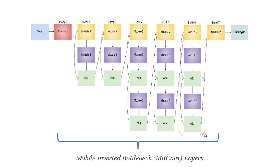
4.1 Stem Layer and Final Layer
The neural network starts at the stem layer, where it performsitsfirstconvolutionsandgetsthedatareadyfor layers that come after. The stem layer of EfficientNet-B0
preparesthegroundforfeatureextractionthatfollows.The retrievedfeaturesarerefinedinthelastlayer,readyingthem forprecisepredictionsintaskslikepicturecategorization.
4.1.1 Input Layer
The inputlayer of EfficientNetB0acceptsinput imagesof size299x299pixels,typicallyinRGBformat.
4.1.2 Rescalling
Changestoanimage'sproportionsorsizearereferredtoas imageprocessing.Itisatypicalpreprocessingmethodthatis utilized for a number of reasons, including improved performance and consistency, uniformity for machine learningalgorithms,andspeedierprocessing.
4.1.3 Normalization
Inordertomodifytherangeofpixelintensityvalueswithin an image, normalization is an essential approach. Normalization assists with efficient categorization by bringingpixelvaluesintoamoreknownorstandardrange andenhancingthecontrast andvisibilityof featuresin an image.
4.1.4 Zero Padding
Inimageprocessing,zeropaddingistheprocessofenlarging animage'sedgeswithadditionalrowsandcolumnsofzeros. It stabilizes computations, avoids artifacts at image boundaries, and guarantees consistent dimensions during operationssuchasconvolution.
4.1.5 Conv2D Layer
InConvolutionalNeuralNetworks(CNNs),theConv2Dlayer applies 2D convolution to pictures. It extracts features by swipingfiltersovertheinput.Kernelsize,strides,padding, and activation function are important characteristics. Conv2D is essential for object detection and image categorization.
4.1.6 Batch
BatchNormisnotdoneonrawdata,butratherinbetween layers of a neural network. During training, it uses minibatchesratherthanstandardizingtheentiredataset.
4.1.7 Activation
Inaneuralnetwork,activationfunctionsarethedecisionmakers. Every neuron has an activation function that controlswhetherornottheneuronneedstobestimulated. Whetherornottheinputaneuronreceivesispertinentto thenetwork'spredictiondetermineswhetherornotitgets activated.
International Research Journal of Engineering and Technology (IRJET) e-ISSN: 2395-0056
Volume: 11 Issue: 04 | Apr 2024 www.irjet.net
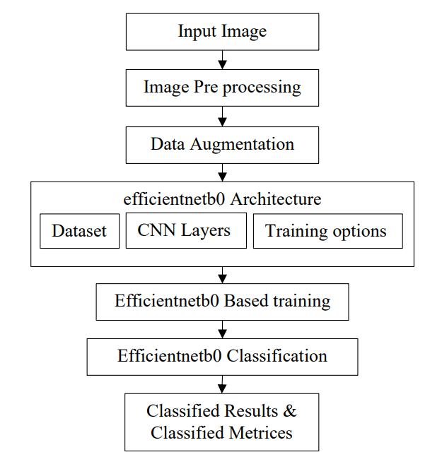
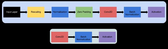
4.2 Mobile Inverted Bottleneck (MBConv) Layers
EfficientNet'sfundamentalcomponents.Mixresidualblocks thatareinvertedanddepth-wiseseparableconvolutions.For feature recalibration, include squeeze-and-excitation (SE) optimization.Astackofdifferent-depthandbreadthMBConv layersispartofthedesign.
EachlayerofMBConvismadeupof:
4.2.1 Depth-wise separable convolution
Reducescomputation.
4.2.2 Inverted residual block
Capturesrichfeatures.
4.2.3 Squeeze-and-excitation mechanism
Enhancesfeatureimportance.
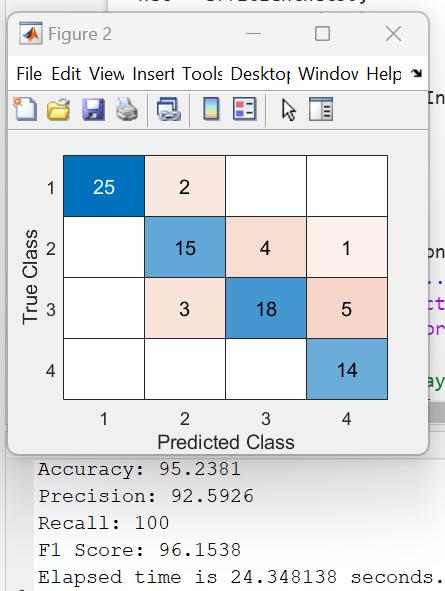
5.RESULTS AND ANALYSIS
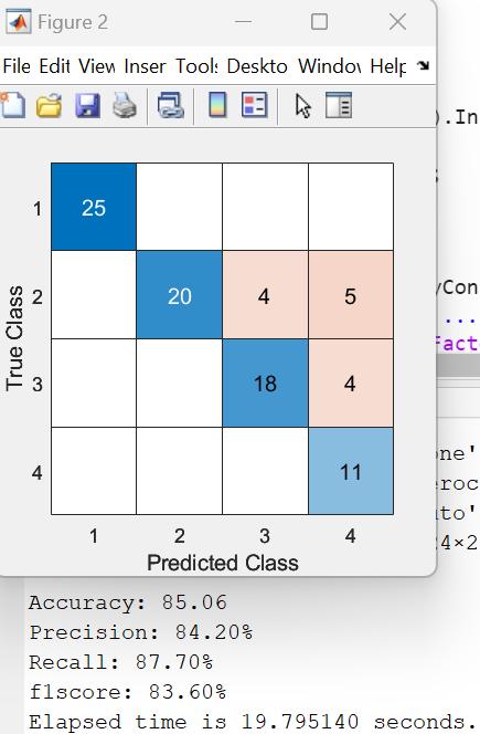
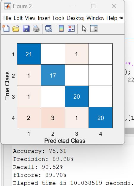
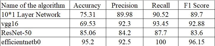
Based on the developed model, the bone tumors are classified.Aftertestinganumberofmodels,weselectedthe mostpracticalone.Thesearethelayernetworkalgorithms for Resenet50, VGG16, EfficientNet B0, and 10*1. By comparing those to other algorithms, EfficientNet B0 was able to accurately identify the bone tumors. With 92.5% precisionand95.2%accuracy,theEfficientNetB0method producesthebestresultsoutofalltheothers.
International Research Journal of Engineering and Technology (IRJET) e-ISSN: 2395-0056
Volume: 11 Issue: 04 | Apr 2024 www.irjet.net p-ISSN: 2395-0072
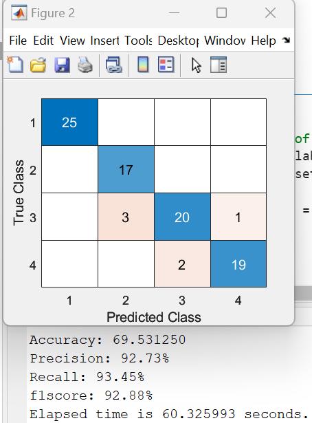
Fig-7:ResultsofVGG
Table -1: AlgorithmsperformanceTable
Onesignificantdevelopmentintheaccurateclassificationof bone tumors is the use of state-of-the-art picture segmentationtechniques,particularlythosethatmakeuseof theEfficientNetB0architecture.Throughthisstudy,wehave demonstratedtheefficacyandreliabilityofthisapproachin enhancingdiagnosticprecisionandefficiencyinhealthcare settings. Thanks to the application of deep learning and sophisticatedalgorithms,tumorcategorizationhasadvanced significantly,enablingmedicalprofessionalstoactswiftlyand decisively.TheimplementationofEfficientNetB0showshow well it can handle complex medical imaging data, outperformingconventionalmethodsand,incertaincases, evenoutperforminghuman-levelaccuracy.Thisadvancement mayspeedupthediagnosisprocedureandimprovepatient care,whichinturnmayleadtobettertreatmentresultsand reducedmedicalexpenses.
[1]Shukla,A.,&Patel,A.(2020).BoneCancerDetectionfrom X-Ray and MRI Images through Image Segmentation Techniques.InternationalJournalofRecentTechnologyand Engineering,8(6),273-278.
[2]D.P.YadavandS.Rathor,“Bonefracturedetectionand classification using deep learning approach,” in 2020
International Conference on Power Electronics & IoT ApplicationsinRenew-ableEnergyanditsControl(PARC), pp.282–285,Mathura,India,2020.
[3]M.M.Ranjitha,N.L.Taranath,C.N.Arpitha,andC.K.Subbaraya,“BonecancerdetectionusingK-meanssegmentation andKnnclassification,”in20191stInternationalConference onAdvancesinInformationTechnology(ICAIT),pp.76–80, Chikmagalur,India,2019.
[4] A. Torki, “Fuzzy rank correlation-based segmentation method and deep neural network for bone cancer identification,”NeuralComputingandApplications,vol.32, no.3,pp.805–815,2020.
[5]B.S.Vandana,P.J.Antony,andR.A.Sathyavathi,“Analysis ofmalignancyusingenhancedgraphcut-basedclusteringfor diagnosis of bone cancer,” in Information and Communication Technology for Sustainable Development, pp.453–462,Springer,2020.
[6]RSMLPatibandla,APGopi,BTRao(2021),Comparative StudyonAnalysisof Medical ImagesUsing DeepLearning Techniques, Deep Learning for Biomedical Applications, 2021.
[7]D.Shrivastava,S.Sanyal,A.K.Maji,andD.Kandar,“Bone cancer detection using machine learning techniques,” in SmartHealthcareforDiseaseDiagnosisandPrevention,vol. 20,pp.175–183,AcademicPress,2020.
[8] W. Li, G. G. Wang, and A. H. Gandomi, “A survey of learning-basedintelligentoptimizationalgorithms,”Archives ofCom-putationalMethodsinEngineering,vol.28,no.5,pp. 3781–3799,2021.
[9] P. A. Kumar and R. Gunasundari, "MR Image ReconstructionfromUndersampledDatausingMultiDense ResidualBlockNetworkwithSpatialandChannelAttention Mechanism and Entropy Loss Function," 2023 Second International Conference on Advances in Computational IntelligenceandCommunication(ICACIC),Puducherry,India, 2023,pp.1-7, doi:10.1109/ICACIC59454.2023.10434987.
[10]PentaAnilKumar,RGunasundari,RAarthi,Fractional SailfishOptimizer withDeepConvolutionNeural Network forCompressiveSensingBasedMagneticResonanceImage Reconstruction,TheComputerJournal,Volume66,Issue2, February 2023, Pages 280–294, https://doi.org/10.1093/comjnl/bxab160.