
Volume: 11 Issue: 04 | Apr 2024 www.irjet.net p-ISSN: 2395-0072
Volume: 11 Issue: 04 | Apr 2024 www.irjet.net p-ISSN: 2395-0072
Mrs.Sumaiya1 , Ms. Amulya HM2 , Ms. Pooja B3 , Ms. Priyanka Kumari Singh4 , Mr. Uday Kumar DR5
1 Assistant Professor, Dept. of Computer Science and Engineering, Maharaja Institute of Technology, Thandavapura
2,3,4,5Students, Dept of Computer Science and Engineering, Maharaja Institute of Technology, Thandavapura ***
Abstract - Mindgrowthlocationthroughclinicalimagingis vital for early conclusion and treatment arranging. This studypresentsaclevermethodologyusing3DConvolutional Brain Organizations (CNNs) for precise cerebrum growth discovery.Dissimilartoregular2Dstrategies,ourproposed 3DCNNengineeringprocessesvolumetricX-rayinformation, catchingspatialconnectionsandperplexingexamplesallthe more thoroughly. We influence information increases and regularizationmethodstoenhancethemodel'sspeculation andmoderateoverfitting.Throughbroadtrialanderrorona different dataset, our methodology accomplishes cutting edgeexecutioninresponsiveness,particularity,andgeneral precision.Nearexaminationsapprovethepredominanceof ourtechniqueoverexistingmethodologies,highlightingits vigorandviability.Theproposedframeworkdisplaysbasic potential for impelling clinical picture assessment, particularlyinfrontalcortexdevelopmentareas.Itscapacity to remove important elements straightforwardly from volumetric information guarantees work on analytic precision and opportune mediations, at last improving patientconsiderationandresultsinclinicalpractice.
Key Words: Perplexing, Regularization, Speculation, Vigor, Viability
Cerebrum growths represent a critical test for general wellbeingaroundtheworld,withtheirinitialidentification beingfundamentalforcompellingtreatmentandworkingon understanding results. As of late, profound learning procedures, especially Convolutional Brain Organizations (CNNs), have shown exceptional progress in different PC vision errands, including clinical picture examination. Conventional CNN designs work on two-layered (2D) pictures, restricting their capacity to catch spatial data intrinsicinvolumetricclinicalinformation.Thisimpediment haspromptedtheinvestigationofthree-layered(3D)CNNs, which straightforwardly process volumetric information, empowering more extensive examination and catching perplexing spatial connections inside the pictures. This paper proposes an original methodology for cerebrum canceridentificationutilizing3DCNNs,intendingtobeatthe impedimentsofconventional2Dtechniquesandupgradethe exactnessandeffectivenessofgrowthdiscoveryfromX-ray examinations.Byutilizingthespatialdataencodedin3DXray volumes, our proposed strategy expects to separate
significanthighlightsandexamplescharacteristicofgrowth presence with further developed awareness and particularity. Therestofthispaperismadeupasfollows: Segment 2 gives an outline of related work in the field of cerebrumcancerrecognitionandtheutilizationofCNNsin clinicalpictureexamination.Area3blueprintsthetechnique, includingtheengineeringoftheproposedframeworkof3D CNNmodelandthedatasetutilizedfortrialanderror.Area4 presentsthetrialresultsandthinksaboutthepresentation oftheproposedapproachasopposedtoexistingprocedures. Atlonglast,Segment5talksabouttheramificationsofour discoveries,possiblefutureheadings,andclosesthepaper.
The target of mind cancer location utilizing 3D Convolutional Brain Organizations (CNNs) is to foster a mechanized and exact technique for recognizing the presence and qualities of cerebrum growths in X-ray examinations.
CNNinAIstrategy,whichincludesnetworkswithmany layers to make expectations. These networks helps in gettingexceptionalresultsandaccuracy.Beforestartingthe implementationandseeingthemagicofCNNalgorithmit’s veryvitaltocomprehendhowtheseorganizationsworkand howtomakethem.
Man-madeintelligenceistheongoingmostcreatingfield andhasshownafewadditionalconventionaloutcomesthat havedemonstrateditssignificance.Utilizationofman-made intelligencehasbeenadjustedinpracticallyallfieldsandany place conceivable. The brain network is one of the little partsunderthishugeumbrellaofman-madeconsciousness. The clinical field has likewise evolved itself throughout historyandhasexpandedintothefuture.Thewholethought behindthistaskistoutilizebrainorganizationsandclinical informationinseparably.
Convolutionalbrainorganizationisakindofsignificant learning that is generally applied to analyzing visual
International Research Journal of Engineering and Technology (IRJET) e-ISSN: 2395-0056
Volume: 11 Issue: 04 | Apr 2024 www.irjet.net p-ISSN: 2395-0072
imagery.CNNsuseanassortmentofmulti-facetperceptron's expected to require immaterial pre-handling. They are in addition suggested as move invariant or space invariant counterfeit brain organization (SIANN), upholding their typicalweightsconstructionandinterpretationinvariance attributes.Convolutionalnetworkswereimpelledbyregular methodology so the accessibility plan between neurons takes after the relationship of the animal visual domain. Individualcorticalneuronsanswerupgradesjustduringa confined locale A CNN comprises an information and a result layer, as well as various secret layers. The secret layers of a CNN regularly contain convolutional layers, pooling layers, totally related layers, and normalization layers.
Mind growth location and characterization are essential errandsinclinicalimaging,supportinganalysis,treatment arrangement, and observing. Customary techniques frequently depend on manual translation by radiologists, which can be monotonous and inclined to emotional mistakes. Nevertheless, late progressions in profound learning, especially 3D Convolutional Brain Organizations (CNNs),have showngoodresultsinrobotizingmindcancer recognition
More work has been finished in this following field, and some of it is [1]. The principal was to demonstrate the outcomesthathadbeengotten,whichwasanormalof0.82 dicesimilitudefile,anditwasbettersinceitshowedbetter cross-over between the separated cancer district with physicallyremovedgrowthbytheradiologists.
PradeepSinghYadavetal.[2]recommendthatintheX-ray reports, the disease-impacted region is of focused energy pixels and typical tissue is of low force pixels. Division involving justpowerasa boundaryiscalledthresholding. Thisisthefundamentalkindofdivision,whichgroupsthe growthatlightanddarklevels.Theessentialmorphological orders,forexample,imerodeandimdilate,arebetterusedto extricate the growth, yet in our proposed technique, alongsidetheseorders,alocaleofinterestisidentifiedanda portionofthehighlightsofthecancerareseparated.
Nishant Verma et al. [3] recommended that locale development be district based picture division. Here the power of the same picture is assembled into one area, utilizing a 4-associated area or an 8-associated area. Assumingthattheforcehasaplacewithasimilarseed,ithas a place with one locale, and the cycle is iterated. District based mathematical dynamic shape models are more resistant to commotion in the X-ray, bringing about unfortunatedivision.
Deepthi Murthy T.S. et al. [4] Utilizing thresholding and morphological tasks, proficient mind growth division is
done.Inanycase,theedgeesteemutilizedisworldwide,and consequently, not completely computerized needs human mediation.
L.Ramyaetal.[5]Acultivatedlocaledevelopingdivisionis utilized to recognize the growth in the X-ray cerebrum picture. Additionally, skull evacuation methodology is utilized,utilizingmorphologicaladministratorstobuildthe precisionofcerebrumgrowthrecognition.
Cerebrumgrowthidentificationisvitalforearlyanalysisand treatment arrangement. Profound learning methods, especially convolutional brain organizations (CNNs), have shownpromisingoutcomesinrobotizingthiscycle.Among CNNdesigns,ResNet50standsapartforitsprofundityand skipassociations,whicheasetheevaporatingangleissueand empowerproficientelementlearning.
ResNet50-based frameworks show significant potential in robotizing cerebrum growth identification from X-ray examines.Utilizingmovelearningandprofoundcomponent extractioncapacities,ResNet50offersapromisingroadfor working on demonstrative exactness and clinical work process effectiveness in neuroimaging applications. Proceededwithinnovativeworkendeavors,itisimportant to address existing restrictions and work with the reconciliation of these frameworks into routine clinical practice.
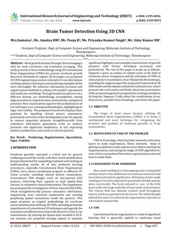
Mind growth discovery utilizing a 3D Convolutional Brain Organization (CNN) utilizes advanced picture handling methodsonthree-layeredclinical sweeps.Theinteraction includespreprocessingthevolumetricinformation,planning specific 3D CNN engineering, and preparing the model on markeddatasets.Bydissectingmultifacetedexamplesinthe three-layered space, the 3D CNN can really recognize and restrictmindgrowths,addingtoadditionalsolidandexact clinical judgments. Mind cancer recognition utilizing a 3D ConvolutionalBrainOrganization(CNN)includesafewkey stages.Atfirst,clinicalimaginginformation,normallyas3D sweeps, for example, X-ray or CT examinations, is preprocessedtoimprovequalityandnormalizeinput.The modelisthenpreparedonanameddataset,figuringouthow to perceive designs demonstrative of cerebrum growths. Assessmentondiscretetestinformationsurveysthemodel's exhibition.
International Research Journal of Engineering and Technology (IRJET) e-ISSN: 2395-0056
Volume: 11 Issue: 04 | Apr 2024 www.irjet.net p-ISSN: 2395-0072
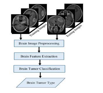
CNNs are a kind of profound learning design that is appropriateforpicturecharacterizationundertakings.They canbeutilizedtoseparateelementsfromcerebrumpictures andarrangethemasoneortheothertypicalordangerous. The framework design utilizes freely delivered clinical datasetstocreateOKlocationresults.Thedesigncansort cerebrum growths into four kinds: ordinary, meningioma, glioma,andpituitary.
1.A dataset is a collection of data, often organized in a structuredorsemi-structuredformat,thatisutilized fora specific purpose, such as research, analysis, or machine learning.
2.Once a dataset is captured, it often undergoes preprocessing steps to clean and format the data
appropriately for the intended use, such as training a machinelearningmodel.
3.Thisdatasetcontains7017imagesofhumancerebrumXray pictures which are classified into 4 classes: glioma, meningioma,notumor,pituitary.
4.Glioma Consists of 300 images, meningioma Consists of 305 images, no tumor Consists of 404 images, pituitary Consists of 300 images. These pictures are utilized for Testing.
5.GliomaConsistsof1320images,meningiomaConsistsof 1338images, no tumor Consists of1594 images,pituitary Consists of 1456 images. These pictures are utilized for Training.
Intheproposedframework,wewillutilizetheadministered 3D CNN, which will further work on the exactness of the prediction. 3D CNN is demonstrated for better exactness, supporting the profound learning strategies. It is too supplemented with the light weight library in Python for picturehandlingasOpenCV,whichassistsuswitharranging the picture and works on the speed of execution. The framework has involved different boundaries for characterizationbetweenordinaryandtumorouscerebrums withaccuracyof98%.
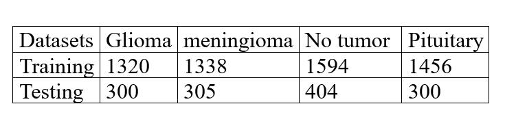
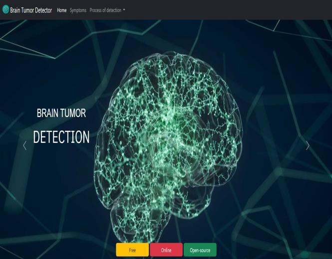
Fig 3: Home Page
International Research Journal of Engineering and Technology (IRJET) e-ISSN: 2395-0056
Volume: 11 Issue: 04 | Apr 2024 www.irjet.net p-ISSN: 2395-0072


Theuseof3DConvolutionalBrainOrganizations(CNNs)for mind cancer identification denotes a huge progression in clinical imaging innovation. Through the combination of profound learning and clinical diagnostics, this approach offers a promising answer for work on the precision furthermore,viabilityofmindcancerdetermination.Allin all, the utilization of 3D CNNs holds gigantic potential for upgradingearlydiscovery,exactlimitation,andportrayalof cerebrumgrowthsfromX-rayfilters.Byutilizingthespatial dataimplantedinthree-layeredinformation,thesemodels cancatchmany-sidedexamplesandelementsessentialfor preciseanalysis.Inaddition,theircapacitytolearnvarious leveledportrayalsempowersthemtoadjustandsumupwell across different datasets. Notwithstanding, further examinationisjustifiedtorefineandimprovethesemodels for true clinical settings, tending to difficulties like information shortage, interpretability, and speculation in inconspicuous patient populaces. Cooperative endeavors between clinical experts, specialists, and information researchersarecrucialforoutfittingthemaximumcapacity of 3D CNNs and making an interpretation of them into compellingapparatusesforworkingonpersistentresultsin neuro-oncology.
We wish to express our deepest appreciation to our esteemed Project Guide, Prof. Sumaiya, whose invaluable guidanceandsuggestionshavepropelledourprojectbeyond ourexpectations.Weextendourheartfeltgratitudetoour Project Coordinator, Dr. HK Chethan, for his unwavering supportanddedicationinhelpinguscompletethisproject withinatighttimeframe.Wewouldalsoliketoacknowledge our Head of Department, Dr. Ranjit KN, for fostering an environment that encourages innovation and practical applicationofouracademiccurriculum.Finally,weextend oursincerestthankstoourPrincipal,Dr.YTKrishneGowda, for providing us with a golden opportunity to carry out project on the topic of 'Brain Tumor Detection using 3D CNN',andforhisunwaveringsupportinourresearchand learningendeavors.
[1] D. Kornack and P. Rakic, “Cell Proliferation without NeurogenesisinAdultPrimateNeocortex,”Science,vol.294, Dec.2001,doi:10.1126/science.1065467.pp.2127-2130
[2]R.Nicole,“Titleofpaperwithonlyfirstwordcapitalized,” J.NameStand.Abbrev.,inpress.
[3]K.Elissa,“Titleofpaperifknown,”unpublished.
[4] Anupurba Nandi, “Detection of human brain tumour using MRI image segmentation and morphological operators” IEEE International Conference on Computer Graphics,VisionandInformationSecurity(CGVIS),2015.


Sumaiya, Professor at Maharaja Instituteof TechnologyThandavapura, Mysore, Department of Computer ScienceandEngineering.
Amulya H.M, Student of Maharaja InstituteofTechnologyThandavapura, Mysore. Pursuing Bachelor’s of Engineering Degree in Computer ScienceandEngineering.
PoojaB,StudentofMaharajaInstituteof Technology Thandavapura, Mysore. Pursuing Bachelor’s of Engineering Degree in Computer Science and Engineering.
International Research Journal of Engineering and Technology (IRJET) e-ISSN: 2395-0056
Volume: 11 Issue: 04 | Apr 2024 www.irjet.net p-ISSN: 2395-0072


Priyanka Kumari Singh, Student of Maharaja Institute of Technology Thandavapura, Mysore. Pursuing Bachelor’s of Engineering Degree in ComputerScienceandEngineering.
Uday Kumar D.R, Student of Maharaja InstituteofTechnologyThandavapura, Mysore. Pursuing Bachelor’s of Engineering Degree in Computer ScienceandEngineering