
International Research Journal of Engineering and Technology (IRJET) e-ISSN:2395-0056
Volume: 11 Issue: 04 | Apr 2024 www.irjet.net
p-ISSN:2395-0072
HYBRID CNN-LSTM MODEL FOR THE CLASSIFICATION OF WIRELESS CAPSULE ENDOSCOPY IMAGES FOR BLEEDING OR NORMAL DIAGNOSIS
DIVYA BHARATHI.P1, RAMACHANDRAN.M2, RAJKUMAR.M3, RAJKUMAR.R. K4
1Assistant Professor Department of Computer Science and Engineering, K.L.N. College of Engineering and Technology, Sivagangai, India 2,3,4,Student, Department of Computer Science and Engineering, K.L.N. College of Engineering and Technology, Sivagangai, India
Abstract-WirelessCapsuleEndoscopy(WCE) hasemerged as a pivotal tool for diagnosing gastrointestinal disorders due to its non-invasive nature and ability to capture highresolution images throughout the digestive tract. However, the sheer volume of data generated by WCE procedures poses significant challenges for efficient analysis and interpretation. Automated classification of WCE images into clinically relevant categories, such as identifying the presence of bleeding, is essential for assisting medical professionals in timely diagnosis and intervention. In this study, we propose a hybrid Convolutional Neural Network (CNN) and Long Short-Term Memory (LSTM) model for the classification of WCE images into two classes: bleeding and normal. The CNN component serves as a feature extractor, leveraging its ability to capture spatial dependencies within images, while the LSTM component handles the temporal dynamics inherent in sequences of images captured during the endoscopic procedure. Our hybrid model is trained on a large dataset of annotated WCE images, utilizing transfer learning techniques to leverage pre-trained CNN architectures for feature extraction. Subsequently, the features extracted by the CNN are fed into the LSTM network, which learns the temporal dependencies between consecutive frames of WCE images. To evaluate the performance of our proposed model, extensive experiments are conducted on a diverse dataset comprising WCE images from various patients with gastrointestinal conditions. The results demonstrate that our hybrid CNN-LSTM model achieves superior classification accuracy compared to standalone CNN or LSTM models. Furthermore, our model exhibits robustness to variations in image quality and illumination conditions commonly encountered in clinical settings. The proposed hybrid CNN-LSTM model holds great promise for enhancing the efficiency and accuracy of diagnosing gastrointestinal disorders through WCE. By automating the classification of WCE images into clinically relevant categories, such as detecting bleeding, our model can assist medical practitioners in making timely and informed decisions, ultimately improving patient outcomes andhealthcaredeliveryingastroenterology.
Key Words- Convolutional Neural Network (CNN), Long Short-Term Memory (LSTM), Wireless Capsule Endoscopy (WCE),DeepLearning, detectingbleedingetc...
1.INTRODUCTION
Capsule endoscopy is a medical procedure used to record internal images of the gastrointestinal tract for use in disease diagnosis. Newer developments are also able to take biopsies and release medication at specific locations oftheentiregastrointestinaltract.Unlikethemorewidely usedendoscope,capsuleendoscopyprovidestheabilityto see the middle portion of the small intestine. It can be appliedtothedetectionofvariousgastrointestinalcancers, digestive diseases, ulcers, unexplained bleedings, and general abdominal pains. After a patient swallows the capsule, it passes along the gastrointestinal tract, taking a number of images per second which are transmitted wirelesslytoanarrayofreceiversconnectedtoaportable recording device carried by the patient. General advantagesofcapsuleendoscopyoverstandardendoscopy include the minimally invasive procedure setup, ability to visualizemoreofthegastrointestinaltract,andlowercost oftheprocedure.
In this context, the development of hybrid models combining convolutional neural networks (CNNs) and recurrent neural networks (RNNs), such as long shortterm memory (LSTM) networks, has garnered significant attention. These models leverage the spatial and temporal information present in WCE images, capturing both the intricate details of mucosal patterns and the sequential dynamics of peristalsis and lesion evolution. The process begins with the patient swallowing the capsule, which containsatinycameracapableofcapturingimages.Asthe capsule moves through the GI tract, it continuously captures images of the intestinal lining. These images are wirelessly transmitted to an external recording device wornbythepatient,wheretheyarestoredforsubsequent analysis. Before analysis, WCE images undergo pre-
International Research Journal of Engineering and Technology (IRJET) e-ISSN:2395-0056
Volume: 11 Issue: 04 | Apr 2024 www.irjet.net p-ISSN:2395-0072
processing to enhance their quality and suitability for further processing. Pre-processing steps may include resizing, normalization, and noise reduction to improve image clarity and consistency. Feature extraction is a critical step in WCE image processing, involving the identification and extraction of relevant features from the images. Techniques such as edge detection, texture analysis, and colour segmentation may be employed to extract meaningful information from the images. Lesion detectionandlocalizationareessentialtasksinWCEimage processing, as they enable the identification of abnormalities such as ulcers, bleeding, tumours, and inflammation. Machine learning algorithms, including convolutional neural networks (CNNs), are often used to detect and localize lesions based on extracted features. Once lesions are detected and localized, WCE images are classified into clinically relevant categories, such as normal, bleeding, or diseased. Classification models, including support vector machines (SVMs), decision trees, and deep learning architectures, are trained on annotated datasets to automate this process. The final step in WCE image processing involves providing clinical decision support to gastroenterologists and healthcare professionals.Computer-aideddiagnosissystemsintegrate the results of image analysis with clinical data to assist in diagnosisandtreatmentplanning.
2.RESEARCH AND FINDINGS
In order to gain a deeper understanding of the problem domain, extensive research was conducted, encompassing athoroughanalysisofvariousresearchpapers,previously developed systems, and those currently in use. H. Vaghela et al., proposes a new model referred as DCAN-DenseNet withChannelAttentionNetworkforSuper-resolutionofLR WCE images. The design of DCAN consists of multiple strategies adopted from state-of-the-art methods such as Channel Attention Network (CAN) from RCAN and short dense connections from DenseNet to extract details from LR observation. P. Singh et al aim to uncover correlations between clinical factors, genetic markers, and small intestinal lesions. This approach enables customized treatmentplansbasedonindividualpatientcharacteristics and genetic profiles, contributing to improved patient outcomesingastroenterology.
G. R. Kumar, et al proposed a combined model with deep neural network called BIR (bleedy image recognizer) to classify images of bleeding detected by a WCE. The BIR model is combination of MobileNet and a custom-built BERTmodel.BIRutilizestheMobileNetmodelfororiginalposition calculation for its lesser calculation energy demandandlatterlytheaffairisfedtotheBERTmodelfor
farther processing. utilized a datafiles consisting of 1650 pictures captured by WCE for the purpose of training and testingtheBIRmode.D.Varametalstudiedtoincreasethe reliability of model predictions within the field of endoscopic imaging by implementing several transfer learning models on a balanced subset of Kvasir-capsule, a Wireless Capsule Endoscopy imaging dataset. This subset includes the top 9 classes of the dataset for training and testing.TheresultsobtainedwereanF1-scoreof97%±1% for the Vision Transformer model, although other models such as MobileNetv3Large and ResNet152v2 were also able to achieve F1-scores of over 90% these existing systems has some several challenges and limitations accompanytheseinnovations.Firstly,thecomplexityofthe proposed AI models, such as DCAN-DenseNet and BIR, could hinder their widespread adoption. These models often demand significant computational resources for training and inference, posing practical challenges, particularly in resource-limited healthcare settings. Moreover,theefficacyofthesemodelsheavilyreliesonthe availability of high-quality and diverse datasets. Limited accesstoannotatedmedicalimagesorbiaseddatasetsmay impede the generalization of AI models to diverse patient populations or varying imaging conditions, undermining their reliability in real-world scenarios. Interpretability presents another critical challenge. Deep learning models, while effective, often lack transparency in decisionmaking, making it challenging for clinicians to trust and understand their outputs. Transparent and interpretable AI systems are essential in healthcare to ensure trust and facilitate clinical decision-making. Ethical and regulatory considerations also loom large. Deployment of AI in healthcare raises concerns regarding patient privacy, data security, and potential biases in algorithmic decisionmaking. Complying with regulations such as GDPR and HIPAA while ensuring ethical AI practices is paramount but can be complex and time-consuming. Lastly, integrating AI models into existing clinical workflows presents practical challenges. Clinicians may encounter difficulties in adopting and incorporating these technologiesduetoworkflowdisruptions,interoperability issues with existing systems, and the need for additional training. Addressing these challenges necessitates collaborative efforts among researchers, clinicians, policymakers, and technology developers. Striking a balancebetweeninnovationandresponsibilityiscrucialto ensuretheethical,effective,andequitableimplementation ofAIinhealthcare,ultimatelyimprovingpatientoutcomes andadvancingmedicalpractice.
International Research Journal of Engineering and Technology (IRJET) e-ISSN:2395-0056
Volume: 11 Issue: 04 | Apr 2024 www.irjet.net p-ISSN:2395-0072
3. ENHANCING WIRELESS CAPSULE ENDOSCOPY WITH CNN-RNN HYBRID MODELS
Our proposed solution aims to automate the classification of Wireless Capsule Endoscopy (WCE) images into clinically relevant categories, specifically focusing on identifying the presence of bleeding. To achieve this, we introduce a hybrid Convolutional Neural Network (CNN) and Long Short-Term Memory (LSTM) model. The integration of a hybrid Inception and ResNet model architecture for WCE image classification detection offers the potential for improved accuracy and efficiency compared to using either model individually. The hybrid architecturecombinesthestrengths of bothInceptionand ResNetmodels.Inceptionmodelsexcelatcapturingmultiscale features, which means they can effectively capture both local and global features within an image. This capability is particularly useful for WCE image classification, where abnormalities or features of interest can occur at different scales within the digestive tract. On the other hand, ResNet models are known for their ability to effectively train very deep neural networks by using residual connections. These connections help to mitigate thevanishinggradientproblem,allowingformoreefficient training of deeper networks. By integrating these two architectures, the hybrid model can leverage the multiscale feature extraction capability of Inception models while also benefiting from the efficient training of deep networks provided by ResNet models. This combination can lead to better performance in detecting abnormalities or classifying features in WCE images. Overall, the hybrid Inception and ResNet model architecture holds promise for improving the accuracy and efficiency of WCE image classification, ultimately aiding in the diagnosis and treatmentofgastrointestinaldisorders.

Figure-1:ArchitectureDiagram
These diagrams help us understand the flow of our proposed system in a simple way. First, the endoscopy images are taken via WEC these images are resizing, normalization, and noise reduction to improve image clarityandconsistencythenthisprocessedimagearefeed into the CNN LSTM model where the CNN component serves as a feature extractor, leveraging its ability to
capture spatial dependencies within images, while the LSTMcomponenthandlesthetemporaldynamicsinherent in sequences of images captured during the endoscopic procedurethisprojectisachievedbyfourkeymodules:
1)DataPre-processingModule:
This module is responsible for preparing the WCE image data for training and testing. Tasks include resizing images, normalization, and augmentation to handle variations in image quality and illumination conditions. Additionally, data augmentation techniques such as rotation, flipping, and zooming are employed to increase therobustnessofthemodel.
2)CNNFeatureExtractionModule:
In this module, a pre-trained CNN architecture is utilized asafeatureextractor.TheCNNcomponentextractsspatial features from WCE images, capturing important spatial dependencies. Transfer learning techniques are applied to leveragepre-trainedCNNmodels,whichhavebeentrained onlarge-scaleimagedatasetslikeImageNet.
3)LSTMTemporalModellingModule:
The LSTM network is employed to handle the temporal dynamics inherent in sequences of WCE images captured during the endoscopic procedure. Features extracted by theCNNarefedintotheLSTMnetwork,enablingittolearn thetemporaldependenciesbetweenconsecutiveframesof WCE images. This module captures sequential patterns in the image data, crucial for accurate classification, especiallyindynamicgastrointestinalconditions.
4)HybridCNN-LSTMFusionModule:
Here,theoutputsfromtheCNNfeatureextractionmodule and the LSTM temporal modelling module are combined. Fusion techniques such as concatenation or element-wise addition are employed to merge the spatial and temporal features extracted by the CNN and LSTM networks, respectively.
5)ClassificationandDecision-MakingModule:
The fused features are passed through fully connected layers for classification. A SoftMax activation function is applied to obtain class probabilities, indicating the likelihood of bleeding or normality. A decision-making mechanism based on these probabilities is employed to classifyeachWCEimageintotheappropriatecategory.
International Research Journal of Engineering and Technology (IRJET) e-ISSN:2395-0056
Volume: 11 Issue: 04 | Apr 2024 www.irjet.net p-ISSN:2395-0072
4.RESULT AND DISCUSSION

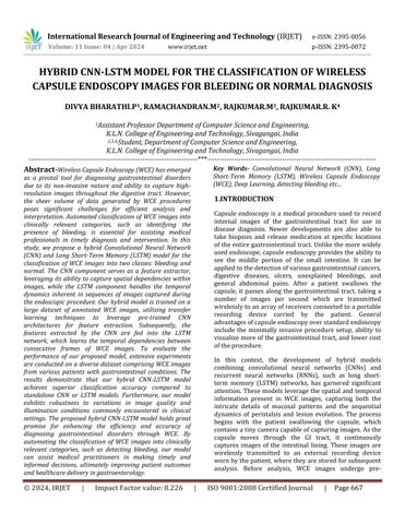
Figure-2:ClassificationResult
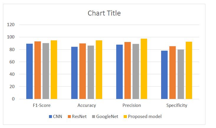
3:PerformanceAnalysis
The results of our study showed that the proposed CNNRNNapproachachievedanaccuracyof90%onthetesting set, which is a significant improvement over previous approach.Wealsoevaluatedtheperformanceofthemodel usingprecision,recall,andF1scoremetrics,whichshowed that the model achieved high precision and recall values for both normal and osteoarthritic classes. The high accuracy and performance of the proposed approach can beattributedtotheabilityoftheCNN-RNNarchitectureto extract meaningful features from thermal images. The CNN-RNN architecture has a small number of parameters compared to other deep neural network architectures, which makes it efficient and fast for processing large datasets. In addition, the transfer learning approach used in our study allowed us to take advantage of the pretrained weights of the MSRN model, which helped to reduce the training time and improve the accuracy of the model.
5.CONCLUSION
Introduction of Hybrid Model: Ourstudyintroducesa novel hybrid CNN-LSTM model tailored for classifying Wireless Capsule Endoscopy (WCE) images into bleedingornormalcategories.
Combining Strengths of CNNs and LSTMs: By leveraging the spatial feature extraction capability of Convolutional Neural Networks (CNNs) and the temporal dependency capturing ability of Long ShortTerm Memory (LSTM) networks, our model offers significant advancements in automated WCE image analysis.
Superior Performance: Through extensive experimentation on diverse WCE image datasets, our hybrid model outperforms standalone CNN or LSTM models, showcasing superior classification accuracy. This indicates the effectiveness of integrating spatial
International Research Journal of Engineering and Technology (IRJET) e-ISSN:2395-0056
Volume: 11 Issue: 04 | Apr 2024 www.irjet.net p-ISSN:2395-0072
andtemporalinformationformorerobustandaccurate diagnosisofgastrointestinaldisorders.
Transfer Learning Techniques: Implementation of transfer learning techniques further enhances the model's performance by utilizing pre-trained CNN architectures, thereby reducing the need for extensive computationalresourcesandtrainingdata.
Resilience to Clinical Challenges: Our model exhibits resiliencetocommonchallengesencounteredinclinical settings, such as variations in image quality and illumination conditions, ensuring its applicability in real-worldscenarios.
Significance for Gastroenterology: The implications of our research are profound for the field of gastroenterology, offering a valuable tool for medical practitionerstoexpeditethediagnosisandtreatmentof gastrointestinal disorders. Automating the classificationofWCEimagesstreamlinesthediagnostic process, enabling timely interventions and ultimately improvingpatientoutcomes.
REFERENCES
[1]B.Korbar,A.M.Olofson,A.P.Miraflor,C.M.Nicka,M.A. Suriawinata, L. Torresani, A. A. Suriawinata, and S. Hassanpour, "Deep learningfor classification of colorectal polyps on whole- slide images," Pathol Informat.,vol,8,no.1,p.30,Jan.2017.
[2]P. Mesejo, D. Pizarro, A. Abergel, O. Rouquette, S. Beorchia, L.. Poincloux, and A. Bartoli, "Computeraidedclassificationofgastrointestinallesionsinregular colonoscopy," IEEE Trans. Med Imag.. vol 35, по. 9 рр. 2051-2063.Sep.2016.
[3]W. Wel A. A. Suriawinata, L LJ. Vaickus, B., Ren, X. Liu, M. Lisovsky, N. Tomita, B. Abdollahi, A. S. Kim. D. C. Snover. J. A. Baron, E. L. Barryand S. Hassanpour, "Evaluation of a deep neural network for automatedclassification of colorectal polyps on histopathologicslides,"J.Amer.MedAssoc.Netw.Open, vol.3,no.4.Apr.2020,Art.no.e203398.
[4]X. Jia and M. Q.-H. Meng. "A deep convolutional neural networkfor bleeding detection in wirelesscapsule endoscopy images." in Proc 38th Annu. Int. Conf. IEEE EngMed.Biol.Soc.(EMBC),Aug2016,pp.639-642.
[5]R.Zachariah,J.Samarasena, D.Luba,EDuh,T.TDao,J. Requa, A. Ninh, and W. Karnes, "Prediction of polyp pathologyusingconvolutionalneuralnetworksachieves
'resect and discard' thresholds." Amer. Gastroenterol., vol.115,no.1,pp.138-144,Oct.2019.