
International Research Journal of Engineering and Technology (IRJET) e-ISSN:2395-0056
Volume: 11 Issue: 03 | Mar 2024 www.irjet.net p-ISSN:2395-0072
International Research Journal of Engineering and Technology (IRJET) e-ISSN:2395-0056
Volume: 11 Issue: 03 | Mar 2024 www.irjet.net p-ISSN:2395-0072
V.Anbarasan1 and K. Arivalagan2
1Department of Chemistry, DMI college of Engineering, Chennai-600123.
2Department of Chemistry, Govt Arts College for Men (A) Nandanam, Chennai.600035.
Iron oxide has attracted a great deal of attention among specialists because of their multivalent oxidation states. The iron oxide nanoparticles have been synthesized by adding Albizia Amara leaf extract into the aqueous solution of ferric chloride. The phytoconstituents of A. Amara leaf extract serve a dual role as reducing, capping and stabilizing agent during the synthesis. GSIron Oxide nanoparticles were characterized by UV-vis absorption Spectroscopy, Fourier Transform Infrared Spectroscopy (FTIR), X-ray Diffraction (XRD) and Transmission Electron Microscope (TEM) and Scanning Electron Microscope (SEM). The existence of the Fe2O3 nanoparticles was revealed by UV-vis spectroscopy. The FTIR spectra of leaf extract and synthesized Fe2O3 nanoparticles identifies the functional groups of the active components. The formation of Fe2O3 nanoparticles has been confirmed by X-ray diffraction and average crystallite size for assign peaks were 37.91 nm. GS- iron oxide nanoparticles serve as potent antibacterial agent in an eco-friendly way by securing naturally biome as nanoparticles usually through target delivery. Thus, A. Amara mediated iron oxide nanoparticles can act as an alternative antimicrobial agent to the antibiotics.
Keywords: Albizia Amara leaves,Extract,ironoxidenanoparticles,Characterization.
Introduction
Nanoparticlesaresubmicronmoietieswithdiametersrangestartingfrom1-100nmmadeupoforganicorinorganicmaterials having novel properties as compared to a large number of materials [1]. The nanotechnology process depends on synthesis, manipulation,anduseofmaterialsthatareofNanoscalesize.Inthenewera,nanoparticlestakemoreattentionduetotheir unique size-dependent properties and applications [2]. Metal nanoparticles gain great attention due to their wide range of applications in the fields of electronics, optoelectronics, antibacterial activity, and medical applications such as therapy, diagnosis,anddrugdelivery[3-4].Thedevelopmentofadequatetechniquesforsynthesizingmetalnanoparticleshasbecome amajorfocusofresearchers.Metallicnanomaterialsuchassilver,gold,zincandironareusedinvariousfieldsbecauseoftheir broad applications; among these nanoparticles, iron nanoparticle is preferred for the following reasons: cost effective, antimicrobialactivity,highreactivity,smallersize;therefore,itgiveshighsurface-area-to-volumeratio,whichallowsinteract with different chemical species and also efficient in binding metal ions [5]. There are a large number of methods (physical, chemical, and biological) to synthesize various types of nanomaterial. When synthesized by chemical and physical methods, these nanoparticles lose their reactivity due to aggregation magnetism, and dispersibility upon air exposure Chemical synthesis methods involve toxic chemicals, the formation of hazardous by-products, and contamination from chemical precursors [6]. Therefore, there is growing interest in developing clean, simple, inexpensive, eco-friendly methods for the synthesisof nanoparticles. Bacteria,fungi,algae, and plantextractscan beusedin modern alternativesforthe production of metal/metal oxidenanoparticles.Plantmediatedsynthesisofnanoparticlesisa revolutionarytechniquethathaswide range ofapplicationsinagriculture,foodindustry,medicineandenvironmentalremediation.Theplantrelatedpartssuchasleaves, stems, roots, shoots, flowers, barks, seeds and their metabolites have been successfully used for the efficient biosynthesis of nanoparticles.Plantextractsusuallycontainsugars,terpenoids,polyphenols,alkaloids,phenolicacids,andprotein,whichare responsible for reducing and stabilizing metal nanoparticles [7]. In the past, green synthesis of Fe2O3 nanostructures using different plant extractssuchasLagenaria siceraria, HordeumvulgareandRumexacetosa plants,peel extractofplantainand Tridax procumbens leaf extract. Therefore, in this study, we have made an attempt on the synthesis of Fe2O3 nanoparticles usingextractofAlbiziaAmaraleaves.
International Research Journal of Engineering and Technology (IRJET) e-ISSN:2395-0056
Volume: 11 Issue: 03 | Mar 2024 www.irjet.net p-ISSN:2395-0072
2.1. MATERIALS
Allchemicalsusedwereofanalyticalreagentgradewithoutanyfurtherpurificationinadditiontodeionisedwater,Ferric Chloride(FeCl3.6H2O),Sodiumhydroxide(NaOH),hydrochloricacid(HCl),ethanol(C2H5OH)andAlbiziaAmaraLeaves.
2.2. Preparation of green synthesized Iron oxide nanoparticles
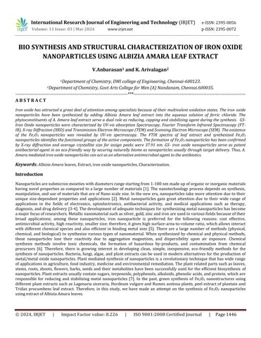
2.2.1. Preparation of Albizia Amara leaf Extract,
Albizia Amara leaves (AAL) were collected from Kallakurichi (DT), Kottaiyur (village) in Tamilnadu. The clean and fresh sources are dried in a shaded place at room temperature for 10 to 15 days and then the leaves were pulverized using commercialblender. Thefinepowderedwasstoredatroomtemperatureforfurtheruse. Ina250mlofconicalflask10gof leaf powder were taken and to this 100 ml of double distilled water is added and it is heated at 80oC for 1 hour. Then the solutionwasfilteredusingWhatmanfilterpaperandkeptasideforfurtherprocess.Theobtainedextractinpalebrowncolor andadjustedtothepHat11byadding0.1Mofsodiumhydroxidesolution.
2.2.2. Preparation of Iron oxide nanoparticles.

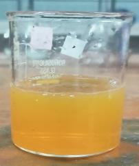


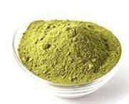
In a 250 ml conical flask, 50ml of Albizia Amara leaves extract was taken and to this 100 ml of 0.1 M FeCl3.6H2O solution is added slowly at room temperature under static conditions. The colour change of the reaction was observed and the time taken for the changes was noted. The solution colour changes immediately from pale brownish to reddish brown indicating the formation of iron oxide nanoparticles (Fe2O3NPs). Further the solution is centrifuged and precipitated is extracted and dried in electrical oven for 24 hours at 100oC. The dried sample kept in muffle furnace for 4 hours at 500oC. The green synthesizedFe2O3NPsisformedatuniformparticlesizeandstoredforfurthercharacterizationanduses[8]
3. CHARACTERIZATION OF ADSORBENT
3.1. UV-Visible spectrophotometer analysis
Synthesized Fe2O3 nanoparticles were subjected to UV-Vis spectroscopy analysis, which confirms the formation of nanoparticles in the initial stage. The Fe2O3 nanoparticles synthesized were subjected to scan UV-Vis spectrophotometer in therange190nm-1100nmusingElicoSL210UVVISSpectrophotometer.
International Research Journal of Engineering and Technology (IRJET) e-ISSN:2395-0056
Volume: 11 Issue: 03 | Mar 2024 www.irjet.net p-ISSN:2395-0072
3.2. FT-IR Spectroscopic analysis
The plant extract and green synthesized Fe2O3 nanoparticles were characterized by FT-IR spectrometer. The spectroscopic technique is based on the analysis of peaks at certain wave numbers. FT-IR data indicates the presence of functionalgroupsintheplantextractandsynthesizednanoparticles. TheFT-IRanalysiscarriedoutinthefrequencyrangeof 4000-400cm-1 usingPerkinElmerinstrument.
3.3. X-ray diffraction analysis (XRD)
X-ray diffractometer (lakjdf) was used to study the average particle size and crystalline nature of the synthesized adsorbents. Thediffraction pattern wasobtained byusingFeKαradiation with wavelengthof λ=1.541Ao. The scanning was donein2θvaluerangeof4o to80o at0.02min-1 andonesecondtimeconstant.
3.4. Scanning Electron Microscopic (SEM)
The SEM analysis provide the details about surface morphology, porosity and particle size distribution of the adsorbents. ThesurfacemorphologyofthesynthesizedFe2O3 nanoparticleswasrecordedusingHitachiinstrument.
3.5. Transmission Electron Microscope (TEM)
TEM is regarded as the best among other electron microscopy techniques for the determination of particle size and morphologicalidentitiesofFe2O3NPsandothermetalnanoparticles.
4. RESULTS AND DISCUSSIONS
4.1. Characterisation study of green synthesized copper oxide nanoparticles.
4.1.1. Ultraviolet –Visible (UV-vis) Spectroscopy.
Thetypeofnanoparticlesthathasbeentargetedforsynthesisfromselectedplantcanbeidentifiedbytwomethods,onewith the visual observation of colour change pattern and another one with UV–Vis analysis. In the present study, iron oxide nanoparticlessynthesizedfromleavesextractshoweda colourchangepatternfrombrowntoreddishbrownincolouratthe timeofsynthesis.
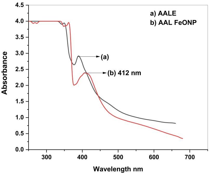
Fig2showsUV-Visspectrumgreensynthesizedironoxidenanoparticles.
International Research Journal of Engineering and Technology (IRJET) e-ISSN:2395-0056
Volume: 11 Issue: 03 | Mar 2024 www.irjet.net p-ISSN:2395-0072
As can be seen from Figure 2, the absorption peaks for Albizia amara leaf extracts are around 370 to 390 nm, which correspondstotheexistenceofseveralnaturalcompoundsintheextracts[9].Thesepeaksarevanishedafterreactingwithan iron salt, indicating that the extract compounds acted as reducing and capping agents to synthesize the iron oxide nanoparticles.Furthermore,andinaccordancewiththeresultsofthepresentstudy,thesurfaceplasmonbandforironoxide nanoparticlesatwavelengthsof412nmindicatetheformationofironoxidenanoparticleswerereportedbypreviousstudies (Figure2)[10].
TheFT-IRanalysiswereperformedontheAlbiziaamaraleafextractandthesynthesizedironoxidenanoparticlestoidentify apossiblechangeinfunctionalgroupbondsduringthereductionprocessandpresentedinFigure3.
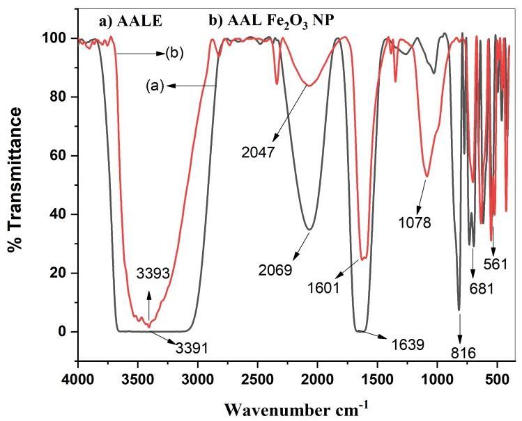
Fig3a&3bshowsFTIRspectrumofAlbiziaAmaraleavesextractandIronoxidenanoparticles.
FTIRspectraoftheleafextractarepresentedinFig.3a.Theabsorptionpeaksat3391and1639cm−1refecttheO–HandN–H stretching vibration in the phenolic compound and protein in the fruit extract. The bands at 2069cm−1 are assigned to C–H stretching in carbohydrates. The bands at 816cm−1 is caused by the (C=O) NH2 stretching [11].FTIR spectra of synthesized ironoxidenanoparticlesusingextractofC.masisshowninFig.3b.Thebandwithhigherintensityassignedtothe -OHgroups indicates water soluble polyphenol compounds that have capped the surface of the prepared iron oxide nanoparticles. The bandat2047cm-1maybeduetoC≡NstretchingfromunreactedimpuritiesorduetoCO2inthesamplecompartment.The bandat3393cm-1and2800cm-1correspondstothe-OHbondstretchinganddenotestheaqueousphase,withanincrease intheabsorption band,indicatingtheferrous sulphate reduction. The existingfindingsagreedwell withthereportedvalues [12]
4.1.3. X - ray diffraction (XRD) analysis.
XRDisananalyticaltoolusedtodistinguishcrystallinephaseandcelldimensionsofsynthesizednanoparticles. Fig(6)Shows theappearanceofdiffractionpatternat2θ= 35.12º,36.63º,40.64º,49.97º,57.08 º and65.49º withcorrespondinglattice plane values at (104), (110), (113), (024), (116) and (300) respectively. The intense and sharp peaks undoubtedly revealed thatironoxidenanoparticlesformedbythereductionmethodusingAlbiziaamaraleafextractwerecrystallineinnature.The crystallinesizewascalculatedwiththehelpofScherrer’sformula,whichisgivenas[13]
D=0.9λβcosθ,(1)
WhereDisthecrystallitesize,βthefull-widthathalfmaximum(FWHM)ofthemostintensediffractionpeakinradians,θthe diffractionangleandλthewavelengthofX-rayradiation.Asharppeakat2θ=35.12and36.63withthediffractionofthe(104)
International Research Journal of Engineering and Technology (IRJET) e-ISSN:2395-0056
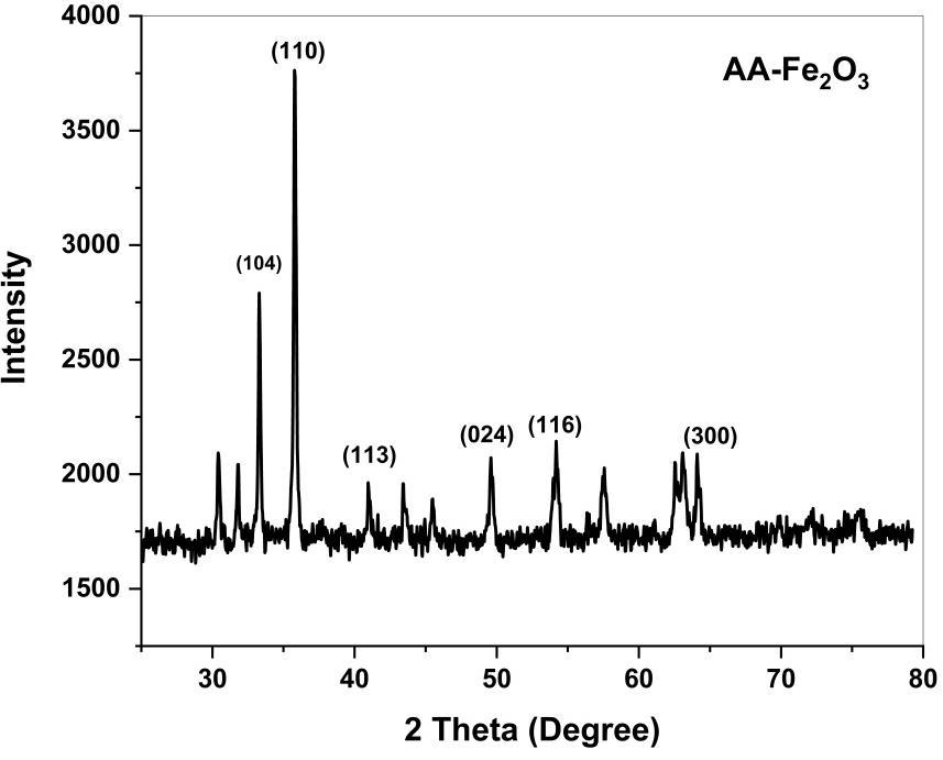
Fig4showsXRDpatternspectrumofGreensynthesizedironoxidenanoparticles.
4.1.4. Scanning Electron Microscope (SEM)
Themorphologyofthepreparednanoparticleswasexaminedusingscanningelectronmicroscopy.Figure5showstheFESEM imageofthesynthesizedFe2O3 nanoparticlesconsistedofnano-sizedparticleswithanearlysphericalshape.Itwasobserved thatthenanoparticlesformedwereagglomeratedwiththeparticlesandappearstoadheretoeachother,formingaggregateof particles, which result in irregular arrangements. The particles sizes measured about 9 to 12 nm, hence supporting the TEM result.
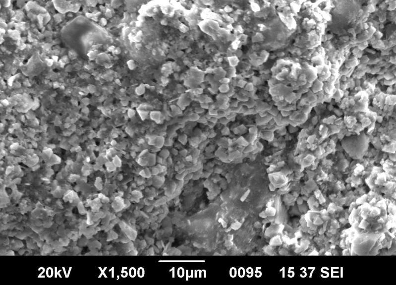
Fig6showsSEMimageofgreenmediatedironoxidenanoparticles
Volume: 11 Issue: 03 | Mar 2024 www.irjet.net p-ISSN:2395-0072 © 2024, IRJET | Impact Factor value: 8.226 | ISO 9001:2008 Certified Journal | Page1450 and (110) plane indicates that confirmation of Fe2O3NPs. The average crystallite size in the samples of Fe2O3NPs is below 37.91nm.
International Research Journal of Engineering and Technology (IRJET) e-ISSN:2395-0056
Volume: 11 Issue: 03 | Mar 2024 www.irjet.net p-ISSN:2395-0072
TheTEMphotographofthesynthesizedIONPsisdepictedinFig.7.Theaverageparticlesizewasfound8.03±8.99nmfor206 individualselections.Asitisseen,thenanoparticlesarealmostsphericallikeandpartlyasahexagonalshape.Theaveragesize ofsynthesizednanoparticlesshowssomedifferencewiththesizeofparticlesfoundbyScherer’sequationintheXRDspectra. It may be due to the wide range size distribution of the nanoparticles, given the SD value calculated from the TEM analysis [15].
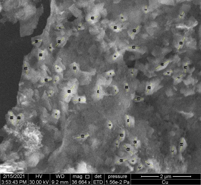
Fig7showsTEMimageofsynthesizedironoxidenanoparticles.
5. Conclusion.
In this present study, we reported the successful use of Albizia Amara as a one-pot green method for the synthesis of iron oxide nanoparticles. The color change, observed instantaneously suggested that the formation of black colored solution indicatedtheformationofironoxidenanoparticles.TherapidreductionprocessprovedtheefficiencyofAlbiziaAmaraextract asreducingandstabilizingagents.TheXRDpatternshowedthecubiccrystalstructureofironoxidenanoparticleswithoutany impurities.FTIRshowedthattheinteractionsthatexistedbetween AlbiziaAmaraandironoxidenanoparticles.TEMshowed the formation of Fe2O3with an average size of 9–12 nm with irregular shape. Green routes for nanoparticle synthesis are of greatinterestbecausetheyareecofriendly,inexpensive,simpleandrapid.Theyalsohaveawiderangeofapplications,suchas innanomedicine,catalysis,andoptoelectronics.
Reference
1. Mandeep Kaur, Dimple Sethi Chopra. Green Synthesis of Iron Nanoparticles for Biomedical Applications. Glob J Nanomed.2018;4(4):68-76.
2. MK Habeeb. Biosynthesis of nanoparticles by microorganisms and their applications, Int. J. Adv. Sci. Technol. Res. 2013;1(3):44–51.
3. P. Christian, V. der Kammer, F. Baalousha, and T. Hofmann, “Nanoparticles structure, properties, preparation, and behaviourinenvironmentalmedia,”Ecotoxicology,vol.17,pp.326–343,2008.
4. J. Virkutyte and R. S. Vara, “Chapter 2 environmentally friendly preparation of metal nanoparticles,” in Sustainable Preparationof Metal Nanoparticles:MethodsandApplications,pp. 7–33,+eRoyal Society ofChemistry,London,UK, 2013.
International Research Journal of Engineering and Technology (IRJET) e-ISSN:2395-0056
Volume: 11 Issue: 03 | Mar 2024 www.irjet.net p-ISSN:2395-0072 © 2024, IRJET | Impact Factor value: 8.226 | ISO 9001:2008 Certified Journal | Page1452
5. HUBER, DL.2005. Synthesis, properties, and applications of iron nanoparticles. Small, 1(5), 482-501. https://doi.org/10.1002/smll.200500006
6. W.Wu,Q.He,andC.Jiang(2008).NanoscaleRes.Lett.3,397.
7. T.Shahwana,S.AbuSirriaha,M.Nairataetal.,“Greensynthesisofironnanoparticlesandtheirapplicationasafentonlike catalyst for the degradation of aqueous cationic and anionic dyes,” Chemical Engineering Journal, vol. 172, pp. 258–266,2011.
8. Saraswathi Ramaiah Chinnasamy , Sujatha Dadhala , Anbarasan Veeran , Priyanka Ravichandran , Lakshmi Loganathan , Arivalagan Kuppusamy. Antibacterial and Antifungal Activity of Plant Mediated Iron Oxide Nanoparticles.Volume13Issue3,March2024.DOI:https://dx.doi.org/10.21275/SR24302102211
9. BuarkiF,AbuHassanH,AlHannanFandHenariF.Z,“Greensynthesisofironoxidenanoparticlesusinghibiscusrosa sinensisflowersandtheirantibacterialactivity,”J.ofNanotechnol.,20222022;2022:1-6.
10. MahdaviM,NamvarF,AhmadM.BandMohmadR,“GreenBiosynthesisandCharacterizationofMagneticIronOxide (Fe3O4)NanoparticlesUsingSeaweed(Sargassummuticum)AqueousExtract,”Molecul.(2013;18:5954–5964.
11. ElhamRostamizadeh, AlirezaIranbakhsh, AhmadMajd, SedighehArbabian , IrajMehregan. Green synthesis ofFe2O3 nanoparticles using fruit extract ofCornus mas L. andits growth-promoting roles inBarley. Journal of NanostructureinChemistry.https://doi.org/10.1007/s40097-020-00335-z
12. DemirezenD.A,YilmazSandYilmazD.D,“GreensynthesisandcharacterizationofironnanoparticlesusingAesculus hippocastanumseedextract,”Inter.J.ofAdv.inSci.Engg.andTech.2018;62:24–29
13. CulityBDandStockSR1978PrinciplesofX-rayDiffraction(Reading:Addision-Wesley).
14. NurDiyana SyazwaniZambri ,Nurul IzzaTaib,*,Famiza Abdul LatifandZakiahMohamed.UtilizationofNeemLeaf ExtractonBiosynthesisofIronOxideNanoparticles.Molecules2019,24,3803;doi:10.3390/molecules24203803.
15. MinaJamzad· MaryamKamariBidkorpeh. Green synthesis ofiron oxide nanoparticles bytheaqueous extract ofLaurus nobilis L. leaves andevaluation oftheantimicrobial activity. Journal of Nanostructure in Chemistry. https://doi.org/10.1007/s40097-020-00341-1