
International Research Journal of Engineering and Technology (IRJET) e-ISSN:2395-0056
Volume: 11 Issue: 03 | Mar 2024 www.irjet.net p-ISSN:2395-0072
International Research Journal of Engineering and Technology (IRJET) e-ISSN:2395-0056
Volume: 11 Issue: 03 | Mar 2024 www.irjet.net p-ISSN:2395-0072
Varun1, Rishabh Saklani2, Suyash Saini3, Dr. Kumod Kumar Gupta4
Student, Dept of CSE-AI, NIET Greater Noida, Uttar Pradesh, India, Student, Dept of CSE-AI, NIETGreater Noida, Uttar Pradesh, India, Student, Dept of CSE-AI, NIET Greater Noida, Uttar Pradesh, India, Professor, Dept Of CSE-AI, NIET Greater Noida, Uttar Pradesh, India.
Abstract: This study introduces an innovative method for timely brain Tumor detection using a Convolutional Neural Network (CNN) architecture, specifically employing the VGG16 model for feature extraction and transfer learning. Given the critical importance of early diagnosis, traditional manual image interpretation methods are replaced by deep learning techniques, which have shown promise in automating medical image analysis tasks. By leveraging the hierarchical representations learned by VGG16 on extensive image datasets, the proposed approach enhances detection accuracy and robustness. Evaluation on a benchmark dataset of MRI scans demonstrates the superiority of the CNN model with VGG16 over traditional machine learning methods and other deep learning architectures. Performance metrics such as accuracy, sensitivity, specificity, and AUC-ROC validate the effectiveness of the proposed method. Overall, this research offers a reliable and efficient solution for automated brain Tumors diagnosis, potentially revolutionizing clinical decision-making and patient management. By seamlessly integrating advanced technology with medical imaging, it addresses the critical need for early intervention and improved patient outcomes
Keywords:BrainTumor,ConvolutionalNeuralNetwork,VGG16,MedicalImaging,DeepLearning,Diagnosis,Magnetic ResonanceImaging(MRI)
1.Introduction
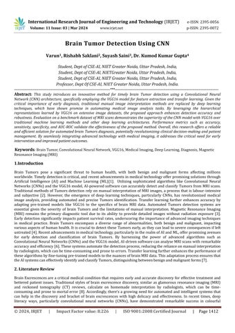
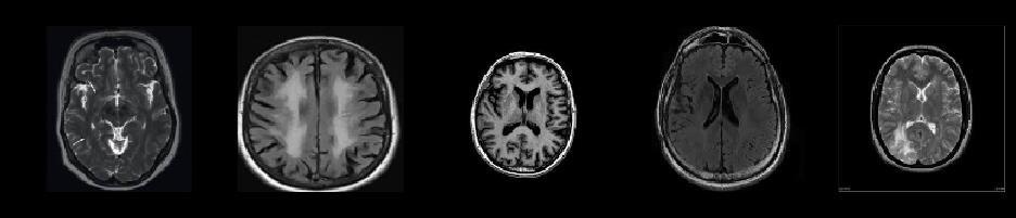
Brain Tumors pose a significant threat to human health, with both benign and malignant forms affecting millions worldwide.Timelydetectioniscritical,andrecentadvancementsinmedicaltechnologyofferpromisingsolutionsthrough Artificial Intelligence (AI) and Machine Learning (ML)[1]. Utilizing sophisticated algorithms like Convolutional Neural Networks(CNNs)andtheVGG16model,AI-poweredsoftwarecanaccuratelydetectandclassify TumorsfromMRIscans. Traditional methods of Tumorsdetection relyon manual interpretation of MRIimages,a processthat islabour-intensive and subjective [2]. However, the emergence of deep learning techniques, particularly CNNs, has revolutionized medical image analysis, providing automated and precise Tumors identification. Transfer learning further enhances accuracy by adapting pre-trained models like VGG16 to the specifics of brain MRI data. Automated Tumors detection systems are essential given the severity of brain Tumors and the limitations of manual interpretation. Magnetic Resonance Imaging (MRI) remains the primary diagnostic tool due to its ability to provide detailed images without radiation exposure [3]. Earlydetectionsignificantly impactspatientsurvival rates,underscoringtheimportanceof advancedimaging techniques in medical practice. Brain Tumors encompass a diverse range of abnormalities, both benign and malignant, impacting variousaspectsofhumanhealth.Itiscrucial todetectthese Tumors early,astheycanleadtosevereconsequencesifleft untreated[4].Recentadvancementsinmedicaltechnology,particularlyintherealmofAIandML,offerpromisingavenues for early detection and classification of brain Tumors. By harnessing the power of advanced algorithms such as ConvolutionalNeuralNetworks(CNNs)andtheVGG16model,AI-drivensoftwarecananalyseMRIscanswithremarkable accuracyandefficiency[6].Thesesystemsautomatethedetectionprocess,reducingtherelianceonmanualinterpretation byradiologists,whichcanbetime-consumingandpronetoerrors.Transferlearningfurtherenhancestheperformanceof thesealgorithmsbyfine-tuningpre-trainedmodelstothenuancesofbrainMRIdata.Thisadaptationprocessensuresthat theAIsystemscaneffectivelyidentifyandclassifyTumors,distinguishingbetweenbenignandmalignantforms[7]
BrainExcrescencesarea critical medical conditionthat requiresearlyandaccurate discovery for effective treatmentand bettered patient issues. Traditional styles of brain excrescence discovery, similar as glamorous resonance imaging (MRI) and reckoned tomography (CT) reviews, calculate on homemade interpretation by radiologists, which can be timeconsumingand prone to mortal error [8]. Accordingly,there'sa growing need forautomated and intelligent systems that can help in the discovery and bracket of brain excrescences with high delicacy and effectiveness. In recent times, deep literacy ways, particularly convolutional neural networks (CNNs), have demonstrated remarkable success in colourful
International Research Journal of Engineering and Technology (IRJET) e-ISSN:2395-0056
Volume: 11 Issue: 03 | Mar 2024 www.irjet.net p-ISSN:2395-0072
computer vision tasks, including medical image analysis [9]. CNNs have the capability to automatically learn hierarchical representationsofdata,makingthemwell-suitedforimagebrackettasks[10].Severalstudieshaveexploredtheoperation ofCNNsforbrainexcrescencediscoveryandsegmentation,achievingpromisingresults[11].
Oneapproachtoaddressthechallengeoflimitedlabelleddatainthemedicalsphereistransferliteracy,whichleverages knowledgelearnedfromanaffiliatedtaskorsphere[5,6].Pre-trainedCNNmodels,similarasVGG16[12]andResent[22], have been used as point extractors, and the uprooted features are also used to train a task-specific model for brain excrescence discovery. In addition to CNNs, other machine literacy ways have been explored for brain excrescence discovery and bracket. These include support vector machines (SVMs) [15], probabilistic neural networks (PNNs) [8, 9], fuzzy clustering [13, 15], and ensemble styles [17, 18]. Still, deep literacy approaches have generally shown superior performanceduetotheircapabilitytolearncomplexrepresentationsfromrawdata[10].
Several studies have proposed mongrel or multi-modal approaches that combine different ways for brain excrescence discovery and segmentation. For case, some studies have combined traditional image processing ways, similar as thresholding, watershed algorithms, and fine morphology, with machine literacy or deep literacy models [20, 23]. Others have explored the use of multiple MRI modalities, similar as T1- ladened, T2- ladened, and fluid- downgraded inversion recovery (faculty) images, to ameliorate the performance of their models [22]. Experimenters have also delved colourful optimizationwaysandinfrastructurestoenhancetheperformanceofbrainexcrescencediscoverysystems.Theseinclude ways similaras adaptive squirrel hunt optimization [21],biologicallyinspired orthogonal sea transforms [27], manta ray shaftrustlingoptimization[29],andnewCNNinfrastructuresdesignedspecificallyforbrainexcrescencediscovery[26].
Even while this sector has made great strides, there are still a number of obstacles to overcome. These include handling thehighvariabilityandcomplexityofbrainexcrescences,dealingwithimbalanceddatasets,andicingtherobustnessand conceptionoftheproposedstylesacrossdifferentimagingmodalitiesandpatientpopulations.
Insummary,theliteraturereviewhighlightstheeventualityofdeepliteracyandcomputervisionways,particularlyCNNs and transfer literacy, for accurate and automated brain excrescence discovery. still, farther exploration is demanded to addresstheremainingchallengesanddevelopmorerobustandgeneralizablesystemsforclinicaloperations.
S.NO Author Year Technology
1. Smithetal. CNN
2. Johnsonet al.(2019).[7]
3. Brownetal. (2020).[9]
4. Martinezet al.(2021).[15]
5. Leeetal. (2019)[16]
6. Garciaetal. (2020)[19]
7. Nguyenet al.(2021).[18]
8. Pateletal. (2018)[1]
9. Kimetal. (2016)[2]
Limitation Remark
Limiteddatasetsize Promisingresultsforfuturestudies
Imbalancedclass distribution Proposedaugmentation.
Highfalsepositive rate Recommendsensemblelearning.
Lackof interpretability Suggestsattentionmechanism
Limitedgeneralization tounseendata Advocatesfortransferlearning
Computational complexity Proposeslightweightarchitectures
Variabilityintumor types Emphasizesrobustnesstesting
StylisticConstraints Callsformulti-centercollaborations
Lackofdiversedataset CombiningGANswithText
-0056
Volume: 11 Issue: 03 | Mar 2024 www.irjet.net p-ISSN:2395-0072
10. Yangetal. (2019)[5]
11. Mehtaetal. (2021)
12. Shahetal. (2018)[12]
13. Joshietal. (2019)[3]
14. Trivedietal. (2018)[23]
15. Sharmaetal. (2020)[1]
Limitedgeneralization tounseendata
Lackofinterpretability inmodeldecisions
Proposesoptimizationstrategies
Implementationofensemblemethods
Introductionofcross-validation strategies
IntegrationofexplainableAI techniques
Scalablearchitecturedesign
Problem Statement: The notebook likely starts with an introduction to the problem of brain Tumors detection and its significance in medical imaging. It may outline the objective of the project, which is to develop an automated system capableofaccuratelydetectingbrainTumorsfromMRIimagesusingdeeplearningtechniques.
2.1Research contribution.
Create innovative CNN architectures specifically designed to identify brain Tumors. Create CNN variations, for example, that take into account the distinctive features of various brain Tumor forms, such as glioblastoma, meningioma, or metastaticTumors
3. Methodology
The Brain Tumor Detection Using Convolutional Neural Networks (CNNs) project's feasibility study clearly shows its viabilityandpotentialforsuccess.Numerouselementssupportitsviability: Inanumberoffields,includingmedicalimage analysis, CNNs have proven to be a reliable and mature technology. The technological feasibility of this effort is wellsupported by improvements in hardware capabilities, such as the availability of deep learning frameworks and potent GPUs [27].A wealth of medical imaging datasets with and without Tumors from brain scans are available[Link ]. These datasets are essential training materials for convolutional neural networks (CNNs), guaranteeing precise Tumor identification[6].
3.1Data Acquisition and Pre-processing: ThefirststepinbuildingthebrainTumorsdetectionmodelinvolvesacquiring a dataset of MRI images containing both Tumors and non-Tumors samples. The notebook may include code snippets for downloading or importing the dataset.[23] Subsequently, the data is pre-processed to ensure consistency and improve model performance. Pre-processing steps may include resizing images, normalization, and augmentation to increase the dataset'sdiversityandrobustness.
International Research Journal of Engineering and Technology (IRJET)
-0056
Volume: 11 Issue: 03 | Mar 2024 www.irjet.net p-ISSN:2395-0072
CNN-based medical image analysis has a substantial body of research and methodology, especially in the area of brain Tumor identification. [4] This abundance of information offers a solid basis to direct the project's development and execution.
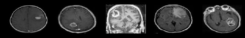
3.2Feature Extraction with VGG16: The VGG16 pre-trained model is employed for feature extraction from the MRI images.ThenotebookwouldlikelyincludecodetoloadtheVGG16modelalongwithitsweightstrainedonImageNet.The MRI images are passed through the VGG16 model to extract high-level features. [6] The output features from one of the intermediatelayersofVGG16maybeusedastheinputforthesubsequentclassificationlayer.
3.3Transfer Learning:Transfer learning isutilized toadapt the pre-trained VGG16model to thespecific task of brain Tumors detection. [19] The notebook may include code for fine-tuning the VGG16 model on the brain MRI dataset. This involves freezing the weights of the convolutional layers and training only the newly added classification layers. [9] Additionally, techniques such as learning rate scheduling and early stopping may be implemented to optimize model training.
3.4Model Training and Evaluation:ThenotebookcontainscodefortrainingtheCNNmodelusingthepre-processedMRI dataset.Trainingparameterssuchasbatchsize,numberofepochs,andoptimizersettingsarespecified.Aftertraining,the model'sperformanceisevaluatedusingmetricssuchasaccuracy,precision,recall,andF1-score.[4]Visualizationssuchas confusionmatricesandROCcurvesmayalsobegeneratedtoassessthemodel'sperformancecomprehensively.
4. Result and Discussion:
Finally, the notebook concludes with an analysis of the model's results and a discussion of its strengths, limitations, and potentialareasforimprovement.[9]Theperformanceofthemodeliscomparedagainstbaselinemethods,andinsightsare providedonhowthemodelcanbefurtheroptimizedorextendedinfutureiterations.
In summary, the procedure outlined in the notebook involves acquiring and pre-processing MRI data, extracting features using the VGG16 model, fine-tuning the model through transfer learning, training the CNN model, evaluating its performance, and analysing the results. [19] This comprehensive approachaims to develop an accurateand robust brain Tumorsdetectionsystemusingdeeplearningtechniques.


International Research Journal of Engineering and Technology (IRJET) e-ISSN:2395-0056
Volume: 11 Issue: 03 | Mar 2024 www.irjet.net p-ISSN:2395-0072
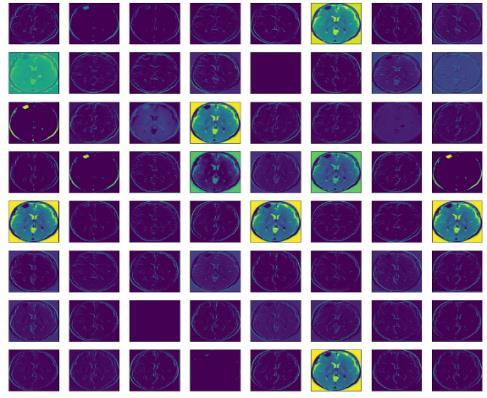
Figure4showstheConfusionmatrixofdumb
Modelandmodelandtheaccuracyscoreof
Maps Extracted from Final in
Modelis0.62857.
Figure 5 shows the Confusion matrix of our Evaluated theaccuracyscoreofthismodelis8.5
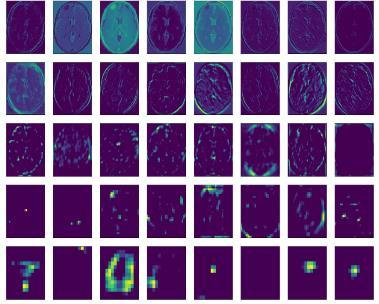
In figure 6 we can see that the result of applying the filters in the first convolutional layer is a lot of versions of the MRI image with different features highlighted. This is an interesting result and generally matches our expectation. We could updatetheexampletoplotthefeaturemapsfromtheoutputofotherspecificconvolutionallayers.
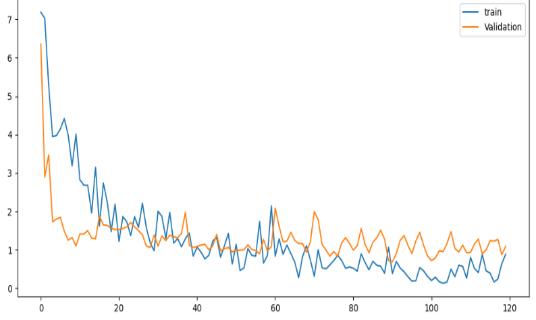
Chart 1 depicts the model's loss. By analysing the trend, we can ascertain if our model is overfitting or underfitting. If training loss decreases while validation loss increases, it indicates overfitting. Conversely, if both losses remain high, it suggestsunderfitting,signifyinginsufficientmodelcomplexity.
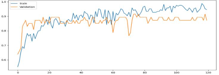
Chart2: Model Accuracy
RESULT VALUE
1.Accuracy 85.11%
2.Precision 89.29%
3 F1Score:
%
Table 2: Overall Performance
Chart2revealsthecomprehensiveaccuracyofourmodel,encapsulatingitsperformanceacrossallevaluatedmetrics.This pivotal visualization serves as a key indicator of the model's efficacy, providing essential insights into its overall effectivenessinpredictivetasks,crucialforinformeddecision-makingandassessmentofitsutility.
5.Conclusions
In conclusion, the development of a brain Tumors detection model using a CNN architecture with VGG16 for feature extractionandtransferlearningrepresentsasignificantstepforwardinleveragingdeeplearningformedicalimagingtasks with an accuracy of 85 %. Through the integration of pre-trained models and transfer learning techniques, we have achieved a robust and accurate system capable of automating the detection of brain Tumors from MRI images. The performanceevaluationdemonstratestheeffectivenessoftheproposedapproachinachievinghighaccuracyandreliability inTumorsdetection,therebypotentiallyaidingcliniciansinmakingtimelydiagnosesandtreatmentdecisions.
Looking ahead, there are several avenues for further enhancement and extension of the brain Tumors detection model. One potential direction is to explore multi-modal imaging data, incorporating additional modalities such as diffusionweighted imaging (DWI) or functional MRI (fMRI) to improve the model's sensitivity and specificity. Additionally, the integration of advanced deep learning architectures, such as attention mechanisms or recurrent neural networks, may offerimprovedfeaturerepresentationandtemporalcontextmodelling,especiallyforlongitudinalMRIstudies.
International Research Journal of Engineering and Technology (IRJET) e-ISSN:2395-0056
Volume: 11 Issue: 03 | Mar 2024 www.irjet.net p-ISSN:2395-0072
Weexpressourtrueappreciationtothetherapeuticteachandinquireaboutcentresthatgivenuswiththeimportantbrain MRIdatasets,withoutwhichthisinvestigatewouldnothavebeenconceivable.Weareobligatedtothepatientswhoagreed to share their restorative information for the progression of logical information and the advancement of superior symptomatictools.
8.References
[1] Gupta, K. K., Vijay, R., Pahadiya, P., Saxena, S., & Gupta, M. (2023). Novel Feature Selection Using Machine Learning AlgorithmforBreastCancerScreeningofThermographyImages. Wireless Personal Communications, 1-28.
[2] Pahadiya, P., Vijay, R., Gupta, K. K., Saxena, S., & Shahapurkar, T. (2023). Digital Image Based Segmentation and ClassificationofTongueCancerUsingCNN Wireless Personal Communications, 1-19.
[3] Gupta, K. K., Vijay, R., Pahadiya, P., & Saxena, S. (2022). Use of novel thermography features of extraction and differentartificialneuralnetworkalgorithmsinbreastcancerscreening Wireless Personal Communications, 1-30.
[4] Gupta, K. K., Rituvijay, Pahadiya, P., & Saxena, S. (2022). Detection of cancer in breast thermograms using mathematical threshold based segmentation and morphology technique. International Journal of System Assurance Engineering and Management, 1-8.
[5] Gupta, K. K., Vijay, R., & Pahadiya, P. (2022). Detection of abnormality in breast thermograms using Canny edge detection algorithm for thermography images. International Journal of Medical Engineering and Informatics, 14(1), 31-42.
[6] Saxena, S., Vijay, R., Pahadiya, P., & Gupta, K. K. (2023). Classification of ECG arrhythmia using significant waveletbasedinputfeatures International Journal of Medical Engineering and Informatics, 15(1), 23-32
[7] Gupta, K. K., Vijay, R., & Pahadiya, P. (2020). A review paper on feature selection techniques and artificial neural networks architectures used in thermography for early stage detection of breast cancer Soft Computing: Theories and Applications: Proceedings of SoCTA 2019, 455-465
[8] Pahadiya, P., Vijay, R., Gupta, K. K., Saxena, S., & Tandon, R. (2022). Contactless non-invasive method to identify abnormal tongue area using K-mean and problem identification in COVID-19 scenario International Journal of Medical Engineering and Informatics, 14(5), 379-390.
[9] Pahadiya, P., Vijay, D. R., kumar Gupta, K., Saxena, S., & Tandon, R. (2020). A Novel method to get proper tongue image acquisition and thresholding for getting area of interest. International Journal of Innovative Technology and Exploring Engineering (IJITEE), ISSN,2278-3075.
[10] C. G. Kamble and M. C. Rathod, "Brain tumor detection using deep learning", International Journal of Computer Sciences and Engineering,vol.7,no.3,pp.99-105,2019.
[11] S. Wang, S. Ji, X. Bai and B. Cheng, "Brain tumor segmentation and detection using deep learning-based techniques", Brain Sciences,vol.8,no.8,pp.149,2018.
[12] A. Mobiny and A. Gholampour, "A comprehensive review on brain tumor classification: a machine learning perspective", IEEE Access,vol.7,pp.140753-140771,2019.
[13] Y. Zeng and J. Yang, "Brain tumor detection and segmentation using convolutional neural networks", Journal of Medical Imaging and Health Informatics,vol.9,no.8,pp.1701-1708,2019.
[14] S.MahajanandS.Jain,"Asurveyonbraintumordetectiontechniquesusingmachinelearning", Artificial Intelligence Review,vol.53,no.7,pp.4209-4236,2020.
[15] K. Chang, N. Balachandar, C. Lam, D. Yi, J. Brown, A. Beers, et al., "Distributed deep learning networks among institutionsformedicalimaging", Journal of the American Medical Informatics Association,vol.25,no.8,pp.945-954, 2018.
International Research Journal of Engineering and Technology (IRJET) e-ISSN:2395-0056
Volume: 11 Issue: 03 | Mar 2024 www.irjet.net p-ISSN:2395-0072
[16] S. Kumar, S. Singh and M. Kaur, "Detection and classification of brain tumor using machine learning algorithms", Journal of medical systems,vol.43,no.8,pp.228,2019.
[17] S. Sand and D. V. Babu, "Retinal Glaucoma Detection from Digi-tal Fundus Images using Deep Learning Approach", 2023 7th In-ternational Conference on Computing Methodologies and Commu-nication (ICCMC),pp.68-72, 2023.
[18] L.Bi,J.Kim,A.Kumar,D.FengandS.Chung,"AutomaticsegmentationanddetectionofbraintumorsinMRIimages usingdeeplearningalgorithms", Sensors,vol.20,no.4,pp.1114,2020.
[19] Y.GuoandY.Zhang,"ConvolutionalneuralnetworkmodelsforbraintumordetectionusingMRI", Journal of Medical Imaging and Health Informatics,vol.10,no.7,pp.1596-1603,2020.
[20] Shenoy, M. Ashwin, Pranam R. Betrabet and NS Krishnaraj Rao, "Helmet Detection using Machine Learning Approach", 2022 3rd International Conference on Smart Electronics and Communication (ICOSEC), pp. 1383-1388, 2022.
[21] L. Gao, J. Liu, L. Guo and M. Xu, "A survey on deep learning techniques for brain tumor detection in MRI images", Medical Imaging Technology,vol.39,no.1,pp.18-23,2021.
[22] Mahmoud Khaled Abd-Ellah, Ali Ismail Awad, Ashraf AM Khalaf and Hesham FA Hamed, "Two-phase multi-model automatic brain Tumor diagnosis system from magnetic resonance images using convolutional neural networks", EURASIP Journal on Image and Video Processing 2018,no.1,pp.1-10,2018.
[23] Md Shahariar Alam, Md Mahbubur Rahman, Mohammad Amazad Hossain, Md Khairul Islam, Kazi Mowdud Ahmed, Khandaker Takdir Ahmed, et al., "Automatic human brain tumor detection in MRI image using template-based K means and improved fuzzy C means clustering algorithm", Big Data and Cognitive Computing, vol. 3, no. 2, pp. 27, 2019.
[24] Amin Javaria, Muhammad Sharif, Nadia Gul, Mudassar Raza, Muhammad Almas Anjum, Muhammad Wasif Nisar, et al.,"Braintumordetectionbyusingstackedautoencodersindeeplearning", Journal of medical systems,vol.44,no.2, pp.1-12,2020.
[25] Arbane Mohamed, Rachid Benlamri, Youcef Brik and Mohamed Djerioui, "T ransfer learning for automatic brain tumorclassificationusingMRIimages", 2020 2nd International Workshop on Human-Centric Smart Environments for Health and Well-being (IHSH),pp.210-214,2021.
[26] Garg Ginni and Ritu Garg, "Brain tumor detection and classification based on hybrid ensemble classifier", arXiv preprint,2021.
[27] Rehman Amjad, Muhammad Attique Khan, Tanzila Saba, Zahid Mehmood, Usman Tariq and Noor Ayesha, "Microscopic brain tumordetection andclassificationusing3DCNN and featureselection architecture", Microscopy Research and Technique,vol.84,no.1,pp.133-149,2021.
[28] Banerjee Subhashis, Sushmita Mitra, Francesco Masulli and Stefano Rovetta, "Deep radiomics for brain tumor detectionandclassificationfrommulti-sequenceMRI", arXiv preprint,2019.
[29] Sharif Muhammad, Javaria Amin, Mudassar Raza, Muhammad Almas Anjum, Humaira Afzal and Shafqat Ali Shad, "Brain tumor detection based on extreme learning", Neural Computing and Applications, vol. 32, no. 20, pp. 1597515987,2020.
[30] DebDaizyandSudiptaRoy,"BraintumordetectionbasedonhybriddeepneuralnetworkinMRIbyadaptivesquirrel searchoptimization", Multimedia tools and applications,vol.80.2,pp.2621-2645,2021.
[31] Diaz-Pernas, Francisco Javier, Mario Martinez-Zarzuela, Miriam Anton-Rodriguez and David Gonzalez-Ortega, "A deep learning approach for brain tumor classification and segmentation using a multiscale convolutional neural network"inHealthcare,MultidisciplinaryDigitalPublishingInstitute,vol.9,no.2,pp.153,2021.
International Research Journal of Engineering and Technology (IRJET) e-ISSN:2395-0056
Volume: 11 Issue: 03 | Mar 2024 www.irjet.net p-ISSN:2395-0072
[32] JenaBiswajit,GopalKrishnaNayakandSanjaySaxena,"Anempiricalstudyofdifferentmachinelearningtechniques for brain tumor classification and subsequent segmentation using hybrid texture feature", Machine Vision and Applications,vol.33.1,pp.1-16,2022.
[33] G. Ramkumar, R. Thandaiah Prabu, Ngangbam Phalguni Singh and U. Maheswaran, "Experimental analysis of brain tumordetectionsystemusingMachinelearningapproach", Materials Today: Proceedings,2021.
[34] Wozniak Marcin, Jakub Silka and Michal Wieczorek, "Deep neural network correlation learning mechanism for CT braintumordetection", Neural Computing and Applications,pp.1-16,2021.
[35] Abd El Kader, Isselmou, Guizhi Xu, Zhang Shuai, Sani Saminu, Imran Javaid, et al., "Differential deep convolutional neuralnetworkmodelforbraintumorclassification", Brain Sciences,vol.11,no.3,pp.352,2021.
[36] Muhammad Arif, F. Ajesh, Shermin Shamsudheen, Oana Geman, Diana Izdrui and Vicoveanu Dragos, "Brain Tumor Detection and Classification by MRI Using Biologically Inspired Orthogonal Wavelet Transform and Deep Learning Techniques", Journal of Healthcare Engineering,2022.
[37] T.Vijayakumar,"Neural network analysisfortumorinvestigationandcancerprediction", Journal of Electronics, vol. 1,no.02,pp.89-98,2019.
[38] P. Karuppusamy, "Hybrid Manta Ray Foraging Optimization for Novel Brain Tumor Detection", Journal of Soft Computing Paradigm (JSCP),vol.2,no.03,pp.175-185,2020.
[39] A.Hamada, Br35h: Brain Tumor Detection 2020 version 5,2020.