
International Research Journal of Engineering and Technology (IRJET) e-ISSN:2395-0056
Volume: 11 Issue: 03 | Mar 2024 www.irjet.net p-ISSN:2395-0072
International Research Journal of Engineering and Technology (IRJET) e-ISSN:2395-0056
Volume: 11 Issue: 03 | Mar 2024 www.irjet.net p-ISSN:2395-0072
K. Himavanth1, B. Sharath2, A. Balaji3 , M. Sujan4,T.Himaja Sumasri5
12345Student, Dept. Of Computer Science Engineering, GITAM Deemed University, Visakhapatnam, AP, INDIA
Abstract - Lung cancer is a common and life-threatening disease with a high mortality rate all over the world. Detecting this early plays an important role in improving patient outcomes and reducing mortality. In recent years, deep learning models have shown significant potential in medical image analysis, especially in the field of cancer detection. This paper presents a new approach for lung cancer detection using the VGG-16 algorithm, a convolutionalneuralnetwork(CNN)architectureknownfor its performance in image classification tasks. We provide a comprehensive methodology that includes lung image preprocessing, VGG-16 model training on an annotated lung scan and model performance evaluation using various metrics such as accuracy, sensitivity and specificity. In addition, we investigate the effect of different hyperparameters and data augmentation techniques on model performance. Experimental results show the effectivenessoftheVGG-16algorithmforaccuratedetection of lung cancer based on medical image data. The proposed approach holds significant promise for improving the early diagnosis and treatment planning of patients with lung cancer, ultimately contributing to improved clinical outcomesandqualityoflife
Keywords: Lung cancer (Bronchogenic Carcinoma) detection,VGG-16,Image classification, Convolutional neural networks
1.INTRODUCTION
Bronchogenic Carcinoma is a vital global haleness problem, requiring urgent attention due to its impact on patient prognosis and survival rates. Timely detection is essential, and computed tomography(CT) scans have emergedasimportantdiagnostictoolsforlungcancerand its stages. To address this, considerable research has focusedonlungcancerdetectionsystemsfromCTimages. This project report presents our attempt to use a deep learning model, specifically the VGG-16 algorithm, to predict lung cancer using CT scan images. Bronchogenic Carcinoma is the major cause of mortality related to cancer worldwide, underscoring the need for accurate diagnostic tools, Although CT scanning provides detailed information,manualinterpretationistime-consumingand prone to errors. Our project uses Deep Learning to automateandimproveBronchogenicCarcinomadetection. ThecoreofthesolutionistheVGG-16algorithm,which is known for its image segmentation capabilities.
Differentiationbetweenbenignandmalignantlunglesions on CT scan images can alter the clinical picture and diagnosis. Using VGG-16, we aim to improve the correctness and efficacy of Bronchogenic Carcinoma detection,emerginginbestfalloutforpatients.Thisreport shows how to implement our system step by step. It focuses on classifying the CT scan images using the VGG16algorithm.
Lung cancer is one of the deadly and most common cancers in the world. Early detection is critical to improving patient outcomes and survival. Traditionally, thedetectionoflungcancerwasbasedonmethodssuchas chest X-ray, computed tomography (CT), and tissue biopsy. However, these methods often require extensive manual inspection by radiologists and may not always detectcanceratanearlystage. Inrecentyears,interest in using deep learning techniques for early detection and diagnosisoflungcancerhasincreased..Deeplearningisa subset of artificial intelligence (AI) that involves training neural networks to recognize patterns and make predictions from large amounts of data. In medical imaging such as CT scanning and X-rays, deep learning algorithms can help radiologists identify suspicious areas that may indicate the presence of cancer. One of the main advantagesofdeeplearningforlungcancerdetectionisits ability to analyze a huge number of medical images quickly and accurately. Deep learning models can be trained on datasets containing thousands of annotated images, allowing them to learn complex patterns associated with cancer lesions. These models can then be used to analyze new images and make recommendations to radiologists for further evaluation. Several deep learning techniques have been developed for lung cancer detection,includingConvolutionalNeuralNetworks(CNN) and Recurrent Neural Networks (RNN). In particular, CNNs have shown promising results in detecting lung nodules and other abnormalities in medical images. Designed to automatically learn hierarchical representationsofimagefeatures,thesenetworksarewell suitedfortaskssuchaslungcancerdetection.
Recent advancements in deep learning and machine learninghavepropelledsignificantstridesinthedetection
International Research Journal of Engineering and Technology (IRJET) e-ISSN:2395-0056
of lung cancer, a critical area of medical research aiming for early diagnosis and improved patient outcomes. Noteworthystudies,suchasthosebyPuvvadaHarshaSiva Prasadetal.[1],Dr.K.Ramanjaneyuluetal.[2],Radhikaet al. [3], and Günaydin et al. [4], shed light on various methodologiesandtechniquesemployedinthisdomain.
In their study, Radhika, P. R., Nair, R. A., & Veena, G. [3], conduct a comparative analysis of lung cancer detection usingmachinelearningalgorithms.Presentedatthe2019 IEEEInternationalConferenceonElectrical,Computerand Communication Technologies (ICECCT), their work explores different machine learning techniques for lung cancerdetection.Thisresearchprovidesvaluableinsights into the comparative performance of various algorithms, contributing to the ongoing efforts to identify optimal approaches for accurate and efficient lung cancer detection.
Similarly,Günaydin,Ö.,Günay,M.,&Şengel,Ö.[4],present acomparativestudyoflungcancerdetectionalgorithmsat the 2019 Scientific Meeting on Electrical-Electronics & Biomedical Engineering and Computer Science (EBBT). Their research delves into the comparison of different algorithms, likely including both traditional machine learning and deep learning approaches, to evaluate their effectiveness in lung cancer detection. Such comparative analyses play a crucial role in benchmarking and advancing the state-of-the-art in lung cancer detection, aiding researchers and clinicians in identifying the most promisingmethodologiesforclinicalapplication.
Collectively,thesestudiesunderscoretheinterdisciplinary nature of lung cancer detection research, drawing upon diverse methodologies and techniques from machine learning, deep learning, and medical imaging. By synthesizing insights from these studies, researchers are better equipped to develop robust and accurate algorithms for early detection and diagnosis of lung cancer,ultimatelyenhancingpatientcareandoutcomesin thefightagainstthisdeadlydisease.
3.1
Lungcancerremainsaformidableworldfitnesschallenge, and timely diagnosis is pivotal for boosting patient prognosisandsurvivorship.ComputedTomographyscans come into view as a critical diagnostic device for identifying
Bronchogenic Carcinoma and its stages. Consequently, notable research has been done to develop a system that can predict Bronchogenic Carcinoma from Computed Tomographyscans. Weare implementinga Deeplearning model to predict Bronchogenic Carcinoma using CT Scans usingVGG16algorithm.
Volume: 11 Issue: 03 | Mar 2024 www.irjet.net p-ISSN:2395-0072 © 2024, IRJET | Impact Factor value: 8.226 |
Explorationinlungcancerdiscoveryhasseenremarkable progress, particularly in the bracket of lung lesions as benign, nasty,ornormalusingmachine literacyanddeep literacy ways. colorful studies have employed convolutional neural networks( CNNs) similar as CheXNet, a modified DenseNet armature, for automatic discovery and bracket of thoracic abnormalities from casket radiographs( Ardila et al., 2019). Li etal.( 2020) proposed a mongrel CNN and intermittent neural network( RNN) frame, achieving high delicacy and area under the wind( AUC) for differencing between benign and nasty nodes on 3D CT reviews. Transfer literacy with pre-trained CNN models like VGG and ResNet has alsobeenexploredforlungbumpbrackets,demonstrating notableadvancementsinbracketperformance(Zhaoetal., 2021). Likewise, Wang etal.( 2019) introduced a multitask deep literacy approach for contemporaneous discovery and bracket of lung nodes on CT reviews, showcasing superior performance in terms of delicacy andcomputational effectivenesscomparedtosingle-task models. Overall, these studies emphasize the eventuality of deep literacy ways in perfecting individual delicacy andcase issuesinlungcancerwebbingand opinion.
WeusedVGG-16todetectthelungcancer fortheIQ-OTH dataset,workingofmodelisdividedintotwophases
3.3.1
Image preprocessing is a convenient technique to refine thestandards ofscansand upgradethem for examination and foradditionalconversion.Geometrictransformations ofimageslikerotation,scaling,translationaredoneinthis phase.
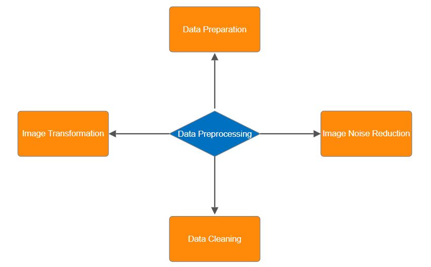
Fig -1: Data Preprocessing Techniques
International Research Journal of Engineering and Technology (IRJET) e-ISSN:2395-0056
Volume: 11 Issue: 03 | Mar 2024 www.irjet.net p-ISSN:2395-0072
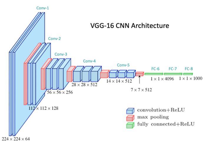
The above diagram represents VGG-16 architecture, vgg16isaCNN(ConvolutionalNeuralNetwork)with16layers which is why it is called VGG-16. It is generally used for largescaleimageRecognition.
Thenetwork'sstraightforwarddesign,characterizedbyits deep stack of convolutional layers with small filter sizes, facilitates powerful feature extraction, allowing it to capture intricate patterns in images effectively. Despite being surpassed by more complex architectures in recent years, VGG-16 remains a benchmark model and a cornerstone in the development of deep learning techniquesforcomputervisiontasks.
The VGG-16 has a total of layers out of which 13 are convolutional layers and remaining 3 layers are fully connected layers. The 13 convolutional layers are again divided into 5 blocks out of which first 2 blocks has two 3x3convolutional layersandremainingthreeblockshave three 3x3 convolutional layers, each convolution is followed Relu activation function to introduce non linearity and also after each convolution block max pooling is introduced to reduce the dimensions of the image. After the convolutional blocks it is introduced to fullyconnectedlayersalsocalledasthelinearlayerwhich isusedtoflattentheimagethatisitisusedtoclassifythe given image into a label.At last an softmax activation functionisusedtoproducetheclassprobabilities.
The objectives of the research paper on lung cancer detection using the VGG-16 architecture are multifaceted. Firstly, it aims to address the urgent need for early detection of lung cancer, a pressing healthcare challenge associated with high mortality rates. Leveraging deep learning techniques, particularly the VGG-16 algorithm, theprojectseekstodevelopanaccurateanduser-friendly systemfordetectinglungcancerfromCTscanimages.
By focusing on improving model interpretability, enhancing image quality, and handling class imbalances through various techniques like noise cancellation, contrastenhancement,andoversampling,theprojectaims toachieveunbiasedandreliablepredictions.Theultimate goal is to create a VGG-16 system capable of accurately detecting benign and malignant lung lesions, thereby improving patient prognosis and treatment outcomes. Additionally, the project aims to develop user-friendly web applications with robust security measures for data encryption and user authentication, ensuring compliance with healthcare regulations while optimizing scalability andtaskefficiency.
Overall, the objectives encompass advancing lung cancer detectionthroughinnovativedeeplearningmethodologies and practical deployment strategies to benefit both patientsandhealthcareprofessionalsalike.
International Research Journal of Engineering and Technology (IRJET) e-ISSN:2395-0056
Volume: 11 Issue: 03 | Mar 2024 www.irjet.net p-ISSN:2395-0072
4.1 Flowchart
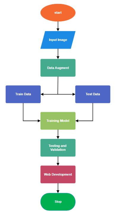
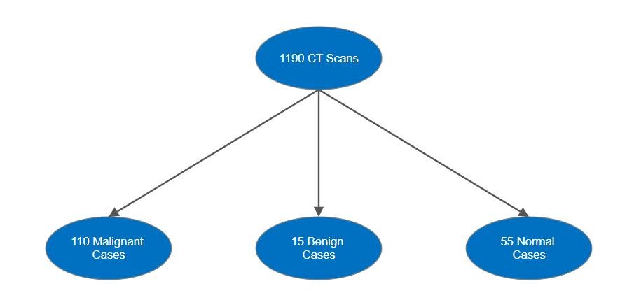
The dataset sourced from kaggle consists of CT scans collected over a 3-month period in the fall of 2019. It includesimagesofpatientsdiagnosedwithvariousstages of lung cancer, as well as images of healthy individuals. The slides were annotated by expert oncologists/radiologists at the IQ-OTH / NCCD. The total size of the dataset is 209 MB and the dataset includes 1190 CT scan slices from 110 cases classified as normal, benign, or malignant classes. In particular, 40 cases were malignant,15werebenign,and55werenormal.Thescans were originally scanned using DICOM format using Siemens’SOMATOMscanner.Thestandardprotocolis120 kV, slice thickness is 1 mm, and the window width and center setting are specified. Each scan includes 80-200 slicesrepresentingvariousanglesofthechest.
We have used some data preprocessing techniques to improvetheoverallqualityoftheimagebeforeusingthem for model training so that model can be more accurate whiletesting.
The dataset is split into training, testing and validation sets.. This distribution is crucial for training a robust model that can accurately identify cancer as Malignant, Benign or Normal from lung images. The initial distribution combines the test and validation sets, ensuring an appropriate distribution of samples into different sets. This layered approach helps preserve the representationofbothmalignantandbenigncasesineach subset.Duringrotation,theVGG-16architectureisusedas a powerful feature collector that exploits its ability to capture complex patterns and features of medical images. Themodelisinitializedwithweightspre-trainedonlargescaleimagedatasetssuchasImageNet,allowingittolearn meaningful representations for lung cancer detection. Fine-tuning the VGG-16 model involves updating its parameters using back propagation and optimization techniques. such as stochastic gradient descent (SGD) or Adam. This process facilitates the adaptation of pretrained features to the specific characteristics of lung cancer image data. In addition, data augmentation strategies such as rotation, scaling, and translation are applied to the training images to increase the diversity of the dataset and improve the model's ability to generalize to unseen data. During training, hyperparameters such as learning. rate, set size and regularization strength are carefullyadjustedtooptimizethemodelperformance.The traininglossisiterativelymonitoredandminimized,while the validation set is used to assess the generalizability of the model and avoid overfitting. Performance metrics including accuracy, sensitivity, specificity, and the area under the receiver operating curve (AUC-ROC) are calculated during training to evaluate the performance of the model in lung cancer lesions. upon detection. This thorough evaluation ensures that the trained model can effectively discriminate between malignant and benign cases.
The trained model is evaluated against an independent test set to provide unbiased estimates of its performance. This final evaluation provides important insights into the
International Research Journal of Engineering and Technology (IRJET) e-ISSN:2395-0056
Volume: 11 Issue: 03 | Mar 2024 www.irjet.net p-ISSN:2395-0072
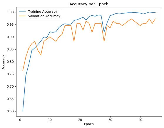
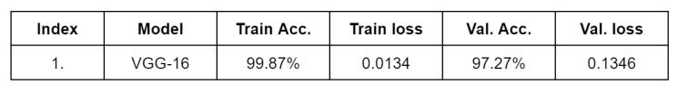
real-worldapplicabilityofthemodelanditsabilitytohelp clinicians accurately diagnose lung cancer based on medicalimagingdata.Themodel testingstepisnecessary to evaluate the performance of the lung cancer detection modelgeneratedbytheVGG-16algorithm.Afterthemodel is trained, accuracy and loss metrics are visualized over successivetime periodsto monitor the learningdynamics of the model and identify potential over- or under-fitting problems. Evaluation metrics such as accuracy and loss are then calculated for both the training and validation sets to measure the generalization ability of the model. Evaluation results, including training, validation, and accuracy on independent test sets, as well as relevant training parameters such as image dimensions, learning rate, and set size, are compiled to provide a comprehensive overview of model performance. In addition, all the time spent in the training and evaluation processes is recorded to evaluate the effectiveness of the calculation. In addition, a confusion matrix is created basedonthetestpredictionsofthemodel,whichprovides insightintothedistributionoftruepositive,truenegative, false positive and false negative predictions in different categories of lung images. This analysis helps identify potential errors or deviations in model predictions and provides valuable feedback for model refinement and improvement. Overall, the model testing phase ensures the readiness of the VGG-16-based lung cancer detection model foruseinclinical settings, whichwill help improve diagnosticaccuracyandpatientcare.
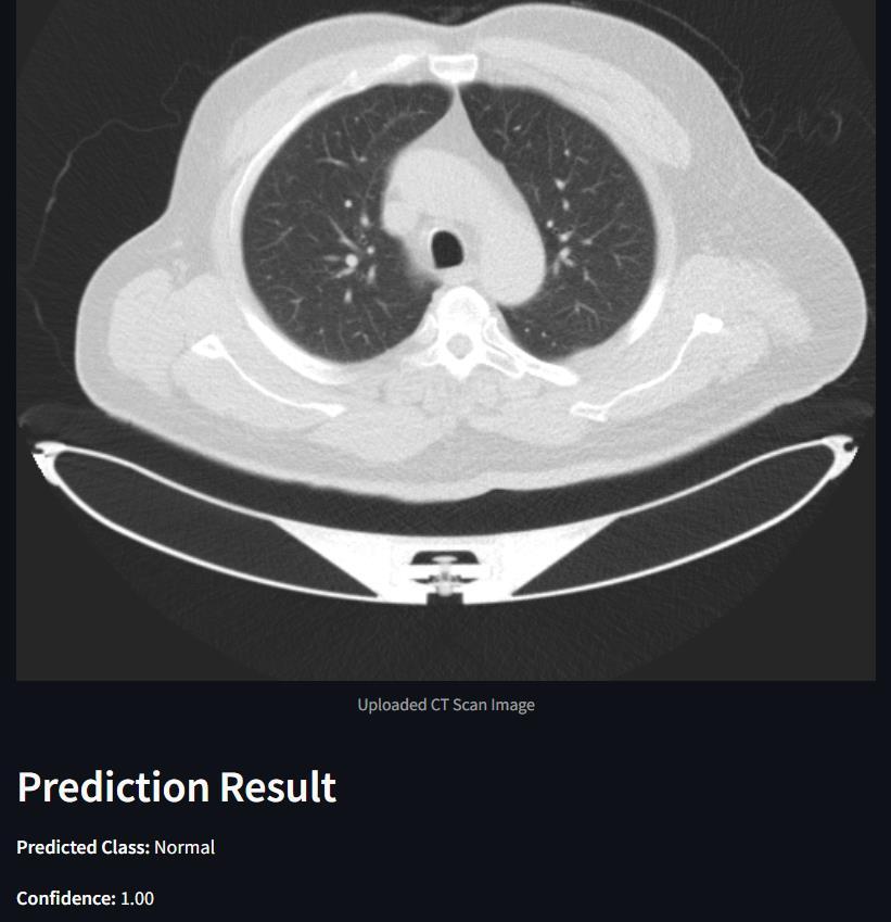
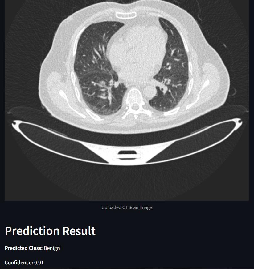
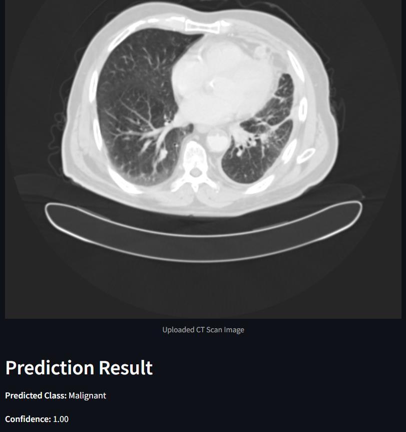
The final accuracy and performance metrics of the model are substantiated visually through a series of graphs and charts, showcasing accuracy and loss metrics across epochsforbothtrainingandtestingdatasets.Additionally, confusion matrices are created to represent classification performanceonbothtrainingandtestingdata,providinga clear and concise understanding of the model's behavior. Complementing these metrics are tumor images displaying predicted classes alongside model confidence scores, offering valuable insights into the model's effectiveness and emotional impact. This comprehensive presentationnotonlyevaluatesefficacybutalsofacilitates furtherrefinementandoptimization,ultimatelyenhancing overallperformanceforend-users.
InourpursuitoflungcancerdetectionusingCTscans,the VGG16algorithmstandsasapivotalcomponentwithinthe broader landscape of machine learning and computer vision. Anchored upon the TensorFlow framework, our research harnesses the power of VGG16, a preeminent convolutional neural network architecture renowned for its effectiveness in image classification tasks. TensorFlow, serving as the bedrock of our methodology, offers a
flexibleenvironmentconducivetotheimplementationand fine-tuning of deep learning models such as VGG16. This synergy enables the seamless integration of advanced neural network architectures into our workflow, facilitatingtheanalysisofintricatepatternswithinCTscan images indicative of lung cancer. Furthermore, OpenCV assumes a central role in our research paradigm, facilitating critical preprocessing tasks and enhancing the interpretability of CT scan data. Through OpenCV, we adeptly extract frames from CT scan images, enabling the creation of comprehensive datasets essential for training the VGG16 model. Moreover, OpenCV's rich suite of functionalities enables seamless color space conversion and feature manipulation, augmenting the quality and utility of input data for subsequent analysis. In essence, the integration of VGG16 within the TensorFlow framework, complemented by the versatile capabilities of OpenCV, forms the cornerstone of our approach to lung cancer detection. Through this amalgamation of cuttingedge algorithms and tools, our research endeavors culminate in a robust framework capable of discerning subtle abnormalities indicative of lung cancer within CT scan images, thereby contributing to advancements in earlydiagnosisandtreatmentofthisdevastatingdisease.
The VGG- 16 model's performance on the IQ- OTH/ NCCD lung cancer dataset is exhaustively presented through a collection of visual aids. These include graphs and charts depicting accuracy and loss metrics over epochs for both trainingandtestingdatasets.Likewise,confusionmatrices are generated to illustrate the classification performance of the model on both training and testing data. This detailed representation offers precious perceptivity into the behavior and efficacy of the VGG- 16 model on the specifieddataset.
International Research Journal of Engineering and Technology (IRJET) e-ISSN:2395-0056
Volume: 11 Issue: 03 | Mar 2024 www.irjet.net p-ISSN:2395-0072
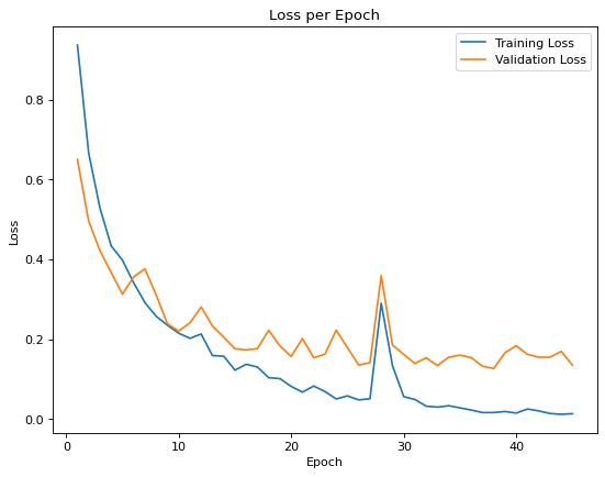
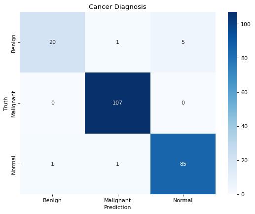
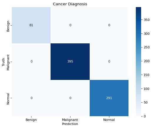
Table-I :Accuracy andLossComparisonofModel
Output:
International Research Journal of Engineering and Technology (IRJET) e-ISSN:2395-0056
Volume: 11 Issue: 03 | Mar 2024 www.irjet.net p-ISSN:2395-0072
In conclusion, the utilization of VGG16 algorithm for lung cancer detection based on CT scan images offers a promising avenue for early diagnosis and intervention. Through this research, it has been demonstrated that the deep learning model can effectively analyze intricate patterns within CT scans, distinguishing between malignant and benign nodules with high accuracy and efficiency. The integration of VGG16 into medical imaging holds tremendous potential for enhancing the diagnostic process, enabling clinicians to make more informed decisionsandimprovepatientoutcomes.
Furthermore, the findings of this study underscore the importance of leveraging cutting-edge technologies in the realm of healthcare. By harnessing the power of artificial intelligence and machine learning, we can augment the capabilitiesofmedicalprofessionals,streamlinediagnostic procedures, and ultimately, save lives. However, it's crucial to acknowledge that the development and implementation of such algorithms necessitate rigorous validation, continuous refinement, and adherence to ethical guidelines to ensure their reliability and safety in clinicalpractice.
Looking ahead, further research and collaboration are needed to enhance the robustness and generalizability of VGG16-basedlungcancerdetectionsystems.Thisincludes exploring larger and more diverse datasets, refining the algorithm's architecture, and investigating potential integration with other modalities for comprehensive
patient assessment. Ultimately, by advancing the field of computer-aided diagnosis in oncology, we can strive towards earlier detection, personalized treatment strategies, and improved prognosis for individuals affectedbylungcancer.Thefuturescopewouldinclude.
EnhancedDiagnosticPrecision:
● Continued refinement and validation of the VGG16 algorithm are expected to result in heightened sensitivity and specificity in detecting lungcancernodulesonCTscans.
● This advancement could significantly improve diagnostic precision, leading to earlier detection of the disease and subsequent treatment initiation.
FacilitationofPersonalizedMedicine:
● The deep learning capabilities of the VGG16 algorithm enable precise characterization of lung cancersubtypesandtumorcharacteristics.
● This could pave the way for personalized treatment approaches tailored to individual patients, resulting in improved therapeutic outcomesandreducedadverseeffects.
IntegrationintoClinicalPractice:
● Integration of the VGG16 algorithm into clinical workflows has the potential to streamline diagnosticprocessesforhealthcareprofessionals.
● By automating nodule detection and characterization tasks, clinicians can allocate more time to patient interaction and treatment planning,ultimately enhancingthe qualityofcare provided.
ExpansionofScreeningPrograms:
● ThesuccessfulapplicationoftheVGG16algorithm in lung cancer detection could support the expansion of population-based screening programs.
● Withimprovedaccuracyinidentifyinghigh-risk individuals,screeningeffortscouldbeoptimized totargetthosemostlikelytobenefitfromearly detection,potentiallyreducingdisease-related mortalityrates.
CatalystforTechnologicalAdvancements:
● The development and validation of the VGG16 algorithm represent a milestone in the
International Research Journal of Engineering and Technology (IRJET) e-ISSN:2395-0056
Volume: 11 Issue: 03 | Mar 2024 www.irjet.net p-ISSN:2395-0072
intersection of artificial intelligence and medical imaging.
● This success is likely to inspire further research and innovation in the field, driving the development of even more sophisticated algorithms capable of detecting various diseases fromimagingdata.
EthicalConsiderationsandChallenges:
● As the use of artificial intelligence in healthcare continues to expand, it is essential to address ethical considerations such as data privacy, algorithmbias,andtransparency.
● Additionally, challenges related to algorithm interpretability, regulatory compliance, and clinician adoption may need to be navigated to ensure the successful integration of the VGG16 algorithmintoclinicalpractice.
FutureDirections:
● Future research directions may include the exploration of multi-modal imaging approaches, such as combining CT with other imaging modalities or genomic data, to further enhance diagnosticaccuracyandpredictivecapabilities.
● Longitudinal studies assessing the impact of VGG16-based lung cancer detection on patient outcomes and healthcare economics could provide valuable insights into its clinical utility andcost-effectiveness.
[1]P.H.S.Prasad,N.M.V.S.Daswanth,C.V.S.P.Kumar,N. Yeeramally,V.M.MohanandT. Satish,"DetectionofLung Cancer using VGG-16," 2023 7th International Conference on Computing Methodologies and Communication (ICCMC), Erode, India, 2023, pp. 860-865, doi: 10.1109/ICCMC56507.2023.10084192. keywords: {Deep learning;Geometry;Visualization;Lung cancer;Training data;Data models;Classification algorithms;Visual Geometry Group16 (VGG16);Convolutional Neural Network;Deeplearning;Machinelearning},
[2] K. Ramanjaneyulu, K. H. Kumar, K. Snehith, G. JyothirmaiandK.V.Krishna,"DetectionandClassification of Lung Cancer Using VGG-16," 2022 International Conference on Electronic Systems and Intelligent Computing (ICESIC), Chennai, India, 2022, pp. 69-72, doi: 10.1109 / ICESIC53714. 2022.9783556. keywords: {Visualization;Computedtomography; Malignanttumors; Standards organizations; Lung cancer;
Lung;Organizations; lung cancer;Image Segmentation;CT Scan;CNN}
[3]Radhika,P.R.,Nair,R.A.,&Veena,G.(2019,February). A comparative study of lung cancer detection using machine learning algorithms. In 2019 IEEE International Conference on Electrical, Computer and Communication Technologies(ICECCT)(pp.1-4).IEEE.
[4] Günaydin, Ö., Günay, M., & Şengel, Ö. (2019, April). Comparison of lung cancer detection algorithms. In 2019 Scientific Meeting on ElectricalElectronics & Biomedical EngineeringandComputerScience(EBBT)(pp.1-4). IEEE