
International Research Journal of Engineering and Technology (IRJET) e-ISSN: 2395-0056
Volume: 11 Issue: 03 | Mar 2024 www.irjet.net p-ISSN: 2395-0072
International Research Journal of Engineering and Technology (IRJET) e-ISSN: 2395-0056
Volume: 11 Issue: 03 | Mar 2024 www.irjet.net p-ISSN: 2395-0072
Shravani Mankar1, Rucha Koshatwar2, Vaishnavi Rathod3, Smit Dafe4 ,Prof. Aditya Sable5
1Student, Dept. of CSE Engineering, PRMITR college Maharashtra, India
2Student, Dept. of CSE Engineering, PRMITR college Maharashtra, India
3Student, Dept. of CSE Engineering, PRMITR college Maharashtra, India
4Student, Dept. of CSE Engineering, PRMITR college Maharashtra, India
5Professor, Dept. of CSE Engineering, PRMIT&R college, Maharashtra, India ***
Abstract - Skin cancer is considered as one of the most dangerous types ofcancers andthereis adrasticincreaseinthe rate of deaths due to lack ofknowledge on the symptoms and their prevention The demand of early opinion of the skin cancer have been increased because of the fast growth rate of Melanoma skin cancer, its high treatment expenses, anddeath rate. This cancer cells are detected manually and it takes time to heal in utmost of the cases. In the recent years, Convolutional Neural Network (CNN) have madeasignificant advancementindetectingskincancertypes from dermoscopic images. The main objective of this project is to develop a CNN basedmodel to automatically classify skin cancer types into melanoma and non-melanoma with high accuracy.
Key Words: Machine learning, CNN, Melanoma Feature Extraction, Classification, Skin Cancer Detection
1.
Skincancerisconsideredasoneofthemostdangeroustypes ofcancersandthere'sadrasticincreaseintherateofdeaths due to lack of knowledge on the symptoms and their forestallment. therefore, early discovery at unseasonable stage is necessary so that one can help the spreading of cancer.Skincancerisfurtherdividedintocolourfultypesout of which the most dangerous bones are Melanoma, rudimentarycellmelanomaandScaledcellmelanomaskin cancer is one of the deadliest cancersand one of the most commoncancersintheworldsincenumerouscountriesdon’t officially record carcinoma cases. This cancer cells are detected manually and it takes time to heal in ultimate of cases.
Traditionally, dermatologists check the following characteristicsoftheskinlesionasymmetry,borders,colors, periphery, and elevation. However, has fuzzy borders, has further than four colors, If the lesion of the case is asymmetric.
This design attempts to automate the identification of carcinoma skin cancer grounded on raw images of skin lesionsinordertoproduceabrisklyandlessprecioussystem ofdetectingthiscomplaintwithoutleavingascar.likewise, thiswouldenabledermatologiststoseefurthercaseseach day,workless,andconcentrateonthemostcriticalcases
Shetu Rani Guhaet.al.[1] proposed a machine literacy groundedfashionusingconvolutionalneuralnetwork(CNN) for classifying seven types of skin conditions. Transfer literacy, along with CNN, has been used to ameliorate the bracket delicacy on the International Skin Imaging Collaboration2018(ISIC)dataset.Theprimaryidealofthis study is to develop a machine literacy- grounded bracket modelforrelatinganddistinguishingbetweensevendifferent typesofskinconditions.
Rashmi Patilet.al [2] proposed exploration paper in this paper,theprimaryidealofthisstudyistoexploretheuseof machine literacy ways for the discovery and potentially staging of carcinoma cancer. Melanoma is a type of skin cancer, and its early discovery and accurate staging are critical for treatment opinions and patient issues. The methodology likely involves the operation of machine learningalgorithmstodissectcarcinoma-relateddata.This data may include clinical information, case records, and conceivablydermatologicalimagesofskinlesions.
Titus J. Brinkeret.al. [3] reviewed that state- of- the- art classifiers grounded on CNNs have demonstrated the capabilitytoclassifyskincancerimagesatapositionsimilar todermatologists.Thishighlightstheeventualityofmachine literacyandCNNsinabettingmedicaljudgments,particularly intheenvironmentofdermatology.Thereferencementions the installation of apps on mobile bias for skin cancer opinion.Thissuggeststheeventualityformobileoperations to bring life- saving and fast judgments to individualities outsideoftraditionalhealthcaresettings,makinghealthcare moreaccessible.
Fabio Santoset.al. [4] proposed exploration paper in this paper,focusesonthecurrentstateofautomatedskinlesion opinion,whilealsofurnishingacomprehensiveviewintothe challenges and openings in dermatology care. The paper discusses the rearmost developments in automated skin lesionopinion,includingadvancementsinmachineliteracy, deepliteracy,andcomputervisionways.Itmaypunctuate thecapabilitiesandlimitationsofcurrentautomatedsystems indiagnosingskinlesions.
International Research Journal of Engineering and Technology (IRJET) e-ISSN: 2395-0056
Volume: 11 Issue: 03 | Mar 2024 www.irjet.net p-ISSN: 2395-0072
Naeemetal.[5]presentedadeepliteracywayforcarcinoma opinion using CNN and give a methodical review for the challenges on the base of parallels and differences. The methodologylikelyinvolvesthedevelopmentandtrainingof CNNbasedmodelsforcarcinomaopinion.Theauthorsmay haveuseddermatologicalimagedatasetsandapplieddeep literacy ways to classify skin lesions as carcinoma ornonmelanoma.
Mahbodetal.[6]proposedanalgorithmthatensemblesdeep featuresfrommultiplepre-trainedandfine-tunedDNNsand fused the attained vaticination values of different models. Ensemble styles can include ways similar as bagging, boosting,ormounding,andeachhasitsownadvantagesin perfecting model performance and the significance of this exploration lies in its implicit to enhance the prophetic delicacyandrobustnessofmodels,particularlyintaskslike carcinomabracket.Bycombiningdeepfeatureslearnedfrom multiplefine-tunedDNNs,thealgorithmcancapturearicher representationofthedata.
Yuetal.[7]recommendedCNNandtheoriginal descriptor garblingapproach.Toprizeskinlesionfeaturesfromimages, the authors employed ResNet101 and ResNet50. Using a Fishervector(FV)andthecollectedResNetfeatures,aglobal image representation was generated. Eventually, a ChisquaredkernelwasappliedinanSVMforbracket.
Jacinth Poornima etal. [8] proposed methodology which includes the dataset, image improvement, image segmentation,pointbirth,bracketandperformanceanalysis. Whichgivesthecomparisonof9differentcasesinbracket.
P N Srinivasu etal. [9] proposed methodology to balance colorful forms of lesions to the same range of images. The proposedmodel,whichisrestedontheLSTMandMobileNet V2approaches,wassetuptobeeffectiveinclassifyingand detecting skin conditions with little trouble and computationalcoffers.
Aya Abu Ali etal. [10] propose a system for classifying carcinomaimagesintobenignandnastyusingConvolutional Neural Networks (CNNs). Having a robotic system for carcinoma discovery will help dermatologists in the early opinionofthistypeofskincancer.Aregularconvolutional networkemployingamodestnumberofparametersisused todescrycarcinomaimages.
Wessam Salma etal. [11] This paper proposes a new automatedComputerbackedopinion(CAD)systemforskin lesionbracketwithhighbracketperformanceusingdelicacy low computational complexity. A pre-processing step grounded on morphological filtering is employed for hair junking and vestiges junking. Skin lesions are segmented automatically using snare- cut with minimum mortal commerce in HSV color space. Image processing ways are delved for an automatic perpetration of the ABCD (asymmetry, border irregularity, color and dermoscopic
patterns) rule to separate nasty carcinoma from benign lesions.
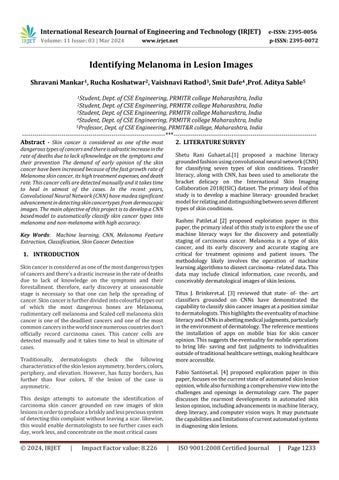
TheproposedmethodologyisshowninFig.1usingablock diagramandeachblockisexplainedindetailbelow.
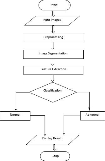
Fig.1.Blockdiagramofproposedmethodology.
Input Image: The proposed framework employments dataset comprises of tall- determinationinjurypictures. ISIC 2019 challengedataset which comprises of diverse classesiscompressedintopicturesandconnectedtothe proposedsystem.
2.InputImage
International Research Journal of Engineering and Technology (IRJET) e-ISSN: 2395-0056
Volume: 11 Issue: 03 | Mar 2024 www.irjet.net p-ISSN: 2395-0072
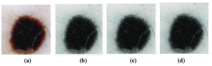
Pre-processing: Thepromotionofpicturespreparemust benon-uniforminafewterms.hence,theprimarythingof thepreprocessingstepistoupgradethepictureparameters comparative as quality, clarity, etc., by expelling or diminishingtheundesirablepassageofthepictureorthe foundation.Thefundamentalwayofthepreprocessingare grayscale change, picture advancement, and commotion junking. In this proposed framework, initially all the picturesarechangedoverintograyscale.tootwopoisons whichareknownasGaussianslimeandmiddleslimeare utilizedforpictureenhancementandclamorjunking.
Pre-processing stage results, (a) Dull image (b) Gray scale image(c)Gaussianfilter(d)Medianfilter
Segmentation: Segmentationistheprocessof separating the region of interest of the image. This separation canbe donebyconsideringeachpixeloftheimagewithaanalogous trait.Themainadvantagethen'sratherofrecyclingtheentire image,theimagewhichisdividedintopartscanbereused. The most common fashion is to indicate the edges of the particular region. The other approaches similar as thresholding,clustering,andregiongrowingusediscoveryof parallelsintheparticularregion.
Feature Extraction: Featureextractionisconsideredasthe mostcrucialpartintheentireprocessofclassification.The extractionofrelevantfeaturesfromthegiveninputdataset for performing computations such as detection and classification further is called feature extraction. Feature extractionreferstotheprocessoftransformingrawdatainto numericalfeaturesthatcanbeprocessedwhilepreserving theinformationintheoriginaldataset.Ityieldsbetterresults than applying machine learning directly to the raw data. Featureextractionforimagedatarepresentstheinteresting partsofanimageasacompactfeaturevector.Inthepast,this wasaccomplishedwithspecializedfeaturedetection,feature extraction,andfeaturematchingalgorithms.
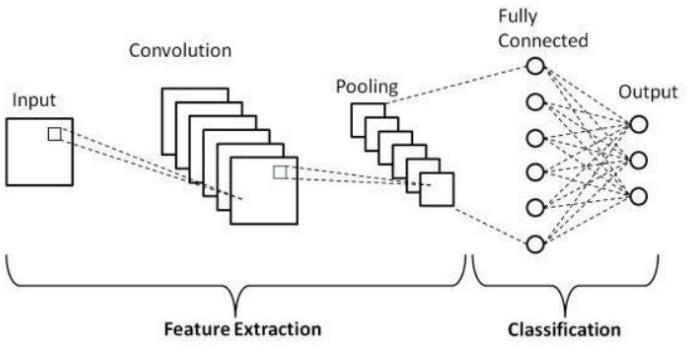
Classification: Aconvolutionneuralnetworkisanessential typeofdeepneuralnetwork,whichiseffectivelybeingused in computer vision. It is used for classifying images, assemblingagroupofinputimages,andperformingimage recognition.
CNNisafantastictoolforcollectingandlearningglobaldata as well as local data by gathering more straightforward features such as curves and edges to produce complex features such as shapes and corners. CNN’s hidden layers consistofconvolutionlayers,nonlinearpoolinglayers,and fullyconnectedlayers.
CNN can contain multiple convolution layers that are followedbyseveralfullyconnectedlayers.Threemajortypes of layers involved in making CNN are convolution layers, poolinglayers,andfull-connectedlayers.
ConvolutionalNeuralNetwork –
A Convolutional Neural Network (CNN) is a deeplearning neural network architecture specifically designed for processing grid-like data, such as images and audio spectrograms. CNNs are characterized by their ability to automaticallylearnandextracthierarchicalfeaturesfromthe input data through a series of convolutional and pooling layers.TheselearnedfeaturesmakeCNNsparticularlywellsuitedfortaskslikeimageclassification,objectdetection,and speechrecognition.Theseneuralnetworksareinspiredbythe humanvisualsystem,mimickingthewaythe humanbrain processes visual information. CNNs excel at capturing intricate patterns, textures, and features within medical images, making them highlyeffective toolsfor recognizing melanomaspecificcharacteristics.
International Research Journal of Engineering and Technology (IRJET) e-ISSN: 2395-0056
Volume: 11 Issue: 03 | Mar 2024 www.irjet.net p-ISSN: 2395-0072
In a Convolutional Neural Network (CNN) architecture designed for a melanoma diagnosis, there are typically several layers, each with a specific purpose in the feature extractionandclassificationprocess–
1. Input Layer:
Theinputlayerrepresentstheskinlesionimagesthatarefed intothenetwork.Theseimagesaretypicallyofafixedsize andarepre-processedtoensureuniformity.
2. Convolutional Layers: Convolutionallayersarethecore oftheCNNandareresponsibleforfeatureextraction.They applyasetoflearnablefilterstotheinputimagetodetect variouspatternsandfeatures.Multipleconvolutionallayers withincreasingcomplexityare usedto capturefeatures at differentscales.
3. Pooling Layers: Poolinglayersdown-samplethespatial dimensions of the feature maps obtained from the convolutional layers. Common pooling operations include max-pooling, which retains the maximum value in a local region,reducingthesizeofthefeaturemapswhilepreserving importantinformation.
4. Fully connected layer: Fully connected layers are densely connected neural layers where each neuron is connectedtoeveryneuroninthepreviousandsubsequent layers.Theselayersareresponsibleforlearninghigh-level representationsandmakingthefinalclassificationdecision.
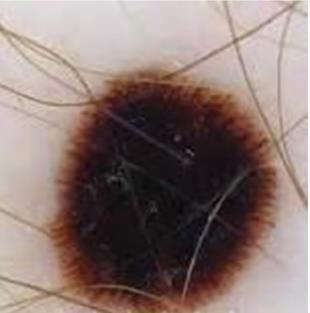
5. Output layer: Theoutputlayerconsistsofoneormore neurons, depending on the specific task. For melanoma diagnosis,ittypicallyconsistsclassificationresults(benignor malignant).
Globally,there'sadrasticincreaseintherateofskincancer cases because of several factors. the development of an automaticcarcinomadiscoverysystemplaysamajorpartinits early opinion. Skincancers are intermittently delicate to identify because of the misconception. Melanoma is one amongthem.Whenanapplicablemotorizedsystemisused, which ease the work of dermatologist to classify the skin lesionswhetherit'sbenignorcarcinoma.
In the proposed model, Image Pre-Processing, Image Segmentation and Image Bracket way are performed for grading skin lesion images into carcinoma or benign. ConvolutionalNeuralNetworkisusedforaddingthenumber ofimageswhich leads to better performance of proposed system.Aconvolutionalneuralnetworkandmachineliteracy classifiers trained with a set of features describing the borders,textureandthecolorofaskinlesion.
1 Shetu Rani Guha, S. M. Rafizul Haque “Convolutional Neural Network Based Skin Lesion Analysis for ClassifyingMelanoma”,December2019
2 Rashmi Patil, Sreepathi Bellary, “Machine learning approachinmelanomacancerstagedetection”,Sept 2020
3 TitusJ.Brinker,AchimHekler,JochenUtikal,Christof von Kalle, “Skin Cancer Classification Using ConvolutionalNeuralNetworks:SystematicReview”, Oct2018
4 Fabio Santos, Filipe Silva and Petia Georgieva, “AutomatedDiagnosisofSkinLesions”,August2020
5 M. Dildar et al., “Skin Cancer Detection: A Review Using Deep Learning Techniques” International JournalofEnvironmentalResearchandPublicHealth, vol.18,no.10,p.5479, May 2021, doi: 10.3390/ijerph18105479.
6 Naeem A, Farooq MS, Khelifi A, Abid A (2020), “Malignant melanoma classification using deep learning: Datasets, performance measurements, challengesandopportunities”,IEEEAccess8:110575–110597
7 Yu, Z. et al. Melanoma recognition in dermoscopy images via aggregated deep convolutional features. IEEETrans.Biomed.Eng.66,1006–1016(2019).
8 Jacinth Poornima, Anitha Jude, Asha Priya, Henry,D. Jude Hemanth, “Melanoma Classification Using MachineLearningTechniques”,January2023
9 PNSrinivasu,JGSivaSai,MFIjaz,AKBhoi,WKim,JJ Kang, Classification of skin disease using deep learning neural networks with MobileNet V2 and LSTM”,Sensors21(8),2852(2021)
10 AyaAbuAli,HasanAl-Marzouqi,“Melanomadetection usingregularconvolutionalneuralnetworks”,January 2018doi:10.1109/ICECTA.2017.8252041
11 Wessam Salma, Ahmed S. Eltrass, “Automated deep learning approach for classification of malignant melanomaandbenignskinlesions”,April2022