
International Research Journal of Engineering and Technology (IRJET) e-ISSN: 2395-0056
Volume: 11 Issue: 02 | Feb 2024 www.irjet.net p-ISSN: 2395-0072
International Research Journal of Engineering and Technology (IRJET) e-ISSN: 2395-0056
Volume: 11 Issue: 02 | Feb 2024 www.irjet.net p-ISSN: 2395-0072
Janhavi Tingre School of Computer Engineering & Technology
MIT World Peace University, Pune, Maharashtra
Vaishnavi Mundada School of Computer Engineering & Technology
MIT World Peace University, Pune, Maharashtra
Harsh Shelke School of Computer Engineering & Technology
MIT World Peace University, Pune, Maharashtra
Abstract Breast cancer is among the main reasons why women die worldwide. Breast cancer mortality rates and treatment expenses can be decreased with early detection and diagnosis. In this effort, we have put forth a novel, affordable, computer vision-based method for detecting and diagnosing breast cancer. Convolutional neural networks are also used for medical image classification. The proposed model is a very simple and cost effective approach with high accuracy and useful outcomes. We have also explored the different challenges faced and the future scope of the project.
Keywords Breast Cancer, Computer vision, Convolutionalneuralnetwork,Detection
Overtaking lung cancer, breast cancer is the most commoncanceramongwomen.InIndiathesurvivalrate for breast cancer patients is about 60% as compared to 90% in the United States, for the last five years [1]. By enhancing treatment options, early detection methods, awareness campaigns, and better diagnostics, we can increasethesesurvivalrates.
Because of its simplicity and practicability, ultrasound has become a standard tool for diagnosing breast disorders. The findings of B-mode ultrasonography, on the other hand, are related to the level of expertise of doctors, poor image quality, benign presentations of malignant tumors, and visual fatigue or neglect on the part of observers [2]. If a huge number of ultrasound mammary images are manually examined, there will be significant flaws. Misdiagnosis is common when lesions that should be properly diagnosed are missed by radiologists[2].
CALC (calcification), CIRC (circumcised masses), SPIC (speculated masses), MISC (other ill-defined masses), ARCH(architectural distortion),andASYM(asymmetry) are the six types of breast cancer [3]. In this paper,
Ayush Chaudhary School of Computer Engineering & Technology
MIT World Peace University, Pune, Maharashtra
Invasive Ductal Carcinoma (IDC), one of the most prevalent forms of breast cancer, is one that we are finding.
There is improvement in the field of diagnosis due to evolvingtechnology.Convolutionalneuralnetworkisthe mostwidelyusedmachinelearningalgorithminthefield of medical image analysis [4]. The fundamental reason for this is because CNN exactly fits the two-dimensional structure of the image in structure and uses this spatial relationshipasthealgorithm'sdirectinputvalue[4].
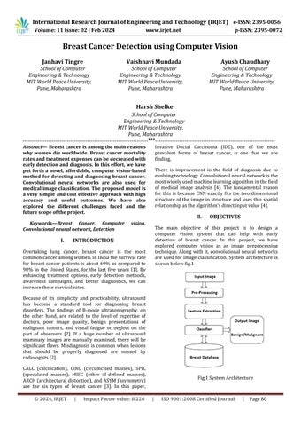
The main objective of this project is to design a computer vision system that can help with early detection of breast cancer. In this project, we have explored computer vision as an image preprocessing technique. Along with it, convolutional neural networks are used for image classification. System architecture is shownbelowfig.1
International Research Journal of Engineering and Technology (IRJET) e-ISSN: 2395-0056
Volume: 11 Issue: 02 | Feb 2024 www.irjet.net p-ISSN: 2395-0072
For the literature survey, we studied multiple research papers based on breast cancer detection and different methods to achieve it. The descriptive details about the samehavebeenmentionedinthesectionsbelow.
A. This research paper presents a method for breast cancer(BC)identificationandcategorizationusingKnearest neighbor (KNN), classification of thermographic images using (SVM) with (SMO), and detection of BC nuclei using Stacked Sparse Autoencoder(SSAE).Thestudysuggeststhattraining on additional datasets can improve the accuracy of the model [5]. Furthermore, the authors emphasize the importance of verifying medical photos using a broad range of applications since some tumor diagnoses can be challenging [5]. Overall, the proposed approach can aid in the accurate detection anddiagnosisofbreastcancer.
B. In this research paper, they used different machine learning algorithms, including multilayer perceptron neural networks (MLPNNs), (SVMs), and (LDA), in variousapplicationsofpatternrecognition.Thestudy highlightsamajorchallengeinthisfield,whichisthe unbalanced dataset problem, where one class has significantly more samples than another [6]. The authors explore different strategies to address this issue, such as resampling techniques and adjusting the cost matrix [6]. Overall, the findings of this research provide insights into the selection of appropriate algorithms and techniques to improve the accuracy of pattern recognition in real-world scenarioswithunbalanceddatasets.
C. This research paper discusses the Breast Imaging ReportandDataSystem(BI-RADS)asastandardized tool for mammography reporting and interpretation. The authors highlight the various categories within the BI-RADS system, including BI-RADS 0, 1, and 2, where BI-RADS 2 denotes a benign finding with a probability of malignancy of 0% [7]. The study emphasizes the importance of considering the possibility of false-negative or false-positive results in interpreting mammography findings and recommends that radiologists incorporate this into their decision-making process. The authors also explore the role of BI-RADS in facilitating communication between radiologists and referring physicians and guiding patient management [7]. Overall,thefindingsofthisresearchcontributetothe understanding and implementation of the BI-RADS system in clinical practice, improving breast cancer detectionanddiagnosis.
D. This research paper presents the use of pre-trained deep neural network (DNN) models, including ResNet, Inception-V3Net, and ShuffleNet, for
conductingbinaryandmulti-classclassifications.The study highlights the importance of labeled data for training these models effectively. However, the authors also acknowledge that the lack of interpretability in DNN models can make it challenging to identify false positives or false negatives [8]. The research demonstrates the potential of pre-trained DNNs in achieving high accuracy in classification tasks, particularly in image analysis. The findings of this study contribute to the understanding of the practical applications of pretrained DNNs and the challenges associated with theiruseinreal-worldscenarios[8].
E. In this research paper, system evaluates computing approachesforbreastcancerdetectionbasedonCAD using mammogram images, focusing on thresholdbased and region-based segmentation. However, the increased computational challenges associated with machine learning (ML) classifiers based on deep learning(DL)asthenumberoflayersincreaseshave been identified as a research gap [9]. DL-based classifiers have shown great potential in breast cancer detection, but their computational challenges make them less practical for clinical settings [9]. Thus, more research is needed to develop efficient and robust computing approaches for breast cancer detectionusingmammogramimages.
Breast Cancer is an ordinary form of cancer among women and immediate assessment is important for successful cure. Convolutional Neural Networks have shown encouraging results in the prognosis of breast cancer using Whole Source Images (WSI). Below is the normalmethodologyofdetectionofBreastCancer:
● AssemblingtheData:Thedatasetshouldhavea gargantuan variety of Whole Source Images ranging from malignant to innocuous tumor images.Consequently,weprepareandconstruct tilesofthoseWholeSourceImages.
● Data Segregation: Further, we separate the aforementioned tiles into cancerous and noncancerousarraysrespectively.
● Data Pre-Processing: Here we preprocess the data using the open computer vision library(cv2) The open computer vision library (cv2) is primarily concerned with image processing,videorecording,andanalysis,which includesfunctionslikefaceandobjectdetection. Here, we make use of this library to resize and redimension all of the photos. Combination of thearrays:Weconjointhetwoabovementioned arraysintoasinglearray.
International Research Journal of Engineering and Technology (IRJET) e-ISSN: 2395-0056
Volume: 11 Issue: 02 | Feb 2024 www.irjet.net p-ISSN: 2395-0072
● Train-TestSplit:Fromthedataset,threesets a trainingset, a validation set, anda testing set were produced. The testing set is used to evaluatetheperformanceoftheCNNafterithas been trained using the training set. In our model, 80% data has been fed into the training setandtheremaining20%isthetestdata.
● Importing CNN libraries and Defining CNN layers:Herewe,usestochasticgradientdescent (SGD) or Adam optimisation methods to train theCNNusingthetrainingdata.Thesemethods canbeusedaswehaveimportedthetenserflow library, which further assists us in performing CNNeffectively.
● ValidationSet:Usingthevalidationset,wefinetune the hyperparameters, such as the learning rate, batch size, number of epochs etc. The epochs represents the number of iterations whichis11andbatchsizerepresentsnumberof subsetsofadatasetwhichis35inourmodel.
● Data Evaluation: Here we use metrics like accuracy to assess how well CNN is performed onthetestingset.
● DataVisualization:Inconclusion,tocomprehend theperformanceoftheCNN,evaluatethelearnt features, and pinpoint areas for development, we analyze the findings.It is possible to apply interpretation techniques like gradient-based attribution, saliency maps, and feature visualization.
In this study, we proposed a breast detection system based on computer vision and convolutional neural networks (CNNs). Our goal was to develop an accurate andefficientsystemthatcanautomaticallydetectbreast regions in medical images, which can help in the early diagnosisandtreatmentofbreastcancer.
We utilised a Kaggle dataset that contained 54 breast cancer patients and 277524 pictures. We divided the dataset into testing (20%) and training (80%) sets. Sequential CNN model training was done using the training set. The model's performance was enhanced by the introduction of a max-pooling layer and a rectified linearunit(ReLU)activationfunction.
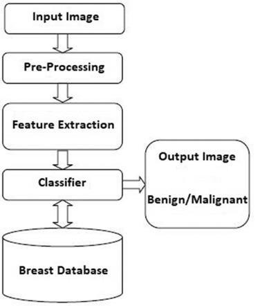
Wetrainedthemodelusingbatchsize35and11epochs. . The training process was stopped when the validation loss did not improve after 11 epochs. As the goal function for optimizing the model presented in fig.2, we usedcross-entropyloss.
Weassessedthemodel'sperformance onthetestingset after training. Our 92.25% accuracy score demonstrates thatourtechnologycanpreciselyidentifybreastareasin medical photos. We also measured other evaluation metrics such as precision, recall, and F1-score, which were all above 0.90 represented using confusion matrix asperfig.4.
Our results show that the proposed system can effectively detect breast regions in medical images with high accuracy. The system can be further improved by using more advanced CNN architectures or by incorporating other features such as texture analysis. Overall,ourapproachcanbeusefulintheearlydetection and diagnosis of breast cancer, which can improve patientoutcomesandsurvivalrates.
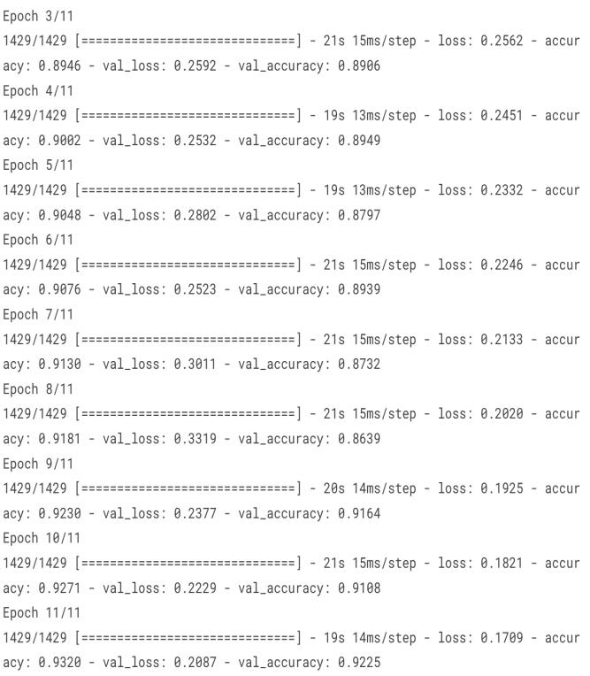
Fig.2Accuracyonvariousepochs
International Research Journal of Engineering and Technology (IRJET) e-ISSN: 2395-0056
Volume: 11 Issue: 02 | Feb 2024 www.irjet.net p-ISSN: 2395-0072
Fig.3.Performanceofproposedmodel
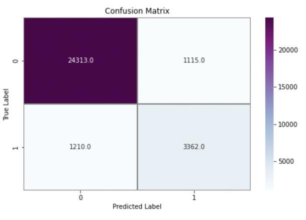
Fig.4.Confusionmatrix
REFERENCES
[1] R.Guzman-Cabrera, J.R.Guzm´an-Sep´ulveda,M.´ 313 Torres-Cisneros, D.A. May-Arrioja, J. RuizPinales, 314 O.G. Ibarra-Manzano, et al., Digital image processing 315 technique for breast cancer detection,
[2] International 316 Journal of Thermophysics 34 (2013),1519–1531.
[3] JacksonV.P.,HendrickR.E.,FeigS.A.,etal.Imaging of the radiographically dense breast.[J]. Radiology, 1993,188(2):297-301.
[4] Sethy, P. K., Pandey, C., Khan, D. M. R., Behera, S. K., Vijaykumar, K., & Panigrahi, D. S. (2021). A costeffective computer-vision based breast cancer diagnosis.JournalofIntelligent&FuzzySystems,1–11.doi:10.3233/jifs-189848
[5] Yunchao, G., & Jiayao, Y. (2019). Application of Computer Vision and Deep Learning in Breast Cancer Assisted Diagnosis. Proceedings of the 3rd International Conference on Machine Learning and Soft Computing - ICMLC 2019. doi:10.1145/3310986.3311010
[6] Sethy, Prabira Kumar et al. ‘A Cost-effective Computer-vision Based Breast Cancer Diagnosis’. 1 Jan.2021:5253–5263.
[7] JalalianA,MashohorS,MahmudR,KarasfiB,Saripan MIB, Ramli ARB. Foundation and methodologies in computer-aideddiagnosissystemsforbreastcancer detection. EXCLI J. 2017;16:113-137. Published 2017Feb20.doi:10.17179/excli2016-701
[8] Al-Tam, Riyadh & Narangale, Sachin. (2021). Breast Cancer Detection and Diagnosis Using Machine Learning: A Survey. JOURNAL OF SCIENTIFIC RESEARCH. 65. 265-285. 10.37398/JSR.2021.650532.
[9] Al-Tam, Riyadh & Narangale, Sachin. (2021). Breast Cancer Detection and Diagnosis Using Machine Learning: A Survey. JOURNAL OF SCIENTIFIC RESEARCH. 65. 265-285. 10.37398/JSR.2021.650532.
[10] AljuaidH,AlturkiN,AlsubaieN,CavallaroL,LiottaA. Computer-aided diagnosis for breast cancer classification using deep neural networks and transfer learning. Comput Methods Programs Biomed. 2022;223:106951. doi:10.1016/j.cmpb.2022.106951
[11] Asma Zizaan, Ali Idri. (2023) Machine learning based Breast Cancer screening: trends, challenges, and opportunities. Computer Methods in Biomechanics and Biomedical Engineering: Imaging &Visualization0:0,pages1-21.
[12] Pawar, R., Ghumbre, S., & Deshmukh, R. (2019). Visual SimilarityUsingConvolutionNeural Network over Textual Similarity in Content- Based Recommender System. International Journal of AdvancedScienceandTechnology,27,137-147.
[13] Pawar, R., Ghumbre, S., & Deshmukh, R. (2020). A Hybrid Approach towards Improving Performance
© 2024, IRJET | Impact Factor value: 8.226 | ISO 9001:2008 Certified Journal | Page83
International Research Journal of Engineering and Technology (IRJET) e-ISSN: 2395-0056
Volume: 11 Issue: 02 | Feb 2024 www.irjet.net p-ISSN: 2395-0072
ofRecommender SystemUsingMatrixFactorization Techniques. International Journal of Future GenerationCommunicationandNetworking,Vol.13, No.4,(2020),pp.467–477.
© 2024, IRJET | Impact Factor value: 8.226 | ISO 9001:2008 Certified Journal | Page84