


Findings
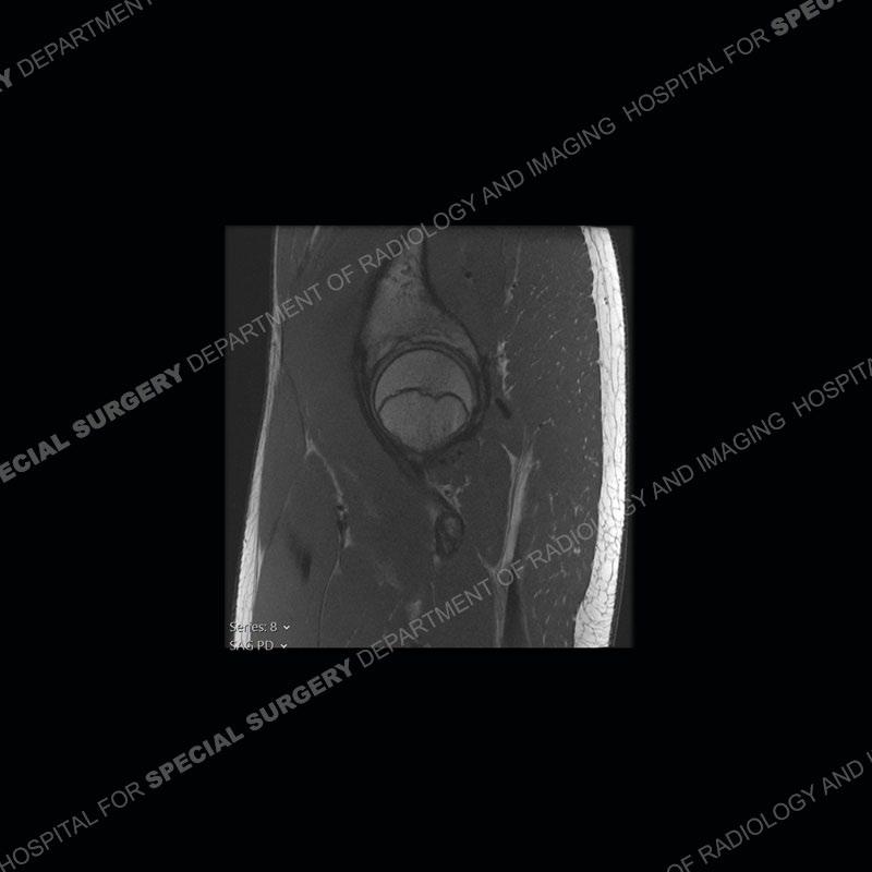
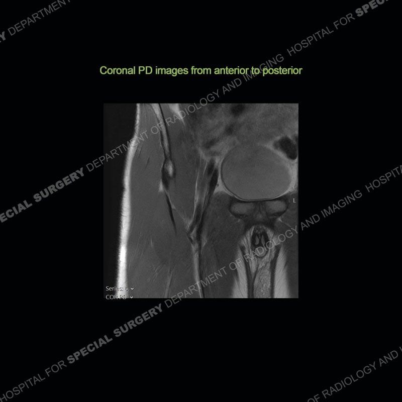
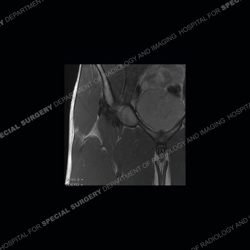
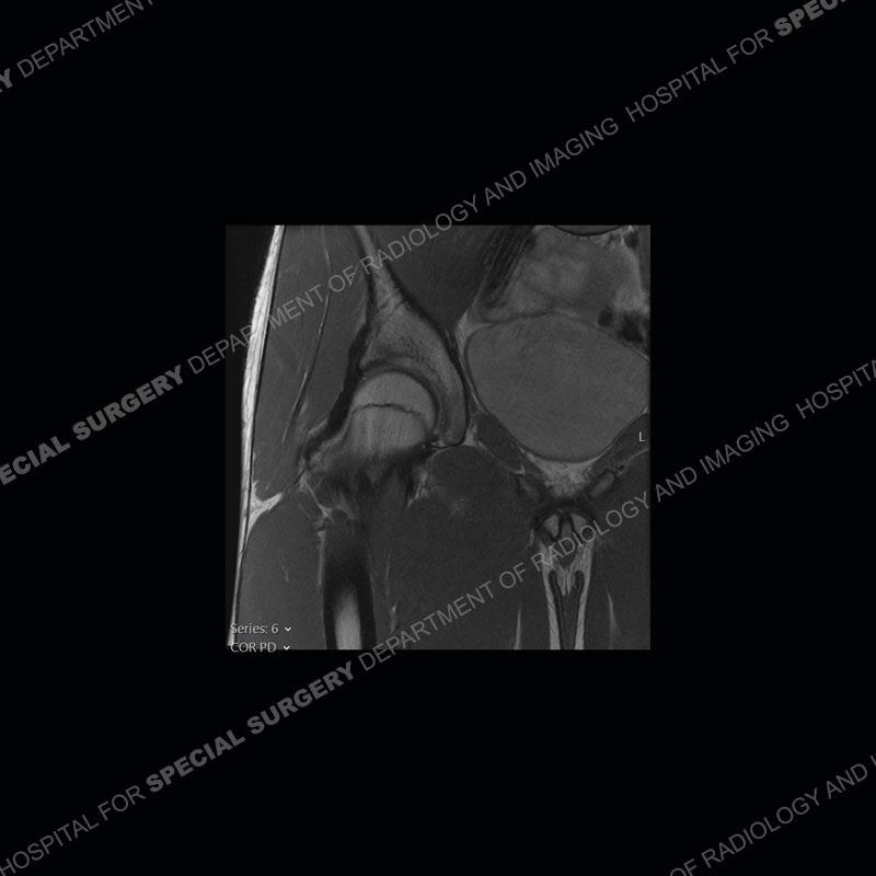
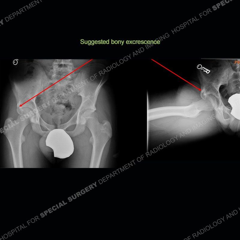
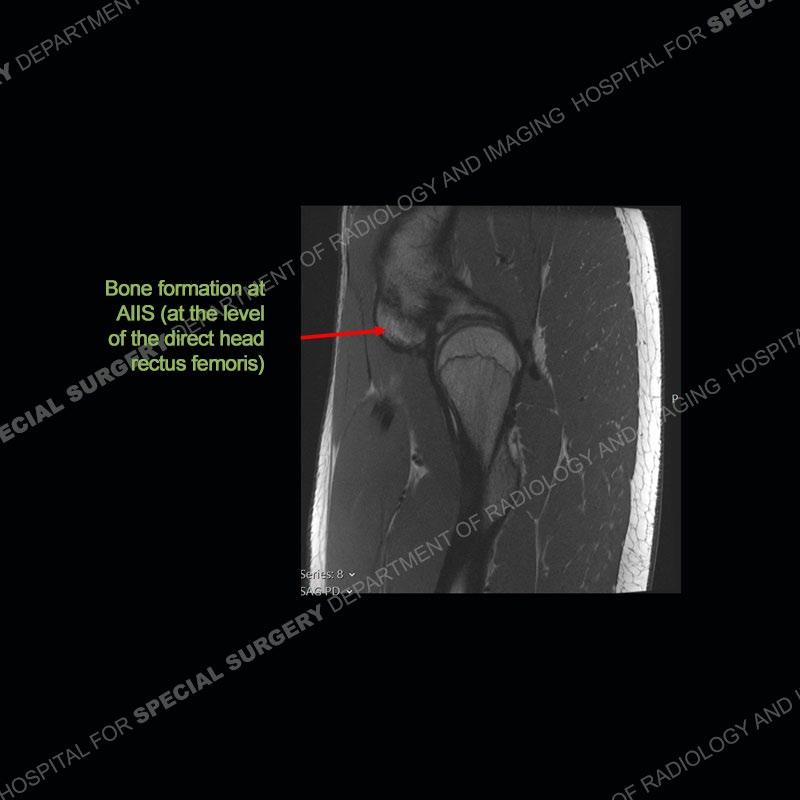
The radiographs demonstrate a suggested bony protuberance or excrescence emanating from the superior aspect of the acetabulum. On the MRI, seen is a large amount of bone about the anterior inferior iliac spine (AIIS) at the level of the origin of the direct head of the rectus femoris. This bone pedunculates inferior to the acetabular rim. No edema pattern is present on the IR sequence and the contralateral left hip acts as a nice internal control where the bony prominence is not present.


Diagnosis: Subspine Femoroacetabular Impingement
Femoroacetabular impingement (FAI) has become increasingly recognized over the last two decades. The pathology may be classified as based on intraarticular or extra-articular processes. The intra-articular relates to the well known cam and pincer type pathology. The extra-articular is slightly less well known and may relate to subspine, ischiofemoral, iliopsoas, or pelvicotrochanteric impingement. The extra-articular type impingement may be concomitant with an intra-articular process or exist on its own.
The anterior inferior iliac spine is the attachment site of the direct head of the rectus femoris. With avulsion, hematoma and heterotopic ossification may form or in some situations, chronic overuse may precipitate a large enthesophyte. Whatever, the etiology, if bone is present about the AIIS with flexion and internal rotation about the hip, impingement may be precipitated between the bone and the proximal femur. This leads to pain and a limited range of motion.
Prior classification of the ASIS has been performed by Kelly et al. A type I has a smooth wall between the AIIS and the acetabular rim, a type II is when the AIIS extends to the level of the acetabular rim, and a type III is when the AIIS extends distally to the rim. Multiple studies have shown that a type II or type III may be associated with impingement. It is important to always remember that the imaging can only demonstrate findings that may be seen in impingement, but the actual impingement remains a clinical diagnosis.
References
Frances Borrego, A., Martinez Garcia, A., Del Baño Barragán, L. et al.
Subspine femoroacetabular impingement: retrospective study of a series of patients treated by hip arthroscopic resection. Arch Orthop Trauma Surg (2023). https://doi.org/10.1007/s00402-022-04761-2
Hetsroni, Iftach MD, Poultsides, Lazaros MD; Bedi, Asheesh MD; Larson, Christopher M. MD; Kelly, Bryan T. MD. Anterior Inferior Iliac Spine
Morphology Correlates With Hip Range of Motion: A Classification System and Dynamic Model. Clinical Orthopaedics and Related Research 471(8):p 24972503, August 2013. | DOI: 10.1007/s11999-013-2847-4