ENDOCRINE
EDITOR:
MICHAEL T. McDERMOTT, MD
Professor of Medicine and Clinical Pharmacy
University of Colorado Denver School of Medicine
Director, Endocrinology and Diabetes Practice
University of Colorado Hospital
Aurora, Colorado
ENDOCRINE SECRETS, SEVENTH EDITION ISBN: 978-0-323-62428-2
Copyright © 2020 by Elsevier, Inc. All rights reserved.
No part of this publication may be reproduced or transmitted in any form or by any means, electronic or mechanical, including photocopying, recording, or any information storage and retrieval system, without permission in writing from the publisher. Details on how to seek permission, further information about the Publisher’s permissions policies and our arrangements with organizations such as the Copyright Clearance Center and the Copyright Licensing Agency, can be found at our website: www.elsevier.com/permissions.
This book and the individual contributions contained in it are protected under copyright by the Publisher (other than as may be noted herein).
Notice
Practitioners and researchers must always rely on their own experience and knowledge in evaluating and using any information, methods, compounds or experiments described herein. Because of rapid advances in the medical sciences, in particular, independent verification of diagnoses and drug dosages should be made. To the fullest extent of the law, no responsibility is assumed by Elsevier, authors, editors or contributors for any injury and/or damage to persons or property as a matter of products liability, negligence or otherwise, or from any use or operation of any methods, products, instructions, or ideas contained in the material herein.
Previous editions copyrighted 2013, 2009, 2005, 2002, 1998, and 1994.
Library of Congress Control Number: 2019937967
Content Strategist: Marybeth Thiel
Content Development Specialist: Angie Breckon
Publishing Services Manager: Shereen Jameel
Senior Project Manager: Umarani Natarajan
Design Direction: Bridget Hoette
in the United States of America
This book is dedicated to Libby, whose strength, courage, and love of life are a daily inspiration, and to Katie Cohen, Emily Cohen, Hayley McDermott, and Henry McDermott, for making life fun.
Meghan Donnelly, MD
Assistant Professor Obstetrics and Gynecology
University of Colorado School of Medicine
Denver, CO, United States
William E. Duncan, MD, PhD, MACP Professor of Medicine Department of Medicine
Uniformed Services University Bethesda, MD, United States
Oliver J. Fackelmayer, MD
Surgical Resident
University of Colorado Anschutz Medical Campus Department of Surgery Aurora, CO, United States
Shari C. Fox, BS, MS, MD, FACE Department of Endocrinology
Colorado Permanente Medical Group Denver, CO, United States
Michele B. Glodowski, MD
Clinical Instructor
New York University School of Medicine
New York, NY, United States
Bryan R. Haugen, MD
Professor of Medicine and Pathology
Head, Division of Endocrinology, Metabolism and Diabetes
Mary Rossick Kern and Jerome H. Kern Chair of Endocrine Neoplasms Research University of Colorado School of Medicine Aurora, CO, United States
Matthew R. Hawkins, BSW, MMSc
Senior Instructor/Physician Assistant Division of Endocrinology, Metabolism and Diabetes University of Colorado School of Medicine Aurora, CO, United States
James Vincent Hennessey, MD
Clinical Director
Endocrinology
Beth Israel Deaconess Medical Center
Associate Professor Medicine
Harvard Medical School
Boston, MA, United States
Thanh Duc Hoang, DO, FACP, FACE Director, NCC Endocrinology Fellowship Program Department of Endocrinology
Walter Reed National Military Medical Center
Associate Professor, Internal Medicine Director, Endocrinology Division
Uniformed Services Health Sciences University Bethesda, MD, United States
Sean J. Iwamoto, MD
Instructor of Medicine
Endocrinology, Metabolism and Diabetes University of Colorado School of Medicine
Rocky Mountain Regional VA Medical Center Aurora, CO, United States
Thomas Jensen, MD
Assistant Professor University of Colorado Denver Aurora, CO, United States
Janice M. Kerr, MD
Associate Professor Endocrinology, Metabolism and Diabetes University of Colorado, Denver Denver, CO, United States
Pratima Kumar, MD, FACE
Assistant Professor, Division of Endocrinology Department of Medicine
Dell Medical School
The University of Texas at Austin
Austin, TX, United States
Helen M. Lawler, MD
Assistant Professor Division of Endocrinology, Metabolism, and Diabetes
University of Colorado
Aurora, CO, United States
Homer J. LeMar Jr., MD
El Paso Veterans Affairs Health Care System
El Paso, TX, United States
Vinh Q. Mai, DO, FACP, FACE
Chief and Associate Professor Endocrinology, Diabetes and Metabolism
Walter Reed National Military Medical Center
Bethesda, MD, United States
Ayesha F. Malik, MD
Fellow Endocrinology
Mayo Clinic
Jacksonville, FL, United States
Roselyn I. Mateo, MD, MSc Endocrinology
Beth Israel Deaconess Medical Center
Boston, MA, United States
Sarah E. Mayson, MD
Assistant Professor Division of Endocrinology, Department of Medicine
University of Colorado School of Medicine
Rocky Mountain Regional Veterans Affairs Medical Center
Aurora, CO, United States
Michael T. McDermott, MD
Professor of Medicine and Clinical Pharmacy
University of Colorado School of Medicine
Director, Endocrinology and Diabetes Practice
University of Colorado Hospital
Aurora, CO, United States
Robert C. McIntyre Jr., MD
Professor Department of Surgery
University of Colorado Anschutz Medical Campus
Aurora, CO, United States
Logan R. McKenna, MD
Surgical Resident
University of Colorado Anschutz Medical Campus
Aurora, CO, United States
Shon Meek, MD, PhD
Assistant Professor
Endocrinology Division
Mayo Clinic
Jacksonville, FL, United States
Richard Millstein, DO
University of Colorado Health Endocrinology
Greeley, CO United States
Kerrie L. Moreau, PhD
Professor Division of Geriatrics
University of Colorado Anschutz Medical Campus
Aurora, CO, United States
Research Health Scientist
Geriatric Research Education Clinical Center (GRECC)
Denver Veterans Administration Medical Center
Denver, CO, United States
Wesley Nuffer, PharmD
Associate Professor
Department of Clinical Pharmacy
University of Colorado Skaggs School of Pharmacy & Pharmaceutical Sciences
Aurora, CO, United States
John J. Orrego, MD
Endocrinologist
Endocrinology and Metabolism
Colorado Permanente Medical Group
Denver, CO, United States
Endocrinology Department Chair
Endocrinology and Metabolism
St. Joseph Hospital
Denver, CO, United States
Roger A. Piepenbrink, DO, MS, MPH, FACP, FACE
Staff Physician
Departments of Adult Endocrinology and Sleep Medicine
Mike O’Callaghan Federal Medical Center
Nellis Air Force Base
Las Vegas, NV, United States
Christopher D. Raeburn, MD
Associate Professor
Department of Surgery
University of Colorado Anschutz Medical Campus
Aurora, CO, United States
Aziz Ur Rehman, MD
Assistant Professor
Division of Endocrinology
Texas Tech University
El Paso, TX, United States
Richard O. Roberts III, MD, MPH
Fellow Physician
Children’s Hospital Colorado
Department of Pediatrics
Section of Endocrinology and Diabetes
University of Colorado Anschutz Medical Campus
Aurora, CO, United States
Kevin B. Rothchild, MD
Assistant Professor
GI, Tumor, and Endocrine Surgery
University of Colorado Hospital
Aurora, CO, United States
Micol Sara Rothman, MD
Associate Professor of Medicine
Endocrinology, Diabetes and Metabolism
University of Colorado School of Medicine
Aurora, CO, United States
Shauna Runchey, MD, MPH Fellow
University of Colorado Anschutz Medical Campus
Denver, CO, United States
Mary H. Samuels, MD
Professor of Medicine
Program Director, Clinical and Translational Research Center
Oregon Health & Science University
Portland, OR, United States
Leonard R. Sanders, MD, FACP, BC-ADM, CDE, CLS Director of Diabetes Care
Endocrinology
Montage Medical Group Monterey, CA, United States
Virginia Sarapura, MD
Associate Professor Medicine-Endocrinology
University of Colorado Anschutz Medical Campus
Aurora, CO, United States
David Saxon, MD, MSc
Assistant Professor
Division of Endocrinology, Metabolism, and Diabetes
University of Colorado Aurora, CO, United States
Jonathan A. Schoen, MD
Associate Professor of Surgery
GI, Tumor, and Endocrine Surgery
University of Colorado Hospital Aurora, CO, United States
Emily B. Schroeder, MD, PhD
Clinician Investigator
Institute for Health Research
Kaiser Permanente Colorado
Assistant Professor
Division of Endocrinology, Metabolism and Diabetes
University of Colorado School of Medicine
Aurora, CO, United States
Stacey A. Seggelke, DNP, APRN, ACNS-BC, BC-ADM
Senior Instructor of Medicine
Department of Medicine, Division of Endocrinology
University of Colorado Denver
Aurora, CO, United States
Kenneth J. Simcic, MD†
Formerly Assistant Professor Division of Endocrinology
Department of Medicine
University of Texas Health Science Center at San Antonio
San Antonio, TX, United States
Robert H. Slover, MD
Director of Pediatrics
The Barbara Davis Center for Diabetes Professor of Pediatrics
Wagner Family Chair in Childhood Diabetes University of Colorado Denver Anschutz Medical Campus Aurora, CO, United States
Robert Smallridge, MD Professor of Medicine
Endocrinology Division
Mayo Clinic
Jacksonville, FL, United States
Christine M. Swanson, MD, MCR, CCD
Assistant Professor Division of Endocrinology, Metabolism and Diabetes University of Colorado
Aurora, CO, United States
Elizabeth A. Thomas, MD
Assistant Professor Division of Endocrinology, Metabolism and Diabetes
University of Colorado School of Medicine
Rocky Mountain Regional Veterans Affairs Medical Center
Aurora, CO, United States
Carlos A. Torres, MD
William Beaumont Army Medical Center El Paso, TX, United States
Sharon H. Travers, MD
Associate Professor Pediatric Endocrinology
Children’s Hospital Colorado University of Colorado Aurora, CO, United States
Jennifer M. Trujillo, PharmD
Associate Professor
Clinical Pharmacy University of Colorado Aurora, CO, United States
Amy M. Valent, DO
Assistant Professor
Obstetrics and Gynecology
Oregon Health and Science University Portland, OR, United States
Nicole Odette Vietor, MD
Department of Endocrinology, Diabetes, and Metabolism
Walter Reed National Military Medical Center
Bethesda, MD, United States
Robert A. Vigersky, MD
Professor of Medicine
Endocrinology Service
Walter Reed National Military Medical Center
Bethesda, MD, United States
Katherine N. Vu, DO
Assistant Professor
Endocrinology Service
Naval Medical Center San Diego
San Diego, CA, United States
Cecilia C. Low Wang, MD, FACP
Associate Professor of Medicine
Associate Director, Fellowship/Education
Division of Endocrinology, Metabolism and Diabetes
University of Colorado School of Medicine
Aurora, CO, United States
Matthew P. Wahl, MD
Assistant Professor of Medicine (Clinical)
Division of Endocrinology Metabolism, and Diabetes
University of Utah School of Medicine
Salt Lake City, UT, United States
Katherine Weber, MD
Endocrinology
Kaiser Permanente
Denver, CO, United States
Margaret E. Wierman, MD
Professor of Medicine
University of Colorado School of Medicine
Aurora, CO, United States
Majlinda Xhikola, MD
Fellow
Endocrinology Division
Mayo Clinic
Jacksonville, FL, United States
Adnin Zaman, MD
Clinical/Research Fellow
Division of Endocrinology, Metabolism and Diabetes
University of Colorado
Aurora, CO, United States
Philip Zeitler, MD, PhD
Professor
Pediatrics
University of Colorado Denver Anschutz Medical Campus
Chair
Endocrinology
Children’s Hospital Colorado
Aurora, CO, United States
†Deceased
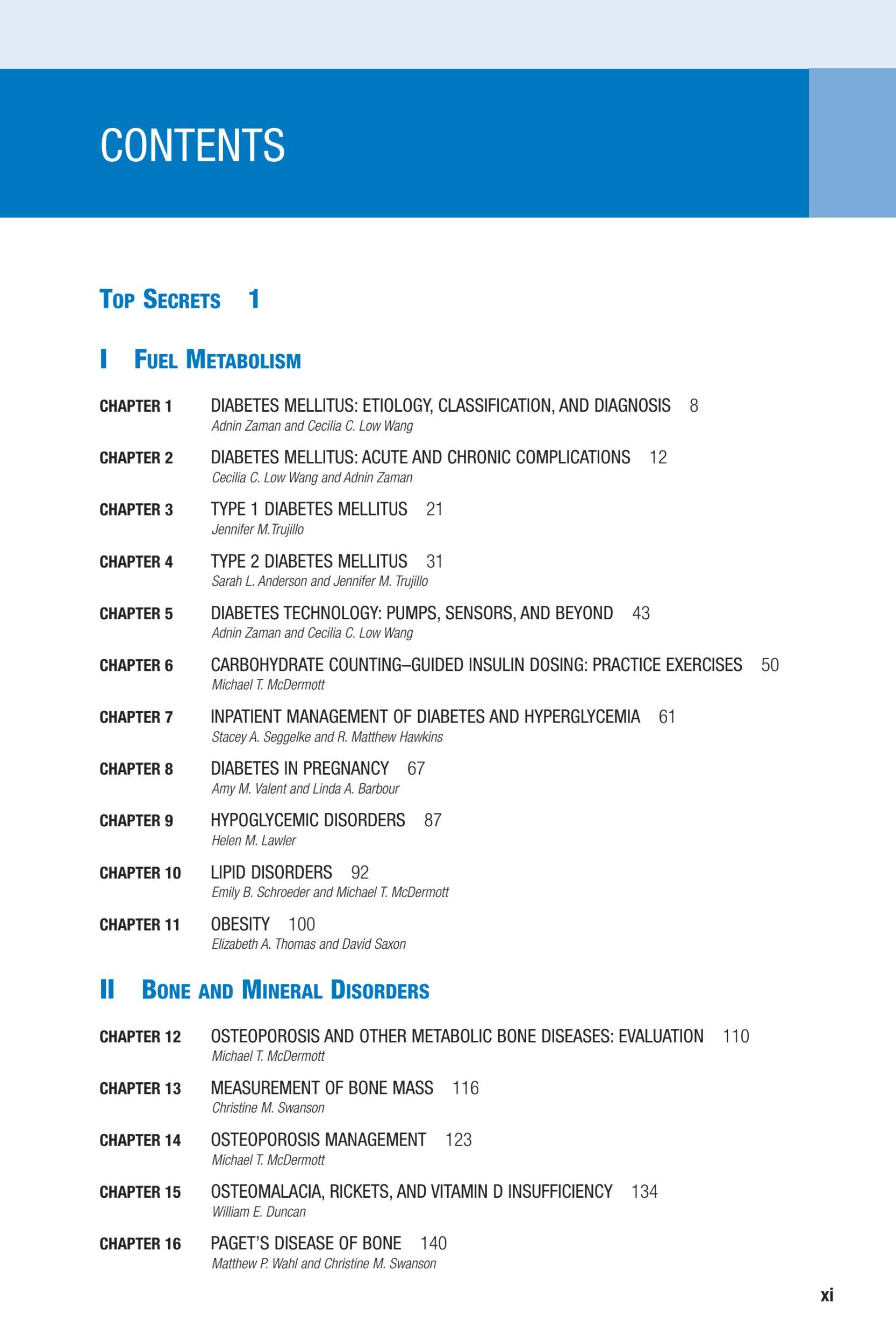
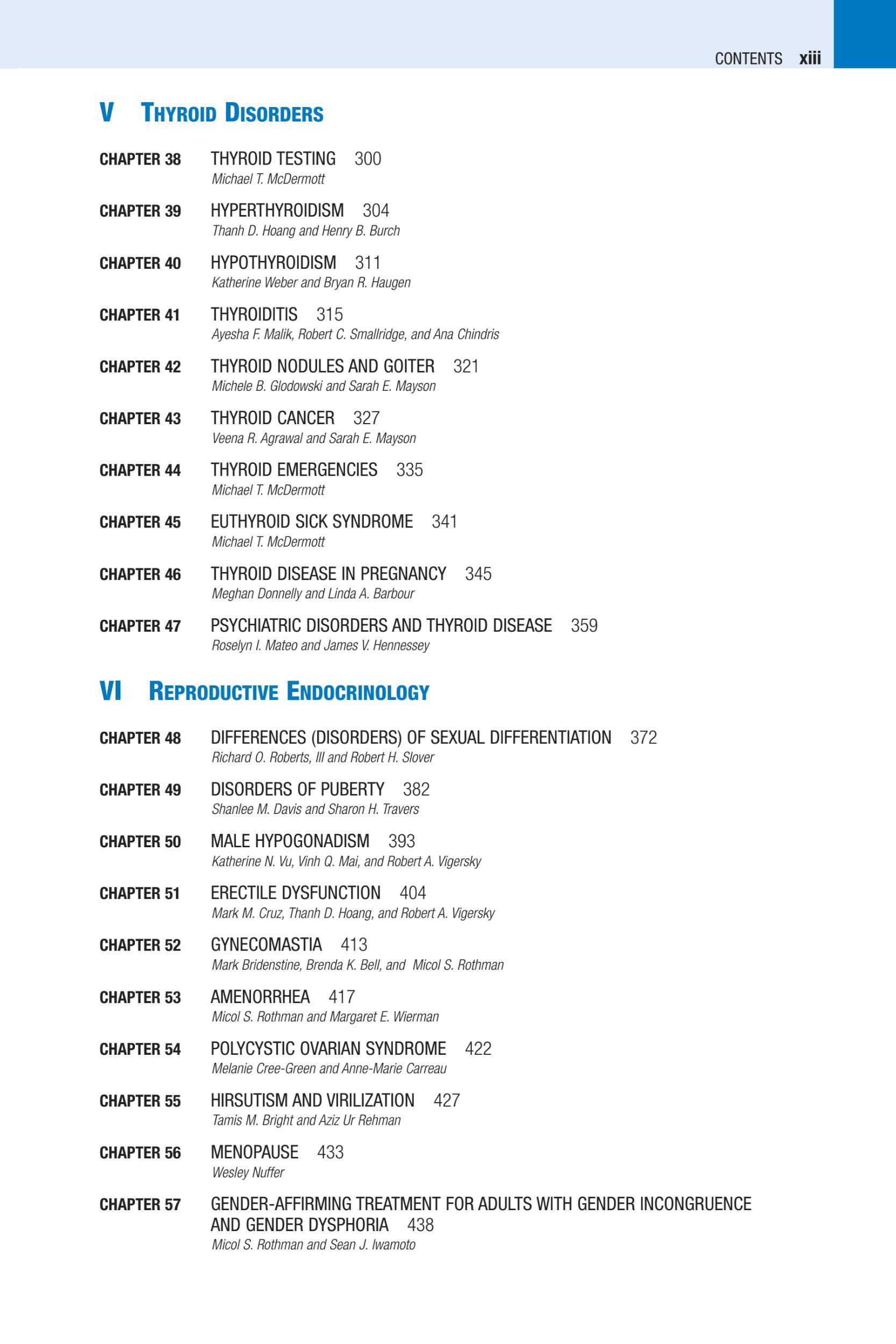
CHAPTER 1 DIABETES MELLITUS: ETIOLOGY, CLASSIFICATION, AND DIAGNOSIS
Adnin Zaman and Cecilia C. Low Wang
1. What is diabetes mellitus?
Diabetes mellitus is a term that encompasses a heterogeneous group of metabolic disorders, all of which are characterized by elevated blood glucose levels.
2. What is the origin of the term diabetes mellitus?
Although diabetes has been described in literature as ancient as the Ebers Papyrus of Egypt by Hesy-Ra (1552 bce), the word diabetes comes from Arateus and the Greek word “diabainein” which means “siphon,” because affected people excreted excessive amounts of urine. The term diabetes mellitus was coined by Thomas Willis of Oxford, England, in 1675, when it was discovered that the urine of people with diabetes was sweet rather than tasteless (insipidus).
3. What is the epidemiology of diabetes?
Approximately 1.5 million Americans are diagnosed with diabetes each year. Thirty million Americans, or nearly 9% of the population, had diabetes mellitus in 2015. Of this, 1.25 million children and adults had type 1 diabetes. However, evidence suggests that out of the 30 million adults with diabetes, 7.2 million remain undiagnosed. The number of individuals with prediabetes is roughly 84 million. American Indians and Alaskan Natives have the highest rate of diabetes (15.1%), whereas non-Hispanic whites have the lowest prevalence.
4. What is the underlying pathophysiology of the two most common types of diabetes?
Type 1 diabetes results from autoimmune destruction of pancreatic beta cells causing complete or nearly complete insulin deficiency. Type 2 diabetes is characterized by excessive hepatic glucose production, tissue insulin resistance, and relative insulin deficiency, resulting in insufficient beta cell insulin production to compensate for the increased insulin requirements. In both cases, it is ultimately the absolute or relative insulin deficiency that results in elevated blood glucose levels.
5. Why do people get diabetes?
Type 1 diabetes occurs in people who have an inherited susceptibility (genetic—HLA related) and later a superimposed environmental trigger (theories have focused on food exposures, viral infections, and alterations of the intestinal microbiome). Type 2 diabetes has an even stronger genetic influence (polygenic, but not yet well defined) and more established environmental triggers (obesity, physical inactivity, glucocorticoid therapy).
6. How is diabetes diagnosed?
Diabetes is diagnosed through laboratory testing; there are several criteria for the diagnosis of diabetes. Ideally, two tests on different occasions are needed to confirm the diagnosis:
a. Hemoglobin A1c (HbA1c) 6.5% (HbA1c of 5.7%–6.4% establishes a diagnosis of prediabetes
b. Fasting plasma glucose 126 mg/dL. Fasting is defined as no caloric intake for at least 8 hours
c. Random plasma glucose 200 mg/dL in a patient with classic symptoms of hyperglycemia
d. Oral glucose tolerance test with a 2-hour plasma glucose 200 mg/dL after a 75-g load of anhydrous glucose dissolved in water
7. What is the current classification for different types of diabetes?
Previously, diabetes was classified as type 1, type 2, gestational, or “secondary” diabetes. The most current classification of diabetes includes type 1, type 2, type 3c (pancreatogenous), gestational (type 4), latent autoimmune diabetes of adulthood (LADA), and maturity-onset diabetes of the young (MODY), among many others.
European investigators have recently proposed that types of diabetes be divided into five relatively distinct groups, which may offer greater predictability for diabetes-related outcomes:
Cluster 1 (severe autoimmune diabetes)—early-onset disease, low body mass index (BMI), poor metabolic control, insulin deficiency, glutamic acid decarboxylase antibody (GAD Ab) positive
Cluster 2 (severe insulin-deficient diabetes)—similar to cluster 1, but GAD Ab negative
Cluster 3 (severe insulin-resistant diabetes)—high BMI and insulin resistance
Cluster 4 (mild obesity-related diabetes)—high BMI, but no insulin resistance
Cluster 5 (mild age-related diabetes)—similar to cluster 4, but higher age at diagnosis, and with modest metabolic derangements
Table
Classification of Diabetes Based in Antibody Status and Beta Cell Function.
A1 A
b1 A1/b1
Presence of autoantibodies but preserved b-cell function (ex. LADA)
b2
A1/b2
Presence of autoantibodies and absent b-cell function (ex. type 1 DM)
DM, Diabetes mellitus; LADA, latent autoimmune diabetes of adulthood.
A /b1
Absence of autoantibodies and preserved b-cell function (ex. type 2 DM)
A /b2
Absence of autoantibodies with absent b-cell function (ex. type 3c DM)
Another way to classify diabetes is to consider antibody status and beta cell function (Ab6), which places all forms of diabetes along a spectrum. In this system, for example, type 1 diabetes would be reclassified as autoimmune positive and beta cell negative (A1/b2) to indicate that it is a disease of hyperglycemia in the presence of autoimmunity with no beta cell production of insulin. Similarly, one might think of LADA as A1/b1 (autoimmune disorder of pancreas but with continued beta cell function) and type 2 diabetes as A /b1 and postpancreatectomy diabetes as A /b2 (Table 1.1).
8. What is the natural histor y of type 1 diabetes?
The natural history of type 1 diabetes involves lifelong requirement for insulin therapy. Soon after the diagnosis of type 1 diabetes is made, there is often a “honeymoon phase,” during which beta cells are still able to produce small amounts of insulin. Patients usually still require insulin during this time but at generally smaller doses than later in the course of their diabetes. Without insulin therapy, patients develop a life-threatening condition known as diabetic ketoacidosis (DKA; see Chapter 2). If hyperglycemia is not adequately controlled, patients may suffer from chronic complications of diabetes, such as retinopathy, diabetic kidney disease, and neuropathy.
9. What is the natural histor y of type 2 diabetes?
In individuals with type 2 diabetes, generally there is some degree of insulin resistance in the initial stages, but eventually, insulin deficiency develops and worsens over time. Diabetes (with hyperglycemia) does not develop until pancreatic beta cells become incapable of producing enough insulin to compensate for the individual’s insulin resistance. Noninsulin medications are often effective in restoring euglycemia initially, but multiple agents are often needed over time to maintain glycemic control. If patients are on several noninsulin agents but have not achieved their goal HbA1c, insulin can be added to the regimen. Some patients with type 2 diabetes may ultimately require basal-bolus insulin therapy (as with type 1 diabetes) to achieve glycemic control. Weight loss is an effective strategy to reduce insulin resistance and usually allows patients to reduce the number of medications needed to achieve and maintain their goal HbA1c
10. Who develops ketosis-prone diabetes?
A less commonly encountered form of diabetes is characterized by a temporary lack of insulin production by pancreatic beta cells. Ketosis-prone diabetes disproportionately affects nonwhite individuals. Patients with DKA require insulin therapy to manage this condition and for a short time after resolution of DKA. However, in most individuals, beta cell function is eventually recovered, and insulin therapy can be tapered off within a few months. In fact, patients tend to become hypoglycemic within weeks of hospital discharge if maintained on their original basal-bolus regimen unless the clinician and patient closely monitor blood glucose values to scale back the insulin regimen. Once the immediate post-DKA period of glucose toxicity is overcome and beta cell function is regained, patients can typically be maintained on one or more noninsulin agents. Without lifestyle modifications and medication adherence, patients are at risk for a repeating cycle of DKA–glucose toxicity–insulin dependence–possible hypoglycemia–recovery.
11. What is LADA?
LADA is a form of autoimmune diabetes with onset later in life compared with typical type 1 diabetes. This was previously thought to be a rare cause of diabetes, and individuals were often misdiagnosed as having type 2 diabetes because of the later age of presentation. However, unlike patients with type 2 diabetes, patients with LADA have positive antibodies (usually GAD Ab). After their initial presentation, they may go through a “honeymoon phase,” during which noninsulin agents are sufficient to achieve and maintain glycemic control. However, as beta cell failure progresses, patients eventually require insulin and ultimately experience a course similar to those with type 1 diabetes.
12. What other causal factors should I be thinking about when I see a patient with high glucose?
When evaluating a patient with high blood glucose, a new diagnosis of diabetes or existing poorly controlled diabetes should always be at the forefront of consideration. However, there are other causes of hyperglycemia that ought to be considered. The most common non–diabetes-related cause is glucocorticoid administration. Although most individuals do not develop hyperglycemia while on steroids, these medications (given via any route)
1.1.
may precipitate hyperglycemia in those with underlying glucose intolerance. Critical illnesses and medical conditions, such as infections, may also cause hyperglycemia because of stress-induced increase in cortisol production. Similarly, hyperglycemia may develop in patients with endogenous hypercortisolism (Cushing syndrome)—either from overproduction of adrenocorticotropic hormone (ACTH) by a pituitary tumor or ectopic tumor or from overproduction of cortisol by an adrenal tumor. Other rare causes of hyperglycemia include acromegaly resulting from a growth hormone–secreting pituitary adenoma or a catecholamine-producing tumor, such as a pheochromocytoma or paraganglioma. In the inpatient setting, patients with glucose intolerance receiving intravenous dextrose as either maintenance fluid or with IV medications may also develop hyperglycemia. Those receiving enteral nutrition or total parenteral nutrition are at particularly high risk for developing hyperglycemia when nutrition is delivered in this nonphysiologic manner.
13. What is type 3c diabetes?
Also known as pancreatogenic or pancreatogenous diabetes, type 3c diabetes is a form of diabetes that develops when nonautoimmune disorders of the pancreas compromise pancreatic endocrine function, resulting in decreased insulin production. Patients who have had recurrent acute pancreatitis or chronic pancreatitis; those who have sustained abdominal trauma, such as from a motor vehicle accident; and those who have undergone partial or complete pancreatectomy are most likely to develop type 3c diabetes.
14. Who should be screened for diabetes?
The United States Preventive Services Task Force (USPSTF) recommends screening for abnormal fasting plasma glucose levels in overweight and obese adults ages 40 to 70 years. People who have a family history of diabetes, those who have a history of gestational diabetes or polycystic ovarian syndrome, and those who are members of certain racial/ethnic groups (African American, American Indian or Alaskan Native, Asian American, Hispanic or Latino American, or Native Hawaiian/Pacific Islander in origin) may develop diabetes at a younger age or at a lower BMI and, therefore, should be screened earlier. The American Diabetes Association (ADA) has made similar recommendations and suggests screening every 3 years starting at age 45 years with a fasting plasma glucose test.
15. Can diabetes be prevented?
The Diabetes Prevention Program (DPP) has demonstrated the significant beneficial effects of intensive lifestyle modifications in patients with prediabetes to prevent progression to diabetes. Randomized controlled trials have shown that pharmacotherapy may also reduce the rates of progression to overt diabetes in individuals at high risk for type 2 diabetes, but the risks of these medications may outweigh their benefits in some patients. The ADA recommends pharmacotherapy for patients who are at high risk for progression to diabetes because of multiple risk factors at baseline and for those who have a persistently elevated HbA1c . 6% despite lifestyle modifications. Although numerous strategies have been evaluated in controlled clinical trials, no therapies have been demonstrated to effectively prevent the progression of type 1 diabetes.
16. What is monogenic diabetes?
Unlike type 1 and type 2 diabetes, which are multifactorial in etiology, monogenic diabetes results from single gene mutations causing pancreatic beta cell dysfunction or insulin signaling defects. Patients are typically young at the time of diagnosis, do not require insulin, and lack autoantibodies. Monogenic diabetes is often inherited in an autosomal dominant pattern, and it is common to have multiple generations of family members affected. Neonatal diabetes and MODY are the two more common forms of monogenic diabetes. Identification of the affected genes is beneficial from a therapeutic standpoint because the various forms of monogenic diabetes are treated differently. For example, MODY3 is caused by a mutation in the hepatocyte nuclear factor-1 alpha and is most effectively treated with sulfonylureas, whereas MODY2 results from a defect in the glucokinase gene and is best treated with dietary changes alone.
17. How can insulin resistance be assessed clinically?
Insulin resistance has a wide array of clinical manifestations, including acanthosis nigricans, skin tags, hirsutism, ovarian hyperandrogenism, and androgenic alopecia. Of these, acanthosis nigricans is the most commonly recognized sign and is described as symmetric, velvety, light brown to black, thickened plaques and accentuated skin marks that appear on knuckles and in intertriginous areas. The pathophysiology is thought to be stimulation of insulin growth factor-1 receptors in fibroblasts and keratinocytes by extremely high insulin levels, resulting in proliferation of these skin cells. Insulin resistance can be estimated in patients without diabetes by using the Homeostatic Model Assessment of Insulin Resistance (HOMA-IR) score after measuring fasting blood glucose and serum insulin levels. Measurements of insulin resistance, such as the hyperinsulinemic euglycemic clamp, are used in the research setting but not in the clinical setting.
18. What is metabolic syndrome?
The diagnosis of metabolic syndrome requires at least 3 out of 5 of the following: hyperglycemia, hypertension, hypertriglyceridemia, low levels of high-density lipoprotein (HDL), or increased abdominal circumference. When present, metabolic syndrome is associated with significantly increased risk of heart disease, stroke, and diabetes (if not already present). Treatment to reduce the risk for cardiovascular events requires intensive lifestyle modification with dietary changes, exercise, and weight loss. Often, medications are needed to treat each individual component of metabolic syndrome.
KEY POINTS
• Diabetes results from absolute or relative insulin deficiency. In type 1 diabetes, beta cells are destroyed, resulting in complete insulin deficiency. In type 2 diabetes, beta cells cannot produce enough insulin to compensate for the underlying insulin resistance.
• Diabetes is diagnosed via blood testing and requires abnormal results on two separate occasions to confirm the diagnosis. The diagnostic criteria include HbA1c 6.5%, fasting plasma glucose 126 mg/dL, random plasma glucose 200 mg/dL in addition to the typical symptoms of diabetes, or plasma glucose 200 mg/dL 2 hours after receiving a 75-g glucose load during an oral glucose tolerance test (OGTT).
• There are several ways to classify the types of diabetes. The first method involves classifying diabetes as type 1, type 2, type 3c, gestational (type 4), and “other.” Diabetes can also be conceptualized as falling on a spectrum of autoimmunity and beta cell function (A 1/b2). More recently, some investigators have proposed that the types of diabetes be divided into five distinct groups, which may be more helpful in predicting diabetes-related outcomes.
• Patients with type 1 diabetes require lifelong insulin therapy, although there is often a short-lived initial “honeymoon phase” during which pancreatic beta cells are still able to produce a small amount of insulin. In comparison, the natural history of type 2 diabetes involves insulin resistance that develops before beta cell dysfunction, at which stage patients can be managed with noninsulin agents, but progressive insulin deficiency ensues. Approximately half the patients with type 2 diabetes in the United States are on insulin therapy with or without noninsulin agent(s).
• When evaluating a patient with hyperglycemia, a new diagnosis of diabetes or preexisting diabetes should be at the forefront of consideration. However, glucocorticoids, critical illness, or medical therapies, such as enteral or parenteral nutrition, may cause stress hyperglycemia. Acromegaly and pheochromocytoma are rare causes of hyperglycemia and diabetes.
BiBliography
Ahlqvist, E., Storm, P., Käräjämäki, A., Martinell, M., Dorkhan, M., Carlsson, A., … Groop, L. (2018). Novel subgroups of adult-onset diabetes and their association with outcomes: a data-driven cluster analysis of six variables. Lancet Diabetes & Endocrinology, 6, 361–369.
American Diabetes Association. (2018). Classification and diagnosis of diabetes: standards of medical care in diabetes—2018. Diabetes Care, 41(Suppl. 1), S13–S27.
Balasubramanyam, A., Nalini, R., Hampe, C. S., & Maldonado, M. (2008). Syndromes of ketosis-prone diabetes mellitus. Endocrine Reviews, 29, 292–302.
Duggan, S. N., & Conlon, K. C. (2017). Pancreatogenic type 3c diabetes: underestimated, underappreciated and poorly managed. Practical Gastroenterology, 163, 14–23.
Fajans, S. S., & Bell, G. I. (2011). MODY: history, genetics, pathophysiology, and clinical decision making. Diabetes Care, 34(8), 1878–1884.
González-Saldivar, G., Rodríguez-Gutiérrez, R., Ocampo-Candiani, J., González-González, J. G., & Gómez-Flores, M. (2017). Skin manifestations of insulin resistance: from a biochemical stance to a clinical diagnosis and management. Dermatologic Therapy, 7(1), 37–51.
U.S. Preventive Services Task Force. (2018, April). Final recommendation statement: abnormal blood glucose and type 2 diabetes mellitus: screening. Rockville, MD: U.S. Preventive Services Task Force.
AbstrAct
Diabetes mellitus is a term that encompasses a heterogeneous group of disorders that are all characterized by elevated blood glucose. The most common type is type 2 diabetes, which has a strong genetic component and environmental contributors, but there are many other types to be aware of including type 1 diabetes, gestational diabetes, and “secondary” forms of diabetes. This chapter provides a general overview of diabetes mellitus including the pathophysiology, classification, diagnosis, screening, and prevention. It also outlines a few of the key types of diabetes including types 1 and 2, latent onset autoimmune diabetes of adults and ketosis-prone diabetes, and monogenic diabetes, as well as features of insulin resistance and metabolic syndrome.
Key Words
diabetes mellitus, beta cell function, autoimmune, diagnosis, insulin resistance, monogenic, metyabolic syndrome
9. How is DKA diagnosed?
The following laboratory findings are also needed for the diagnosis of DKA:
• Hyperglycemia (blood glucose usually 250 mg/dL)
• Anion gap metabolic acidosis (pH # 7.3; HCO3 # 18 mEq/L; anion gap . 15)
• Positive serum or urine ketones
Blood glucose levels are usually over 250 mg/dL but may be lower than this (termed “euglycemic DKA”) in the following situations: pregnancy, starvation, alcohol use, insulin therapy, and use of sodium-glucose cotransporter-2 (SGLT-2) inhibitors. A definitive determination of anion gap metabolic acidosis can only be made with simultaneous measurement of blood gases (partial pressure of carbon dioxide [PaCO2] and pH are decreased) and serum chemistries (basic metabolic panel). Ideally, arterial blood gas measurement is done for the diagnosis, but venous blood gas measurements may be used to monitor therapy. The anion gap is calculated using the equation: sodium [Na1] chloride [Cl ] bicarbonate [HCO3 ]. Ketonemia may be documented by using the serum betahydroxybutyrate method, but it may be underestimated if ketones are measured by using the nitroprusside method.
10. What are the basic principles of DKA management?
Key measures for treating DKA include:
1. Volume repletion
2. Correction of electrolyte abnormalities
3. Insulin therapy to correct hyperglycemia and ketonemia
4. Identification and treatment of the precipitating event(s)
DKA often resolves within 10 to 18 hours if appropriate treatment is instituted promptly. Patients must be monitored every 1 to 2 hours initially and then every 2 to 4 hours, with assessment of serial vital signs, physical examination to determine volume status and mental status, urine output, basic metabolic panels (for bicarbonate, potassium, anion gap) and venous blood gas measurements (pH) to assess the effectiveness of treatment. Changes in the rate of intravenous fluid administration and insulin doses are often needed.
11. Should ketones be monitored in the management of DKA?
When point-of-care beta-hydroxybutyrate measurements are available, these can be used to diagnosis ketosis and to monitor therapy. However, measurement of ketones by using the nitroprusside method helps detect acetoacetate and acetone but not beta-hydroxybutyrate, which is the main ketone body produced in DKA. Serial measurement of ketones in DKA, therefore, is not accurate if the nitroprusside method is used.
12. When can patients be transitioned off intravenous insulin infusion after treatment for DKA?
Intravenous insulin can be discontinued when the patient’s acidemia (determined by both the absolute bicarbonate concentration and the elevated anion gap) has resolved, the patient shows clinical improvement (off vasopressors), the patient is able to tolerate oral intake, and the precipitating cause(s) have been addressed. Factors that predict successful transition from intravenous insulin to a subcutaneous regimen include a stable infusion rate of , 2.0 to 2.5 units/hr with blood glucose levels consistently , 130 mg/dL.
13. What is HHS?
HHS is an abbreviation for hyperglycemic hyperosmolar state.
14. Who develops HHS?
HHS generally occurs in elderly patients with type 2 diabetes who have an impaired thirst mechanism or are unable to access free water for any reason.
15. What are symptoms and signs of HHS?
Patients often report typical symptoms of hyperglycemia, including polyuria, polydipsia, blurred vision, and weakness. They may have decreased mental status and often will have signs of dehydration, such as dry mucous membranes and decreased skin turgor. Patients with HHS are often hemodynamically unstable with significantly decreased blood pressure and tachycardia.
16. Why do people with HHS generally not have ketoacidosis?
Patients with HHS have sufficient insulin production to suppress lipolysis and subsequent generation of ketones. In addition, these patients usually have lower circulating levels of the counterregulatory hormones.
17. How does one distinguish between DKA and HHS? (Table 2.1)
Patients with DKA have increased acidemia with ketonemia, whereas patients with HHS have little to no ketones, normal serum bicarbonate levels, and elevated serum osmolality of . 320 mOsm/kg. Patients with HHS also usually have more severe hyperglycemia compared with those with DKA (occasionally presenting with blood glucose levels . 1000 mg/dL) and have a more severe volume deficit.
18. Can a patient present with both DKA and HHS?
Yes. Patients may have features of both ketoacidosis and hyperosmolarity. Patients with HHS may have mild to moderate ketonemia and may have a concomitant metabolic acidosis caused by lactic acidosis, uremia, or alcoholic ketoacidosis.
Table 2.1. Diagnostic Criteria for DKA and HHS.
.
.
,
Urine to serum ketonesa Positive Positive Positive Small
Urine or serum b-hydroxybutyrate (mmol/L) . 3.0 . 3.0 . 3.0 , 3.0
Effective serum osmolalityb Variable Variable Variable . 320 mOsm/kg
Anion gap . 10 . 12 . 12 Variable
Mental status Alert Alert/drowsy Stupor/coma Stupor/coma
aNitroprusside reaction.
bEffective serum osmolality: 2[measured Na1(mEq/L) 1 glucose (mg/dL)/18.
DKA, Diabetic ketoacidosis; HHS, hyperglycemic hyperosmolar state.
Adapted from Kitabchi, A. E., Umpierrez, G. E., Miles, J. M., & Fisher, J. N. (2009). Hyperglycemic crises in adult patients with diabetes. Diabetes Care, 32(7), 1335–1343. With permission.
19. What is needed for the diagnosis of HHS?
The diagnosis of HHS is made by the following findings:
• Serum glucose of . 600 mg/dL
• Serum osmolality . 320 mOsm/kg
• Ketones absent
Effective osmolality [ sodium ion (mEq/L) 3 2 1 glucose (mg/dL)/18 1 blood urea nitrogen (BUN) (mg/dL) 4 2.8
20. What is key to the treatment of HHS?
Patients with HHS are extremely volume depleted; therefore, the key to treatment is adequate volume repletion.
21. What role does insulin play in the treatment of HHS?
Insulin therapy is important but is secondary. Unlike the treatment for DKA, in which insulin deficiency is the most important driving factor, correction of volume loss is the critical element in the management of HHS.
22. Describe the signs and symptoms of hypoglycemia
Common symptoms of hypoglycemia include shakiness, diaphoresis, irritability, hunger, and fatigue. Neuroglycopenic symptoms, such as confusion, unusual behavior, visual disturbances, and loss of consciousness, may also occur. Patients with severe hypoglycemia may appear to be intoxicated, with slurred speech and clumsiness, and seizures can develop. Hypoglycemia can be fatal, with recent reports indicating that 4% to 10% of deaths in type 1 diabetes were caused by hypoglycemia.
23. What are common reasons patients with diabetes become hypoglycemic?
Hypoglycemia in patients with diabetes most often results from missed or delayed meals after taking rapidacting insulin, a sulfonylurea, or a meglitinide; excessive prandial and/or correctional insulin; too much basal insulin; physical activity without medication or nutritional adjustments; and renal disease resulting in decreased insulin clearance and loss of renal gluconeogenesis. Other causes are similar to those in patients with diabetes and in those without diabetes: liver failure, cardiac failure, sepsis, critical illness, untreated adrenal insufficiency, untreated hypothyroidism, and starvation or malnutrition. Patients with diabetes rarely have insulinomas, but this is still in the differential diagnosis for hypoglycemia.
24. Which diabetes medications are most commonly associated with hypoglycemia?
Insulins, sulfonylureas, and meglitinides (repaglinide, nateglinide) account for the vast majority of cases of hypoglycemia in patients with diabetes.
25. What diabetes medications are associated with a low risk of hypoglycemia?
Metformin, acarbose, thiazolidinediones, dipeptidyl peptidase-4 inhibitors, glucagon-like peptide-1 receptor agonists, and SGLT-2 inhibitors rarely cause hypoglycemia, unless used in combination with one of the medications listed in question 24. Two of the more recently approved, but less often used, “old drugs for a new indication” (bromocriptine and colesevelam) may occasionally lead to mild hypoglycemia.
26. Do patients with type 1 diabetes and type 2 diabetes have the same risk of hypoglycemia?
Patients with type 1 diabetes are two to three times more likely to develop hypoglycemia compared with patients with type 2 diabetes, and the risk increases with increasing duration of diabetes. Other risk factors for hypoglycemia include advancing age, intensive glycemic control, renal disease, and decreased cognitive function.
27. What is “hypoglycemia unawareness”?
Patients with diabetes typically develop adrenergic symptoms of hypoglycemia when blood glucose levels drop below 70 mg/dL; lower glucose levels may also be associated with neuroglycopenic symptoms. “Hypoglycemia unawareness” (absence of hypoglycemic symptoms when blood glucose levels decrease into the hypoglycemic range) typically occurs when a patient has had multiple, recurrent episodes of hypoglycemia, resulting in lowering of the glucose threshold that triggers counterregulatory hormone release. Affected patients often need assistance from others to recognize and treat hypoglycemia. Hypoglycemia-associated autonomic failure (HAAF) is a term that is used to describe hypoglycemia unawareness and impaired counterregulatory hormone responses.
28. Is hypoglycemia unawareness reversible?
Yes. With strict avoidance of hypoglycemia for a certain period (weeks to months), patients often regain hypoglycemia awareness.
29. How is hypoglycemia treated in a conscious patient?
Hypoglycemia in a conscious patient should be treated with 15 g of oral carbohydrates (glucose tablets or gel; half cup, or 4 ounces, of juice or regular soda; 1 cup, or 8 ounces, of milk; 1 tablespoon of honey; three 3-packs of SweeTARTS; or 3 rolls of Smarties).
30. What should be done if a patient is unconscious and severe hypoglycemia is suspected?
For severe hypoglycemia, when a patient is unconscious or otherwise unable to take anything by mouth, glucagon (available as a glucagon kit) should be injected subcutaneously or intramuscularly to stimulate immediate glycogenolysis in the liver. Because glucagon is injected in these situations by a family member, friend or coworker, individuals who may potentially need to administer glucagon should be trained on when and how to inject this hormone. An emergency 911 call should also be made.
31. What are the key elements of education needed to prevent and avoid hypoglycemia?
Patient education is essential to decrease the risk of iatrogenic hypoglycemia. Key elements of patient education include recognizing the symptoms of hypoglycemia, proper treatment of hypoglycemia, and being familiar with situations in which one might anticipate a higher risk for hypoglycemia. These elements need to be reviewed at every visit. In addition, patients may require guidance on dietary and exercise modifications, medication adjustments, and sensible glucose monitoring methods. Meticulous surveillance by the health care provider is imperative.
32. What are the common long-term complications of diabetes mellitus?
The long-term complications of diabetes are often divided into microvascular and macrovascular types; the former include retinopathy, diabetic kidney disease (DKD), and neuropathy, whereas the latter includes atherosclerotic cardiovascular disease (coronary heart disease, cerebrovascular disease, and peripheral arterial disease presumed to be of atherosclerotic origin). Macrovascular disease is the leading cause of morbidity and mortality among individuals with diabetes and is the largest contributor to the direct and indirect costs of diabetes, but microvascular disease also causes significant morbidity and decreased quality of life. Diabetic foot ulcers are multifactorial and cannot be classified neatly into a “microvascular versus macrovascular” complication.
Diabetes is associated with a number of comorbid conditions, including obesity, obstructive sleep apnea, fatty liver disease, fractures, pancreatitis, hearing loss, certain cancers (liver, pancreas, endometrium, colon/ rectum, breast, bladder), depression, anxiety, cognitive impairment and dementia, eating disorders, and periodontal disease; these will not be discussed in this section.
33. Why do these complications develop in individuals with diabetes?
The basic mechanisms underlying the development of microvascular and macrovascular complications are complex and are under intensive investigation by researchers around the world. Uncontrolled hyperglycemia, insulin resistance, and hyperinsulinemia, as well as associated conditions, such as dyslipidemia, hypertension, and obesity, conspire to create a “perfect storm” leading to increased oxidative stress and inflammation and promoting the development of these complications. With microvascular complications, hyperglycemia leads to activation of protein kinase C and reactive oxygen species and to the generation of toxic products of abnormal glucose metabolism (e.g., advanced glycation end-products and methylglyoxal).
34. What fundamental factors need to be managed to prevent microvascular complications?
Hyperglycemia, hypertension, and dyslipidemia must all be well controlled to prevent the development and/or progression of these conditions.
35. How common is diabetic retinopathy?
Diabetic retinopathy is the most common microvascular complication of diabetes. Globally, diabetic retinopathy affects almost 100 million people. In developed countries, it is the most common cause of new cases of blindness among adults 20 to 74 years of age. Glaucoma and cataracts also occur earlier and at a higher frequency in patients with diabetes.
amitriptyline, dextromethorphan, topical capsaicin, isosorbide dinitrate spray, and typical opioids. Nonpharmacologic therapies, other than percutaneous electrical nerve stimulation (PENS), lack data to support their use. These therapies all target the symptoms, and not the underlying mechanisms.
45. What foot problems can patients with diabetes experience?
Diabetic foot ulcers; insensate feet; foot infections; foot deformities, including Charcot foot; peripheral artery disease; and amputations can all occur in patients with diabetes.
46. What are the important strategies for preventing diabetic foot ulcers?
Patients should be aware of the risk factors for diabetic foot ulcers and amputations; these include poor glycemic control, peripheral neuropathy with loss of protective sensation, cigarette smoking, foot deformity, preulcerative callouses and corns, peripheral artery disease, history of previous foot ulcers, prior amputations, visual impairment, and DKD (particularly ESRD on dialysis). Proper foot care includes daily assessment of skin and nails, palpation or visual surveillance (including use of an unbreakable mirror, if needed) to monitor the condition of the feet, use of well-fitting shoes (custom-fitted in select circumstances), and proper wound care if an ulcer develops.
47. What are the clinical features of diabetic autonomic neuropathy?
Autonomic neuropathy can cause a variety of clinical manifestations, including hypoglycemia unawareness, resting tachycardia, orthostatic hypotension, gastroparesis, constipation, diarrhea, fecal incontinence, erectile dysfunction, neurogenic bladder, and sudomotor dysfunction with either increased or decreased sweating.
48. How do you detect cardiac autonomic neuropathy?
Early cardiac autonomic neuropathy is usually asymptomatic and only manifests as decreased heart rate variability with deep breathing. Later, it causes resting tachycardia and orthostatic hypotension. Cardiac autonomic neuropathy is independently associated with mortality.
49. When do patients with diabetes develop gastroparesis, and when should it be suspected?
Patients with type 1 diabetes generally do not develop gastroparesis until 10 to 15 years after diabetes is diagnosed; the condition takes even longer to develop in those with type 2 diabetes. Gastrointestinal neuropathies can manifest along any portion of the gastrointestinal tract, resulting in esophageal dysmotility, gastroparesis, diarrhea with or without fecal incontinence, and constipation. Patients who have poorly controlled blood glucose (especially a pattern of frequent postprandial hypoglycemia, followed by prolonged hyperglycemia) and unexplained gastric or esophageal symptoms should be suspected of having gastroparesis.
50. How is gastroparesis diagnosed?
Gastric-emptying scintigraphy is the gold standard for diagnosis but mechanical obstruction, and gastric or peptic ulcer must first be excluded. Patients must discontinue any drugs that might affect gastric emptying, fast overnight, and then consume a standard low-fat radiolabeled meal within 10 minutes. Imaging is performed at baseline and then 1, 2, and 4 hours later with the patient in the standing position. Glucose values should be , 275 mg/dL for a valid result because hyperglycemia itself acutely inhibits gastric emptying. Delayed gastric emptying is defined as . 60% retention at 2 hours or . 10% retention at 4 hours. Limitations include low sensitivity for detecting mild or moderate gastroparesis, potential for overdiagnosis in women who have a physiologic delay of gastric emptying, and intraindividual variation of up to 24%. Furthermore, the low-fat, low-fiber test meal used to standardize testing conditions may not be similar to actual meals that patients normally consume and may, therefore, lead to underdiagnosis of gastroparesis. An alternative is a gastric emptying breath test with use of a radiolabeled carboncontaining test meal (carbon13–labeled Spirulina platensis or octanoic acid). After consuming this meal, radiolabeled carbon is released during digestion and is detected as exhaled radiolabeled carbon dioxide 4 to 6 hours later. Comparative studies suggest that the breath test may be as accurate as scintigraphy. Another approach is a wireless motility capsule (a “smart pill”), but evidence supporting its use is of low quality.
51. What is diabetes-related distress?
This is relatively new term for an increasingly recognized affective state resulting from constant worry about adherence to a strict regimen of diet, exercise, and frequent blood glucose monitoring, while feeling afraid, anxious, overwhelmed, at times angry, and eventually burned out. There is no standard definition for this condition, but it may be defined simply as “decreased quality of life resulting from a combination of the medical and psychological burden of diabetes as a complex and chronic condition creating emotional distress often hidden from providers and sometimes from the sufferer.” Diabetes-related distress can negatively impact diabetes management and outcomes. The level of diabetes-related distress does not appear to be associated with the duration of diabetes but has been noted to be much higher in younger patients, females, nonwhite patients, those with a higher body mass index (BMI), and patients treated with insulin compared with those not treated with insulin.
52. How common is depression in people with diabetes?
Depression is two to three times more common in people with diabetes than in the general population. There is now emerging evidence that depression may actually be a risk factor for the development of diabetes (up to 60% increased risk). Conversely, the pooled relative risk (RR) for developing depression in individuals with preexisting diabetes is 1.15.
53. How can macrovascular disease be prevented in persons with diabetes?
Standard measures for the prevention of atherosclerotic cardiovascular disease (ASCVD) include smoking cessation and lowering of blood pressure and low-density lipoprotein (LDL) cholesterol levels. The recommended intensity of LDL lowering is greater for individuals with diabetes compared with those of low to moderate ASCVD risk without diabetes. Regular physical activity and a healthy dietary pattern that emphasizes intake of fruits and vegetables, reduced saturated fat, and low-fat dairy products is recommended; the Mediterranean diet and the Dietary Approaches to Stop Hypertension (DASH) diet are both consistent with those recommendations. Low-dose aspirin (75–162 mg per day) in those individuals with a 10-year ASCVD risk of 10%, avoidance of hypoglycemia, and prevention of DKD are additional important preventive measures.
54. Should people with diabetes be screened for coronary artery disease and peripheral arterial disease?
Resting electrocardiography, ankle-brachial index, and electron beam computed tomography (CT) for measurement of the coronary artery calcium (CAC) score are reasonable choices to assess for the presence of coronary artery disease (CAD) and peripheral arterial disease (PAD). Stress myocardial perfusion imaging, however, is not indicated in asymptomatic individuals at low or intermediate ASCVD risk, unless there is a strong family history of CAD or if previous risk assessment testing suggests a high individual risk of CAD (e.g., high CAC).
55. What is the best strategy for the secondary prevention of ASCVD in individuals with diabetes? Smoking cessation, regular physical activity, and a heart-healthy diet, with weight loss, if needed, are the cornerstones of secondary ASCVD prevention. In addition, patients should maintain good blood pressure control, and most should take a statin and aspirin. Glucose-lowering therapy with an agent indicated for reduction of ASCVD risk (glucose-dependent insulinotropic peptide-1 [GLP-1] analogues, SGLT-2 inhibitors) should also be considered.
56. How important is glycemic control in preventing the chronic complications of diabetes mellitus?
The Diabetes Control and Complications Trial/Epidemiology of Diabetes Interventions and Complications (DCCT/ EDIC) and the UKPDS are landmark trials that demonstrated the importance of good glycemic control in prevention of diabetic microvascular complications; long-term follow-up of these studies (DCCT/EDIC and UKPDS) also showed a legacy effect, demonstrating a beneficial impact on macrovascular complications. More recent trials (Action to Control Cardiovascular Risk in Type 2 Diabetes [ACCORD] and Action in Diabetes and Vascular Disease: Preterax and Diamicron MR Controlled Evaluation [ADVANCE]) also confirmed the importance of good glycemic control for the prevention of microvascular complications (particularly kidney disease).
57. What are the clinical implications of the ACCORD, ADVANCE, and VADT trials?
The ACCORD trial was published in 2008 and was the first of three large contemporary randomized controlled trials designed to determine whether intensive glycemic control would further reduce the risk of major adverse cardiovascular events in individuals with diabetes. The study population consisted of patients with type 2 diabetes of long duration and mostly with established ASCVD. There was no difference in the primary outcome of three-point major adverse cardiovascular event (MACE; nonfatal myocardial infarction, nonfatal stroke, or death from cardiovascular causes; P 5 0.13) between the intensive treatment group and the conventional treatment group. However, the trial was stopped early by the Data Safety Monitoring Board because all-cause mortality was increased (driven primarily by cardiovascular-related death, especially congestive heart failure) in the intensive treatment group. Because the trial was stopped early, there was insufficient power to draw other definitive conclusions, but much has been learned from post hoc analyses of data, including differential relationships between hemoglobin A1c (HbA1c) and mortality in the intensive versus conventional treatment groups, and the relationships between hypoglycemia and cardiovascular events and mortality. For example, in the intensive-treatment group only subjects with a baseline HbA1c . 8.5% were at higher risk for mortality. Increased mortality was also associated with history of neuropathy and higher on-treatment HbA1c (i.e., those unable to achieve the target glycemia).
The ADVANCE and VADT (Veterans Affairs Diabetes Trial) studies were two other large randomized controlled trials of intensive versus conventional glycemic control in subjects with longstanding type 2 diabetes that were published around the same time. These trials did not show increased mortality or adverse cardiovascular outcomes but also did not show improved cardiovascular outcomes with intensive glycemic control targeting HbA1c , 7% in patients with longer duration (. 10–15 years) type 2 diabetes with or without established or high risk for ASCVD.
KEY POINTS
• The basic principles of diabetic ketoacidosis (DKA) management are (1) volume repletion, (2) correction of electrolyte abnormalities, (3) insulin therapy for hyperglycemia and ketonemia, and (4) identification and treatment of the precipitating event(s).
• Patients may be transitioned off the intravenous insulin infusion after resolution of DKA (the acidemia and elevated anion gap have resolved, the patient is clinically improved and able to tolerate oral intake, and the precipitating cause(s) have been addressed) AND when the insulin infusion rate has been stable for at least 6 hours at 2-3 units/hr or lower, and glucose is well-controlled.
• Patients with DKA are significantly acidemic and have ketonemia, while patients with hyperglycemic hyperosmolar state (HHS) have little to no ketones, normal serum bicarbonate levels, and serum osmolality
of 320 mOsm/kg. Patients with HHS often have more severe hyperglycemia (occasionally presenting with blood glucose levels . 1000 mg/dL) compared with patients with DKA and have a more severe volume deficit. To diagnose HHS, the patient should have serum osmolality 320 mOsm/kg, serum glucose . 600 mg/dL, and no (or only small) ketones. A significant proportion of individuals may have components of both HHS and DKA.
• Hypoglycemia unawareness occurs when a patient has had recurrent hypoglycemia because repeated episodes of hypoglycemia lower glucose levels, triggering the release of counterregulatory hormones. It is reversible with strict avoidance of hypoglycemia for a period (weeks to months). Hypoglycemia-associated autonomic failure (HAAF) consists of hypoglycemia unawareness and impaired counterregulatory hormone responses.
• Diabetic kidney disease (DKD) may manifest in various ways. These include albuminuria (urinary albumin excretion), decreased glomerular filtration rate (GFR), glomerular hematuria, other abnormalities of the urinary sediment, or abnormalities on imaging studies. Not all individuals with DKD and reduced estimated GFR (eGFR) have increased albuminuria.
• Diabetic neuropathies have two distinct patterns of progression. Sensory and autonomic neuropathies generally progress gradually, with increasing duration of diabetes. In comparison, mononeuropathies, radiculopathies, and acute painful neuropathies present acutely, are short-lived, and resolve completely.
• The best strategy for both primary and secondary prevention of ASCVD in patients with diabetes includes smoking cessation, regular physical activity, and a heart-healthy diet, with weight loss, if needed. Patients should also take a statin, aspirin, and maintain good blood pressure control; for high-risk patients or those with established ASCVD, use of a glucose-lowering agent is indicated for the reduction of CVD risk in diabetes. KEY POINTS—cont’d
BiBliography
American Diabetes Association. (2019). 11. Microvascular complications and foot care: standards of medical care in diabetes - 2019. Diabetes Care, 42(Suppl. 1), S124–S138.
Barrett, E. J., Liu, Z., Khamaisi, M., King, G. L., Klein, R., Klein, B. E. K., … Casellini, C. M. (2017). Diabetic microvascular disease: an endocrine society scientific statement. Journal of Clinical Endocrinology and Metabolism, 102(12), 4343–4410.
Benoit, S. R., Zhang, Y., Geiss, L. S., Gregg, E. W., & Albright, A. (2018). Trends in diabetic ketoacidosis hospitalizations and in-hospital mortality - United States, 2000-2014. Morbidity and Mortality Weekly Report, 67(12), 362–365.
Beulens, J. W., Patel, A., Vingerling, J. R., Cruickshank, J. K., Hughes, A. D., Stanton, A., … Stolk, R. P. (2009). Effects of blood pressure lowering and intensive glucose control on the incidence and progression of retinopathy in patients with type 2 diabetes mellitus: a randomised controlled trial. Diabetologia, 52(10), 2027–2036.
Bril, V., England, J., Franklin, G. M., Backonja, M., Cohen, J., Del Toro, D., … Zochodne, D. (2011). Evidence-based guideline: treatment of painful diabetic neuropathy: report of the American Academy of Neurology, the American Association of Neuromuscular and Electrodiagnostic Medicine, and the American Academy of Physical Medicine and Rehabilitation. Neurology, 76(20), 1758–1765. (Erratum in: Neurology, (2011). 77(6), 603. Dosage error in article text.)
Cefalu, W. T., Kaul, S., Gerstein, H. C., Holman, R. R., Zinman, B., Skyler, J. S., … Riddle, M. C. (2018). Cardiovascular outcomes trials in type 2 diabetes: where do we go from here? Reflections from a diabetes care editors’ expert forum. Diabetes Care, 41(1), 14–31. Desai, D., Mehta, D., Mathias, P., Menon, G., & Schubart, U. K. (2018). Health care utilization and burden of diabetic ketoacidosis in the U.S. over the past decade: a nationwide analysis. Diabetes Care, 41(8), 1631–1638. doi:10.2337/dc17-1379.
Duh, E. J., Sun, J. K., & Stitt, A. W. (2017). Diabetic retinopathy: current understanding, mechanisms, and treatment strategies. JCI Insight, 2(14), 93751.
Fayfman, M., Pasquel, F. J., & Umpierrez, G. E. (2017). Management of hyperglycemic crises: diabetic ketoacidosis and hyperglycemic hyperosmolar state. Medical Clinics of North America, 101(3), 587–606. Fox, C. S., Golden, S. H., Anderson, C., Bray, G. A., Burke, L. E., de Boer, I. H., … Vafiadis, D. K. (2015). Update on prevention of cardiovascular disease in adults with type 2 diabetes mellitus in light of recent evidence: a scientific statement from the American Heart Association and the American Diabetes Association. Circulation, 132(8), 691–718.
Hingorani, A., LaMuraglia, G. M., Henke, P., Meissner, M. H., Loretz, L., Zinszer, K. M., … Murad, M. H. (2016). The management of diabetic foot: a clinical practice guideline by the Society for Vascular Surgery in collaboration with the American Podiatric Medical Association and the Society for Vascular Medicine. Journal of Vascular Surgery, 63(Suppl. 2), 3S–21S.
Iqbal, Z., Azmi, S., Yadav, R., Ferdousi, M., Kumar, M., Cuthbertson, D. J., … Alam, U. (2018). Diabetic peripheral neuropathy: epidemiology, diagnosis, and pharmacotherapy. Clinical Therapeutics, 40(6), 828–849.
Ismail-Beigi, F., Craven, T., Banerji, M. A., Basile, J., Calles, J., Cohen, R. M., … Hramiak, I. (2010). Effect of intensive treatment of hyperglycaemia on microvascular outcomes in type 2 diabetes: an analysis of the ACCORD randomised trial. Lancet, 376(9739), 419–430. (Erratum in: Lancet, (2010). 376(9751), 1466.)
Kumar, M., Chapman, A., Javed, S., Alam, U., Malik, R. A., & Azmi, S. (2018). The investigation and treatment of diabetic gastroparesis. Clinical Therapeutics, 40(6), 850–861.
Lee, A. K., Warren, B., Lee, C. J., McEvoy, J. W., Matsushita, K., Huang, E. S., … Selvin, E. (2018). The association of severe hypoglycemia with incident cardiovascular events and mortality in adults with type 2 diabetes. Diabetes Care, 41(1), 104–111.
Low Wang, C. C., Hess, C. N., Hiatt, W. R., & Goldfine, A. B. (2016). Clinical update: cardiovascular disease in diabetes mellitus: atherosclerotic cardiovascular disease and heart failure in type 2 diabetes mellitus - mechanisms, management, and clinical considerations. Circulation, 133(24), 2459–2502.
National Center for Chronic Disease Prevention and Health Promotion, Division of Diabetes Translation, & Centers for Disease Control and Prevention. Chronic kidney disease: kidney disease and diabetes. Retrieved from https://www.cdc.gov/diabetes/pdfs/programs/ fact-sheet-chronickidneydiseasekidneydiseasediabetes.pdf. Accessed March 30, 2019.
AbstrAct
Diabetes mellitus is associated with acute life-threatening complications including diabetic ketoacidosis (DKA), hyperglycemic hyperosmolar syndrome (HHS), and hypoglycemia. Chronic complications of diabetes often have significant impact on quality of life such as loss of vision, renal impairment or need for dialysis, neuropathy, markedly increased risk for atherosclerotic cardiovascular disease and congestive heart failure, peripheral artery disease, autonomic neuropathy, gastroparesis, and depression. This chapter outlines key information for screening, recognizing, managing, and preventing these complications of diabetes.
Key Words
diabetic ketoacidosis, hyperglycemic hyperosmolar syndrome, hypoglycemia, retinopathy, neuropathy, kidney disease, microvascular, macrovascular, cardiovascular
4:00 8:00 12:00 16:00 20:00 24:00 4:00
5. What are the risks of IIT?
Hypoglycemia and weight gain are the most common adverse effects of insulin therapy. IIT in the DCCT resulted in a threefold increased risk of severe hypoglycemia compared with conventional treatment (62 episodes per 100 patient-years of therapy). Since the completion of the DCCT, newer rapid-acting and long-acting insulin analogues have been developed and are associated with less hypoglycemia compared with the short-acting and intermediate-acting human insulin products used in the DCCT. Frequent episodes of hypoglycemia can lead to loss of clinical warning symptoms (e.g., palpitations, sweating, hunger) with hypoglycemia (known as hypoglycemia unawareness). A unique risk of insulin pump therapy is diabetic ketoacidosis (DKA) as pump malfunctions or infusion site problems can interrupt insulin delivery. Finally, IIT requires time and commitment from the patient and may have negative psychosocial and economic implications.
6. What is the difference between basal insulin coverage and bolus insulin coverage?
IIT is designed to mimic the normal insulin secretion pattern, which includes continuous basal coverage in addition to bursts of insulin to regulate the rise in glucose after food intake (Fig. 3.1). Basal insulin secretion suppresses hepatic glucose production to control blood glucose levels in the fasting state and premeal periods. Normal basal insulin secretion from the pancreas varies slightly throughout the day, responding to changes in activity, blood glucose levels, and regulatory hormones. Basal insulin coverage in IIT is usually accomplished with injections of long-acting insulin analogues or with the basal infusion function on the insulin pump. Bolus insulin doses consist of two components, the nutritional dose, which is the amount of insulin required to manage glucose excursions after meals, and the correction dose, which is the amount of insulin required to reduce a high glucose level detected before a meal. Bolus coverage is accomplished by administration of rapid-acting or short-acting insulin preparations or using the bolus function on the insulin pump. Physiologic insulin secretion requirements are approximately 50% basal and 50% bolus.
7. How are basal and bolus insulins used with an MDI regimen?
A long-acting insulin is injected either once or twice daily to provide the basal insulin portion of an MDI regimen, which is approximately 50% of a patient’s total daily dose. Ideally, basal insulin should cover background insulin needs only, independent of food intake. A rapid-acting or short-acting insulin is injected before meals to provide the bolus insulin portion of an MDI regimen (see Fig. 3.1). Rapid-acting insulin is preferred because of the rapid onset and short duration of action. A patient can adjust each bolus dose to match the carbohydrate intake and to correct for high glucose levels before the meal, whereas the basal dose remains constant from day to day. Premixed “biphasic” insulin preparations combine either a rapid-acting insulin analogue or regular human insulin with a crystalline protaminated form of the analogue or regular human insulin in an attempt to imitate basal or bolus therapy with fewer injections.
8. What are the currently available bolus insulin preparations?
Bolus insulin options include rapid-acting analogues (aspart, glulisine, and lispro), short-acting regular human insulin, and ultrarapid-acting agents (faster-acting insulin aspart and inhaled insulin). See Table 3.1 for a complete list of products and their pharmacodynamic profiles. All bolus insulin agents are effective at lowering PPG levels and HbA1C. Rapid-acting agents have a faster onset of action and shorter duration of action compared with shortacting insulin. Because of this, current guidelines recommend the use of rapid-acting agents over short-acting agents in patients with type 1 diabetes to reduce the risk of hypoglycemia; however, cost may necessitate the use of regular insulin in some patients. Ultrarapid-acting agents may be an option for patients who have rapid rises
Fig. 3.1. Intensive insulin therapy pattern with multiple daily injections (MDIs).


