THE BIG PICTURE Physiology
Medical Course & Step 7 Review
Second Edition
a LANGE medical book
Jonathan
D. Kibble, PhD Professor
of Physiology
Assistant Dean for Students College of Medicine
University of Central Florida Orlando, Florida
Copyright © 2020 by McGraw Hill. All rights reserved. Except as permitted under the United States Copyright Act of 1976, no part of this publication may be reproduced or distributed in any form or by any means, or stored in a database or retrieval system, without the prior written permission of the publisher.
ISBN: 978-1-26-012251-0 MHID: 1-26-012251-4
The material in this eBook also appears in the print version of this title: ISBN: 978-1-26-012250-3, MHID: 1-26-012250-6.
eBook conversion by codeMantra Version 1.0
All trademarks are trademarks of their respective owners. Rather than put a trademark symbol after every occurrence of a trademarked name, we use names in an editorial fashion only, and to the benefit of the trademark owner, with no intention of infringement of the trademark. Where such designations appear in this book, they have been printed with initial caps.
McGraw-Hill Education eBooks are available at special quantity discounts to use as premiums and sales promotions or for use in corporate training programs. To contact a representative, please visit the Contact Us page at www.mhprofessional.com.
Notice
Medicine is an ever-changing science. As new research and clinical experience broaden our knowledge, changes in treatment and drug therapy are required. The authors and the publisher of this work have checked with sources believed to be reliable in their efforts to provide information that is complete and generally in accord with the standards accepted at the time of publication. However, in view of the possibility ofhuman error or changes in medical sciences, neither the authors nor the publisher nor any other party who has been involved in the preparation or publication of this work warrants that the information contained herein is in every respect accurate or complete, and they disclaim all responsibility for any errors or omissions or for the results obtained from use of the information contained in this work. Readers are encouraged to confirm the information contained herein with other sources. For example and in particular, readers are advised to check the product information sheet included in the package of each drug they plan to administer to be certain that the information contained in this work is accurate and that changes have not been made in the recommended dose or in the contraindications for administration. This recommendation is of particular importance in connection with new or infrequently used drugs.
TERMS OF USE
This is a copyrighted work and McGraw-Hill Education and its licensors reserve all rights in and to the work. Use of this work is subject to these terms. Except as permitted under the Copyright Act of 1976 and the right to store and retrieve one copy of the work, you may not decompile, disassemble, reverse engineer, reproduce, modify, create derivative works based upon, transmit, distribute, disseminate, sell, publish or sublicense the work or any part of it without McGraw-Hill Education's prior consent. You may use the work for your own noncommercial and personal use; any other use of the work is strictly prohibited. Your right to use the work may be terminated if you fail to comply with these terms.
THE WORK IS PROVIDED "AS IS." McGRAW-HILL EDUCATION AND ITS LICENSORS MAKE NO GUARANTEES OR WARRANTIES AS TO THE ACCURACY, ADEQUACY OR COMPLETENESS OF OR RESULTS TO BE OBTAINED FROM USING THE WORK, INCLUDING ANY INFORMATION THAT CAN BE ACCESSED THROUGH THE WORK VIA HYPERLINK. OR OTHERWISE, AND EXPRESSLY DISCLAIM ANY WARRANTY, EXPRESS OR IMPLIED, INCLUDING BUT NOT LIMITED TO IMPLIED WARRANTIES OF MERCHANTABILITY OR FITNESS FOR A PARTICULAR PURPOSE. McGraw-Hill Education and its licensors do not warrant or guarantee that the functions contained in the work will meet your requirements or that its operation will be uninterrupted or error free. Neither McGraw-Hill Education nor its licensors shall be liable to you or anyone else for any inaccuracy, error or omission, regardless of cause, in the work or for any damages resulting therefrom. McGraw-Hill Education has no responsibility for the content of any information accessed through the work. Under no circumstances shall McGraw-Hill Education and/ or its licensors be liable for any indirect, incidental, special, punitive, consequential or similar damages that result from the use of or inability to use the work, even if any of them has been advised of the possibility of such damages. This limitation of liability shall apply to any claim or cause whatsoever whether such claim or cause arises in contract, tort or otherwise.
DEDICATION
In loving memory of my brother Gary.
This page intentionally left blank
Acknowledgments
Anatomy
Principles
Endocrine
Renal
Urine
Regulation
The goal of this textbook is to help medical students to efficiently learn and review physiology. The text oifers a complete yet concise treatment of the major topics in medical physiology Several design features are included to make the text easy to use.
High-yield clinical pearls 'Y are integrated throughout to the text; and c:linical examples highlight the relevance and application of physiologic concepts.
Key concep ts are highlighted using italics, and buic term& are shown in bold when first used.
Full-color tigures illustrate essential processes; explanatory figure legends allow figures to be used for review.
Bullets and numbering are used to break down complex processes
Study questions and answers are provided at the end of each chapter. A final examination is also provided, which is organized by body system to allow either comprehen sive testing or focused review.
This page intentionally left blank
Particular
thanks to my first edition coauthor Colby Halsey and artist Matt Chamky; to the second edition editorial team from McGraw- Hill. especially Tuuseen Qadri and Kirti Sharma Kaistha; and to the project leader Michael Weitz for his patience and support
This page intentionally left blank
JonathanKibble is a professor of physiology at the University of Central Florida, College ofMedicine in Orlando. He was recognized by the American Physiological Society in 2018 as the Arthur C. Guyton Physiology Educator of the Year and also received the Alpha Omega Alpha. Robert J. Glaser Distinguished Teacher Award in 201 S from the Association of American Medical Colleges. Jon trained in the United Kingdom in the early 1990s and also worked in the Caribbean and Canada before moving to the United States in 2008. Dr. Kibble brings 25 years of experience in teaching medical phygiology to write a ten that is both accessible and relevant for student. of medicine.
This page intentionally left blank
Homeostasis
1. Medical physiology is about how the body systems function and how they are controlled.
2. HomeoltU:la is the maintenance of a stable internal environment and requires integration of organ system functions (Table 1-1).
3. Negative feedback controL
a. The stability of the body's internal environment is defined by the maintenance of phyaiologk controlled ftriabl.ea within narrow normal ranges (Table 1-2) .
b. Minimal variation in a controlled variable iB explained by the presence of negative feedback control mechani!ms.
c. Negative feedback respon1es counter deviations of a controlled variable from ita normal range; this is the major control process used to maintain homtostasis.
1-1. Major Components and Functions of the Body Systems
C.rclionscular Heart, blood Transport of materials throughout the body vessels, blood
Digestive Gastrointestinal Assimilation of nutrients; elimination of tntct. liver, some wastes panaeas
Endocrtne Endocrtne glands Coordination of body functions through release of regulatory molecules
Immune Thymus, spleen, Defense against pathogens lymphatic system, white blood cells
lntegumentlirJ Skin Protection against external environment
Musculos•letal Skeleta l muscle Movement and support and bones
Nervous Brain, spinal a>rd, Coordination d body functions throuvh peripheral neTYeS electrical signals and release of molecules; cognition
Repraductln Gonads. penis. Procreation vagina, uterus
ite.pl'9torJ Lungs
Urtn.ry Kidneys, bladder
OXygen and carbon dioxide and exchange with external environment
Homeostasis of ion concentrations in internal environment; elimination of wastes
Table
Table 1-2. Some Examples of Physiologic Controlled Variables
Controlled Variable (Arterial Blood Typical Set Point Sample) Value
Arterial OJ partial pressure 100mmHg
Arterial COJ partial pressure 40mmHg
Arterial blood pH JA
Glucose 90 mg/dl (S mM}
Core body temperature 98A°F (37"C)
Serum Na+ 140mM
SerumK+ 4.0mM
SerumcaJ+ 2.SmM
Mean arterial blood pressure 90mmHg
Glomerular filtratlon rate 120ml/mln
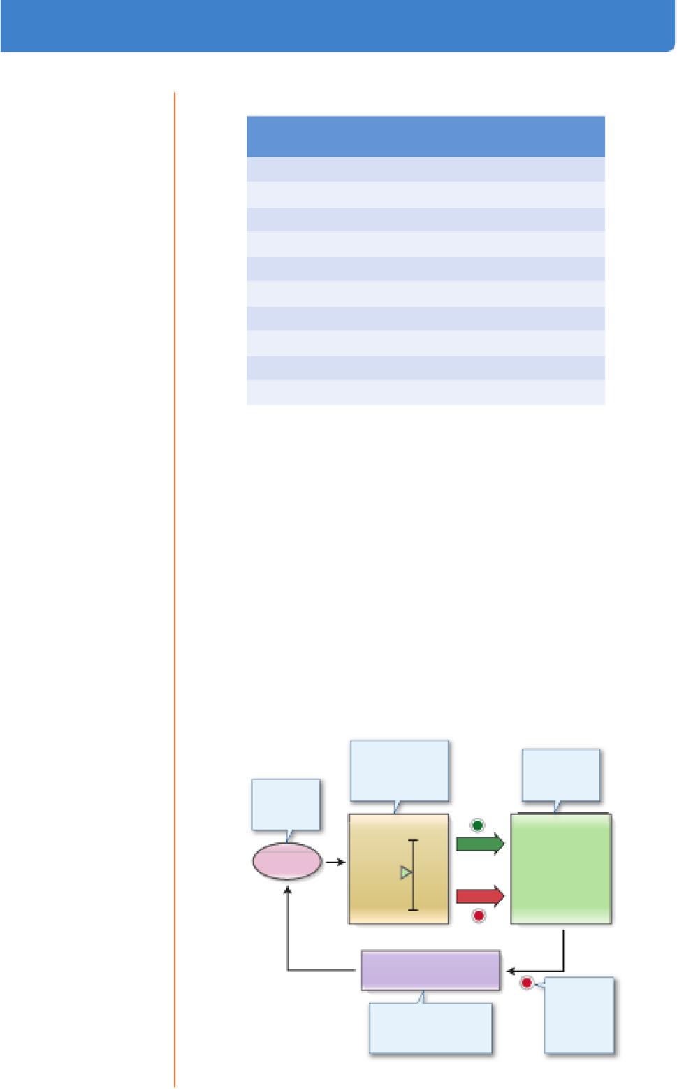
d. A negative feedback control system has the following elements (Figure 1-1):
i A set point value, which is at the cente.r of the normal range and is treated by the control system as the target value.
ii Senson that monitor the controlled variable.
ill. A comparator, which interprets input from the sensors to determine when deviations from the set point have occurred. The comparator initiates a counter response.
iv: Eirectors are the mechanisms that restore the set point
e. Using the control of blood pressure as an example:
i 1he confl'olled variable is mean arterial blood pressure (MAP).
ii 1he normal set point for MAP is approximately 95 mm Hg.
ill. Pressure sensors are located in the carotid sinus and relay information to a comparator located in the central nervous system. Measures controlled varlable
Figure 1-1. Components of a negative feedback control system.
iv. If MAP suddenly changes, the activity of effectors (e.g., cardiac contractility, vascular tone, and urinary fluid excretion) is altered to restore normal blood pre5sure.
4. The internal environment.
a. The purpose ofhomeostasis is to provide an optimal fluid environmentfor cellular function.
b. The body fluids are divided into two major functional compartment&: i Intraccllular fluid (ICF) is the fluid inside cells.
ii Emacellalar fluid (BCF) is the ftuid outside cells, which is subdivided into the lnterstitlal fluid and the blood plasma.
c. The concept of an internal environment in the body correlates with the interstitial fluid bathing cells.
d. There is free exchange of water and small solutes in the ECF between interstitial fluid and plasma across the blood capillaries.
e. Exchange between interstitial ftuid and ICF is highly regulated and occurs across cell membranes.
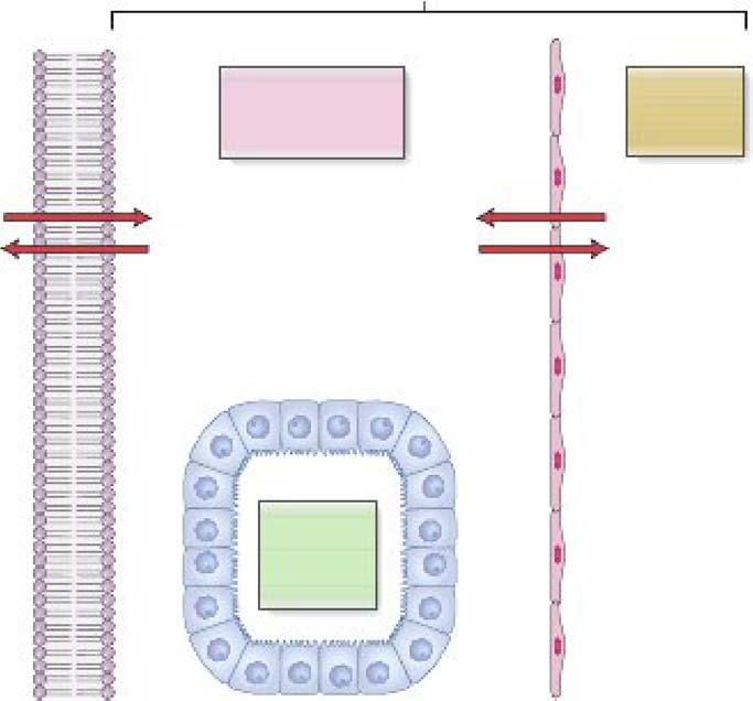
f. The volume of total body water is approximately 60% of the body weight in men and 50% in women.
i About 60% of the total body water is ICF and 40% is ECF (Figure 1-2).
INTRACELLULAR FLUID
25L
[Na+J=15mM
[K+]= 120 mM
[CrJ=20mM
[ProlBln] = 3 mM
OSmolarlty =285 mOSM
EXTRACELLULAR FLUID
INlERSTITIAL FLUID 13L
(Na+]= 145 mM
(K+]=4.2mM
(Ct"] = 113 mM
(Protein] = OmM
OSmolarlty = 285 mOsM
Cell membranes
Epithelial cells I • capillary endo1hella
BLOOD PLASMA
3L
(Na+] = 142 mM
(K+]=4mM
(Ct"] = 103 mM
(Protein] = 1 mM
0Smolar1ty = 285 mOsM
Figure 1-2. Body fluid compartments. Intracellular fluid (ICF} is separated from extracellular fluid (ECF) by cell membranes. ECF is composed of the interstitial fluid bathing cells and the blood plasma within the vascular system. Interstitial fluid is separated from plasma by capillary endotnelia. Transcellular fluid is part of the ECF and includes epithelial secretions such as the cerebrospinal and extraccular fluids. ECF has a high [Na+] and a low [K+J, whereas the opposite Is true of ICF. All compartments have the same osmolarlty at steady state.
ii Approximately 80% of the ECF is interstitial fluid and the remaining 20% is blood plasma.
iii ECF is high in NaCl and low in K+, whereas ICF is high in K+ and low in NaCL
iv. Interstitial fluid is similar in composition to plasma. except that interstitial fluid has almost no protein.
v. Osmolarity is the same in all compartments.
g. 'Y Fluid can move freely from the interstitial to plasma compartments and helps to maintain blood volume during hemorrhage.
i Because approximately 8096 of the ECF is interstitial :fluid and 20% is blood plasma. a hemorrhaging patient must lose about 5 L of ECP before the plasma volume is deaeased by 1 L.
ii 'Ihe reverse is also tru.e; to replace 1 L of plasma volume, approximately 5 L of intravascular isotonic saline must be infused. Y
Membrane Transport Mechanisms
1. lhe transport of solutes across cell membranes is fundamental to the survival of all cells. Specializations in membrane transport mechanisms often underlie tissue function. For example, voltage-sensitive ion channels account for the ability to generate electrical signals.
2. Cell membranes separate the cytosol from the ECF.
a. Cell membranes are formed from phospholipid.s that are an effective barrier against the free movement of most water-soluble solutes.
b. Most biologically important substances require a protein-mediated pathway to cross cell membranes.
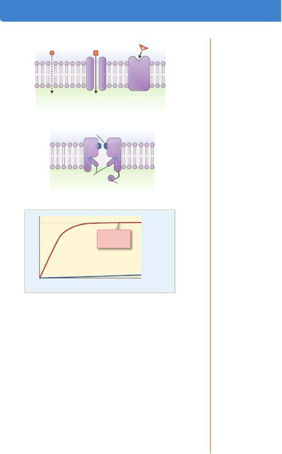
3. Solute transport can be categorized based on the use of cellular energy or the type of transport pathway (Figure 1-3):
a. Active transport requires adenosine triphosphate (ATP) hydrolysis.
i Primary active tranaport occurs via membrane proteins that directly coupk ATP hydrolysis to solute movement.
ii Secondary active transport couples the transport of two or more solutes together. Energy is used to develop a favorable electrochemical driving force for one solute, which is then used to power the transport of other solutes (e.g., the inwardly directed Na+ gradient is used to drive glucose uptake from the intestine).
b. Passive transport does not require ATP hydrolysis or coupling to another solute.
c. Primary active transporters (Figure l-4A):
i. The Na+/I{+-ATPaae (known as the •sodium pump•) is present in all cells and transports 3Na+ out of a cell in exchange for 2K+, using 1 ATP molecule in each transport cycle. The action of sodium pumps accounts for high Na+ concentration in BCP and high K+ conuntration inICP.
ii <A.i+-ATPases are located in the plasma membrane and endoplasmic reticulum membrane and function to maintain very low intracellular [or+].
iii ff+JK+-ATPasea pump H+ out of cells in exchange for K+ and are present in several epithelia. H+/K+ -ATPase is responsible for the secretion of acidic gastric juice in the stomach.
ENERGY REQUIREMENT
PASSIVE
PRIMARY
AC'TlVE TRANSPORT
SIMPLE FACILIU\TED SECONDARY
ENDOCYTOSIS DIFFUSION DIFFUSION ACTIVE EXOCYTOSIS TRANSPORT
LIPID TRANSPORT PROTEIN VESICLE BILAYER
TRANSPORT MECHANISM
Figure 1-3. Classrflcatlon of membrane transport systems.
Plasma 3Na+ t 7.:"oP+P1 2K+
ADP+P; ATP
Selected organic D solutes
Cytoplasm
B.
Plasma membrane \
Cotransporters Na+ Glucose Na+ Cr
Smooth endoplasmlc reticulum Na+ Peptide Na+
Exchangers 3Na+ er
Cytoplasm
Figure 1-4. Active transport. A. Examples of primary active transporters (ATPases) in the plasma membrane and in organelles. B. Examples of secondary active transporters; cotransporters transport solutes in the same direction, and exchangers transport solutes in opposite directions. ADP, adenosine di phosphate; ATP, adenosine triphosphate; MOR, multidrug resistance.
iv. ff+-ATPases are mainly expressed inside cells, including the vacuolar ff+-ATPase, which acidifies lysosomes; ATP sy:nthase is a form ofH+-ATPase. which operates in reverse to synthesize ATP in mitochondria.
v. The multidrug resistance (MDR) transporters are ATPases that extrude a wide variety of organic molecules from cells. MDRs are physiologically expressed in the liver, kidney. and blood-brain barrier.
• 'T The expression ofMDR transporters (e.g., P-glycoprotein) is one mechanism by which bacteria and cancer cells can become drug resistant The effectiveness of a drug will be reduced if it is transported out of the target cell by MDR transporters. •
d. There are many examples of secondary active transporters (Figure 1-4B):
i. Cotransporters (symporters) couple the movement of two or more solutes in the same direction.
• Examples of Na+-driven cot:ransporters include Na+/glucose uptake in the intestine and diuretic-sensitive Na+JK+/Cl- and Na+/Cluptake in the kidney.
• H+/peptide cotransport in the intestine is an example ofNa+independent cotransport
ii Euhangen (antiporters) couple the movement of two solutes in the opposite direction.
• Na+-driven antiporters include Na+/Ca2+ and Na+IH+ exchange, which are important for maintaining low intracellular [Cal+] and [H+], respectively.
• c1-1Hco3 - exchange is an example of an anion exchanger. It is widely expressed, for example, in red blood cells, where it assists in HCO, - transport into and out of the cell as part of the blood-C02 transport system.
e. Passive transport can only occur along a favorable electrochemical gradient
f. Simple passive transport is characterized by a linear relationship between the transport rate and the electrochemical driving force.
g. Pathways for simple passive transport include diffusion through the lipid bilayer or via pores or channels in the membrane (Figure 1-SA).
h. Fidt's law of ditfasion describes the simple diffusion of an uncharged solute (s):
J, = Net flux per unit area
P, = Permeability
.6.C, = Concentration difference of s across the membrane
i. Permeability is a single coefficient relating the driving force for diffusion to net11ux.
i The membrane permeability to a solute is proportional to the lipid solubility of the solute and inversely proportional to its molecular size.
ii Gases are an example of molecules that are able to move through the lipid bilayer ofcell membranes by simple diffusion because they are small and lipid soluble.
Equation 1-1
A. Roulu avall•ble far pmlve tranaport
Simple diftusion through lipid bilayer
Simple diffusion through channel
B. Companentll at Ion ch•nne18
Facili1at&d diffusion through unlpor1Br
C. 1<1ne11e11 at pM8M! 1n11111pan
Facilitated dtTualon
Simple diffusion through External solute concentration bilayer
Figure 1-5. A. Passive transport pathways. B. General components of Ion channels components. C. Kinetics of passive transport. Note the linear relatlonshlp between simple diffusion and flux; facllltated diffusion via unlporters Is faster than simple diffusion but Is saturable
j The passive transport of ions and other small water-soluble molecules across a cell membrane requires transport proteins that span the membrane.
k. Ion cb•nnela are the most numerous example of passive transporters . Ion channels have the following general components (Figure 1-S B):
i A pore through which ions diffuse.
ii A leledhity filter within the pore, causing the channel to be highly selective for a particular ion (e g., Na+ channels)
iii A pting .mec;haniam that opens and closes the channel; gates may be controlled by membrane voltage (voltage-gated dtaoneh) , chemicals (ligand-gated c;hannel1), or mechanical forces in the membrane (e.g., atretch- activated c;hannela).
I. Passive transport can also occur via unfporten, which selectively bind a single solute at one side of the membrane and undergo a conformational change to deliver it to the other side.
i Solute transport via uniporters is called fadlitated. diffaaion because it is faster than simple diffusion (Figure 1-SC).
ii A characteristic feature offacilitated diffusion is the saturation of the transport rate at high solute conantrations.
ill. The GLUT family are examples of uniporters for glucose transport that are expressed in many tissues.
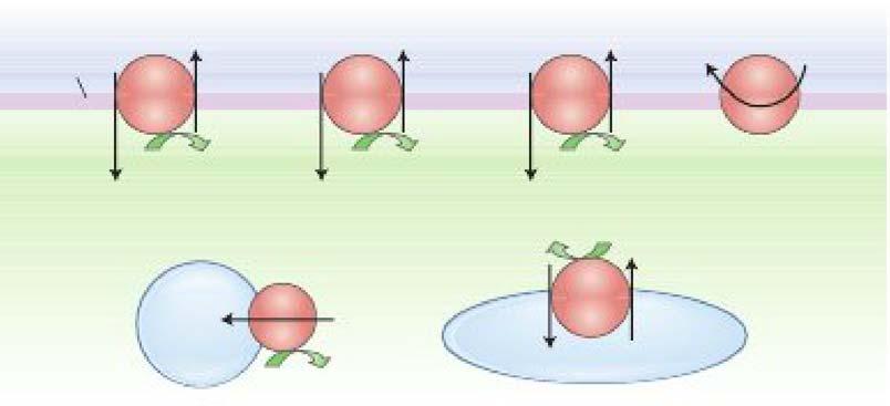
m. Macromolecules are transported between tlu! ICP and the ECF using membrane-limited vesicles.
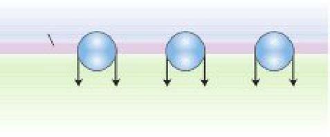
i Endoc:ytollls is the ingestion of extrac:ellular material to form endocytic vesicles Jnside a cell. There are three typea of endocytosit:
• Pinocytosis is the ingestion of small particles and ECF that occurs constitutively in most cells.
• Phagoc:ytosis is the uptake oflarge particles (e.g., microorganisms) that occurs in specialized immune cells.
• Receptor-mediated endocytosis allows uptake of specific molecules and occurs at specialized areas of membrane called d.athrin-coated pits (e.g., uptake of cholesterol from low-density lipoproteins).
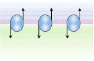
ii Emqtosis is export of soluble proteins into the extracellular space by vesicular transport When vesicles containing proteins fuse with the plasma membrane, the soluble proteins are secreted and the vesicle membrane is incorporated in the plasma membrane. There are two pathways for a:ocytoais:
• The constitutive pathway is present in most cells and is used to export extracellular matrix proteins.
• The regulated pathway is present in cells that are specialized for the secretion of proteins such as hormones, neurotransmitters, and digestive enzymes. An increase in tlu! intracellular c;aa+ conuntration is a event that triggers regulated exocytosis.
• 'Y Lambert-Eaton syndrome is a neurologic condition resulting from autoantibodies that bind to and block Ca* channels on the presynaptic motor nerve terminals. By blocking the cau channels, the Ca2+-dependent exocytosis of vesicles filled with acetylcholine (a neurotransmitter needed for muscle contraction) is inhibited, resulting in muscle weakness. 'f'
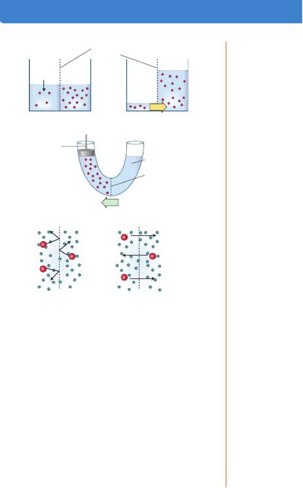
4. Osmosis.
a. Water transport across a barrier is always passive, driven eitlu!r by a diffusion gradU!nt or by a hydrostatic pressure gradient.
b. Osmosis is water movement that is driven by a water concentration gradient across a membrane (Figure l-6A).
c. Water concentration is expressed in terms of total solute concentration; the more dilute a solution, the lower its solute concentration and the higher its water concentration.
d. When two solutions are separated by a semipermeable membrane (i.e., one that allows the transport of water but not solutes), water moves by osmosis awayfrom the more dilute solution.
e. Omwlarity is an expression of the osmotic strength of a solution and is the total solute concentration.
i Osm.olarity is the product of the molar solute c:onc:entration and the number ofparticles that the solute dissociates into when dWolved. For example:
A. Oammla Semipermeable membrane -... _
Dilute
so lution
llme=O SlBady state
B. Concept of oemollc preuure
Force per unit =
water area (preBSure) needed to p1'9119nt osmotic watBr flow = osmotic pressure
Semipenneable
membrane
Oemotlo force for walllr flux
C. Rdecllon caetllclent
Figure 1-6. Osmosis. A. Illustration of osmotic water movement across a semi permeable membrane. L The concept of osmotic pressure. C. Reflection coefficients: Solutes that do not permeate the membrane exert all their osmotic pressure (a = 1); freely permeable solutes (a = O) do not exert any osmotic pressure.
• 1 mol of glucose dissolved in 1 L of water produces a solution of 1 Osm/L.
• 1 mol of NaCl dissolved in 1 L of water produces a solution of approximately 2 Osm/L
iL Two solutions of the same osmolarity are termed ilo111Dotic. A solution with a greater osmolarity than a reference solution is said to b e hyperoamotic, and a solution of lower osmolarity is described as hypollDlOtic.
iii Osmolarity can be converted into units of pressure, which allow osmotic and hydrostatic pressure gradients to be mathematically combined; for example, when considering fluid filtration across capillary walls (see Chapter 4).
iv. The concept of osmotic pressure (n) is illustrated in Figure l-6B and is calculated by vuit Hoff law: 1t = gxCxRT
Equation 1-2
g = Number of particlet produced when the solute dissociates in solution
C = Molar solute concentration
R = Gas constant
T = Temperature
f. Although blood plasma contains many solutes, a simplified clinical estimate ofp1aama osm.olarity can be obtained by considering only the Na+, glucose, and urea concentrations:
Po.m =2P11 +(Pp.a... /18)+(P_ /2.8)
Po.m. = Plasma osmolarity (mOsm/L)
PNa= Plasma [Na] (mEq/L)
P.,..._=Plasma [glucose] (mg/dL)
P = Plasma [urea] (mg/dL)
Equation 1-3
g. A difference between the measured. and estimated osmolarity is called an oamolar gap and is caused by the presence ofadditional solutes in plasma.
i Y Patients with alc:ohol lnto:lication or ethylene glycol poisoning will have an increased osmolar gap. Y
h. The concept of effective osmolarity (tonicity} includes the effect of solute permeation through membranes (Figure l-6C).
i Most biologic membranes are not completely semipermeable.
ii When the membrane is permeated by the solute, the observed osmotic pressure gradient is reduced.
ill. The re&ection coefficient (CJ') is the fraction of the measured osmolarity actually applied:
• a = 1.0 fur solutes that do not permeate a membrane.
• CJ = 0 when the membrane is freely permeable to the solute.
• The effective osmolarity of a solution is calculated as the product of the osmolarity and the refiection coefficient (e.g.• ifu = 0.5, the effective osmolarity is only 50% of the measured osmolarlty).
iv. The terms isotonic, hypotonic, and hypertonic are used to describe the effective osmolarity of a solution relative t.o a cell:
• An Isotonic solution has the same effective osmolarity as the cell and causes no net water movement.
• A hypotonic solution has a smaller effective osmolarity and causes cells to swell.
• A hype:rtonic solution has a larger effective osmolarity and causes cells to shrink.
v: Y At steady state the ECF is isotonic with respect to the ICF because water moves freity across most all numbranes. When the steady state is temporarily disrupted. water moves witil ICP and ECP tonidty again becomes equal. For example, if a patient is intravenously infused with a h}'potonic saline aolnlion, the ECF tonicity is initially decreased and some water moves into the ICF by osmosis (ie., cells swell). T
Membrane Potentials
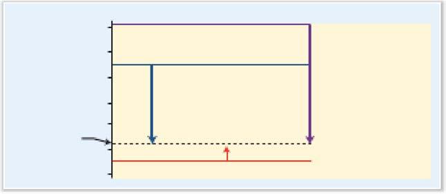
1. All living cells have a membrane potential difference in which the cytoplasm is negative with respect to the ECF (Figure 1-7A).
A. ANdng membrane poiMtlal
C. Electrochemlcal gradle!Q
("our)
=+123mV
ENa =+61 mV
(Vm-Ec:.) =-197 mV
(Inward
Figure 1-7. A. The resting membrane potential; all cells have a negative intracellular potential. B. Generation of a K+ diffusion potential. In this example, K+ is the only permeable ion; a small amount of K+ diffuses to the lower compartment, creating a negative potential in the upper compartment The Nernst equation predicts the equ!llbrlum potentlal (voltage), based on the size of the K+ concentration ratio between compartments. c. Electrochemlcal gradients. Membrane potentlal (V.) Is shown by the dashed llne. Downward arrows Indicate gradients for cation flux Into the cell; the upward arrow Indicates a gradient for catton efflux.
2. Membrane potentials arise because there are stable ion diffusion gradients across the membrane and because cell membranes contain ion channels that provide seleatve ion permeability.
3. The following steps are involved in the development of a diffiW.on potential:
a. In the example in Figure 1-78. two potassium. chloride (KCl) solutions are separated by a membrane that is permeable to K+ but not to et-.
b. x:+ diffiues from the upper to the lower compartment, down its concentration gradient. In this case. Cl- cannot follow.
c. A voltage d11ference develops as the K+ ions leave the upper compartment, leaving a net negative charge behind. A ..,ery slight separation ofKCI ion pairs is enough to generate physiologic voltages.
d. 1he negative potential in the upper compartment attracts K+ ions and opposes the K+ concentration gradient.
e. An equilibrium potentlal is established when the voltage difference and the concentration gradient are equal but opposite driving forces. At the f4.UIUbrium potenttai there l.s no net mcwement ofK+.
f. 1his simulated example is analogous to most resting cells, which contain a high [K+] and have numerous open K+ channels at rest (note, however, that the major intracellular anion in cells is protein, not Cl-).
4. The equilibrium potential is a function of the size of the ion concentration gradient (Table 1-3) and is calculated using the Nern1t equation: