The material in this eBook also appears in the print version of this title: ISBN: 978-1-25-983433-2, MHID: 1-25-983433-6.
eBook conversion by codeMantra Version 1.0
All trademarks are trademarks of their respective owners. Rather than put a trademark symbol after every occurrence of a trademarked name, we use names in an editorial fashion only, and to the benefit of the trademark owner, with no intention of infringement of the trademark. Where such designations appear in this book, they have been printed with initial caps.
McGraw-Hill Education eBooks are available at special quantity discounts to use as premiums and sales promotions or for use in corporate training programs. To contact a representative, please visit the Contact Us page at www.mhprofessional.com.
Notice
Medicine is an ever-changing science. As new research and clinical experience broaden our knowledge, changes in treatment and drug therapy are required. The authors and the publisher of this work have checked with sources believed to be reliable in their efforts to provide information that is complete and generally in accord with the standards accepted at the time of publica-tion. However, in view of the possibility of human error or changes in medical sciences, neither the authors nor the publisher nor any other party who has been involved in the preparation or publication of this work warrants that the information contained herein is in every respect accurate or complete, and they disclaim all responsibility for any errors or omissions or for the results obtained from use of the information contained in this work. Readers are encouraged to confirm the informa-tion contained herein with other sources. For example and in particular, readers are advised to check the product information sheet included in the package of each drug they plan to administer to be certain that the information contained in this work is accurate and that changes have not been made in the recommended dose or in the contraindications for administration. This recommendation is of particular importance in connection with new or infrequently used drugs.
TERMS OF USE
This is a copyrighted work and McGraw-Hill Education and its licensors reserve all rights in and to the work. Use of this work is subject to these terms. Except as permitted under the Copyright Act of 1976 and the right to store and retrieve one copy of the work, you may not decompile, disassemble, reverse engineer, reproduce, modify, create derivative works based upon, transmit, distribute, disseminate, sell, publish or sublicense the work or any part of it without McGraw-Hill Education’s prior consent. You may use the work for your own noncommercial and personal use; any other use of the work is strictly prohibited. Your right to use the work may be terminated if you fail to comply with these terms.
THE WORK IS PROVIDED “AS IS.” McGRAW-HILL EDUCATION AND ITS LICENSORS MAKE NO GUARANTEES OR WARRANTIES AS TO THE ACCURACY, ADEQUACY OR COMPLETENESS OF OR RESULTS TO BE OBTAINED FROM USING THE WORK, INCLUDING ANY INFORMATION THAT CAN BE ACCESSED THROUGH THE WORK VIA HYPERLINK OR OTHERWISE, AND EXPRESSLY DISCLAIM ANY WARRANTY, EXPRESS OR IMPLIED, INCLUDING BUT NOT LIMITED TO IMPLIED WARRANTIES OF MERCHANTABILITY OR FITNESS FOR A PARTICULAR PURPOSE. McGraw-Hill Education and its licensors do not warrant or guarantee that the functions contained in the work will meet your requirements or that its operation will be uninterrupted or error free. Neither McGraw-Hill Education nor its licensors shall be liable to you or anyone else for any inaccuracy, error or omission, regardless of cause, in the work or for any damages resulting therefrom. McGraw-Hill Education has no responsibility for the content of any information accessed through the work. Under no circumstances shall McGraw-Hill Education and/or its licensors be liable for any indirect, incidental, special, punitive, consequential or similar damages that result from the use of or inability to use the work, even if any of them has been advised of the possibility of such damages. This limitation of liability shall apply to any claim or cause whatsoever whether such claim or cause arises in contract, tort or otherwise.
Emil A. Tanagho,
& Tom F. Lue, MD, ScD (Hon), FACS
Emil A. Tanagho, MD; Hiep T. Nguyen, MD; & Michael DiSandro, MD
Benjamin N. Breyer, MD, MAS,
Mary
Anobel Y. Odisho, MD, MPH; Sima P. Porten, MD, MPH; & Kirsten L. Greene, MD, MS
Daniela Franz, MD; Scott Gerst, MD; & Hedvig Hricak, MD,
Benjamin
David B. Bayne, MD,
Joachim W. Thüroff, MD; Rolf Gillitzer, MD; & Thomas Chi,
Badrinath R. Konety, MD,
& Peter R. Carroll, MD, MPH
Anobel Y. Odisho, MD,
& Kirsten L. Greene, MD, MS
Matthew R. Cooperberg, MD,
Samuel L. Washington III, MD; & Peter R. Carroll, MD,
Sima P. Porten, MD, MPH; & Joseph C. Presti, Jr.,
Maxwell V.
Peter R. Carroll, MD,
Vadim
Michelle L. McDonald, MD; & Christopher J. Kane, MD, FACS
David B. Bayne, MD, MPH; Jack W. McAninch, MD, FACS, FRCS(E)(Hon); & Thomas Chi, MD
Brian K. Lee,
Flavio G. Vincenti, MD
Brian K. Lee,
& Flavio G. Vincenti, MD
Brian K. Lee, MD; & Flavio G. Vincenti, MD
John M. Barry,
Arpita Desai,
Yun Rose Li, MD, PhD; Alexander R. Gottschalk, MD, PhD; & Mack Roach III, MD
Barry A. Kogan,
Samuel L. Washington III, MD; & Katsuto Shinohara,
Amanda B. Reed-Maldonado, MD, FACS; & Tom F. Lue, MD
Alan W. Shindel, MD, MAS ; & Tami S. Rowen, MD, MS
Benjamin N. Breyer, MD, MAS, FACS; & Jack W. McAninch, MD, FACS, FRCS(E)(Hon)
Laurence S. Baskin,
Thomas J. Walsh, MD, MS; & James F. Smith, MD, MS
James F. Smith, MD, MS; Bogdana Schmidt, MD, MPH; & Thomas J. Walsh, MD, MS
June M. Chan, ScD; David Tat, DO; & Stacey Kenfield, ScD
This page intentionally left blank
Contributors
Karl-Erik Andersson, MD, PhD
Institute for Regenerative Medicine
Wake Forest University School of Medicine
Winston Salem, North Carolina
Susan Barbour, RN, MS, WOCN
Palliative Care Services
UCSF School of Medicine
San Francisco, California
John M. Barry, MD
Professor of Urology and Professor of Surgery
Division of Abdominal Organ Transplantation
Organ Health and Science University
Portland, Oregon
Laurence S. Baskin, MD
Chief of Pediatric Urology
University of California Children’s Medical Center
UCSF School of Medicine
San Francisco, California
Attending Urologist
Children’s Hospital Oakland Oakland, California
David B. Bayne, MD, MPH
Endourology Fellow
Department of Urology
UCSF School of Medicine
San Francisco, California
Benjamin N. Breyer, MD, MAS, FACS
Associate Professor and Vice Chair
Department of Urology
UCSF School of Medicine
San Francisco, California
Peter R. Carroll, MD, MPH
Professor
Ken and Donna Derr-Chevron Endowed Chair in Prostate Cancer
Department of Urology
UCSF School of Medicine
San Francisco, California
June M. Chan, ScD
Program Director, Genitourinary Cancer Epidemiology and Population Sciences
Department of Urology
UCSF School of Medicine
San Francisco, California
Thomas Chi, MD
Associate Professor and Katzman Endowed Professor in Clinical Urology
Department of Urology
UCSF School of Medicine
San Francisco, California
Matthew R. Cooperberg, MD, MPH
Associate Professor Department of Urology
Helen Diller Family Comprehensive Cancer Center
UCSF School of Medicine
San Francisco, California
Hillary L. Copp, MD, MS
Associate Professor of Urology and Pediatric Urology Fellowship Director
Benioff Children’s Hospital
UCSF School of Medicine
San Francisco, California
Donna Y. Deng, MD, MS
Neurourology Lead, Kaiser Permanente Northern California
Medical Director, Kaiser NorCal Regional Spina Bifida Program
Associate Fellowship Director, Female Pelvic Medicine
Reconstructive Surgery, Kaiser East Bay/UCSF
Oakland, California
Arpita Desai, MD
Clinical Instructor
Department of Genitourinary Medical Oncology
Helen Diller Family Comprehensive Cancer Center
UCSF School of Medicine
San Francisco, California
Michael DiSandro, MD
Professor of Urology Department of Urology
UCSF School of Medicine
San Francisco, California
Daniela Franz, MD
Department of Diagnostic and Interventional Radiology
Klinikum rechts der Isar Munich Technical University Munich, Germany
Thomas W. Gaither, MD, MAS
Urology resident University of California Los Angeles, California
Maurice M. Garcia, MD, MAS
Associate Professor of Urology and Anatomy (Adjunct) Departments of Urology and Anatomy
UCSF Medical Center
San Francisco, California
Director, Cedars-Sinai Transgender Surgery and Health Program
Division of Urology
Cedars-Sinai Medical Center
Los Angeles, California
Scott Gerst, MD
Associate Attending Physician Department of Radiology
Memorial Hospital, Memorial Sloane-Kettering Cancer Center
New York, New York
Rolf Gillitzer, MD Clinical Director Department of Urology
Johannes Gutenberg University Medical Center Mainz Mainz, Germany
Roy L. Gordon, MD
Professor of Interventional Radiology Department of Radiology
UCSF School of Medicine
San Francisco, California
Alexander R. Gottschalk, MD, PhD
Professor of Radiation Oncology
Director of CyberKnife Departments of Radiation and Oncology
UCSF School of Medicine
San Francisco, California
Kirsten L. Greene, MD, MS
Professor and Chair Department of Urology University of Virginia Charlottesville, Virginia
Hedvig Hricak, MD, PhD Chair
Department of Radiology
Memorial Sloan-Kettering Cancer Center Professor of Radiology
Cornell University
New York, New York
Christopher J. Kane, MD, FACS
Dean of Clinical Affairs
UC San Diego School of Medicine
CEO, UC San Diego Health Physician Group La Jolla, California
Stacey A. Kenfield, ScD
Associate Professor Department of Urology
UCSF School of Medicine San Francisco, California
Barry A. Kogan, MD
Professor, Surgery and Pediatrics Falk Chair in Urology
Albany Medical College Albany, New York
Ryan Kohlbrenner, MD
Assistant Professor of Interventional Radiology Departments of Radiology and Biomedical Imaging
UCSF School of Medicine San Francisco, California
Badrinath R. Konety, MD, MBA
Associate Dean for Innovation Professor of Urology
Director of the Institute for Prostate and Urologic Cancers University of Iowa Iowa City, Iowa
Vadim S. Koshkin, MD
Assistant Clinical Professor Genitourinary Medical Oncologist Departments of Hematology and Oncology
UCSF School of Medicine San Francisco, California
John N. Krieger, MD
Professor of Urology
University of Washington School of Medicine
Seattle, Washington
Brian K. Lee, MD
Professor of Medicine
The Connie Frank Kidney Transplant Center
UCSF School of Medicine
San Francisco, California
Yun Rose Li, MD, PhD
Resident Physician
Departments of Radiation and Oncology
UCSF School of Medicine
San Francisco, California
Tom F. Lue, MD, FACS, ScD (Hon) Professor of Urology
Emil Tanagho Endowed Chair in Clinical Urology Department of Urology
UCSF School of Medicine
San Francisco, California
Kristin Madden, PharmD
Pharmacist
Department of Veterans Affairs
San Antonio, Texas
Jack W. McAninch, MD, FACS, FRCS(E)(Hon)
Professor of Urology
UCSF School of Medicine
San Francisco, California
Michelle L. McDonald, MD Urologist
San Diego, California
Maxwell V. Meng, MD, MPH
Professor Department of Urology
UCSF School of Medicine
San Francisco, California
Hiep T. Nguyen, MD
Associate Professor Surgery and Urology
Harvard Medical School and Children’s Hospital Boston, Massachusetts
Anobel Y. Odisho, MD, MPH
Assistant Professor Department of Urologic Oncology
UCSF School of Medicine
San Francisco, California
Sima P. Porten, MD, MPH
Assistant professor Department of Urology
UCSF School of Medicine
San Francisco, California
Joseph C. Presti, Jr., MD
Lead for Urologic Oncology
Kaiser Permanente Northern California
Oakland, California
Amanda B. Reed-Maldonado, MD, FACS
Chief, Male Reproductive Urology Department of Urology
Tripler Army Medical Center
Honolulu, Hawaii
Mack Roach III, MD
Professor of Radiation Oncology and Urology Department of Urology
UCSF School of Medicine
San Francisco Comprehensive Cancer Center
San Francisco, California
Tami S. Rowen, MD, MS
Assistant Professor Departments of Obstetrics, Gynecology, and Reproductive Sciences
UCSF School of Medicine
San Francisco, California
Bogdana Schmidt, MD, MPH
Urologic Oncology Fellow
Stanford University Medical Center Stanford, California
Alan W. Shindel, MD, MAS
Associate Professor Department of Urology University of California Davis, California
Katsuto Shinohara, MD Professor
Helen Diller Family Chair in Clinical Urology Department of Urology
UCSF School of Medicine
San Francisco, California
Eric J. Small, MD
Professor of Medicine and Urology Urologic Oncology Program and Program Member, Comprehensive Cancer Center
UCSF School of Medicine
San Francisco, California
James F. Smith, MD, MS
Associate Professor Director, Male Reproductive Health Departments of Urology, Obstetrics, Gynecology, and Reproductive Sciences
UCSF School of Medicine San Francisco, California
Marshall L. Stoller, MD Professor of Urology Department of Urology
UCSF School of Medicine
San Francisco, California
Anne M. Suskind, MD, MS, FACS
Associate Professor of Urology, Obstetrics, Gynecology, and Reproductive Sciences Director, Neurourology, Female Pelvic Medicine & Reconstructive Surgery
UCSF School of Medicine
San Francisco, California
Emil A. Tanagho, MD Professor of Urology Department of Urology
UCSF School of Medicine San Francisco, California
David Tat, DO Infectious Disease Specialist
Moses H. Cone Memorial Hospital Greensboro, North Carolina
Joachim W. Thüroff, MD Professor Department of Urology University Medical Center Mannheim, Germany
Flavio G. Vincenti, MD Professor of Medicine
The Connie Frank Kidney Transplant Center
UCSF School of Medicine San Francisco, California
Thomas J. Walsh, MD, MS Associate Professor Department of Urology University of Washington School of Medicine Seattle, Washington
Mary K. Wang, MD Childrens’ Urology Austin, Texas
Samuel L. Washington, III, MD Urologic Oncology Clinical Fellow Department of Urology
UCSF School of Medicine San Francisco, California
J. Stuart Wolf, Jr., MD, FACS Professor, Department of Surgery and Perioperative Care
Dell Medical School
The University of Texas at Austin Austin, Texas
Preface
Smith & Tanagho’s General Urology, nineteenth edition, provides the updated information for the understanding, diagnosis, and treatment of urological diseases in a concise and well-organized format. The book is up-to-date, to the point, and readable. Medical students will find this book useful because of its concise, easy-to-follow format, and its breadth of information on common urological diseases. Residents, as well as practicing physicians in urology, family practice, or general medicine, will find it an efficient and current reference, particularly because of its emphasis on diagnosis and treatment.
This nineteenth edition has been thoroughly updated with clinical information and current references. The reader will find that this edition is written in an uncomplicated, straightforward manner that provides relevant clinical information and guidelines for diagnosis and management of urologic conditions. Chapters on immunotherapy in urologic malignancies, radiotherapy of urologic tumors, urinary incontinence, and vascular interventional radiology have all undergone extensive revision. For this current edition, we have added two chapters on the timely topic of gender dysphoria and introduction to clinical research design.
Many illustrations and figures have been modernized and improved with added color. The classic fine anatomic drawings demonstrate well the important clinical findings.
This book has been one of the leading sources of information for students, trainees, and urologists around the world. In addition to English, this book has been published in many other foreign languages, like Chinese, French, Greek, Italian, Japanese, Korean, Portuguese, Russian, Spanish, and Turkish.
We greatly appreciate the patience and efforts of our McGraw-Hill staff, the expertise of our contributors, and the support of our readers.
Jack W. McAninch, MD, FACS, FRCS(E) (Hon) Tom F. Lue, MD, FACS, ScD (Hon) San Francisco, California, January 2020
This page intentionally left blank
Anatomy of the Genitourinary Tract
Emil A. Tanagho, MD; & Tom F. Lue, MD, ScD (Hon), FACS
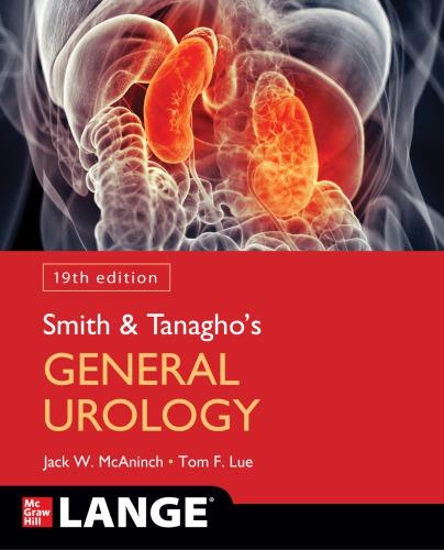
Urology deals with diseases and disorders of the adrenal gland, the male genitourinary tract, and the female urinary tract. These systems are illustrated in Figures 1–1 and 1–2.
ADRENALS
▶ Gross Appearance
A. Anatomy
Each kidney is capped by an adrenal gland, and both organs are enclosed within Gerota’s (perirenal) fascia. Each adrenal gland weighs 4–5 g. The right adrenal is triangular in shape; the left is more rounded and crescentic. The average dimensions are 3 cm width, 5 cm length, and 1 cm thickness. Each gland is composed of a cortex, chiefly influenced by the pituitary gland, and a medulla derived from chromaffin tissue (Avisse et al, 2000; O’Donoghue et al, 2010).
B. Relations
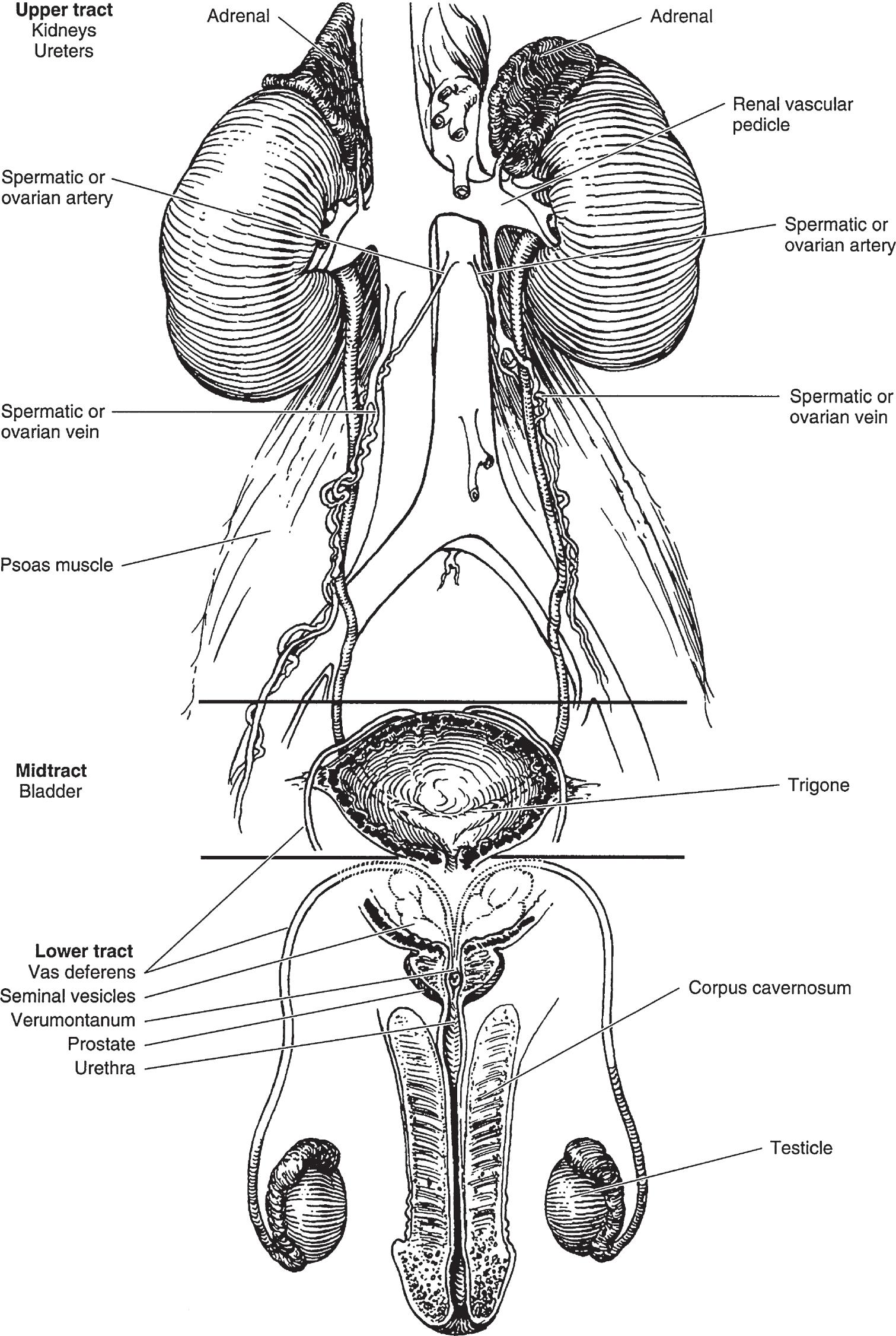
Figure 1–2 shows the relationships between the adrenals and other organs. The right adrenal lies between the liver and the vena cava. The left adrenal lies close to the aorta and is covered on its lower surface by the pancreas. The spleen lies superior and lateral to it.
▶ Histology
The adrenal cortex, which makes up 85% of the mass, is composed of three distinct layers: the outer zona glomerulosa, the middle zona fasciculata, and the inner zona reticularis. The medulla lies centrally and is made up of polyhedral cells with hormone-containing granular cytoplasm. These chromaffin cells are accompanied by a small number of sympathetic ganglion cells.
▶ Blood Supply
A. Arterial
Each adrenal gland receives three arteries: one from the inferior phrenic artery, one from the aorta, and one from the renal artery.
B. Venous
Blood from the right adrenal gland is drained by a very short vein into the vena cava; the left adrenal vein terminates in the left renal vein.
▶ Lymphatics
The lymphatic vessels accompany the suprarenal vein and drain into the lumbar lymph nodes.
KIDNEYS
▶ Gross Appearance
A. Anatomy
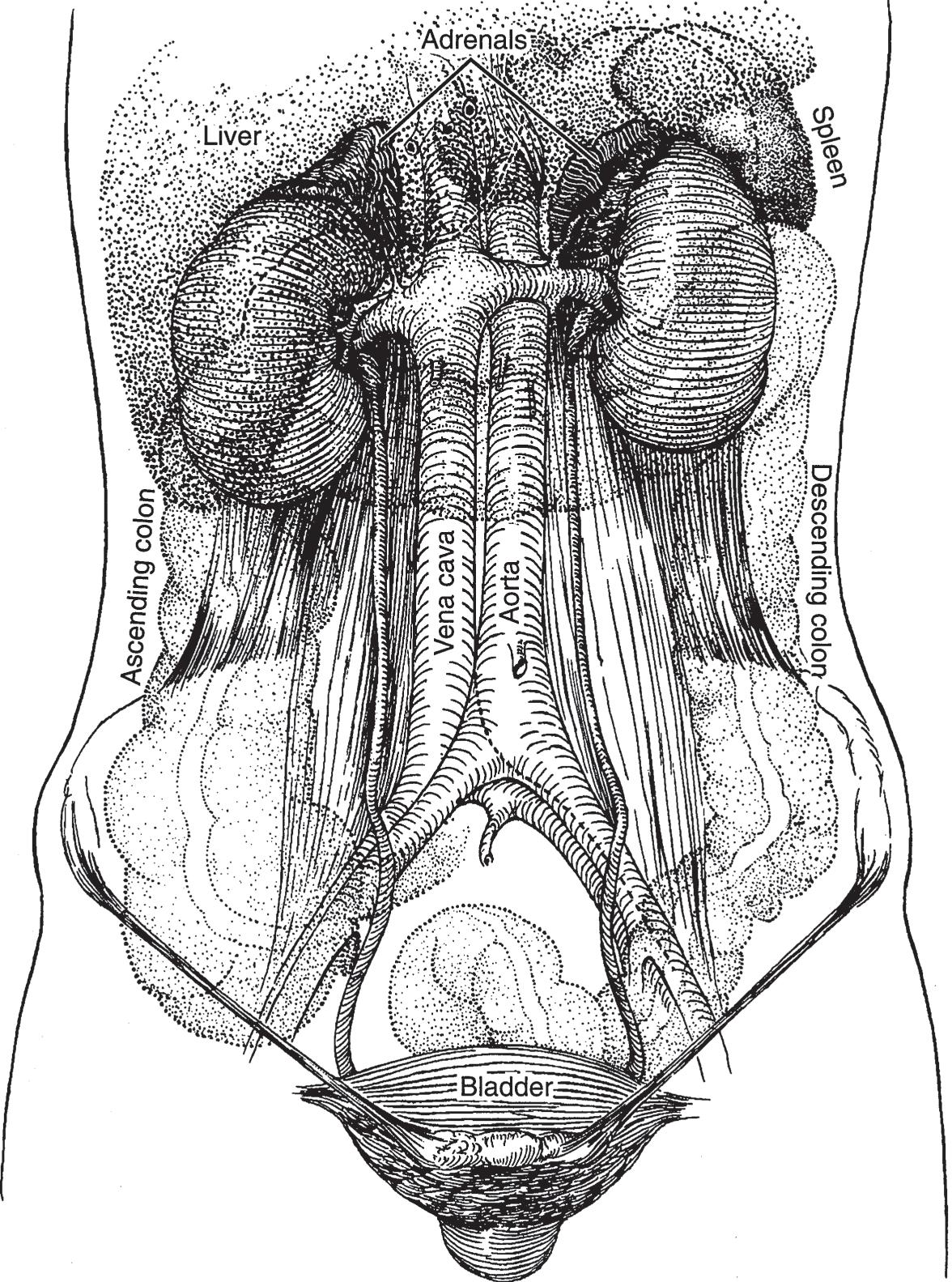
The kidneys lie along the borders of the psoas muscles and are therefore obliquely placed. The position of the liver causes the right kidney to be lower than the left (Figures 1–2 and 1–3). The adult kidney weighs between 125 and 170 g in men and 115 and 155 g in women. It is about 10–12 cm long, 5–7 cm wide, and 3–5 cm thick.
The kidneys are supported by the perirenal fat (which is enclosed in the perirenal fascia), the renal vascular pedicle, abdominal muscle tone, and the general bulk of the abdominal viscera (Rusinek et al, 2004). Variations in these factors permit variations in the degree of renal mobility. The average descent on inspiration or on assuming the upright position is 4–5 cm. Lack of mobility suggests abnormal fixation (eg, perinephritis), but extreme mobility is not necessarily pathologic.
▲ Figure 1–1. Anatomy of the male genitourinary tract. The upper tract and midtract have urologic function only. The lower tract has both genital and urinary functions.
▲ Figure 1–2. Relations between the kidneys, ureters, and bladder (anterior aspect).
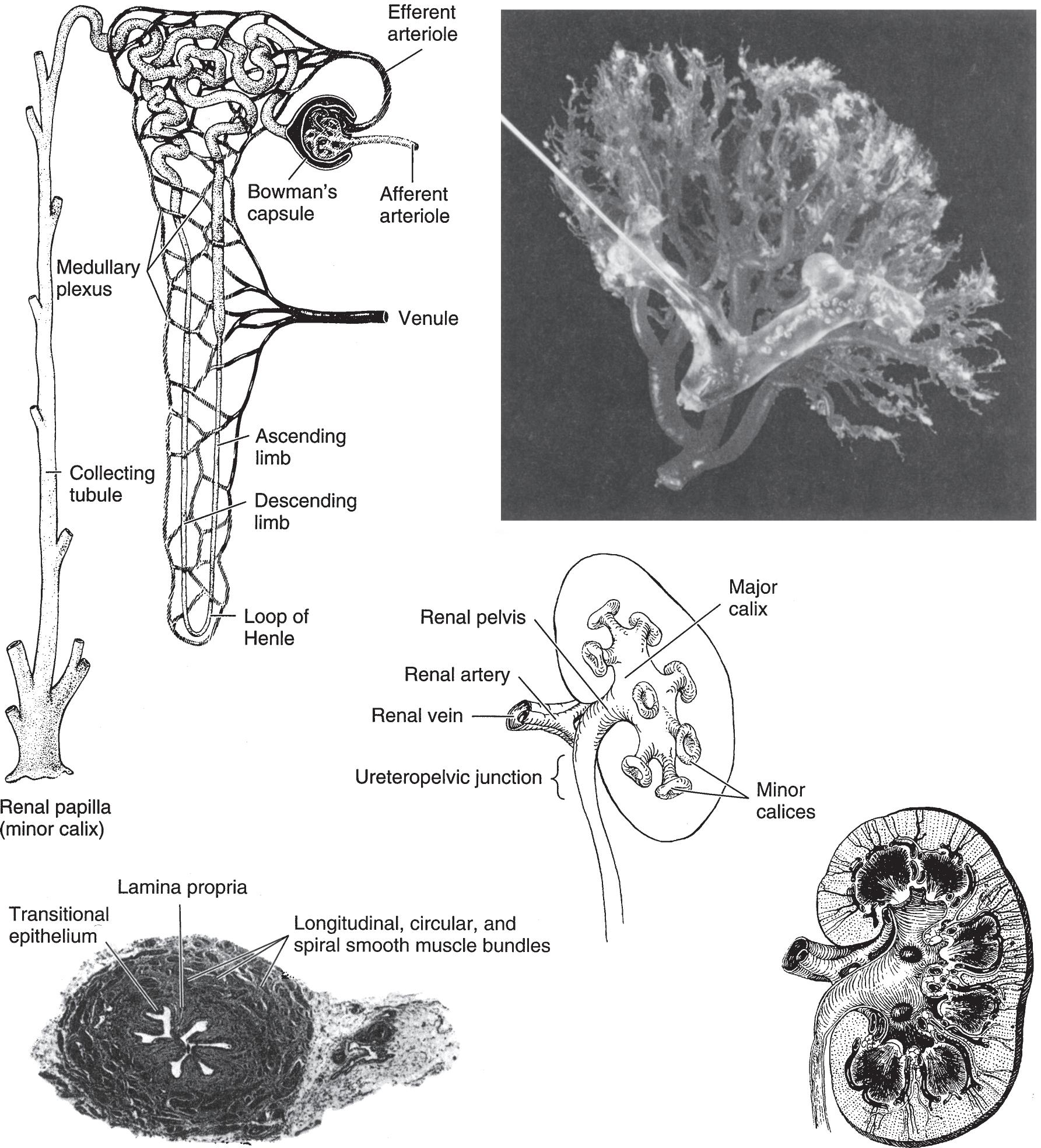
On longitudinal section (Figure 1–4), the kidney is seen to be made up of an outer cortex, a central medulla, and the internal calices and pelvis. The cortex is homogeneous in appearance. Portions of it project toward the pelvis between the papillae and fornices and are called the columns of Bertin. The medulla consists of numerous pyramids formed by the converging collecting renal tubules, which drain into the minor calices at the tip of the papillae.
B. Relations
Figures 1–2 and 1–3 show the relationships between the kidneys and adjacent organs and structures. Their intimacy with intraperitoneal organs and the autonomic innervation that they share with these organs explain, in part, some of the
gastrointestinal symptoms that accompany kidney diseases (Glassberg, 2002).
▶ Histology
A. Nephron
The functioning unit of the kidney is the nephron, which is composed of a tubule that has both secretory and excretory functions (Figure 1–4). The secretory portion is contained largely within the cortex and consists of a renal corpuscle and the secretory part of the renal tubule. The excretory portion of this duct lies in the medulla. The renal corpuscle is composed of the vascular glomerulus, which projects into Bowman’s capsule, which, in turn, is continuous with the epithelium of the proximal convoluted tubule. The secretory portion of the renal
▲ Figure 1–3. Relations between the kidneys (posterior aspect). The dashed lines represent the outline of the kidneys, where they are obscured by overlying structures.
tubule is made up of the proximal convoluted tubule, the loop of Henle, and the distal convoluted tubule.
The excretory portion of the nephron is the collecting tubule, which is continuous with the distal end of the ascending limb of the convoluted tubule. It empties its contents through the tip (papilla) of a pyramid into a minor calyx.
B. Supporting Tissue
The renal stroma is composed of loose connective tissue and contains blood vessels, capillaries, nerves, and lymphatics.
▶ Blood Supply (Figures 1–2, 1–4, and 1–5)
A. Arterial
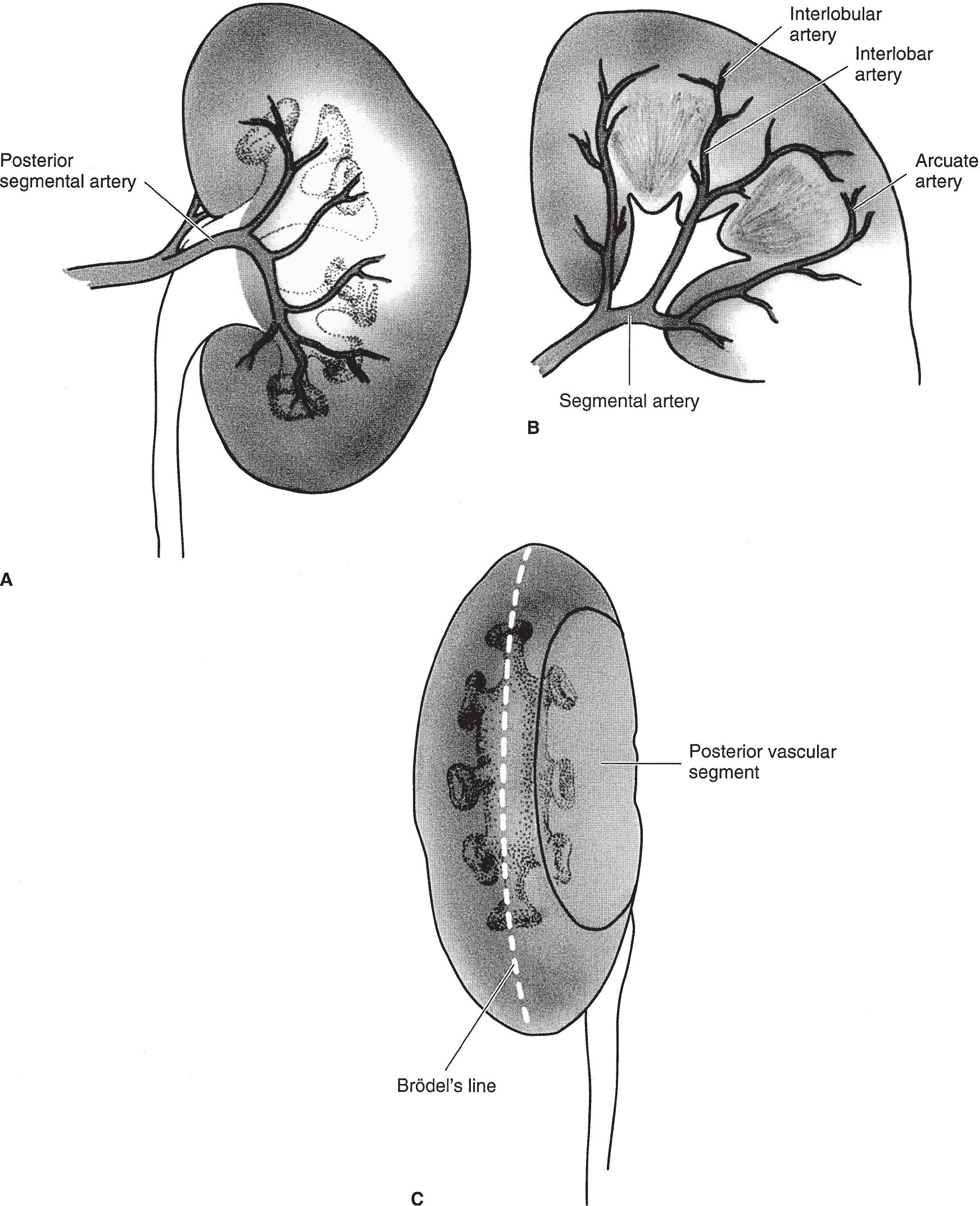
Usually there is one renal artery, a branch of the aorta that enters the hilum of the kidney between the pelvis, which normally lies posteriorly, and the renal vein. It may branch before it reaches the kidney, and two or more separate arteries may be noted (Budhiraja et al, 2010). In duplication of the pelvis and ureter, it is common for each renal segment to have its own arterial supply.
The renal artery divides into anterior and posterior branches. The posterior branch supplies the midsegment
of the posterior surface. The anterior branch supplies both upper and lower poles as well as the entire anterior surface. The renal arteries are all end arteries.
The renal artery branches further divide into interlobar arteries, which travel in the columns of Bertin (between the pyramids) and then arch along the base of the pyramids (arcuate arteries). These arteries then divide as interlobular arteries. From these vessels, smaller (afferent) branches pass to the glomeruli. From the glomerular tuft, efferent arterioles pass to the tubules in the stroma.
B. Venous
The renal veins are paired with the arteries, but any of them will drain the entire kidney if the others are tied off.
Although the renal artery and vein are usually the sole blood vessels of the kidney, accessory renal vessels are common and may be of clinical importance if they are so placed so as to compress the ureter, in which case hydronephrosis may result.
▶ Nerve Supply
The renal nerves derived from the renal plexus accompany the renal vessels throughout the renal parenchyma.
▲ Figure 1–4. Anatomy and histology of the kidney and ureter. Upper left: Diagram of the nephron and its blood supply. (Courtesy of Merck, Sharp, Dohme: Seminar. 1947; 9[3].) Upper right: Cast of the pelvic caliceal system and the arterial supply of the kidney. Middle: Renal calices, pelvis, and ureter (posterior aspect). Lower left: Histology of the ureter. The smooth-muscle bundles are arranged in both spirally and longitudinally. Lower right: Longitudinal section of kidney showing calices, pelvis, ureter, and renal blood supply (posterior aspect).
▲ Figure 1–5. (A) The posterior branch of the renal artery and its distribution to the central segment of the posterior surface of the kidney. (B) Branches of the anterior division of the renal artery supplying the entire anterior surface of the kidney as well as the upper and lower poles at both surfaces. The segmental branches lead to interlobar, arcuate, and interlobular arteries. (C) The lateral convex margin of the kidney. Brödel’s line, which is 1 cm from the convex margin, is the bloodless plane demarcated by the distribution of the posterior branch of the renal artery.
▶ Lymphatics
The lymphatics of the kidney drain into the lumbar lymph nodes.
CALICES, RENAL PELVIS, AND URETER
▶ Gross Appearance
A. Anatomy
1. Calices—The tips of the minor calices (8–12 in number) are indented by the projecting pyramids (Figure 1–4). These calices unite to form two or three major calices that join to form the renal pelvis (Sozen et al, 2008).
2. Renal pelvis—The pelvis may be entirely intrarenal or partly intrarenal and partly extrarenal. Inferomedially, it tapers to join the ureter.
3. Ureter—The adult ureter is about 30 cm long, varying in direct relation to the height of the individual. It follows a rather smooth S curve. Areas that stones are often impacted are (a) at the ureteropelvic junction, (b) where the ureter crosses over the iliac vessels, and (c) where it courses through the bladder wall.
B. Relations
1. Calices—The calices are intrarenal and are intimately related to the renal parenchyma.
2. Renal pelvis—If the pelvis is partly extrarenal, it lies along the lateral border of the psoas muscle and on the quadratus lumborum muscle; the renal vascular pedicle is just anterior to it. The left renal pelvis lies at the level of the first or second lumbar vertebra; the right pelvis is a little lower.
3. Ureter—On their course downward, the ureters lie on the psoas muscles, pass medially to the sacroiliac joints, and then swing laterally near the ischial spines before passing medially to enter the base of the bladder (Figure 1–2). In females, the uterine arteries are closely related to the juxtavesical portion of the ureters. The ureters are covered by the posterior peritoneum; their lowermost portions are closely attached to it, while the juxtavesical portions are embedded in vascular retroperitoneal fat (Koff, 2008).
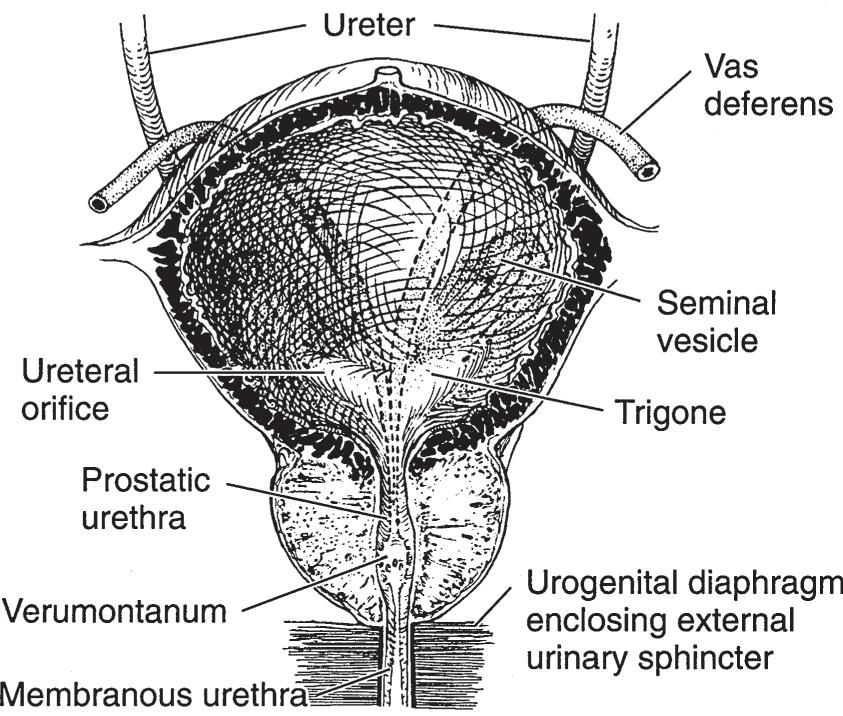
The vasa deferentia, as they leave the internal inguinal rings, sweep over the lateral pelvic walls anterior to the ureters (Figure 1–6). They lie medial to the latter before joining the seminal vesicle and penetrating the base of the prostate to become the ejaculatory ducts.
▶ Histology (Figure 1–4)
The walls of the calices, pelvis, and ureters are composed of transitional cell epithelium under which lies loose connective tissue (lamina propria). External to these are a mixture of
▲ Figure 1–6. Anatomy and relations between the ureters, bladder, prostate, seminal vesicles, and vasa deferentia (anterior view).
helical and longitudinal smooth-muscle fibers. They are not arranged in discrete layers. The outermost adventitial coat is composed of fibrous connective tissue.
▶
Blood Supply
A. Arterial
The renal calices, pelvis, and upper ureters derive their blood supply from the renal arteries; the midureter is fed by the internal spermatic (or ovarian) arteries. The lowermost portion of the ureter is served by branches from the common iliac, internal iliac (hypogastric), and vesical arteries.
B. Venous
The veins of the renal calices, pelvis, and ureters are paired with the arteries.
▶ Lymphatics
The lymphatics of the upper portions of the ureters as well as those from the pelvis and calices enter the lumbar lymph nodes. The lymphatics of the midureter pass to the internal iliac (hypogastric) and common iliac lymph nodes; the lower ureteral lymphatics empty into the vesical and hypogastric lymph nodes.
BLADDER
▶ Gross Appearance
The bladder is a hollow muscular organ that serves as a reservoir for urine. In women, its posterior wall and dome are invaginated by the uterus. The adult bladder normally has
a capacity of 400–500 mL. The wall of the bladder is about 3–5 mm in thickness; it is thinner when it is distended.
A. Anatomy
When empty, the adult bladder lies behind the pubic symphysis and is largely a pelvic organ. In infants and children, it is situated higher (Berrocal et al, 2002). When it is full, it rises well above the symphysis and can readily be palpated or percussed. When overdistended, as in acute or chronic urinary retention, it may cause the lower abdomen to bulge visibly.
Extending from the dome of the bladder to the umbilicus is a fibrous cord, the median umbilical ligament, which represents the obliterated urachus. The ureters enter the bladder posteroinferiorly in an oblique manner and at these points are about 5 cm apart (Figure 1–6). The orifices, situated at the extremities of the crescent-shaped interureteric ridge that forms the proximal border of the trigone, are about 2.5 cm apart. The trigone occupies the area between the ridge and the bladder neck.
The internal sphincter, or bladder neck, is not a true circular sphincter but a thickening formed by interlaced and converging muscle fibers of the detrusor as they pass distally to become the smooth muscle component of the urethra.
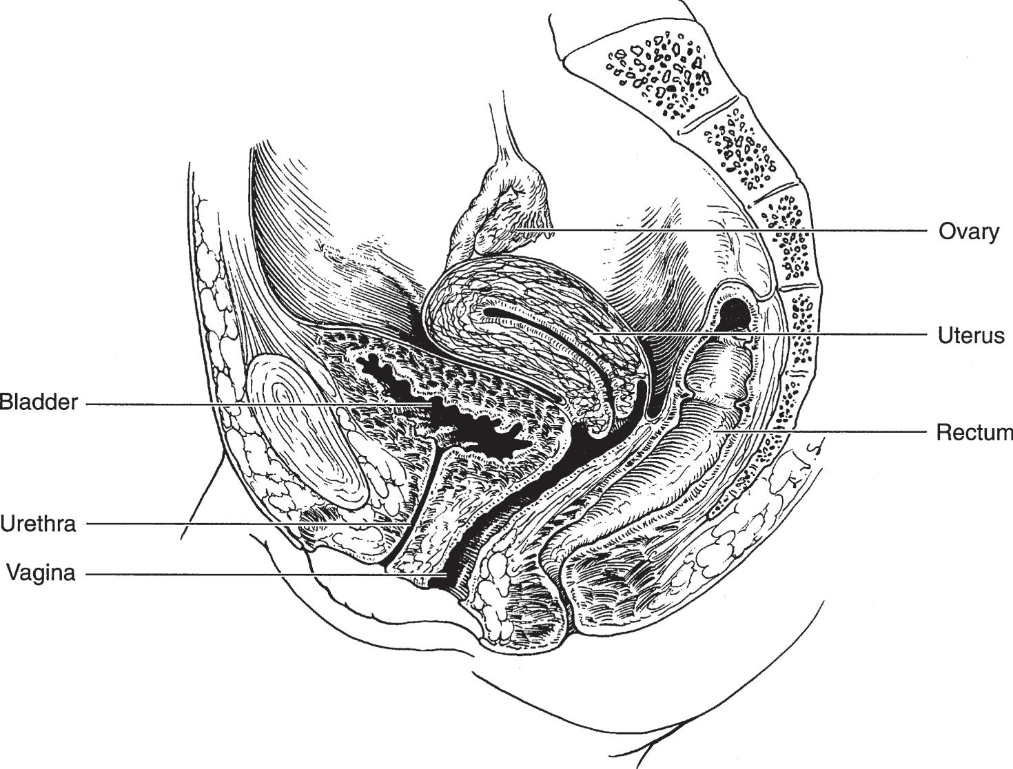
B. Relations
In males, the bladder is related posteriorly to the seminal vesicles, vasa deferentia, ureters, and rectum (Figures 1–7 and 1–8). In females, the uterus and vagina are interposed between the bladder and rectum (Figure 1–9). The dome and posterior surfaces are covered by peritoneum; hence, in this area, the bladder is closely related to the small intestine and sigmoid colon. In both males and females, the bladder is related to the posterior surface of the pubic symphysis, and, when distended, it is in contact with the lower abdominal wall.
▶ Histology (Figure 1–10)
The mucosa of the bladder is composed of transitional epithelium. Beneath it is a well-developed submucosal layer formed largely of connective and elastic tissues. The mucosa may be considered as a single functional unit that consists of the epithelial layer, basement membrane, and lamina propria. Physical or chemical stress on the bladder elicits releases of multiple factors that modulate afferent and efferent nerve activities (Fry and Vahabi, 2016). External to the submucosa is the detrusor muscle that is made up of a mixture of smooth-muscle fibers arranged at random in a longitudinal, circular, and spiral manner without any layer formation or specific orientation except for proximity to the internal meatus, where the detrusor muscle assumes three definite layers: inner longitudinal, middle circular, and outer longitudinal (John et al, 2001).
▶ Blood Supply
A. Arterial
The bladder is supplied by the superior, middle, and inferior vesical arteries, which arise from the anterior trunk of the internal iliac (hypogastric) artery, and by smaller branches from the obturator and inferior gluteal arteries. In females, the uterine and vaginal arteries also send branches to the bladder.
B. Venous
Surrounding the bladder is a rich plexus of veins that ultimately empties into the internal iliac (hypogastric) veins.
▶ Nerve Supply
The bladder receives innervation from sympathetic and parasympathetic nervous systems. The sensory afferent of the bladder originates from both subepithelial nerve endings and nerve fibers between detrusor muscle bundles (Andersson, 2010; Birder et al, 2010; McCloskey, 2010).
▶
Lymphatics
The lymphatics of the bladder drain into the vesical, external iliac, internal iliac (hypogastric), and common iliac lymph nodes.
PROSTATE GLAND
▶ Gross
Appearance
A. Anatomy
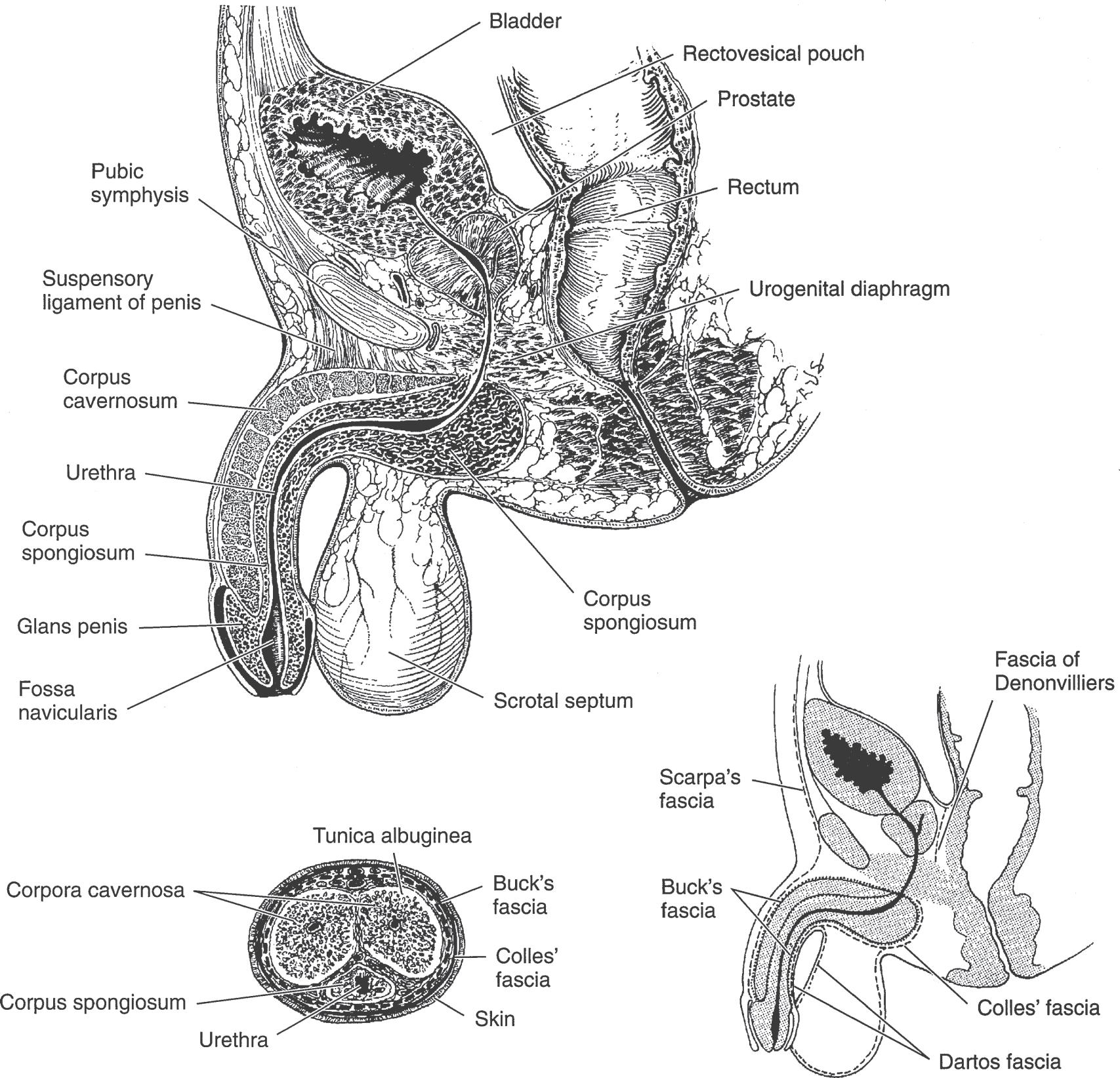
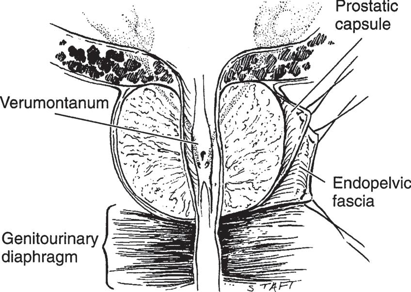
The prostate is a fibromuscular and glandular organ lying just inferior to the bladder (Figures 1–6 and 1–7). The normal prostate weighs about 20 g and contains the posterior urethra, which is about 2.5 cm in length. It is supported anteriorly by the puboprostatic ligaments and inferiorly by the urogenital diaphragm (Figure 1–6). The prostate is perforated posteriorly by the ejaculatory ducts, which pass obliquely to empty through the verumontanum on the floor of the prostatic urethra just proximal to the striated external urinary sphincter (Figure 1–11).
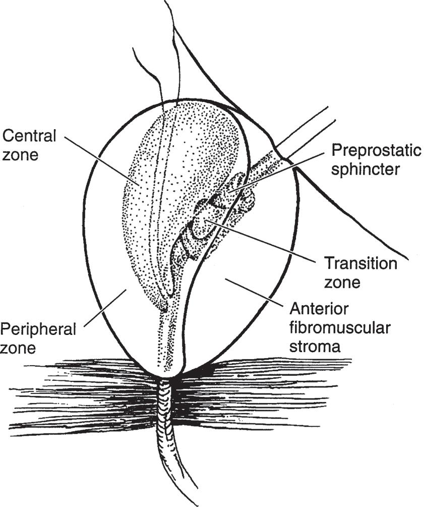
The prostate can be subdivided into two ways: by lobe or by zone. The lobe classification is often used in cystourethroscopic examinations and consists of five lobes: anterior, posterior, median, right lateral, and left lateral. The zone classification is often used in pathology. McNeal (1981) divides the prostate into four zones: peripheral zone, central zone (surrounds the ejaculatory ducts), transitional zone (surrounds the urethra), and anterior fibromuscular zone (Myers et al, 2010) (Figure 1–12). The segment of urethra that traverses the prostate gland is the prostatic urethra. It is lined by an inner longitudinal layer of muscle (continuous with a similar layer of the vesical wall). Incorporated within the
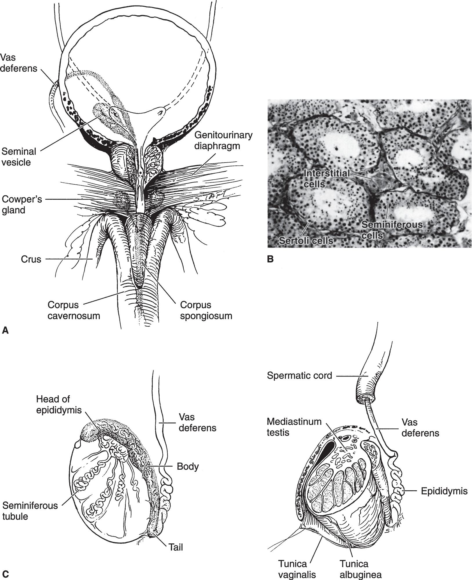
▲ Figure 1–7. (A) Anatomic relationship between the bladder, prostate, prostatomembranous urethra, and root of the penis. (B) Histology of the testis. Seminiferous tubules lined by supporting basement membrane for the Sertoli and spermatogenic cells. The latter are in various stages of development. (C) Cross sections of the testis and epididymis. (Images [A] and [C] reproduced with permission from Walsh PC, Campbell MF: Campbell’s Urology, 6th ed. Philadelphia, PA: Saunders; 1992.)
▲ Figure 1–8. Top: Relations between the bladder, prostate, seminal vesicles, penis, urethra, and scrotal contents. Lower left: Transverse section through the penis. The paired upper structures are the corpora cavernosa. The single lower body surrounding the urethra is the corpus spongiosum. Lower right: Fascial planes of the lower genitourinary tract. (After Wesson.)
prostate gland is an abundant amount of smooth musculature derived primarily from the external longitudinal bladder musculature. This musculature represents the involuntary smooth muscle sphincter of the posterior urethra in males.
B. Relations
The prostate gland lies behind the pubic symphysis. Located closely to the posterosuperior surface are the vasa deferentia and seminal vesicles (Figure 1–7). Posteriorly, the prostate is
separated from the rectum by the two layers of Denonvilliers’ fascia, serosal rudiments of the pouch of Douglas, which once extended to the urogenital diaphragm (Raychaudhuri and Cahill, 2008) (Figure 1–8).
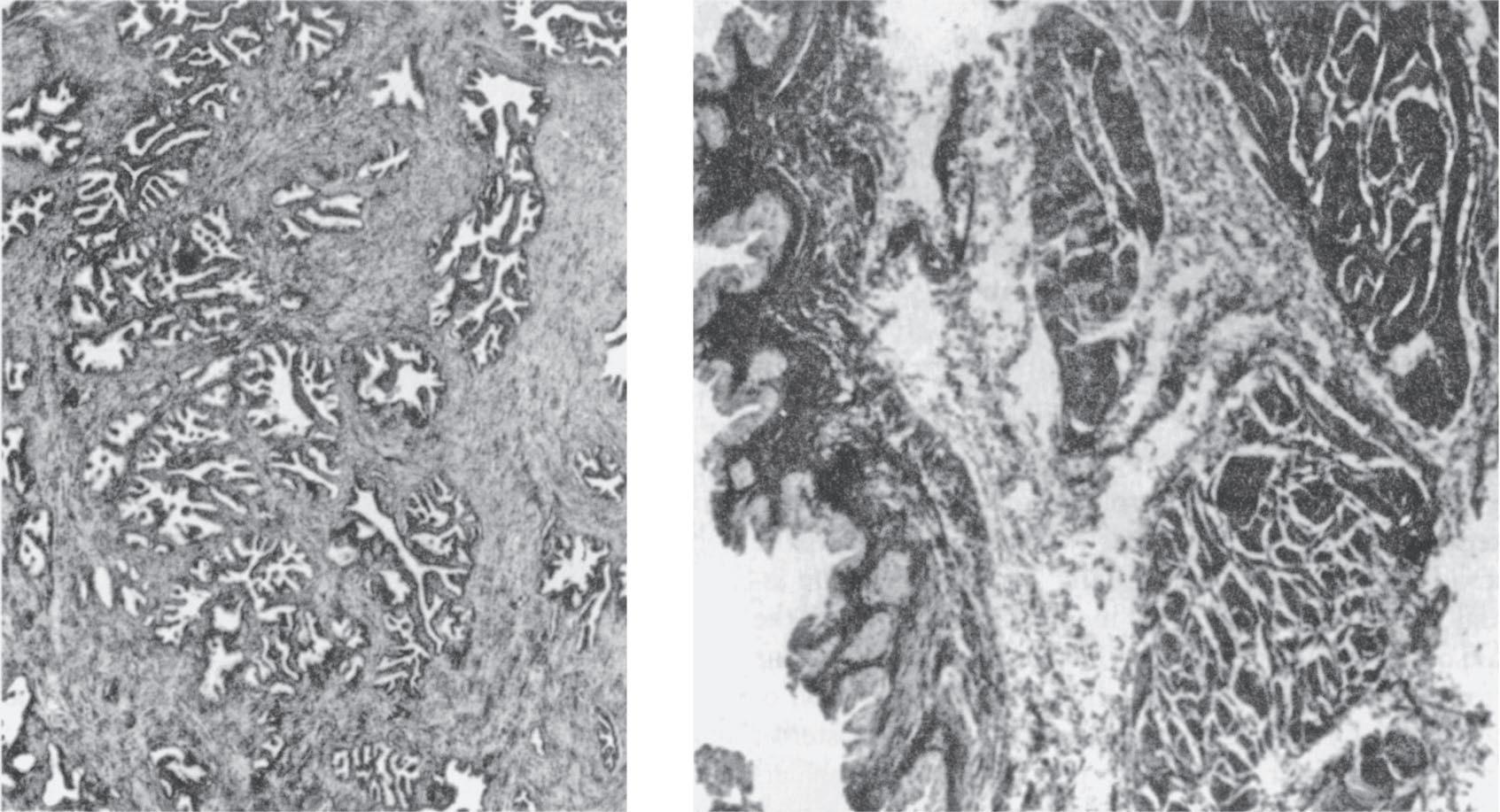
▶ Histology (Figure 1–10)
The prostate consists of a thin fibrous capsule under which lie circularly oriented smooth-muscle fibers and collagenous tissue that surrounds the urethra (involuntary
▲ Figure 1–9. Anatomy and relations of the bladder, urethra, uterus and ovary, vagina, and rectum.
▲ Figure 1–10. Left: Histology of the prostate. Epithelial glands embedded in a mixture of connective and elastic tissue and smooth muscle. Right: Histology of the bladder. The mucosa is transitional cell in type and lies on a well-developed submucosal layer of connective tissue. The detrusor muscle is composed of interlacing longitudinal, circular, and spiral smooth-muscle bundles.
▲ Figure 1–11. Section of the prostate gland shows the prostatic urethra, verumontanum, and crista urethralis, in addition to the opening of the prostatic utricle and the two ejaculatory ducts in the midline. Note that the prostate is surrounded by the prostatic capsule, which is covered by another prostatic sheath derived from the endopelvic fascia. The prostate is resting on the genitourinary diaphragm. (Reproduced with permission from Walsh PC, Campbell MF: Campbell’s Urology, 6th ed. Philadelphia, PA: Saunders; 1992.)
sphincter). Deep in this layer lies the prostatic stroma, composed of connective tissues and smooth-muscle fibers in which are embedded the epithelial glands. These glands drain into the major excretory ducts (about 25 in number), which open chiefly on the floor of the urethra between the verumontanum and the vesical neck. Just beneath the transitional epithelium of the prostatic urethra lie the periurethral glands.
▶ Blood Supply
A. Arterial
The arterial supply to the prostate is derived from the inferior vesical, internal pudendal, and middle rectal (hemorrhoidal) arteries.
B. Venous
The veins from the prostate drain into the periprostatic plexus, which has connections with the deep dorsal vein of the penis and the internal iliac (hypogastric) veins.
▶ Nerve Supply
The prostate gland receives a rich innervation from the sympathetic and parasympathetic nerves of the inferior hypogastric plexus.
▲ Figure 1–12. Anatomy of the prostate gland. Prostatic adenoma develops from the periurethral glands at the site of the median or lateral lobes. The posterior lobe, however, is prone to cancerous degeneration. (Adapted with permission from McNeal JE: The zonal anatomy of the prostate. Prostate 1981;2(1):35–49.)
▶ Lymphatics
The lymphatics from the prostate drain into the internal iliac (hypogastric), sacral, vesical, and external iliac lymph nodes (Saokar et al, 2010).
SEMINAL VESICLES
▶ Gross Appearance
The seminal vesicles lie just cephalic to the prostate under the base of the bladder (Figures 1–6 and 1–7). They are about 6 cm long and quite soft. Each vesicle joins its corresponding vas deferens to form the ejaculatory duct (Kim et al, 2009). The ureters lie medial to each, and the rectum is contiguous with their posterior surfaces.
▶ Histology
The mucous membrane is pseudostratified. The submucosa consists of dense connective tissue covered by a thin layer of muscle that, in turn, is encapsulated by connective tissue.
▶ Blood Supply
The blood supply of the seminal vesicles is similar to that of the prostate gland.
▶ Nerve Supply
The nerve supply is mainly from the sympathetic nerve plexus.
▶ Lymphatics
The lymphatics of the seminal vesicles are those that serve the prostate.
SPERMATIC CORD
▶ Gross Appearance
The two spermatic cords extend from the internal inguinal rings through the inguinal canals to the testicles (Figure 1–7). Each cord contains the vas deferens, the internal and external spermatic arteries, the artery of the vas, the venous pampiniform plexus (which forms the spermatic vein superiorly), lymph vessels, and nerves (Jen et al, 1999). The entire cord contents are enclosed in investing layers of thin fascia. A few fibers of the cremaster muscle insert on the cords in the inguinal canal (Bhosale et al, 2008; Kim et al, 2009).
▶ Histology
The fascia covering the cord is formed of loose connective tissue that supports arteries, veins, nerve, and lymphatics.
The vas deferens is a small, thick-walled tube consisting of an internal mucosa and submucosa surrounded by three welldefined layers of smooth muscle encased in a covering of fibrous tissue. Above the testes, this tube is straight. Its proximal 4 cm tends to be convoluted.
▶ Blood Supply
A. Arterial
The external spermatic artery, a branch of the inferior epigastric, supplies the fascial coverings of the cord. The internal spermatic artery passes through the cord on its way to the testis. The deferential artery is close to the vas.
B. Venous
The veins from the testis and the coverings of the spermatic cord form the pampiniform plexus, which, at the internal inguinal ring, unites to form the spermatic vein.
▶ Lymphatics
The lymphatics from the spermatic cord empty into the external iliac lymph nodes.
EPIDIDYMIS
▶ Gross Appearance
A. Anatomy
The upper portion of the epididymis (globus major) is connected to the testis by numerous efferent ducts from the testis (Figure 1–7). The epididymis consists of a markedly coiled duct that, at its lower pole (globus minor), is continuous with the vas deferens. An appendix of the epididymis is often seen on its upper pole; this is a cystic body that in some cases is pedunculated, but in others, it is sessile.
B. Relations
The epididymis lies posterolateral to the testis and is nearest to the testis at its upper pole. Its lower pole is connected to the testis by fibrous tissue. The vas lies posteromedial to the epididymis.
▶ Histology
The epididymis is covered by serosa. The ductus epididymidis is lined by pseudostratified columnar epithelium throughout its length.
▶ Blood Supply
A. Arterial
The arterial supply to the epididymis comes from the internal spermatic artery and the artery of the vas (deferential artery).
B. Venous
The venous blood drains into the pampiniform plexus, which becomes the spermatic vein.
▶ Lymphatics
The lymphatics drain into the external iliac and internal iliac (hypogastric) lymph nodes.
TESTIS
▶ Gross Appearance
A. Anatomy
The average testicle measures about 4 × 3 × 2.5 cm (Figure 1–7). The volume can be measured by an orchidometer or by a formula with ultrasonic measurement (length × width × height × 0.71). The average volume is 18 mL (ranging from 12 to 30 mL). The testicle has a dense fascial covering called the tunica albuginea testis, which, posteriorly, is invaginated somewhat into the body of the testis to form the mediastinum testis. This fibrous mediastinum sends fibrous septa into the testis, thus separating it into about 250 lobules.
The testis is covered anteriorly and laterally by the visceral layer of the serous tunica vaginalis, which is continuous with the parietal layer that separates the testis from the scrotal wall (Bidarkar and Hutson, 2005). A small amount of fluid normally exists within the tunica vaginalis sac. At the upper pole of the testis is the appendix testis, a small pedunculated or sessile body similar in appearance to the appendix of the epididymis.
B. Relations
The testis is closely attached posterolaterally to the epididymis, particularly at its upper and lower poles (Klonisch et al, 2004).
▶ Histology (Figure 1–7)
Each lobule contains one to four markedly convoluted seminiferous tubules, each of which is about 60 cm long. These ducts converge at the mediastinum testis, where they connect with the efferent ducts that drain into the epididymis.
The seminiferous tubule has a basement membrane containing connective and elastic tissue. This supports the seminiferous cells that are of two types: (1) Sertoli (supporting) cells and (2) spermatogenic cells. The stroma between the seminiferous tubules contains connective tissue in which the interstitial Leydig cells are located.
▶ Blood Supply
The blood supply to the testes is closely associated with that to the kidneys because of the common embryologic origin of the two organs.
A. Arterial
The arteries to the testes (internal spermatics) arise from the aorta just below the renal arteries and course through the spermatic cords to the testes, where they anastomose with the arteries of the vasa deferentia that branch off from the internal iliac (hypogastric) artery.
B. Venous
The blood from the testis returns in the pampiniform plexus of the spermatic cord. At the internal inguinal ring, the pampiniform plexus forms the spermatic vein.
The right spermatic vein enters the vena cava just below the right renal vein; the left spermatic vein empties into the left renal vein.
▶ Lymphatics
The lymphatic vessels from the testes pass to the lumbar lymph nodes, which, in turn, are connected to the mediastinal nodes.
SCROTUM
▶ Gross Appearance
Beneath the corrugated skin of the scrotum lies the dartos muscle. Deep to this are the three fascial layers derived from the abdominal wall at the time of testicular descent. Beneath these is the parietal layer of the tunica vaginalis (Kim et al, 2007).
The scrotum is divided into two sacs by a septum of connective tissue. The scrotum not only supports the testes but also, by relaxation or contraction of its muscular layer, helps to regulate their temperature.
▶ Histology
The dartos muscle, under the skin of the scrotum, is nonstriated. The deeper layer is made up of connective tissue.
▶
Blood Supply
A. Arterial
The arteries to the scrotum arise from the femoral, internal pudendal, and inferior epigastric arteries.
B. Venous
The veins are paired with the arteries.
▶ Lymphatics
The lymphatics drain into the superficial inguinal and subinguinal lymph nodes.
PENIS AND MALE URETHRA
▶ Gross Appearance
The penis is composed of two corpora cavernosa and the corpus spongiosum, which contains the urethra. The corpus spongiosum enlarges distally and forms the glans penis. Each corpus is enclosed in a fascial sheath (tunica albuginea), and all three corpora are surrounded by a thick fibrous envelope known as Buck’s fascia. A covering of skin, devoid of fat, is loosely wrapped these bodies. The prepuce forms a hood over the glans.
Beneath the skin of the penis (and scrotum) and extending from the base of the glans to the urogenital diaphragm is Colles’ fascia, which is continuous with Scarpa’s fascia of the lower abdominal wall (Figure 1–8).
The proximal ends of the corpora cavernosa are attached to the pelvic bones just anterior to the ischial tuberosities. The ischiocavernosus muscles insert into the lateral surface of the tunica albuginea at the proximal corpora cavernosa. Occupying a depression of their ventral surface in the midline is the corpus spongiosum, which is connected proximally to the undersurface of the urogenital diaphragm, below which