


Lonneke Vervelde1 (l.vervelde@gdanimalhealth.com), Thijs Manders1,2 (t.t.m.manders@uu.nl), Samira Kammourieh1 (s.kammourieh@gddiergezondheid.nl), Jeanine Wiegel1 (j.wiegel@gdanimalhealth.com)
1 Royal GD, Deventer, The Netherlands, 2 Faculty Veterinary Medicine, University Utrecht, The Netherlands
Introduction
Enterococcus cecorum are members of the normal enterococcal microbiota in the gastrointestinal tract of poultry. Strains have emerged that cause spondylitis and bacterial osteomyelitis. The translocation and bacteraemia are pivotal to the pathogenesis and clinical disease. Virulence typing to distinguish extra-intestinal disease of lesion from cloacal strains remains difficult. The aim of our study was to develop an in vitro model to quantify invasion in organoids mimicking the translocation in vivo. This would allow differentiation between E. cecorum strains that are more or less virulent and would provide an in vitro model to test intervention strategies such as feed additives that prevent translocation.
Table 1. Summary of the origin and characteristics of the Enterococcus cecorum inoculation strains used.
Materials and Methods
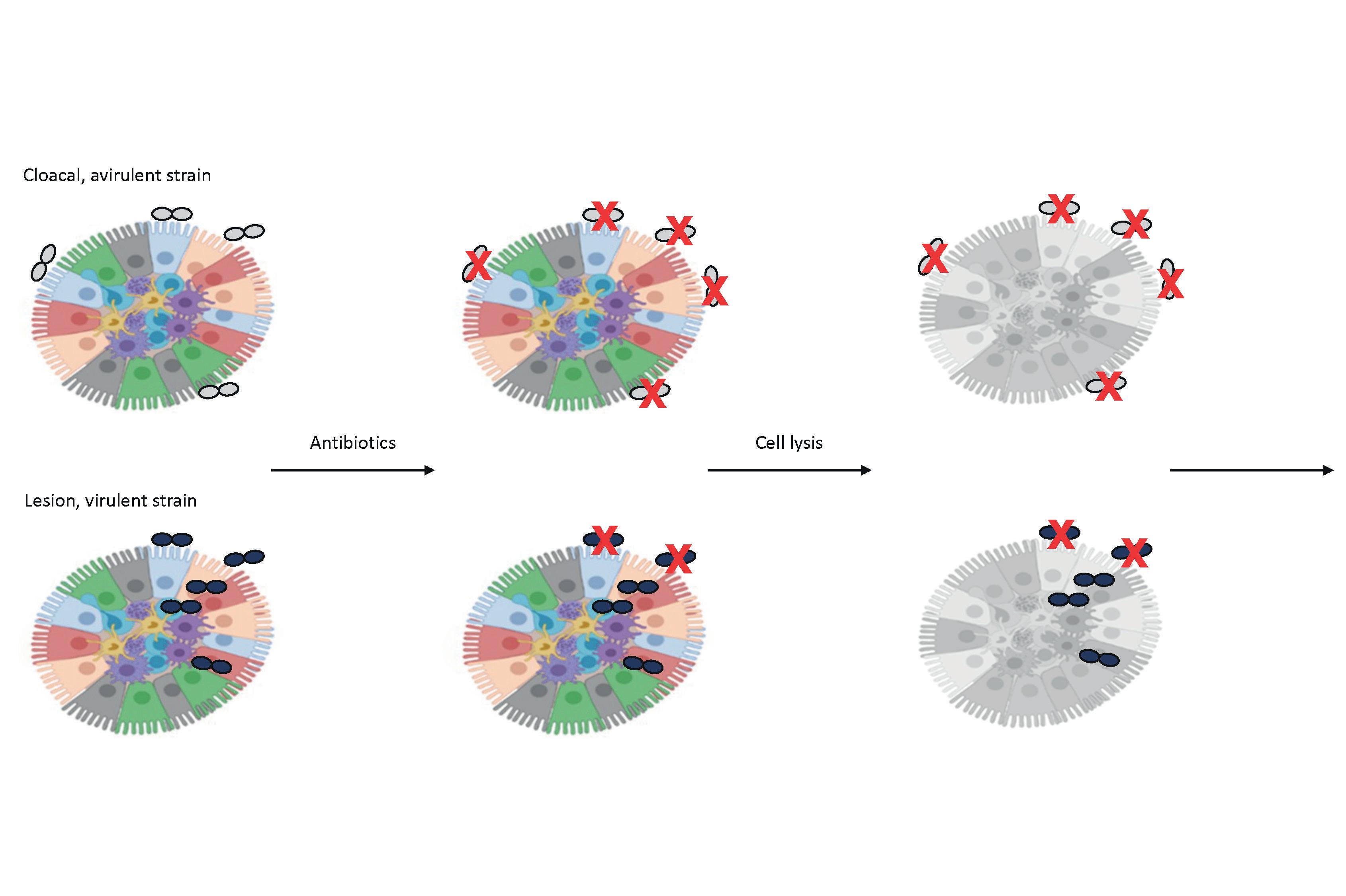
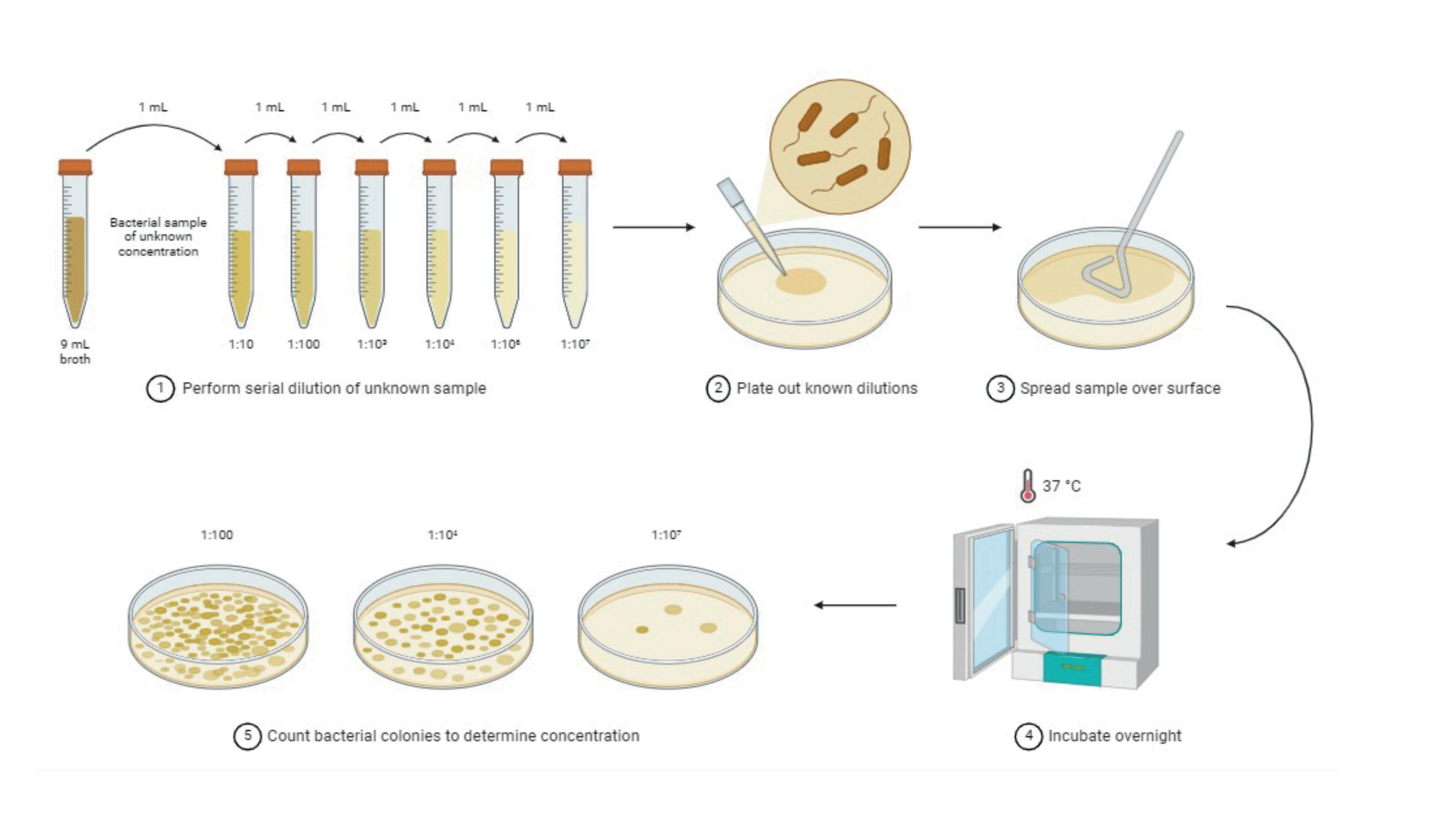
Intestinal villi were collected from 18-day-old SPF layer embryos and grown in Floating Organoid Medium (FOM) as described previously by Nash et al. (2021). Four E. cecorum strains were selected based on the site of isolation, ability to ferment mannitol, embryo mortality rate and resistance against lysozyme, Table 1. To determine if invasion is a discriminating factor for cloacal versus lesion strains, an invasion assay in chicken intestinal organoids was developed (Figure 1). Organoids were inoculated with bacteria, 103 to 107 CFU/well, and analysed at 3 and 6 hours post inoculation. Bacteria were quantified by RT-PCR or by bacterial colony counting of ten-fold serial dilutions bacterial plating on Columbia agar supplemented with sheep blood.
Results
1No lesions
2Range of cumulative embryo mortality is given, three embryo lethality assays were performed and described by Manders et al. (2022).
3Minimum Inhibitory Concentration. 4Sensitive to the tested antibiotic. 5Resitant to the tested antibiotic.
Independent of the bacterial strain, the morphology of the organoids was not affected by a high inoculum dose of E. cecorum (Figure 2). Substantial budding of the organoids indicated rapid growth which was seen independent of the treatment. Since the quantification of E. cecorum by qPCR could not distinguish between cloacal and lesion strains, likely due to bacteria sticking to the intestinal epithelial cells, an invasion assay was set up as shown in Figure 1.
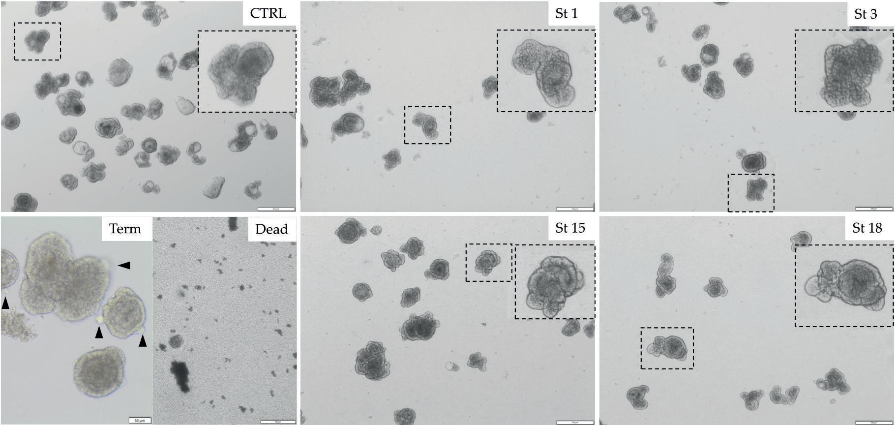
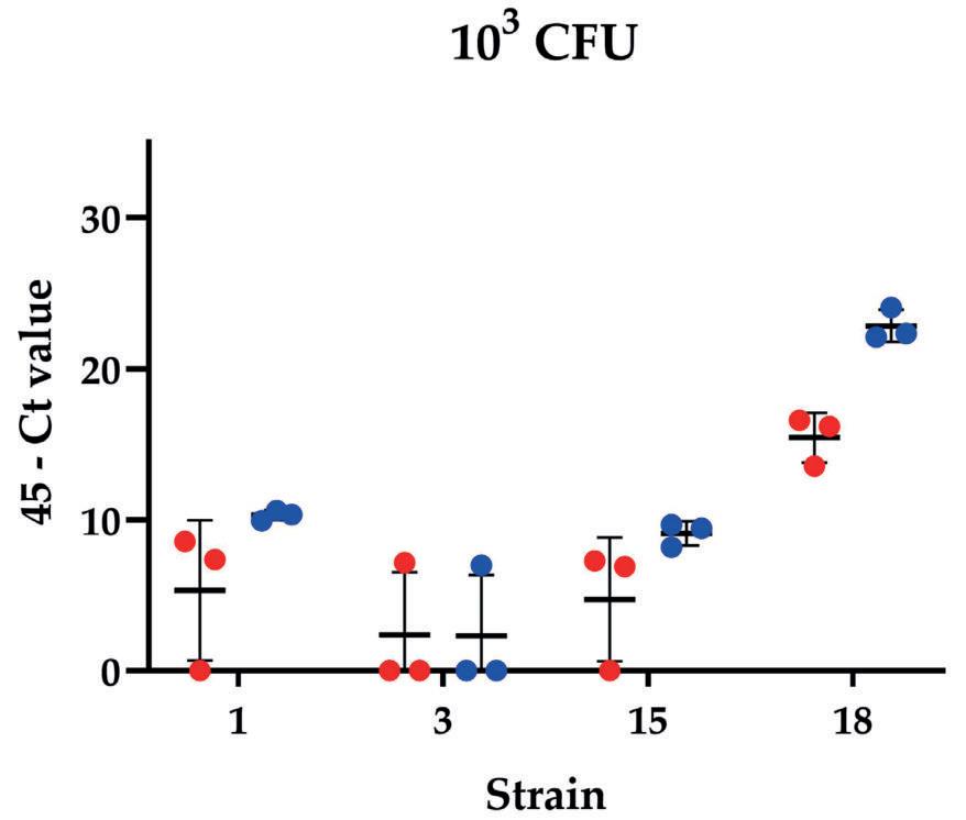
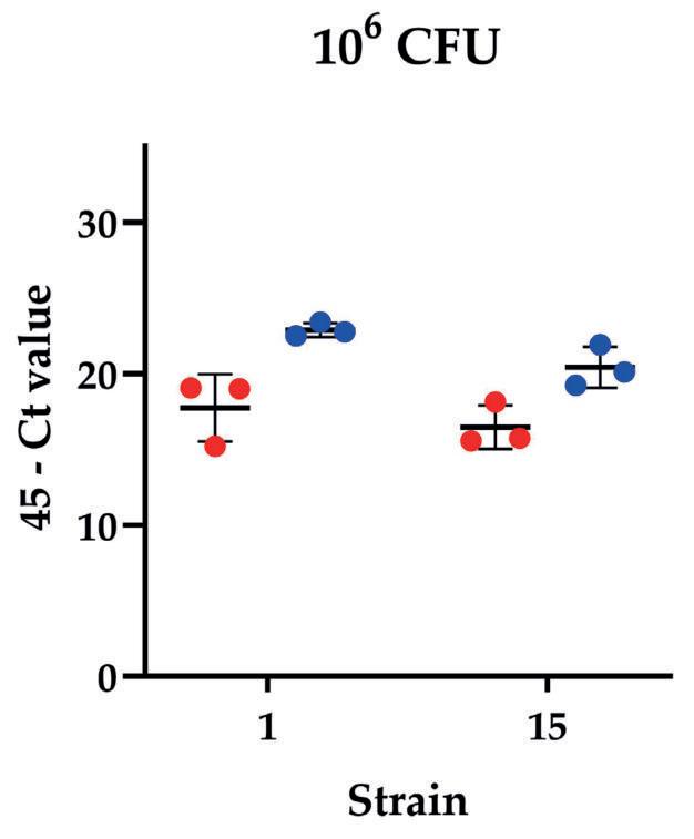
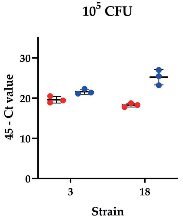
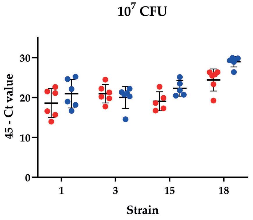

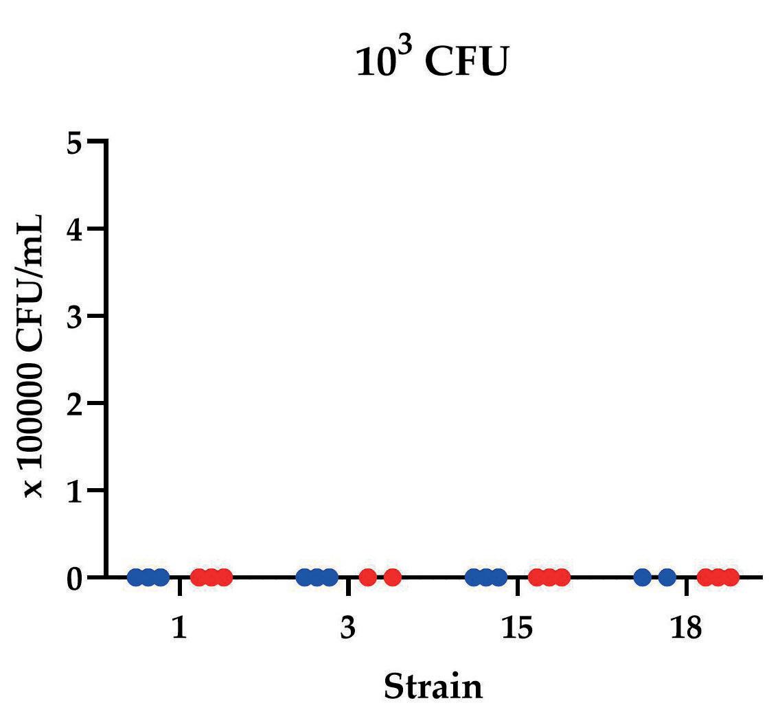
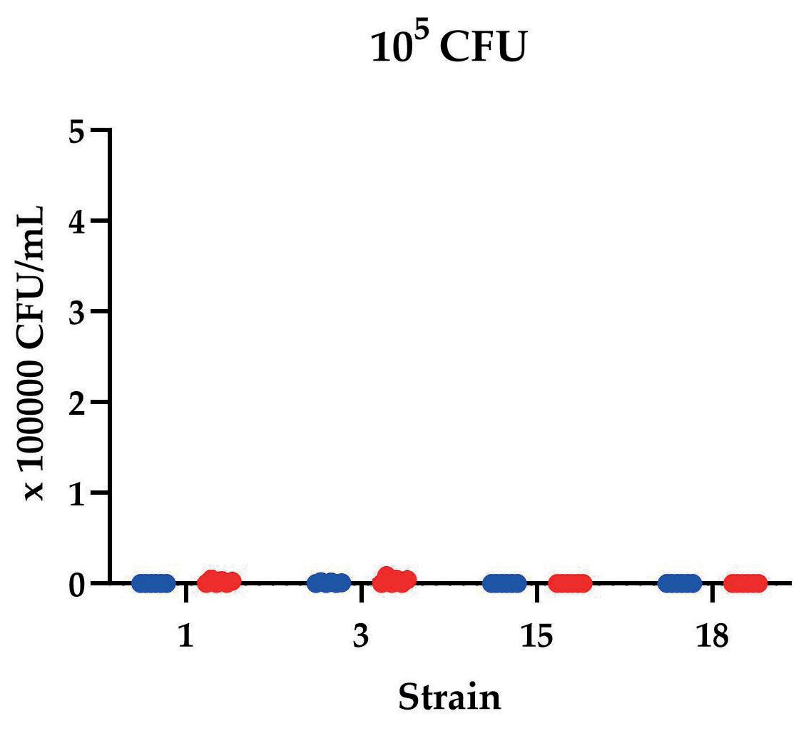
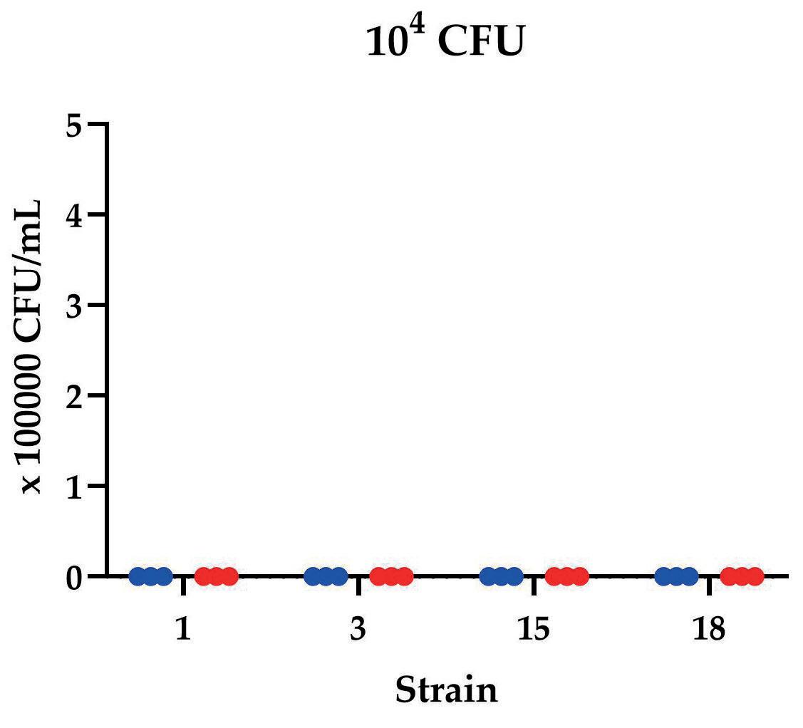
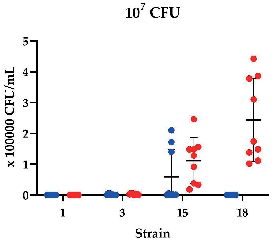
Figure 4. Differential invasion of cloacal and lesion E. cecorum strains can be quantified by colony counting. Invasion assay using chicken intestinal organoids inoculated E. cecorum per well. Organoids were inoculated with with 103 to 105 CFU per well and treated at 3 (blue) or 6 (red) hours post inoculation as shown in figure 1. Data are presented as means with standard deviation.
Conclusion
• Cloacal (avirulent) and lesion (virulent) strains of E. cecorum can be distinguished based on invasion in chicken intestinal organoids.
• The intestinal organoid model will enable us to address outstanding questions on pathogenesis, translocation and local immune responses to E. cecorum
• Chicken intestinal organoids facilitate screening of novel preventive and therapeutic intervention strategies in vitro before in vivo trials are performed.