


Thijs Manders1,2, Arjan Baijense2, Naomi de Bruijn1, Lonneke Vervelde1, Jeanine Wiegel1 1 Royal GD, Deventer, The Netherlands, 2 Faculty Veterinary Medicine, University Utrecht, The Netherlands l.vervelde@gdanimalhealth.com t.manders@gdanimalhealth.com j.wiegel@gdanimalhealth.com
Introduction
Enterococcus cecorum are members of the normal enterococcal microbiota in the gastrointestinal tract of poultry, but virulent strains have emerged causing clinical outbreaks with major economic impact. Strains are considered virulent when recovered from E. cecorum lesions in femur and free thoracic vertebra, whereas commensal or avirulent strains are isolated from cloaca of healthy birds. The pathogenesis of E. cecorum infections is not fully understood and the early stages after infection and site of translocation within the gastrointestinal tract have not been assessed. This study investigated the kinetics and location of E. cecorum translocation. Two animal model have been published with varying total reisolation rates from organs, depending on the dose and strain [Borst et al 2017; Scheier et al., 2021]. The relatively low prevalence of macroscopic BCO lesions in these models may reflect the complexity of BCO pathogenesis. One of the predisposing factors for BCO is mechanical stress on the metaphyseal plate cartilage. We investigated the influence of a wire obstacle on development of typical lesions, on intestinal colonization and on systemic infection in specific pathogen free (SPF) broilers inoculated with E. cecorum.
Materials and Methods
Two groups of 160 SPF broilers each (bred at Royal GD) were orally inoculated with 107.6 colony-forming units of E. cecorum in peptone physiological saline (PPS) per bird on the day of hatch and 64 control birds were inoculated with PPS. The characterisation of the strain is shown in Table 1 and experimental design in Figure 1.
Table 1. Origin and characterisation of the E. cecorum strain used in studie.
Strain Origin Embryo mortality1 Lysozyme MIC level (mg/ml)2
Antibiotic sensitivity
Site Lesion Penicillin Streptomycin Gentamycin 15 FTV Spondylitis 100% Resistant S3 R5 R
1Range of cumulative embryo mortality is given, three embryo lethality assays were performed and described by Manders et al. (2022). 2Minimum Inhibitory Concentration 3Sensitive to the tested antibiotic 4Resistant to the tested antibiotic
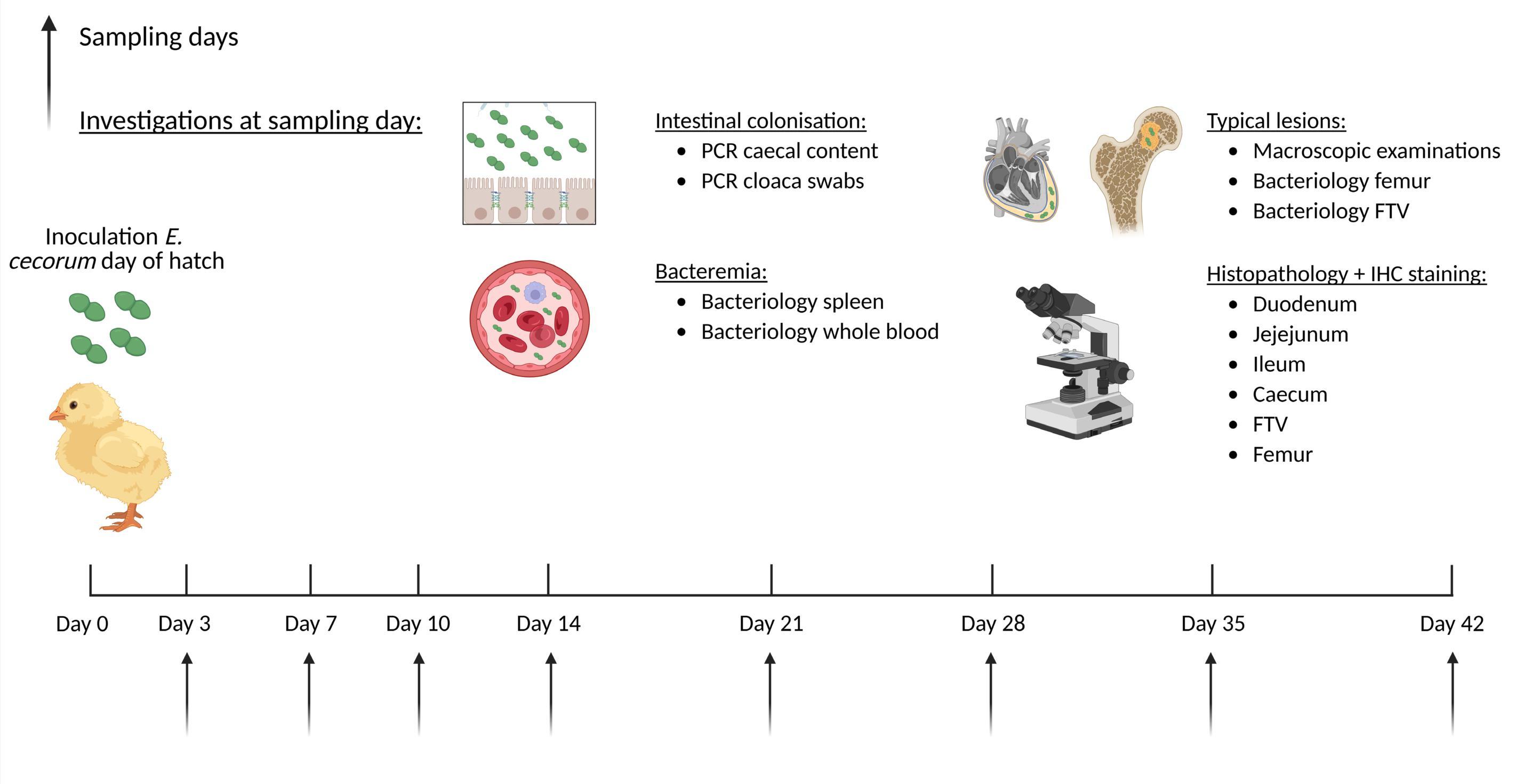
1. Schematic overview of the experimental design. Day-old SPF broilers are orally inoculated with E. cecorum and at different timepoints post inoculations tissues, blood and intestinal contents are collected. Created with BioRender.com
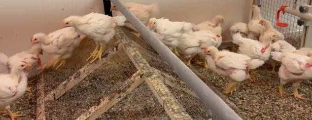
2. Birds were housed in pens with solid floors and wood shavings. To induce mechanical stress a wire obstacle was placed at the centre of each pen, spanning its full width to separate feeders from water nipples. To encourage crouching when crossing the apex and prevent birds from jumping off, a limbo bar was mounted above the apex. The obstacles were covered with chick paper during the first 2 weeks of the study, after which any remaining paper was removed.
Results
Clinical and post mortem examinations. Overall mortality hazard ratios and weight gain did not significantly differ. The overall proportion of birds with ≥1 lesion and the overall prevalence of pericarditis were significantly higher in group EC compared to group CON (p < 0.001) and in group EC_OBS compared to group CON_OBS (p < 0.001). Remarkably, E. cecorum-inoculated broilers without obstacles showed significantly more lameness, in contrast to earlier studies, which reported higher lameness incidence in birds exposed to wire flooring or ramped obstacles.
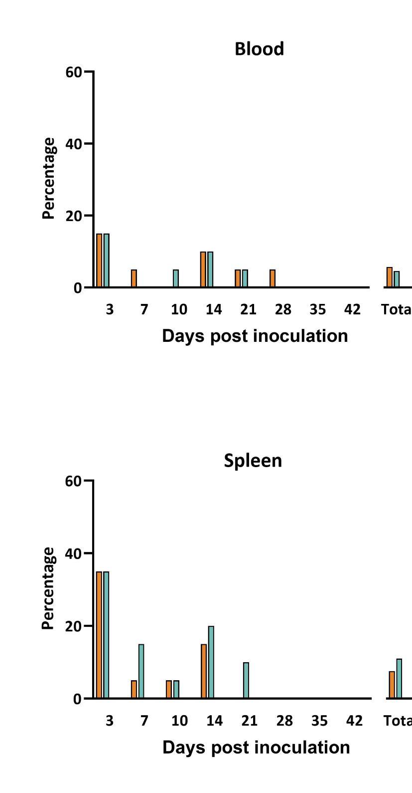
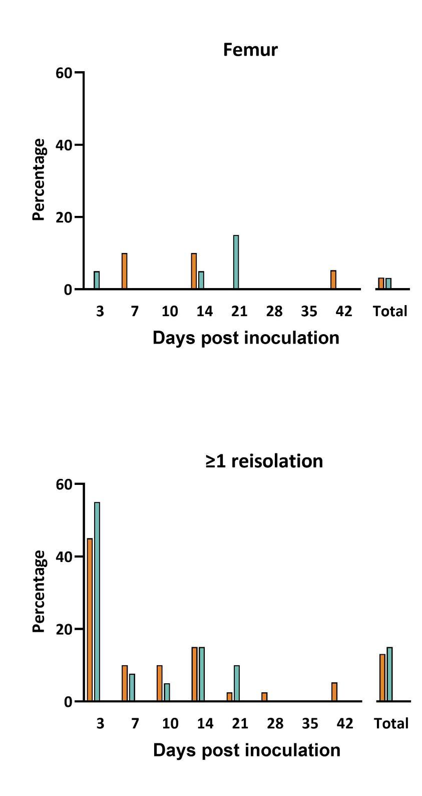
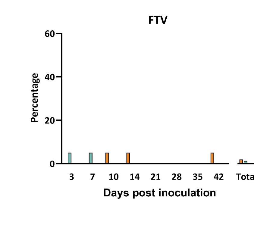
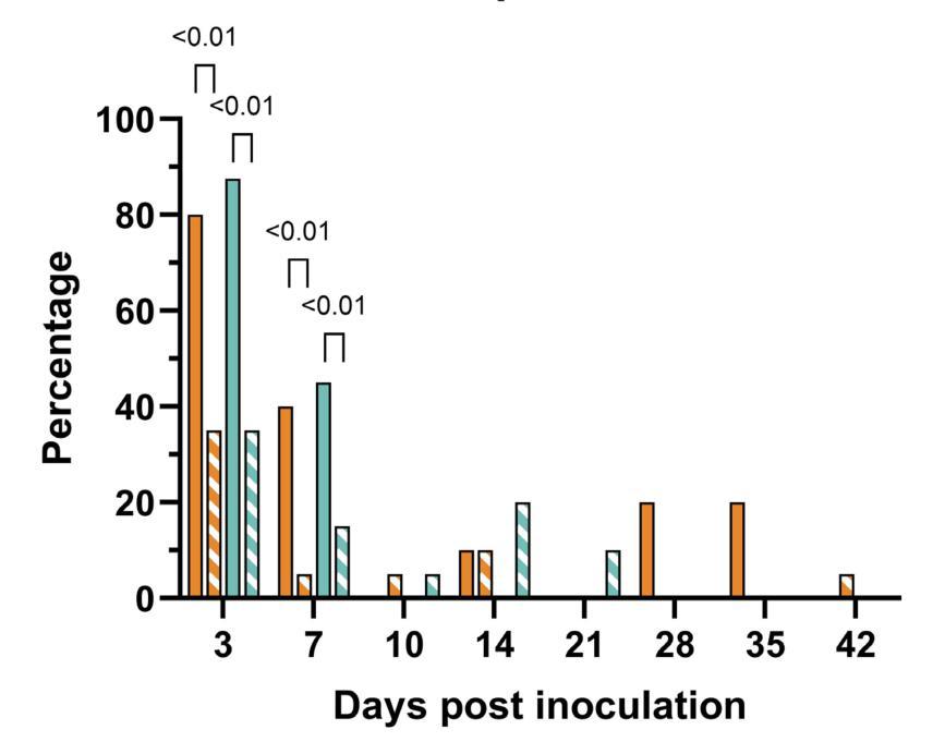
Bacteriological reisolation (Figure 3). E. cecorum was never isolated from birds in the control groups. In both challenged groups, nearly all birds tested positive for E. cecorum by real-time PCR in caecal content samples throughout the entire study period indicating successful colonization.


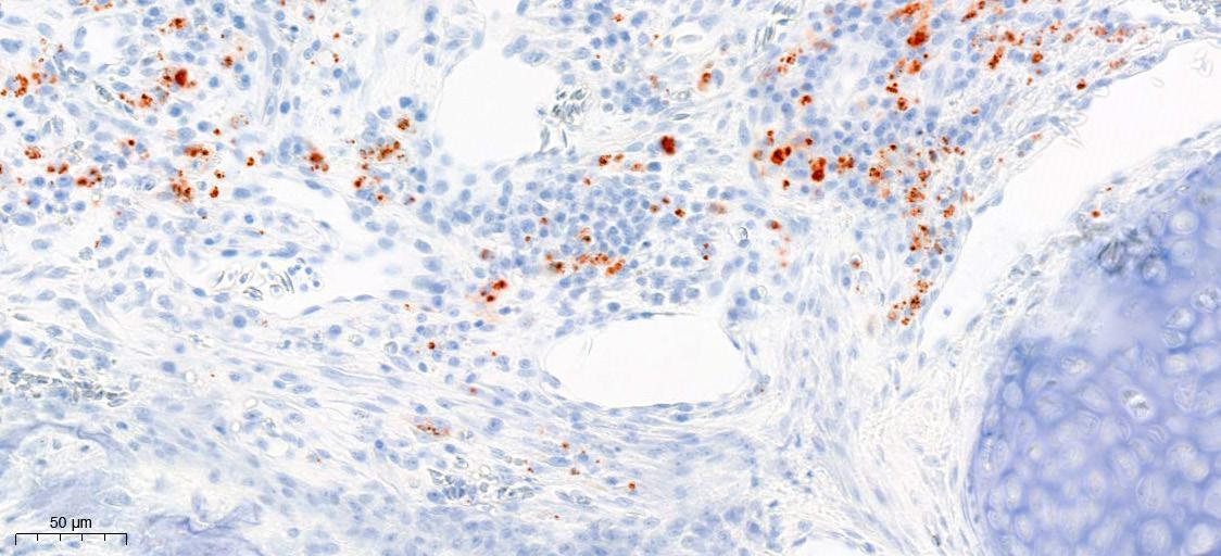
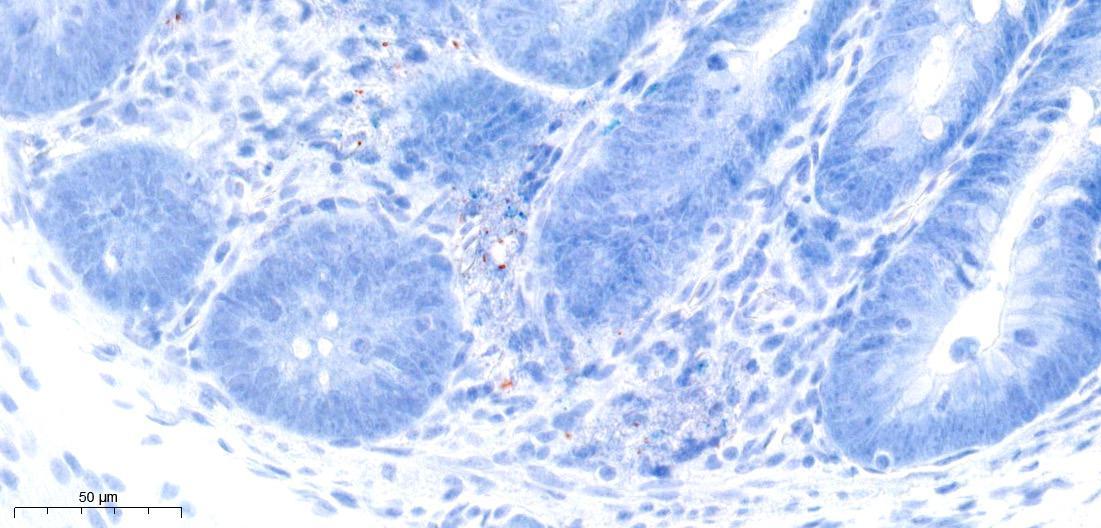
AFigure 4. Heatmap of immunohistochemical analysis of 5 E. cecorum (EC) inoculated and 5 control (PPS) inoculated birds at 3 d.p.i. A. The number of stained E. cecorum were counted and categorized into five groups: 0) no E. cecorum, 1) 1-5 bacteria, 2) 6-10 bacteria, 3) 11-49 bacteria and 4) >50 bacteria. Counting was performed at four different locations: lumen, brush border, intraepithelial and lamina propia. B. Immunocytochemical staining of EC in free thoracic vertebra (top) and in the lamina propria of the caecum (lower panel). Scale bar 50 μm.
Conclusions
• Translocation of E. cecorum to peripheral organs was highest on 3 days post infection.
• The exclusive occurrence of ascites E. cecorum-inoculated birds was not being previously described as a typical E. cecorum lesion.
• The wire obstacle did not affect lesion development or bacterial detection.
• Bacteraemia and development of typical lesions were achieved in SPF broilers inoculated with E. cecorum, enabling further investigation into its pathogenesis, potential prevention methods, and interventions.
Figure 3. Bacteriological reisolation was performed on blood and swabs of femur, free thorac vertebra and spleen. Bacterial reisolation was compared with detection of E. cecorum by qPCR. Translocation of bacteria based on qPCR was shown in 80% of the infected birds with no significant difference due to mechanical stress. References Borst