COLOPHON
Production and Publication
Research & Training, Dept. of Radiology & Nuclear Medicine, Erasmus MC
Editor
Lieke Visser
Assistant Editors
Emar Thomasa
Shemara Mendes
Design & Photography
Steven Ensering
Frank van der Panne
Vincent Blinde
Maartje de Sonnaville
Printing GROENPRINT
Aristotelesstraat 20 3076 BD Rotterdam
The Netherlands
For this Scientific Report GROENPRINT and Trees for All plants serveral trees in Costa Rica to restore tropical rain forest.
Visiting Address
Department of Radiology & Nuclear Medicine
Erasmus MC
Dr. Molewaterplein 40 3015 GD Rotterdam
The Netherlands
Telephone: +31 10 703 5372 research.radiology@erasmusmc.nl
Post Address
Department of Radiology & Nuclear Medicine
Erasmus MC
P.O. Box 2040
3000 CA Rotterdam
The Netherlands
Website
Radiology & Nuclear Medicine – Department –Erasmus MC

Cover photo
Exterior of Sophia Children’s Hospital seen from the Erasmus MC passage, Rotterdam, The Netherlands.
a a a a a a a a a a a a a a a a a a a a a a a a a a a a a
2023 Scientific Report
department of radiology & nuclear medicine
2 a a a a a a a a a a a a a a a a a a a a a a a a a a a a a a a a a a a a a a a a a a a a a a a a a a a a a a a a a a a CONTENTS Preface 5 Highlights 2023 6 Convergence 14 Research Staff 20 Research Support 24 Research Strategy and Targets 28 Imaging Facilities 32 BIOMEDIC AL IMAGE ACQUISITION & ANALYSIS 39 Magnetic Resona nce Physics in Medicine 41 J uan A Hernández Tamames, PhD Physics in CT Technology 51 Marcel van Straten, PhD Quantitative MRI Reconstruction 55 Dirk Poot, PhD AI in Medical Image Analysis 61 Marleen de Bruijne, PhD Image Guidance in Interventions and Therapy 67 Theo van Walsum, PhD Applied Medical Image Analysis 77 Stefan Klein, PhD NeuroImage Analysis & Machine Learning 85 Esther E Bron, PhD Computational Population Biology 93 Gennady Roshchupkin, PhD Artificial Intelligence for Integrated Diagnostics 99 Martijn Starmans, PhD Machine and Deep Learning 105 Jifke F Veenland, PhD MOLECULAR IMAGING & THERAPY 109 Radiobiology of Radionuclide Therapy 111 Julie Nonnekens, PhD Radiotracer Interactions Group 119 Simone Dalm, PhD Translational Nuclear Medicine 129 Frederik A Verburg MD, PhD Optical Molecular Imaging 133 Clemens WGM Löwik, PhD Genetic Engineering for Multimodality Imaging 137 Laura Mezzanotte, PhD
Esther AH Warnert, ir,
Sophie Veldhuijzen van Zanten, MD,
Ricardo Budde, MD, PhD & Alexander Hirsch, MD,
Ivo G Schoots, MD, PhD
Daan Caudri, MD, PhD & Pierluigi Ciet, MD,
Meike W Vernooij, MD,
Jacob J Visser, MD, PhD
MG Myriam Hunink, MD, PhD
Ryan Muetzel, PhD
3 a a a a a a a a a a a a a a a a a a a a a a a a a a a a a a a a a a a a a a a a a a a a a a a a a a a a a a a a a a a Clinical Nuclear Medicine: Imaging and Therapy in Oncology 143
PhD Radiopharmaceutical Chemistry 149 Yann Seimbille, PhD CLINICAL IMAGING 157 Imaging in Neurovascular Disease 159 Aad van der Lugt, MD, PhD Neuroradiology 167
Smits, MD, PhD Bench-to-Bedside MR Imaging Biomarkers 175
Astrid van der Veldt, MD, PhD & Tessa Brabander, MD,
Marion
PhD Theranostics of CNS and H&N deseases 181
PhD Cardiac Imaging 187
PhD Abdominal Imaging 195
Advanced Musculoskeletal Imaging Research Erasmus Mc (ADMIRE) 203
Oei, MD, PhD Advanced Thoracic Imaging Research 217
IMAGING IN HEALTH SCIENCES 225 Population Imaging 227
PhD Imaging of Arteriosclerosis: From Population to Clinical Practice 239 Daniel Bos, MD, PhD Value-based imaging 247
Edwin HG
PhD
Assessment of Radiological Technology (ART) 255
Pediatric Population Neuroimaging 261
I NPUT & OUTPUT 269 Conference Contributions 2023 270 P ublications 2023 272 Index 276 Social Activities 2023 280

a a a a a a a a a a a a a a a a a a a a a a a a a a a a a a a a a a a a a a a a a a a a a a a a a a a a a a a a a a
In 2023, Erasmus MC revised its research strategy, aligning it with the European Commission’s vision for addressing society’s most urgent issues. In response, our department adapted its research focus to highlight critical societal concerns, including aging populations, rising healthcare costs, and workforce shortages. We have prioritized research topics such as reducing scan times, enhancing data acquisition reliability, automating biomarker extraction, and developing minimally invasive treatments and theranostics.
With this strategic shift, we are eager to make significant contributions to the advancement of society and confront the challenges that lie ahead. Our dedication to impactful and high-quality research remains unwavering, and we are excited about the future possibilities that await us.
Over the past year, our department’s research has thrived, resulting in remarkable accomplishments. We are proud to announce that 17 PhD students successfully defended their theses – a milestone for each of them, marking their dedication and scientific perseverance. Their theses will serve as a stepping stone in their careers, supported by education in scientific methods and imaging technologies.
Furthermore, we have secured multiple significant grants from various sources, enabling us to further expand our research initiatives. These grants empower us to tackle pressing challenges and make a meaningful impact on society. Among them are personal grants for talented researchers within the department, as well as multiple grants from the Dutch Research Council, charities, and international funders. Marion Smits received a prestigious personal grant from the Dutch Research Council – an esteemed VICI grant focused on “Virtual Brain Biopsy, paving the way toward reality”.
The achievements mentioned above resulted from an important pillar of our department’s research strategy: the implementation of a talent development plan. This plan emphasizes the creation of an environment where young researchers can grow and receive support to take their research to the next level. We have adopted a new approach for rewarding individuals, which considers not only the number of papers and grants but also acknowledges academic leadership, education, and impact.
Jifke Veenland was appointed as an associate professor with an educational profile, based on her major contribution to the development of the bachelor program in Clinical Technology and the master program in Technical Medicine. In recent years, we have also witnessed the growth of young researchers within our department. Talented individuals like Julia Neitzel,
Martijn Starmans, and Maarten Leening have advanced to assistant professorships. These promotions reflect their excellent achievements and dedication to research.
This year, we continued to strengthen our collaboration with the Technical University (TU) Delft, aligning with the Convergence initiative of Erasmus MC, TU Delft, and Erasmus University. This collaboration has created new avenues for the use and evaluation of technology in medicine. Our department is well-positioned to contribute to this project. To reflect its broad scope, Meike Vernooij has been appointed as the Medical Delta Professor, holding a joint position in both Erasmus MC and TU Delft. Additionally, Jukka Hirvasniemi was appointed in a shared position with the Department of Biomedical Engineering at TU Delft.
Collaboration with other departments is essential for excellent research in the imaging domain. We already had strong ties with the departments of Epidemiology, Molecular Genetics, Cardiology, Obstetrics & Gynaecology and Pediatric Pulmonology. Harm Tiddens, professor of pediatric pulmonology and the link between the two departments, has retired. Daan Caudri has taken over his position and is now leading the research line on Advanced Thoracic Imaging Research, alongside Pierluigi Ciet.
In 2023, we initiated two new collaborations. First, with the Department of Ophthalmology and the Eye Hospital Rotterdam, our department established the Eye Image Analysis Group Rotterdam (EyeR). Danilo Andrade de Jesus and Luisa Sánchez Brea serve as principal investigators, sharing positions across the three founding institutions. Second, in partnership with the Department of Pathology, we formed a research group focused on artificial intelligence for Integrated Diagnostics. This endeavor received a kick start with the AiNed Fellowship Grant of 2 million euros awarded to Martijn Starmans by the Dutch Research Council.
I take pride in our researchers and staff for their hard work and dedication. Research is a collaborative effort, and our team is both strong and committed. I also extend my gratitude to our collaborators, including other departments within Erasmus MC, universities in the Netherlands, universities abroad, and partners in industry.
Enjoy reading this annual report.
Aad van der Lugt, Professor and Chairman April 2024
5 a a a a a a a a a a a a a a a a a a a a a a a a a a a a a a a a a a a a a a a a a a a a a a a a a a a a a a a a a a a
PREFACE
HIGHLIGHTS 2023
Appointments
Maarten Leening was appointed as assistant professor in Preventive Cardiology and Non-invasive Cardiac Imaging.
Julia Neitzel was appointed as assistant professor in Population Brain Health.
Martijn Starmans was appointed as assistant professor in AI for Integrated Diagnostics (AIID), focused on Medical Imaging in Oncology.
Jifke Veenland was appointed as Associate professor in Radiomics and Deep Learning for prostate cancer.
Pierluigi Ciet was appointed as chair of the chest MRI standardization group of the European Society of Cystic Fibrosis (ECFS).
Pierluigi Ciet was appointed chair of the Photon Counting standardization group within the European Society of Pediatric Radiology (ESPR).
Daan Caudri was appointed as member of the European Board for Accreditation in Pneumonology (EBAP).
Ivo Schoots completed his MBA in Healthcare in May 2023 at the Amsterdam Business School.
Erik de Blois was appointed as Scientific Expert at International Atomic Energy Agency (IAEA) for his expertise on Ac-225 labeled radiopharmaceuticals.

Meike Vernooij was appointed as Medical Delta professor at the TU Delft (Faculty of Applied Sciences).
Meike Vernooij was re-elected as Chair of the Diagnostic Committee of European Society of Neuroradiology (ESNR), as well as appointed as Subcommittee Chair for Neuroradiology at European Congress of Radiology (ECR) 2024.

6 a a a a a a a a a a a a a a a a a a a a a a a a a a a a a a a a a a a a a a a a a a a a a a a a a a a a a a a a a a a

Theo van Walsum was officially appointed as Adjunct Professor at VNU-EUT in Hanoi, Vietnam.
Erik de Blois was appointed as Scientific Expert at IAEA for his expertise on Ac-225 labeleld radiopharmaceuticals.
David Hanff was appointed as Board Member of MSK section of the Dutch society of Radiology (NVVR) member of Hip and groin injuries team Erasmus MC
Collaborations
In 2023 the pioneer collaboration, Eye Image Analysis Group , between the Departments of Radiology & Nuclear Medicine and Ophthalmology at Erasmus MC and The Rotterdam Eye Hospital started. Luisa Sanchez Brea and Danilo Andrade de Jesus are leading this initiative and have and have a position in the participating institutions in this collaboration.
Radiology & Nuclear Medicine and Pathology join forces with Artificial Intelligence for Intergrated Diagnostics. Martijn Starmans was appointed in a shared position between the department of Radiology & Nuclear Medicine and the department of Pathology for the AiNed Fellowship Grant project.
Jukka Hirvasniemi has a shared appointment at TU Delft, faculty of Biomedical Engineering for the Technical Medicine MSc programme.
Contributions to Guidelines
Daniel Bos and Dianne van Dam-Nolen contributed to a novel carotid plaque classification system (Carotid Plaque-RADS: A Novel Stroke Risk Classification System) in JACC Cardiovasc Imaging. DOI: 10.1016/j. jcmg.2023.09.005.
Pierluigi Ciet contributed to Italian imaging guidelines for management of respiratory tract exacerbations in people with Cystic Fibrosis patients in Front Pediatr.. 2023;10:1084313.
Theo van Walsum and the AO-VISION team contributed to the development of new image acquisition protocols on AO of patients with inherited retinal diseases (IRDs), namely Retinitis Pigmentosa. The results were published in relevant national and international ophthalmic conferences (e.g. NOG, EURETINA, ARVO).
Erik de Blois contributed to the TecDoc guidelines on Ac-225 labelled pharmaceuticals; at the International Atomic Energy Agency (IAEA) TecDoc.
7 a a a a a a a a a a a a a a a a a a a a a a a a a a a a a a a a a a a a a a a a a a a a a a a a a a a a a a a a a a a
Ivo Schoots received his MBA in Healthcare.
Ricardo Budde led the working group on Coronary CT: standardization from patient preparation to reporting” of the “Kennisinstituut” of the “Federatie Medisch Specialisten”. Aim of the working group was to provide guidance for hospitals that want to start a Coronary CT program by writing a comprehensive “how-to-do-it” document including scan protocols.
Ricardo Budde chairs the Society of Cardiovascular Computed Tomography (SCCT) writing group for the consensus document on “Prosthetic Heart Valve Imaging”.
Marion Smits and Wouter Teunissen contributed to the Dutch national guideline for glioma.
Marion Smits contributed to the joint recommendations from four RANO groups for a framework for standardised tissue sampling and processing during resection of diffuse intracranial glioma, published in Lancet Oncology, 2023; 24:e438-e450.
Marion Smits contributed to the essential requirement of quality cancer care in adult glioma on behalf of the European Society of Radiology, published by the European Cancer Organisation in J Cancer Policy 2023; 38:100438.
Marion Smits contributed to guidance on arterial spin labeling perfusion MRI in clinical neuroimaging by the International Society for Magnetic Resonance in Medicine (ISMRM) Perfusion Study group.
François Willemssen contributed to the Dutch guidelines for diagnostic abdominal imaging in Hepatocellular carcinoma (HCC) and cholangiocarcinoma.
Maarten Thomeer was co-author for the Dutch guidelines in cervical and endometrial carcinoma .
Ivo Schoots contribured as board member European Association Urology (EAU) Prostate Cancer guideline panel and as board member PI-RADS steering committee, on prostate MR imaging.
Astrid van der Veldt is the chair of Dutch Melanoma Guideline which is currently revised completely.
Erik Verburg contributed to the European Association of Nuclear Medicine (EANM) guideline on radioiodine therapy of benign thyroid disease as well as the revised Dutch guideline on the treatment of differentiated thyroid cancer.
Societal Impact
Marleen de Bruijne contributed to the handbook of Medical Image Analysis, a volume in The MICCAI Society book Series.
Daniel Bos organized and hosted debates on Recognition & Rewards during the WEON conference (Werkgroep Epidemiologisch Onderzoek Nederland) and the Dutch Cardiovascular Alliance.
Vapen #JouwKeuze. Together in the fight for a nicotine free generation! Dr. Daan Caudri gave lessons at Rotterdam high schools on the topic of vaping and smoking. Together with >600 respiratory specialists in the Netherlands well over a 1000 lessons have been given already at high school classes, as well as some primary schools.
Theo van Walsum , patent granted: Methods and Systems for Dynamic Coronary Roadmapping Application, Publication/Patent Number: US20200222018A1 (2023-07-25).

Celebrating the start of ICAI Stroke Lab. This project will be led by Sandra Sülz and Theo van Walsum
Jacqueline Claus appeared on national television (NPO1) in an interview on her research by Alzheimer Nederland.
Frank Wolters gave lectures for the general public at the prestigious Alzheimer's Association International Conference (AAIC) and at the ABOARD consortium public-day meeting (organized by Jacqueline Claus ).
8 a a a a a a a a a a a a a a a a a a a a a a a a a a a a a a a a a a a a a a a a a a a a a a a a a a a a a a a a a a a
Ricardo Budde was a member of the working group “Kennisagenda 2023-2027” of the Dutch Society of Radiology. The working group composed a document outlining the top 10 knowledge gaps for Dutch radiology practices.
Julie Nonnekens was invited to participate in a round table discussion on women in science, organised by the Association of Spanish Scientists, Utrecht, The Netherlands. Sep 2023.
Simone Dalm made an educational video in collaboration with Universiteit van Nederland for breast cancer awareness and the potential of targeted radionuclide therapy for breast cancer management.
Patrick Tang was appointed by the KNAW as one of the Faces of Science, to communicate about his research and his live as doctoral students in blogs, vlogs, articles, lectures, media appearances and social media activities. He was interviewed on Dutch national radio (NPO radio 1, Omroep Max).
Ivo Schoots gave an interview for MemoRad (NVVR, MemoRad, November 16, 2023) with the title 'Vroegdiagnostiek bij prostaatkanker ‘MRI bespaart pijnlijke prikken en vermindert zorgkosten’.
Ivo Schoots gave an update of PI-RADS v2.1 (July 1, 2023) on the educational platform Radiology Assistant, with worldwide outreach . (www.radiologyassistant.nl)
Based on the results of the manuscript ‘The meaning of screening: detection of brain metastasis in the adjuvant setting for stage III melanoma’ by Sophie Derks et al, national consensus was achieved during the annual WINO-meeting to stop MRI screening of patients with stage III melanoma.
Gennady Roshchupkin publication about new AI methods for children's facial shape was covered in 425 stories in total in a number of different languages and around the world.
Honors & Awards
Sophie Veldhuijzen van Zanten was selected by the New Scientist as finalist for Science Talent Prize awarded every two years to an inspiring researcher of the Netherlands and Belgium.
Sophie Veldhuijzen van Zanten presented, upon invitation, the ‘Highlights lecture’ during opening ceremony of the 36th European Association of Nuclear Medicine (EANM) Annual Congress, welcoming >7000 international colleagues to Vienna, Austria on the 9th of September 2023.
David Hanff won the More prize Award of Master teacher of the year 2023.
Simone Dalm won the Daniel den Hoed Award for a preclinical project to study Tandem Radionuclide Therapy. The project was highlighted in an interview and on various websites, e.g. Stichting Dutch Uro-Oncology Studygroup (DUOS) and Medische Oncologie.
Ruisheng Su was awarded a prestigious DAAD AIntet fellowship for the Postdoc-NeT-AI 04/2023 Networking Week – Genertive Models in Machine Learning, which allowed him to visit several excellent research groups in Germany.
Wietske Bastiaansen won the yearly Wladimiroff research award of the Department of Obstetrics and Gynecology for the best presentation on “Artificial intelligence to automatically measure the embryonic and head volume in first trimester ultrasound scans: The Rotterdam Periconception cohort”.
Savine Minderhoud won the Medical Delta Thesis Award for her thesis “Biomechanics in congenital heart disease: Using advanced imaging techniques”.
Grants 2023
Personal Grants / Fellowships
Marie Curie Fellowship
Justine Perrin
Title: ‘Impact of BRCA2 deficiency on the DNA damage response and Immunogenicity of Prostate cancer after radioligant therapy’.
Erasmus MC Fellowship
Julia Neitzel
Title: ‘Impact of Modifiable Factors on Dementia Risk across the Adult Lifespan’.
Dutch Research Council - VICI
Marion Smits
Title: ‘Virtual biopsy: paving the way towards reality’.
Alzheimer Nederland - Early Career Grant
Eline Vinke
Title: ‘Unraveling brain aging patterns predictive of AD or ADRD’.
Dutch Heart Foundation - Dekker Beurs
Eline Vinke
Title: ‘Personalized MRI-based Cerebral Small Vessel Disease (SVD) burden quantification, for more accurate diagnosis and prognosis’.
9 a a a a a a a a a a a a a a a a a a a a a a a a a a a a a a a a a a a a a a a a a a a a a a a a a a a a a a a a a a a scientific report 2023 | HIGHLIGHTS
Dutch Research Council - AiNed Fellowship Grant
Martijn Starmans
Title: ‘Radiology and pathology join forces through Artificial Intelligence for Integrated Diagnostics (AIID)’.
Erasmus MC Fellowship
Gennady Roshchupkin
Title: ‘GenetiX: Decoding the Genetic Puzzle of Complex Traits with Federated learning and Explainable AI’.
Dutch Ministry of Education, Culture and Science (OCW)
Starting Grant
Gennady Roshchupkin
Title: ‘AI for multi-omics’.
Erasmus MC2 Research Synergy Grant
Ryan Muetzel
Title: ‘Understanding Cortico-cerebellar maturation trajectories’.
Erasmus MC2 Research Synergy Grant
Daniel Bos
Title: ‘Unraveling the vascular biomechanics of stroke and dementia: the navier-stokes study’.
Primary Ciliary Dyskinesia Foundation - Early Career
Investigator Award
Daan Caudri
Title: ‘Structural lung disease in children with PCD: utilizing an automated airway-artery method to detect disease progression’.
National Grants
Dutch Research Council - Knowledge and Innovation
Covenant
Marion Smits
Title: ‘Bringing tractography into daily neurosurgical practice’.
Dutch Research Council - TTW Perspectief
Esther Bron
Title: ‘CHIME: Cerebral hemodynamics, metabolism and clearance: a comprehensive, non-invasive brain imaging approach to characterize key biological processes in dementia’.
Dutch Research Council - Open Technology Program
Stefan Klein
Title: ‘Liver Artificial Intelligence’.
Health-Holland LSH-TKI PPP
Martijn Starmans, Jacob J. Visser and Frans Vos
Title: ‘An artificial intelligence (AI)-based model for detection of incidental pulmonary embolism in chest CTs’.
Health-Holland LSH-TKI PPP
Erik de Blois
Title: ‘ Tb-161-PSMA’.
Dutch Research Council - ROBUST program and GE
HealthCare
Edwin Oei
Title: ‘Innovation Center for Artificial Intelligence (ICAI) lab Trustworthy AI for Magnetic Resonance Imaging’.
Dutch Research Council - ROBUST program and Philips
Healthcare
Daniel Bos and Theo van Walsum
Title: 'Innovation Center for Artificial Intelligence (ICAI)
Stroke Lab: from 112 to Rehabilitation'.
Health Holland LSH multicenter PPP
Esther Bron
Title: ‘Scan2Go: autonomous MRI for large scale diagnostics of brain integrity ’.
Stichting Wetenschappelijk Onderzoek het Oogziekenhuis (SWOO)
Luisa Sanchez Brea and Daniel Andrade de Jesus
Title: ‘Adaptive optics in patiënten na netvliesloslating’.
Dutch supercomputer computational grant - Computing time
Jing Yu
Title: ‘Brain Imaging Genetics’.
Dutch supercomputer computational grant - Computing time
Xianjing Liu
Title: 3D Facial Shape Analysis’.
International Grants
ISIDORe Joint Research Activities PROGRAMME
Stefan Klein, Marcel Koek, Hakim Achterberg, Adriaan
Versteeg and Martijn Starmans
Title: ‘PATH2XNAT: Covid19 Pathomics meets XNAT ’.
NIHR Research Design Service for Yorkshire and Humber
Matthew Marzetti
Title: ‘Novel Applications for Sarcoma Assessment (NASA)’.
Cystic Fibrosis Foundation Grant
Daan Caudri
Title: ‘Real world outcomes with novel modifier therapy combinations in children with CF (ENHANCE study)’.
10 a a a a a a a a a a a a a a a a a a a a a a a a a a a a a a a a a a a a a a a a a a a a a a a a a a a a a a a a a a a
World Cancer Research Fund
Daniel Bos
Title: ‘Treatment tolerance and prognosis in survivors with non-metastatic colorectal cancer: a matter of liver fa(c)t?’.
EU - Erasmus+ Learning Agreement (KA171)
Ryan Muetzel
Title: ‘Population neuroimaging mobility program, Erasmus MC, Harvard, University of Minnesota’.
FDA Award
Frederik Verburg
Title: ''Aprospective study to support validation of lung deposition models with nuclear medicine imaging methods''.
Charitable Organisations
Wishdom Foundation
Daniel Andrade De Jesus and Luisa Sanchez Brea
Title: ‘Voorspellen van ernstige prematurenretinopathie met behulp van kunstmatige intelligentie: uitbreiding en klinische validatie van een werkend model’.
Dutch Heart Foundation, Brain Foundation Netherlands
Aad van der Lugt
Title: ‘CONTRAST 2.0’.
KWF Dutch Cancer Society
Maarten Thomeer
Title: ‘Effect of FAPI PET-CT on management in patients with potentially resectable biliary tract cancers: prospective multicenter study and cost-effectiveness analysis’.
KWF Dutch Cancer Society
Astrid van der Veldt
Title: ‘In-depth study to provide safe long-term survivorship care to survivors of metastatic melanoma (SURVIVOR)’.
KWF Dutch Cancer Society
Ivo Schoots
Title: ‘ Towards clinical implementation of novel diagnostic tools in oncologic imaging: an opensource web-based reviewing infrastructure for validation studies’.
KWF Dutch Cancer Society
Ivo Schoots
Title: ‘ADMINISTRATE (Advanced Diagnostic Modalities in ImagiNg Impacting on diagnosiS, TReatment And paTient outcomE) on prostate cancer’.
Dutch Foundation for Asthma Prevention
Marleen de Bruijne
Title: ‘Origins of childhood asthma: focus on the developmental lung structure pathway using nonradiant imaging and artificial intelligence’.
Alzheimer Nederland
Daniel Bos, Meike Vernooij, Julia Neitzel, Frank Wolters and Maarten Leening
Title: ‘ Trajectories of Vascular Disease in Aging to Predict Dementia’.
Maarten van der Weijden Foundation
Daniel Bos and Ricardo Budde
Title: ‘Statins to Prevent Immune checkpoint inhibitor-induced pRogression of AtheroscLerosis: the SPIRAL study’.
Institutional Grants
Vrienden van het Sophia
Gennady Roshchupkin
Title: ‘Machine learning for postoperative shape prediction in craniosynostosis treatment using 3D models’.
Vrienden van het Sophia
Ricardo Budde
Title: ‘Development of Photon Counting CT protocols for pediatric cardiovascular imaging’.
Erasmus MC Foundation
Clemens Lowik
Title: ‘PDT + Immune therapy research’.
Investigator initiad industry sponsored grants
GE HealthCare
Pierluigi Ciet
Title: ‘Magnetic Resonance Imaging in Interstitial Lung Disease (M-ILD)’.
GE HealthCare
Edwin Oei
Title: ‘Pinpointing the source of chronic pain and therapy response with wholebody 18F FDG-PET/MRI’.
GE HealthCare
Edwin Oei
Title: ‘ RSNA QIBA MSK Profile Stage 3 and 4 Conformance Testing’.
Enlitic Inc.
Jacob J. Visser
Title: ‘Assessing the value of an AI-algorithm to standardize DICOM-data based on imaging recognition’.
11 a a a a a a a a a a a a a a a a a a a a a a a a a a a a a a a a a a a a a a a a a a a a a a a a a a a a a a a a a a a scientific report 2023 | HIGHLIGHTS
New facilities
Digital ring SPECT system
At the end of 2023, the first patient scans were acquired using our new GE Starguide SPECT/CT system. This system features a novel design with 12 digital CZT SPECT detectors, capturing full 3D information for all patient scans. The utilization of a fixed collimator eliminates the time-consuming process of swapping collimators between acquisitions. This innovative design is anticipated to significantly reduce acquisition times and facilitate new applications, such as 3D dynamic imaging.
In conjunction with the new XELERIS software platform, which includes an AI-assisted dosimetry application, a more efficient workflow for theranostics, such as Lu-177 DOTATE and Lu-177 PSMA, is expected.
Bucky System
A fluoroscopy and bucky system were due for replacement in 2023. A combination was chosen, which can be used for radiography and fluoroscopy. We are working with the Luminos Lotus Max of Siemens Healthineers. The system was installed in December 2023 and put into use in January 2024.


12 a a a a a a a a a a a a a a a a a a a a a a a a a a a a a a a a a a a a a a a a a a a a a a a a a a a a a a a a a a a
MRI scanner - 3 Tesla Premier
We are pleased to announce the completion of the upgrade for one of the MRI systems, now operating on the 3 Tesla Premier platform with the latest DV30 software. This upgrade represents a significant advancement in our hospital's imaging capabilities, establishing this scanner as the most modern within our MRI department.
The integration of the DV30 software brings a suite of enhancements, including the extension of AIR Recon DL to 3D imaging, PROPELLER sequences, and FSE Flex. These enhancements result in improved image quality, a more streamlined patient experience across various exam types, and reduced scan times.
Our collaboration with GE's application specialists has been instrumental in optimizing protocols to maximize the performance of the upgraded system. While initial optimization sessions have concluded, ongoing collaboration with GE's specialists will be needed to refine our imaging protocols, ensuring the optimal utilization of the technology.

Contrast injectors
In 2021 we published a European tender for the replacement of our CT contrastinjectors. Our goal was to get new injectors that were specifically designed for multipatient use.
The winner of the tender was Rembrandt Medical, that offered us the Ulrich CT-Motion injector. Finally, after a long testing and preparation phase in 2022, we started to use the contrastinjectors in May 2023.
The radiographers can work more efficient with contrast agents. There are triple checks for air bubbles with automatic detection at three different points. Instead of 4-hour systems, we now work with 24-hour systems. This is also a great benefit for the CT scanner in the ER department.
The injector has also an RIS/PACS integration which means that documentation about the volume/brand of the contrast agent automatically will be sent to PACS/ HiX.

13 a a a a a a a a a a a a a a a a a a a a a a a a a a a a a a a a a a a a a a a a a a a a a a a a a a a a a a a a a a a scientific report 2023 | HIGHLIGHTS
CONVERGENCE

 Figure: Ten Flagships started in 2022.
Brain Tumors – CONVERGENCE
Erasmus MC
Martin van den Bent
Juan Hernandez-Tamames
Stefan Klein
Pieter Kruizinga
Alejandra Mendez Romero
Dirk Poot
Marion Smits
Krishnapriya Venugopal
Esther Warnert
Karen van der Werff
Expertise
TU Delft
Chi-Hsien Tseng
FransVos
Erasmus University Rotterdam
Justien Dingelstad
Iris Wallenburg
Marion Smits co-initiated and was main lead of the Convergence Flagship ‘Deep Medical Imaging of Structure, Physiology and Function’, in which brain tumor imaging features prominently. She stepped down in Summer 2023, and handed over to Juan Hernandez-Tamames to lead the Flagship on behalf of Erasmus MC. Marion Smits is also scientific lead of the Medical Delta Cancer Diagnostics 3.0 scientific program, which currently focuses primarily on brain tumor diagnostics. Radiology & Nuclear Medicine prominently features in both scientific programs providing expertise on the full spectrum from image acquisition and image analysis to data management and diagnostic clinical neuroimaging in brain tumors. The noninvasive diagnosis of cancer at the tissue level through (advanced) imaging techniques and (big data) analysis is at the core of these programs. See:
Grants and funding

Contribution and Added Value
Cross-pollination of clinical, technical and social sciences, use of specific equipment (e.g., PET-MRI at Erasmus MC, 7T at LUMC, proton therapy at HPTC). In the recently awarded Convergence incentive grant this convergence of expertise is exemplified: the project focuses on the clinical implementation, prospective validation, and interaction of a previously developed AI algorithm to predict brain tumor diagnosis in a true clinical setting.

Figure: Brain tumour with high vascularisation imaged with perfusion MRI.
2019 Convergence: Quantitative Susceptibility MRI: Deep insights in cardio- and neuro-vasculature
2021 Convergence Open mind call: O2-Sense, converging on wearable oxygen monitoring for brain tumor patients
2021 Convergence Open mind call: Neurodegeneration beyond DTI
2022 ICAI lab ROBUST: Trustworthy AI for MRI – brain tumors
2022 Convergence Incentive Grant PIRL: Real-world assessment of ‘PrognosAIs’ for measuring, typing and grading of presumed adult-type diffuse glioma
2022 KWF: Early detection of brain tumor progression with amide proton transfer weighted CEST MRI
2023 ZonMW Vici: Virtual biopsy: paving the way towards reality
15 a a a a a a a a a a a a a a a a a a a a a a a a a a a a a a a a a a a a a a a a a a a a a a a a a a a a a a a a a a a
Musculoskeletal Imaging – CONVERGENCE
Erasmus MC
Sita Bierma
Jaap Harlaar
Rianne van der Heijden
Jukka Hirvasniemi
Stefan Klein
Joyce van Meurs
Edwin Oei
Gerjo van Osch
Gennady Roshchupkin
Jos Runhaar
Dieuwke Schiphof
Expertise
TU Delft
Samantha Copeland
Jaap Harlaar
Jesse Krijthe
Marco Loog
Marcel Reinders
Amir Zadpoor Ajay Seth
Erasmus University Rotterdam
Inge Merkelbach
Sandra Sülz
Jukka Hirvaniemi, jointly appointed at the Department of Radiology & Nuclear Medicine of Erasmus MC and the Department of Biomechanical Engineering of TU Delft, has advanced expertise in the field of musculoskeletal image analysis using artificial intelligence and radiomics. As example extraction of quantitative imaging biomarkers for assessment of osteoarthritisis depicted in the figure below. We also contribute using advanced image acquisition techniques: MRI and PET/ MRI, linking with biomechanics measurements in the new MOtionBiomechanics & Imaging (MOBI) lab.
Contribution and Added Value
The new MOtionBiomechanics & Imaging (MOBI) lab which is being set up in the Department of Radiology & Nuclear Medicine as a joint initiative between Erasmus MC (Oei, Bierma-Zeinstra) and TU Delft (Harlaar) is considered a showcase for the Convergence program as it unites the expertise of technical and medical sciences with the need of collaboration between scientists from various disciplines (engineering, biomechanics, imaging physics, image analysis, clinical orthopedics, radiology).

Grants and funding
2019 ZonMW Open: Biomechanical precision diagnostics in osteoarthritis
Figure: Schematic presentation of a quantitative imaging biomarker extraction pipeline.
2020 Dutch Research Agenda Research along routes by Consortia (NWA-ORC): Healthy Loading to combat osteoarthritis: Leveraging molecular variations in load bearing capacity for individualized movement aDvice: The LoaD project
2022 Convergence Flagship: Healthy Joints
16 a a a a a a a a a a a a a a a a a a a a a a a a a a a a a a a a a a a a a a a a a a a a a a a a a a a a a a a a a a a
Image-guided therapy – CONVERGENCE
Erasmus MC
Tessa van Ginhoven
Aad van der Lugt
Kees Verhoef
Theo van Walsum
Bart Cornelissen
Eppo Wolvius
Expertise
TU Delft
Nandini Bhattacharya
Jenny Dankelman
Frank Gijssen
Benno Hendriks
Ricardo Guerra Marroquim
Aimee Sakes
Theresia van Essen
The success of integration smart instruments with augmented navigation is leveraged by the complementary expertise of the project members, that covers the domains of all aforementioned challenges. Integrating smart instruments with augmented navigation leads to novel solutions that cannot be accomplished with only one of the groups. To develop and implement the SMART Surgical knife in clinical practice, expertise of building surgical instruments with incorporated optical fibers and analysis of the signals (Biomechanical Engineering, TU Delft) has to be combined with surgical expertise on safe removal of tumor tissue (Surgical Oncology group, Erasmus MC). Moreover, to augment navigation real time in an intuitive way preoperative information need to be adapted to the surgical setting (Biomedical Imaging Group Rotterdam, Erasmus MC) and transferred back to the AR environment (Computer Graphics Group, TU Delft). Combining these approaches will provide a more robust and safer way to enhance the surgical procedure, as the visualization can be finely aligned with the surgical procedure using the guidance of the smart instrument, and the feedback from the smart instrument can be enhanced through visualization.

Grants and funding
Erasmus University Rotterdam
Sandra Sülz
Martina Buljac
Contribution and Added Value
Two flagships from 2021, entitled I-GUIDE: Image guided minimally invasive interventions and Smart Surgery in Smart OR, were not granted in the first round. However, both collaborations are still ongoing, collaborative projects are being established and potential subsidies identified. The research group of Theo van Walsum focuses on improving image guidance by integrating pre-operative image information in various interventional procedures. Challenges addressed are the modeling and tracking of motion and deformation of the anatomy, and the instruments. Such trackerless navigation approaches have been implemented for ultrasound and x-ray guided procedures such as TIPS, TACE and ablation of liver lesions. Currently, this research is extended with the integration of augmented reality devices to integrate the information in the direct view of the clinician.
More recently, the application of AI in image guided therapy is being investigated, in the ICAI Stroke Lab, where Erasmus MC collaborates with EUR, and in the Smart OR 2030 project, which a.o. addresses automated virtual planning and intraoperative assistance.
2019 Flagship Augmenting Humans – Smart instruments and interventions: Combining the smart Knife with Augmented Reality
2019 Koers23 TUD-EMC grant: Smart Surgery Lab
2019 Flagship Augmenting Humans – Smart instruments and interventions: Optically guided endovascular thrombectomy in patients with large-vessel ischemic stroke
2022 ICAI lab ROBUST: Stroke
17 a a a a a a a a a a a a a a a a a a a a a a a a a a a a a a a a a a a a a a a a a a a a a a a a a a a a a a a a a a a
scientific report 2023 | CONVERGENCE
Figure: a projection of vessels and structures in the brain (via AR), aligned with a skull phantom.
Theranostics – CONVERGENCE
Erasmus MC
Julie Nonnekens
Yann Seimbille
Laura Mezzanotte
Simone Dalm
Erik Verburg
Gerard van Rhoon
Miranda Christianen
Mark Konijnenberg
TU Delft
Freek Beekman
Antonia Denkova
Marlies Goorden
Antonia Denkova
Kristina Djanashvili
Rienk Eelkema
Elisabeth Carroll
Alina Rwei
Zoltan Perko
Yann Seimbille
Expertise
In a project together with TU Delft (TUD) to develop a system allowing to image alpha-labeled radiopharmaceuticals TUD was working on the development of the detector and software, while we provided actinium-labeled peptides and tissue samples. The data will be used as pilot data for a new grant application.
TU Delft and Erasmus MC have worked on a project to develop a scanning confocal nuclear microscope for improved radiopharmaceutical imaging. TU Delft was providing technical input and physically building the collimator for higher resolution imaging and we provided biological samples to be used during the testing phase and we will in the future implement the new technology in our experimental work.
Erasmus University Rotterdam
Lucas Goossens
Esther de Bekker-Grob
Ken Redekop
The group at the TU Delft reactor institute produces radioactive isotopes that we use for biological assays. For the production, some optimization has been done upfront and we are currently in the phase of receiving biweekly radioactive compounds to perform the biological experiments.
Contribution and Added Value
By collaborating with TU Delft, it is possible to advance the technological side of our medical oriented work and to have access to facilities that we do not have at the Erasmus MC. By sharing students and facilities, such a collaboration will be a perfect example of convergence between technology and clinics, while accounting for economic and societal aspects.

Figure: Theranostics, a concept in which a molecule can be used sequentially as an imaging agent and a therapeutic, has recently revolutionized nuclear medicine.
Grants and funding
2021 Convergence Open Mind call: Scanning Confocal Nuclear Microscope for improved Radiopharmaceutical Imaging 2021 Convergence Open Mind call: Advancing cancer treatment with CERN technology
18 a a a a a a a a a a a a a a a a a a a a a a a a a a a a a a a a a a a a a a a a a a a a a a a a a a a a a a a a a a a

19 a a a a a a a a a a a a a a a a a a a a a a a a a a a a a a a a a a a a a a a a a a a a a a a a a a a a a a a a a a a scientific report 2023 | CONVERGENCE
RESEARCH STAFF
Gennady Roshchupkin, PhD
Gyula Kotek, MD, PhD
Henri Vrooman, PhD
Jacob Visser, MD, PhD
Jifke Veenland, PhD
Julia Neitzel, PhD
Maarten Leening, MD, PhD
Marcel van Straten, PhD
Martijn Starmans, PhD
Pierluigi Ciet, MD, PhD
Full Professors
Aad van der Lugt, MD, PhD
Clemens Löwik, PhD
Edwin Oei, MD, PhD
Frederik Verburg, MD, PhD
Gabriel Krestin, MD, PhD, FACR, FRCR
Harm Tiddens, MD, PhD
Juan Hernández Tamames, PhD
Marion Smits, MD, PhD
Marleen de Bruijne, PhD
Meike Vernooij, MD, PhD
Myriam Hunink, MD, PhD
Pim de Feyter, MD, PhD
Ricardo Budde, MD, PhD
Willem Helbing, MD, PhD
Wiro Niessen, PhD
Associate Professors
Antonia Denkova, PhD visiting professor
Alexander Hirsch, MD, PhD
Daniel Bos, MD, PhD
Filippo Cademartiri, MD, PhD, visiting professor
Frans Vos, PhD
Hieab Adams, PhD
Ivo Schoots, MD, PhD
Julie Nonnekens, PhD
Koen Nieman, MD, PhD, visiting professor
Laura Mezzanotte, PhD
Qian Tao, PhD, visiting professor
Stefan Klein, PhD, ius promovendi
Theo van Walsum, PhD, ius promovendi
Yann Seimbille, PhD
Assistant Professors
Adriaan Moelker, MD, PhD, 31-7-2023
Astrid van der Veldt, MD, PhD
Daan Caudri, MD, PhD
Dirk Poot, PhD
Esther Warnert, PhD
Esther Bron, PhD
Frank Wolters, MD, PhD
Ryan Muetzel, PhD
Simone Dalm, PhD
Stijn Koolen
Sophie Veldhuijzen van Zanten, MD, PhD
Tessa Brabander, MD, PhD
Post-Docs
& Junior Researchers
Arlette Odink, MD, PhD
Ties Mulders, MD, PhD
Danilo Andrade de Jesus, PhD
Eline Vinke, PhD
Erik de Blois, PhD
Erik Vegt, MD, PhD
Galied Muradin, PhD
Giorgia Zambito, PhD
Giulia Tamborino, PhD
Hanyue Ma, PhD
Hyunho Mo, PhD
Hoel Kervadec, PhD
Ilva Klomp, PhD
Ivo Wagensveld, MD, PhD
Jan-Willem Groen,PhD
Joana Campeiro, PhD
Jukka Hirvasniemi, PhD
Justine Perrin, PhD
Kay Pieterman, MD, PhD
Kathleen Harrison, PhD
Laura Nunez Gonzalez, PhD
Luisa Sánchez Brea, PhD
Luu Manh Ha, PhD
Maarten Thomeer, MD, PhD
Mariangela Sabatella, PhD
Mark Konijnenberg, PhD
Mark de Wolf, MD, PhD
Maryana Handula, PhD
Muhammad Arif, PhD
Noor Samuels, MD, PhD
Rebecca Steketee, PhD
Renske Gahrmann, MD, PhD
Rianne van der Heijden, MD, PhD
Rob van de Graaf, MD, PhD
20
a a a a a a a a a a a a a a a a a a a a a a a a a a a a a a a a a a a a a a a a a a a a a a a a a a a a a a a a a a a
Ronald Booij, PhD
Roy Dwarkasing, MD, PhD
Samy Abo Seada, PhD
Shuai Chen, PhD
Stef Levolger, PhD
Tavia Evans, PhD
Winnifred van Lankeren, MD, PhD
PhD Students
Abdullah Thabit, MSc
Adriaan Coenen, MD, MSc PhD 2023
Adine de Keijzer, MSc
Ahmad Alafandi, MD, MSc
Aikaterini Tziotziou, MSc
Alexander Wakker, MD, MSc
Alexandra Cristóbal Huerta, MSc, Alicia Furumaya, MSc
Alireza Samadifardheris, MSc
Ann Hogenhuis, MSc
Angelina Pieters, MD, MSc
Anna Streiber, MSc
Anna Lavrova, MD, MSc
Anouk de Jong, MD, MSc
Antonio Garcia-Uceda Juarez, MSc
Arno van Hilten, MSc
Asabi Leliveld, MSc
Bart-Jan Boverhof, MSc
Bas Dille, MSc
Bianca Dijkstra, MSc
Bina Tariq, MD, MSc
Bo Li, MSc
Brian Berghout, MSc
Bridget Schoon, MD, MSc
Brigit van Dijk, MD, MSc
Britt van Dijk, MSc
Camiel Box, MD, MSc
Carolline Ntihabose, MSc
Céline van de Braak, MSc
Chih Hsien Jerry Tseng, MSc
Chintan Chawda, MSc
Christian di Noia, MSc
Christina Cretu
Circe van der Heide, MSc
Claudia van Waardhuizen, MSc
Danielle van Dorth
Danny Feijtel, MSc
David Hanff, MD, MSc
Desirée de Vreede, MD, MSc
Dennis Ton, MSc
Dianne van Dam-Nolen, MD, MSc,
Dorottya Papp, MSc
Douwe Spaanderman, MSc
Duygu Harmankaya, MD, MSc
Duygu Kilinc, MSc
Dylan Chapeau, MSc
Eefje Dalebout, MD, MSc
Eline Hooijman, MSc
Eline Zoetelief, MSc
Eline AM Ruigrok, MSc, PhD 2023
Eline Vinke, MSc, PhD 2023
Ethell-Marjorie Dubois, MSc
Emanoel Sabidussi, MSc
Erik Kemper,Msc
Érika Murce Silva, MSc
Esther Droogers,MD, Msc
Eveline Molendijk, MSc
Fatemehsadat Arzanforoosh, MSc
Federico Mollica, MSc
Felipe Gama Franceschi
Fjorda Koromani, MD, MSc
Frank Heijboer, MSc, MD
Frank te Nijenhuis, MSc
Gerda Verduijn, MD, MSc
Gigi Vissers, MSc
Gonzalo Mosquera Rojas, MSc
Gökhan Günay, MSc
Hannelore Coerts, MSc
Hazel Zonneveld, MD, MSc
Huib Ruitenbeek, MSc
Ieva Aliukonyte, MSc
Ilanah Pruis, MSc
Imren Ozdamar, MD, MSc
Ilva Klomp, MSc, PhD 2023
Ingrid Bakker, MSc
Ivan Dudurych
Isabelle van der Velpen, MD, PhD 2023
Jacqueline Claus, MD, MSc
Jamie Verwey, MSc
Jan van der Voet, MD,MSc
Jarno Steenhorst, MSc
Jasika Paramasamy, MSc
Jendé Zijlmans, MD, MSc, PhD 2023
Jessica de Jong, MD, MSc
Jiahang Su, MSc, PhD 2023
Jie Deng, MD, MSc
Jing Yu, MSc
Joep van de Sanden, MSc
Joost Verschueren, MD, MSc
Jos é Castillo Tovar, MSc
Josephine Janssen, MSC
Joyce van Arendonk, MSc
Juancito van Leeuwen, MSc
Judith van der Bie, MSc
21 a a a a a a a a a a a a a a a a a a a a a a a a a a a a a a a a a a a a a a a a a a a a a a a a a a a a a a a a a a a
a a a a a a a a a a a a a a a a a a a a a a a a a a a a a a a a a a a a a a a a a a a a a a a a a a a a a a a a a a a
Julie Hamm, MSc
Justien Dingelstad, MSc
Kaouther Mouheb, MSc
Karen van der Werff, MSc
Karin van Garderen, MSc
Karlijn de Joode, MD, MSc
Katrien Bracké, MD, MSc
Kim van Wijnen, MSc
Kristina Dilba, MD, MSc
Konstantinos Ntatsis, MSc
Koen Willemsen, MD, MSc
Krishnapriya Venugopal, MSc
Laura Kemper, MSc
Laurens Topff, MD, MSc
Lennard Wolff, MD, MSc
Le Li, MSc
Lotte van Rijn, MSc
Lisa Bokhout, MSc
Lonneke Elzinga, MSc
Luke Terlouw, MD, MSc
Mara Veenstra, MSc
Matthew Marzetti, MSc
Marguerite Faure, MD, MSc
Marijn Mostert, MSc
Marjolein Dremmen, MD, MSc
Marjolein Verhoeven, MSc
Mark van den Dorpel,MD, MSc
Marleen van den Heuvel, MD, MSc
Maryana Handula, MSc, PhD 2023
Mathijs Rosbergen, MSc
Matthijs van der Sluijs, MD, MSc
Mirthe Kamphuis, MSc
Meedie Ali, MSc
Merel de Vries, MSc
Mohamed Benmahdjoub, MSc
Myrthe van Haaften, MSc
Nadinda van der Ende, MD, MSc
Neslisah Seyrek, MD, MSc
Niels Dur, MD, MSc
Nienke Sijtsema, MSc
Nikki Boodt, MD, MSc,
Nikki van der Velde, MD, MSc
Nina Becx, MSc
Nina Overdevest, MSc
Noémie Minczeles, MD, PhD 2023
Patrick Tang, MSc
Peter van Hulst, MSc
Pinar Yilmaz, MD, MSc
Pleun Engbers, MSc
Pranali Raut, MSc
Priciana Paraiso, PharMD
Qianting Lv, MSc
Raluca Chelu, MSc, PhD 2023
Rahi Alipour Symakani, MD, MSc
Riwaj Byanju, MSc
Robin Camarasa, MSc
Roisin McMorrow, MSc
Rosemarijn Paassen, MSc
Ruisheng Su, MSc
Sanne Boeren, MD, MSc
Sara Boccalini, MSc
Sanne Steltenpool, MSc
Savine Minderhoud, MD, PhD 2023
Sijie Liu, MSc
Shishuai Wang, MSc
Simran Sharma, MD, MSc
Sophie Derks, MD, MSc
Sonja Katz, MSc
Stephan Breda, MD, MSc, PhD 2023
Stijntje Dijk, MD, MSc,
Subhradeep Kayal, MSc
Sven Luijten, MD, MSc
Swaaij Ling, MD, MSc
Tareq Abdel Alim, MSc
Taihra Zadi, MSc
Theresa Feddersen, MSc
Thom Reuvers, MSc
Tijmen van Zadelhoff, MD, MSc
Tijmen de Wolf, MSc
Tiny Cox, BSc
Tong Wu, MD, MSc
Tyrillshall Damiana, MSc
Vicky Chalos-Andreou, MD, MSc, PhD 2023
Wenjie Kang, MSc
Wiebe Knol, MD, PhD 2023
Wietske Bastiaansen, MSc
Wouter Teunissen, MD, MSc, PhD, 2023
Wouter van Genuchten, MSc
Wouter van der Steen, MD, MSc, PhD 2023
Wytse van den Bosch, MD, MSc
Xi Li, MSc
Xianjing Liu, MSc
Xinyi Wan, MSc
Yao Yao, MSc, PhD 2023
Yahong Wu, MScYulun Wu, MSc
Yuxin Chen, MD, MSc
Yvette Grootjans, MSc
Zoë Keuning, MSc
Zuqi Li, MSc
22 a a a a a a a a a a a a a a a a a a a a a a a a a a a a a a a a a a a a a a a a a a a a a a a a a a a a a a a a a a a
Unit Research &Training
Monique de Waard – Director of Research & Training
Lieke Visser – Secretary Research & Training
Imaging Trialbureau
Amos Pomp - Student Assistant
Daan van der Velden – Post Processing CT
Ilva van Houwelingen – Process coordinator Imaging Office
Ivar Jole – Research Assistant
Joelle Hollemans – Research Assistant
Laurens Groenendijk – Data manager, Research Assistant
Leontien Heiligers – Coordinator Imaging Trial Office
Milja de Bruine – Research Assistant
Miranda Slotboom – Trial Monitor
Mohamed Sheikh - Student Assistant
Nicole Vos van Avezathe – Research Assistant
Renée Broeren – Foekens – Research Assistant
Renée Leenaars – Research Assistant
Sharida Ibrahim – Administrative Assistant
IT Architects and Research Software Engineers
Adriaan Versteeg
Alexander Harms
Hakim Achterberg
Ivan Bocharov
Mahlet Birhanu
Marcel Koek
Ruben van Oosterhoudt
Student Assistant MRI ERGO/GenR
Anne-Sterre Schutter
Akin Sonmezdag
Celine Tuik
Eileen Kikkert
Esra Hemmelder
Fengli Bottema
Freya Huijsmans
Gaia Hermans
Hafsa Tozkoparan
Hajar el Moussati
Hoa Nguyen
Issrae Affani
Jill Liu
Laura Oudshoorn
Levy Schimmel, Team Leader
Lieke Bouvy
Lucas de Groot
Martijn van der Meer
Mehdi Badaoui
Michiel van den Akker
Ouidad Oujjit
Paula Rijs Alonso
Sevket Kilic
Suheda Yuce
Technicians
Amber Piet – Research Technician
Corrina de Ridder – Biotechnican
Debra Stuurman – Biotechnican
Ivo Que – Research Technician
Jan de Swart – Imaging Specialist
Lilian van den Brink – Research Technician
Lisette de Kreij-de Bruin – Research Technician
Marcel Dijkshoorn – Research Technologist CT
Savanne Beekman – Research Technician
Coordinators Research & Innovation
Dennis Kuijper – Nuclear Medicine technologist
Jean-Baptiste van Aarsen – Nuclear Medicine technician
Joël de Groen – Computer Tomografie
Luud Rijnen – Magnetic Resonance Imaging
Michelle de Bloeme-Hus – Intervention
Sylvia Bruininks – Magnetic Resonance Imaging
Others
Anita A. Harteveld – Technical Physician
Eliza Moya Saez – Research assistant
Evelien Spaan – Research assistant
Fin van Uum – Research assistant
Jair van Nes – Research & Training assistant
Julianna Lopez – Research and Training assistent
Mika Vogel – MRI Scientist GE Healthcare
Rachida Hadouch – Radiology Assistant MRI Ommoord
Piotr Wielopolski – MR Physicist
Thom Korthals – Student Assistant
Additional Scientific Support Staff Advisors
Britt Gulpen – Staff Advisor
Marjolein van Laere – Legal Counsel
Maureen van Duin – Staff Advisor
Maurice Cats – Staff Advisor
Selma de Vries – Advisor HRM
Sonja Anker – HR officer
Tim Malherbe – Advisor HRM
Finance
Fridjof Berdowski – Financial Advisor
Lyda Kramp – Financial Advisor
Mohamed el Ouassghiri – Project Controller
Natasja Gouweleeuw – Business Controller
23 a a a a a a a a a a a a a a a a a a a a a a a a a a a a a a a a a a a a a a a a a a a a a a a a a a a a a a a a a a a
scientific report 2023 | RESEARCH STAFF a a a a a a a a a a a a a a a a a a a a a a a a a a a a a a a a a a a a a a a a a a a a a a a a a a a a a a a a a a a
RESEARCH SUPPORT
The department Radiology & Nuclear Medicine contains two large sections, Patient Care and Research & Education. Monique de Waard is director of Research & Training and is responsible for managerial, financial, and strategic issues and responsible for research support, the main contact point for advice regarding research content and legal matters. She provides management reports for several output overviews and plays an important role in project management. Lieke Visser works as her secretary and has a huge role in supporting Monique, but she also supports researchers with organizational issues. Maurice Cats, Maureen van Duin and Britt Gulpen are staff advisors. Mohammed el Ouassghiri , Fridjof Berdowski , Lyda Kramp, Natasja Gouweleeuw and Tim Malherbe , staff from the management office of the Theme Diagnostics & Advice, support us regarding project administration, financial administration and human resource management. The staff office together with the unit Research production provides individual researchers with topquality support for organizational, management, legal, ethical, financial, administrative, or other research issues. This way our researchers can focus fully on their research projects.

The Research Committee forms the center of all research activities of the department and meets once every two months. Members of the committee are full professors, associate professors and assistant professors and are leading a research group as Principal Investigator. In 2023 31 research groups were organized within four main research focus areas (Figure 1).
A research group is defined by a distinct research topic within a focus area with its own strategic plan, coordinated by a Principal Investigator in a tenured position at the level of assistant professor or above, with substantial external funding and a group of at least two PhD students. The research committee discusses new research opportunities and strategies and monitors the quality of research within the department. To encourage collaboration within the department, a member of the committee presents his/ her long and short-term research plans during Research Committee meetings. The committee consists of several subcommittees and working groups like research strategy, data management, scientific integrity and communication. The Research Committee gets advice from several working groups, who, for example, prepare policy documents, communication items or analyze output factors.
Our PhD students have a hierarchal appointment within the section Research & Training. Their operational appointment is within the research group they work in. PhD student review meetings are organized regularly with a sub-committee of the Research Committee. The students are asked to present their research, education and thesis planning. The subcommittee advices, asks questions related to research integrity and data management, and observes whether the student complies with the departmental and institutes procedures and policies. Once a year, the Research Committee invites all PhD students for the Graduate Student Dinner. This dinner aims to bring PhD students and members of the Research Committee closer together. In 2023 this dinner was held at restaurant ‘Dudok in het park’ in Rotterdam.
24 a a a a a a a a a a a a a a a a a a a a a a a a a a a a a a a a a a a a a a a a a a a a a a a a a a a a a a a a a a a
Monique de Waard

Figure 1: The individual research lines (31) are organised within four main research focus areas.
Facts & Figures 2023
12 Full Professors
110 Total PhD Students
33 New PhD Students

17 Dissertations
Studies
22 Clinical studies started
50 Clinical studies
76 Service projects started
320 Service projects
533 Scientific Publications
External Subsidies Trail Office
2756 Anonymizations requests
77 Image processing requests
77 MRI volunteers
€ 5.1m Second
€ 2.1m Third
€ 2.1m Fourth
25 a a a a a a a a a a a a a a a a a a a a a a a a a a a a a a a a a a a a a a a a a a a a a a a a a a a a a a a a a a a
€
The unit Research production consists of the following groups of employees with a role in research support:
Imaging Trial Office (ITO)
The office provides high-quality scientific research support to all researchers from the department and from other departments. The ITO prepares Institutional Review Board (IRB) protocols and functions as the primary contact point for the IRB. They provide study volunteers, take oral questionnaires, liaise with the clinic to arrange logistics, and assemble, enter, and track data, and anonymize images and perform image analysis. They also advice on laws and regulations and perform quality controls to assure performance levels, monitor projects and they manage all aspects of service projects freeing our researchers and radiologists of this burden. The data manager is specialized in data safety and privacy, and development of (clinical trial) databases, which extends the level and range of support offered. The clinical trial monitor oversees the conduct of clinical trials and ensures that these trials are conducted according to protocol, GCP, SOPs and regulatory requirements.
Research technicians
Research technicians at our department work within the pre-clinical research groups. They support and execute fundamental research and animal experiments and carry out histological, radiochemical, molecular and imaging techniques.
Research Radiographer
Research Radiographer are (specialized) radiographers and medical nuclear technicians executing data collecting at the different modalities. They guide the introduction of new technologies. They scan study participants for diagnostic- or therapeutic research projects, collect data for scientific projects and provide post processing of radiologic images. This involves, for example, volumetric measurements of liver and lung measurements on CT images and a variety of other services for patient care as well as research projects.
Coordinators Research & Innovation
Each Imaging Modality Unit has its own Coordinator Research & Innovation who is responsible for the organization of research support within their own unit as well as the translation of research results into clinical practice. Together with colleagues like researchers, PhD students, ITB, but also research Radiographer, radiologists and clinical physicists they take care of development and optimization of research protocols and give advice on the use of the protocols. In 2023 six coordinators for the units MRI, CT, Intervention and Nuclear Medicine were available.
ICT administrators
ICT support staff, part of the Unit Technical Support, maintains our Picture Archiving and communication System (PACS) 24/7. They are also responsible for other software, varying from general office programs to medi-

26 a a a a a a a a a a a a a a a a a a a a a a a a a a a a a a a a a a a a a a a a a a a a a a a a a a a a a a a a a a a
Figure 2: Reference infrastructure for handling data and analysis in projects and studies involving medical imaging data.
cal software to specific research applications, and maintain and troubleshoot the hundreds of laptops, desktops, workstations, servers, and other computer equipment used in our department. Large scale medical studies pose technical and administrative challenges.
IT developers and research infrastructure
Large scale medical studies pose technical and administrative challenges. Infrastructure Group design and develop an IT infrastructure to solve these challenges and make medical imaging research reproducible, more robust and more consistent. They are applying their infrastructure and knowledge in local Erasmus MC projects (e.g. RSS, GenR, Research Suite), national projects (e.g. CVON, BBMRI, CONTRAST, Health-RI) and international projects (Euro-BioImaging, EuCanImage, EuCanShare). They are also responsible for hosting the medical imaging archive XNAT in Erasmus MC and Health-RI. They deliver software and infrastructure that support researchers and work together with a long list of researchers in and out of the department to create the best possible solutions. Notable are indicated below.
They created a reference IT infrastructure using a modular approach, so they can suit all projects and studies that
Notable achievements/efforts/milestones for 2023:
• Involvement with the Research Suite has intensified and jointly developed infrastructure for automating the availability of de-identified and consent checked (linked) clinical imaging data in the Health Data Platform based on research questions. [HDP, Research Suite]
• Developing metadata models (e.g. DICOM-MIABIS, HealthDCAT-AP) for DICOM data in catalogs together with EIBIR, this helps data become findable. [EUCAIM, EuCanImage, euCanSHare, (local) Health-RI]
• Build the infrastructure for translating the Low-Grade Glioma analysis pipeline of Karin van Garderen, Sebastian van der Voort and Marion Smits to the clinic for research purposes [EASE, PIRL]
• Involvement in setting up the Erasmus MC Imaging Office, an initiative from our department to handle imaging-related requests from internal and external partners.
need to deal with medical imaging data and data analysis. The modules can be rearranged and configured to fit the specific needs. In Figure 2 schematic overview of the infrastructure is given.
Biomedical engineers
Our biomedical engineers, part of the Unit Technical Support, play an important role in the acquisition and installation of imaging equipment, both for clinical work and research. The technical support team tests and validates new equipment before it is used for patient care or research, assuring image quality and patient safety. Their work allows researchers to acquire validated and reliable data for their research projects.
ERGO and Generation R
At the ERGO center in Ommoord MRI scans for the Rotterdam study are performed. The medical student team of Generation R and ERGO support our research organization. For the Generation R study they make MRI scans of children and their parents. For the ERGO study they assist with the acquisition of MRI scans. After the MRI they are responsible for taking movement tests to screen for Parkinson, a walking test and a polyneuropathy screening including an EMG and a questionnaire.
• Pushing innovations and contributed to a national trust framework in Federated and Distributed Learning [NCDC, Health-RI: Personal Health Train]
• Build DICOM Data ingestion systems from different data sources e.g. CMRad, PACS, VNA and various other DICOM based archives [EuCanImage]
• Development of data models that allow us to link imaging (XNAT) and non-imaging data (EGA-CRG) [EUCAIM, EuCanImage]
• Setup and maintain the Erasmus MC GPU Cluster [Research Suite]
• We have become known as one of the key expert groups on AI and HPC in Co-developing the Research Suite Kubernetes cluster [Research Suite]
• Performing a leading role in the Health-RI Imaging Community [Health-RI]
• 2nd Line Support and driving innovations for the Health-RI XNAT Service [Euro-BioImaging, Health-RI]
27 a a a a a a a a a a a a a a a a a a a a a a a a a a a a a a a a a a a a a a a a a a a a a a a a a a a a a a a a a a a scientific report 2023 | RESEARCH SUPPORT
RESEARCH STRATEGY AND TARGETS
The focus of Erasmus MC’s research in the coming years will be on socially driven research. Four goals have been formulated, all of which are in line with Koers28 and the core values of Erasmus MC: connecting, responsible and entrepreneurial.
Strategic research goal 1:
The Erasmus MC will develop innovative strategies to promote health by preventing disease, disease progression, and the consequences of disease.
Strategic research goal 2:
The Erasmus MC will unravel the mechanisms that are associated with a healthy life course and involved in disease, and applies this knowledge in new interventions.
Strategic research goal 3:
The Erasmus MC will take the lead in the development of strategies for dealing with emerging health threats.
Strategic research goal 4:
The Erasmus MC will develop innovative methods and technologies that contribute to tailored healthcare, inclusive accessibility and sustainable healthcare.
Based upon Erasmus MC’s research strategy the department defined seven research targets for the coming years.
Target 1. Development and validation of new imaging techniques that result in rapid and accurate diagnostics which are cost-effective and sustainable.
The department will focus on developing and/or validating new techniques across multiple imaging modalities. Special emphasis will be placed on novel MRI pulse sequences, CT, and new nuclear radiotracers. Two MRI pulse sequences will be developed to either provide new diagnostic information/imaging biomarkers or deliver the same information in much shorter scan times. Additionally, a specific MRI technique will be developed to reduce the use of MRI contrast agents without compromising diagnostic information. Two new tracers for radionuclide imaging will be developed based on the identification of novel molecular targets. The goal is to improve early detection of diseases, monitoring disease progression, and potentially facilitate targeted radionuclide therapy. Optimal imaging strategies for these tracers with PET-CT and PET-MRI will be developed. The department expects innovations from industrial partners and anticipates early access to cutting-edge technology for testing the clinical value of five new techniques. The department has a longstanding focus on cost-effectiveness assessment. Studies will be conducted in collaboration with the EUR Health Technology Assessment group to evaluate the cost-effectiveness of developed imaging techniques.
28 a a a a a a a a a a a a a a a a a a a a a a a a a a a a a a a a a a a a a a a a a a a a a a a a a a a a a a a a a a a
1
2 3
Target 2. Development and validation of new quantitative imaging biomarkers which will be used for grading disease, monitoring disease progression, and assessment of the effects of treatment.
Imaging-derived biomarkers could serve as key indicators of normal biological processes, pathologic processes or responses to an exposure or intervention. We will optimize and validate ten novel quantitative imaging biomarkers across various imaging modalities and diseases, focusing particularly on MRI and PET-MRI (for musculoskeletal tissue composition, dementia, Parkinson disease, and oncological applications) and the latest photon-counting CT techniques (bone quality, vascular disease and cancer).
We will assess the robustness of imaging biomarker extraction with a focus on accuracy, repeatability and reproducibility. Diagnostic accuracy will be evaluated by comparing these new imaging biomarkers to reference standards such as histopathology or other established imaging modalities. Clinical relevance will be determined by assessing the correlation between these imaging biomarkers and clinical outcomes. Impact on clinical decision-making will be evaluated, demonstrating the practical application of these biomarkers in healthcare settings. Imaging biomarkers will be integrated in multicenter (clinical) trials to assess their role in evaluating disease activity, disease progression, and response to treatment.
Target 3. Development, validation and implementation of artificial intelligence for capacity planning, image acquisition, automated image analysis and interpretation, and creation of image reports aiming at increasing productivity and reducing costs.
Planning and acquisition : AI algorithms will be developed to enhance radiology planning by determining the appropriate modality, acquisition protocol, and preparation. We will improve a) MRI acquisition through automated adaptive planning based on real-time information gathered during scanning resulting in tailored and shorter image acquisition; b) deep learning reconstruction of images leading to shorter data acquisition and correction of motion artefacts reducing the likelihood of scan failures. Image analysis: we will develop and validate eight AI algorithms for disease detection, diagnosis, subtyping, and quantification across all body parts. Applications include fracture detection, tumor subtyping, early differential dementia diagnosis, and grading of osteoarthritis, atherosclerosis, and lung disease. AI method development: we concentrate on novel approaches to make AI work reliably in challenging clinical scenarios, such as limited data to train models, highly heterogeneous data, incorrect annotations, and non-representative training data. Image reports: we will develop, and test structured reporting based on automated image analysis, aiming to provide more consistent and rapid radiological information to referring physicians. Industry-developed AI algorithms : We will test fifteen new techniques with a rigorous study methodology considering diagnostic accuracy, clinical value (impact on clinical management and patient outcomes), costs, and cost-effectiveness within the framework of value-based healthcare.
29 a a a a a a a a a a a a a a a a a a a a a a a a a a a a a a a a a a a a a a a a a a a a a a a a a a a a a a a a a a a
Target 4. Development of novel personalized targeted therapies for oncological diseases by studying cellular and molecular targets for diagnosis of disease and radionuclide therapy in a non-invasive manner.
Novel personalized targeted radionuclide therapies are currently on the rise, attracting considerable attention for their potential to enhance therapeutic effectiveness, alleviate unnecessary burdens, and enhance the overall quality of life for patients with oncological diseases. The department will actively address these challenges through the identification of disease biomarkers and innovative drugs, exploration of cellular and molecular mechanisms associated with these treatments, and research into optimizing their efficacy and safety. We will perform in-depth investigations into the mechanisms of action for these personalized therapies to unveil the critical cellular and molecular components for treatment success. This, in turn, will lead to the discovery of two new biomarkers for radionuclide therapy and the development of two strategies that promise greater efficacy with fewer side effects. Notably, the implementation of new clinical guidelines based on dosimetry models is underway. Simultaneously, we will initiate a research program centered on combinatorial drug discovery approaches and AI, to uncover novel drugs targeting key biomarkers, coupled with payloads to facilitate radionuclide therapy, image-guided surgery, or targeted chemotherapy. In parallel, three clinical studies will be initiated, including a phase 1 investigation into the combination of radionuclide therapy and a PARP1 DNA repair inhibitor.
We will bridge the gap between clinical modalities and departments by developing and implementing multimodal AI algorithms with a specific emphasis on the integration of radiological and histopathological data using AI (RadioPathomics). We will develop RadioPathomics models to improve molecular glioma typing in the Viciawarded Virtual Biopsy project, and to improve treatment stratification of sarcoma in the AiNed-awarded AI for Integrated Diagnostics (AIID) research program. These models are the basis for generalizable multimodal machine learning methods, which will be applied in liver cancer, colorectal cancer, breast cancer, and melanoma. We will (prospectively) validate our findings, including an in-silico and randomized controlled (planned) trial in patients with suspected primary brain tumors, and an insilico trial to validate our sarcoma treatment stratification models. In collaboration with various departments, our research aims to integrate imaging modalities with multi-omics and genetics. Utilizing advanced genomics, medical imaging and AI, we will enhance understanding of -omics on complex traits to improve disease diagnosis, prevention, and treatment: projects include digital pathology and spatial transcriptomics in infectious disease research, and development of infrastructure/framework for distributed multi-omics AI. 5 4
Target 5. Develop and validate tools and platforms for integration of radiological imaging, pathologic imaging and laboratory exams utilizing the multi-modal data to predict outcomes and treatment responses for personalized medicine.
30 a a a a a a a a a a a a a a a a a a a a a a a a a a a a a a a a a a a a a a a a a a a a a a a a a a a a a a a a a a a
Target 6. Develop robust imaging biomarkers to elucidate disease etiology and to identify targets for early (lifestyle) interventions for disease prevention.
Through the large-scale application of quantitative imaging, combined with automated image analysis techniques, we aim to unravel etiology and pathophysiology of common age-related diseases. For the next 6 years we focus on highly prevalent diseases with major societal burden: dementia, arteriosclerosis and osteoarthritis. For dementia, we aim to unravel how modifiable-risk factors (e.g. lifestyle) contribute to resistance and resilience to dementia. Specifically, we will determine if a favorable modifiable-risk profile can attenuate a higher (genetic) predisposition of developing neuropathology, and/or mitigate the adverse effect of neuropathology on cognition. For arteriosclerosis, we apply a translational approach from population to clinical practice to elucidate the causes and consequences of (imaging-defined) arteriosclerosis. We focus on intracranial arteriosclerosis which we have established as one of the major potentially modifiable risk factors for dementia and stroke. We will determine the risk factor profile for intracranial arteriosclerosis to identify potential modifiable targets for (lifestyle) interventions. Secondly, we will focus on developing more advanced imaging sequences (MRI) and reconstructions (Photon Counting CT) to further understand the composition of arteriosclerosis in intracranial arteries. For osteoarthritis, we apply imaging in population-based cohorts to unravel the risk factors (including genetics), abnormal joint development, subtypes of disease, trajectories of disease progression, and associations with other diseases, applying longitudinal imaging of various joints.
7
Target 7. Development, implementation and improvement of (minimally invasive) imageguided treatments in clinical pathways to treat or reduce the effects of disease.
The department aims to develop, implement and improve minimally invasive image-guided treatments for neurovascular disease with a focus on endovascular thrombectomy (EVT) in ischemic stroke patients. This procedure involves removing blood clots from occluded intracranial blood vessels with catheters. We will develop image analysis methods to quantify the effect of EVT during the intervention, such as perfusion-based metrics in digital subtraction angiography (DSA) imaging. Accurately measuring EVT effects during and after the intervention may result in optimization of treatment strategies. We will further improve the interpretation and quantification based on X-ray and DSA imaging, by integrating information from pre-operative 3D imaging in the intervention, aiding in decision-making during the intervention. We will investigate whether improved visualization and data integration contribute to better procedural planning and assessment, and overall patient outcomes. Accurate measurements of EVT effects will be used to evaluate modifications in device design and EVT procedures. The department is also at the forefront of testing new tumor targeting imaging probes. We will target the fibroblast activation protein (FAP) or fatty acid metabolism for cutting-edge fluorescence-guided surgery (FGS). We will also set up the first-in-human studies with fatty-acid indocyanine green (FA-ICG) and a FAP-targeted probe for FGS of glioblastoma and pancreatic cancer in the coming years to support optimal resection of tumor tissue.
31 a a a a a a a a a a a a a a a a a a a a a a a a a a a a a a a a a a a a a a a a a a a a a a a a a a a a a a a a a a a scientific report 2023 | RESEARCH STRATEGY AND TARGETS
6
IMAGING FACILITIES
Magnetic Resonance Imaging
Brand Equipment
GE Healthcare
7.0T Discovery MR901 (pre-clinical) 2010 AMIE Facility
3.0T Discovery MR750W 2012 Sophia
3.0T Signa Premier 2023 Central Hospital
3.0T Signa Premier 2018 Central Hospital
1.5T Signa Explorer 2016 Sophia
1.5T Signa Artist 2023 Central Hospital
1.5T Discovery MR450W 2011 Cancer Institute
1.5T Signa Artist 2018 Central Hospital
1.5T Signa Artist 2018 Central Hospital
1.5T Signa Explorer 2019 Population Imaging Center
X-Ray Computed Tomography
Brand Equipment
Siemens
Somatom Definition DRIVE 2016 Sophia
Somatom Definition Edge Twinbeam 2016 Central Hospital
Somatom Force 2014 Central Hospital
Somatom Definition Edge 2012 Central Hospital
Somatom Definition Edge 2018 Central Hospital
Somatom Definition Edge Plus 2017 Central Hospital
Naeotom Alpha Photon Counting CT 2021 Central Hospital
Somatom On.Site 2022 Central Hospital
32 a a a a a a a a a a a a a a a a a a a a a a a a a a a a a a a a a a a a a a a a a a a a a a a a a a a a a a a a a a a
Year of acquisition Location
Year
of acquisition Location
Single Photon Emission Computed Tomography (SPECT)-based Imaging
Brand
Siemens Symbia T16 SPECT-CT
GE Starguide
Positron-Emission Tomography (PET)-based Imaging
Brand
GE 3T SIGNA PET-MR
Siemens Biograph mCT 40 PET-CT
Biograph mCT 128 PET-CT
Angiography, Interventional Radiology, and Fluoroscopic Imaging
Brand
Philips Allura Xper FD 20
Allura Xper FD 20/10
Siemens Axiom Artis Zee MP
Luminos Lotus Max
Artis Q-Ceiling
Artis Icono Biplane
Mammography
Affirm Prone Biopsy
33 a a a a a a a a a a a a a a a a a a a a a a a a a a a a a a a a a a a a a a a a a a a a a a a a a a a a a a a a a a a
Equipment Year of acquisition Location
2011 Central Hospital 2011 Central Hospital
2023 Central Hospital
Equipment Year of acquisition Location
2019 Central Hospital
2011 Central Hospital
2011 Central Hospital
Equipment Year of acquisition Location
2010 Sophia
2010 Central Hospital
2018 Central Hospital 2009 Sophia
2023 Central Hospital
2018 Central Hospital
2021 Central Hospital
Brand Equipment Year of acquisition Location
3Dimensions 2017 Central Hospital 2018 Central Hospital
Hologic
system 2017 Central Hospital
DEXA systems
Ultrasonic Imaging
Photo Camera Equipment
34 a a a a a a a a a a a a a a a a a a a a a a a a a a a a a a a a a a a a a a a a a a a a a a a a a a a a a a a a a a a
Brand Equipment Year of acquisition Location GE iDEXA Dual-Energy X-ray Absorptiometry System 2014 Central Hospital
Brand Equipment Year of acquisition Location Siemens ABVS 2012 Central Hospital Philips Epiq 5 2014 Central Hospital 2014 Central Hospital Epiq Elite 2022 Central Hospital 2022 Central Hospital 2022 Sophia 2022 Sophia Epiq 7 2019 Central Hospital 2019 Central Hospital 2019 Central Hospital 2019 Central Hospital 2019 Central Hospital Esaote My Lab Twice 2015 Central Hospital
Brand Equipment Number Year of acquisition Location Nikon D750 photo camera 3 2015 All D300 photo camera 8 2011 All 3DMD 3D equipment 1 2013 Central Hospital 2 2009 Sophia Broncolor Lightning system 10 2010 All Quest Medical Imaging Quest Spectrum platform 2.0 1 2020 Burn Center Beverwijk/ AMIE facility
Conventional X-Ray Imaging
Brand
Siemens Mobilett MiraMax
Ysio wi-D
Ysio Max
Cios Alpha
Carestream
Philips C-arm Veradius
C-arm Pulsera
C-arm Unity
Digital Diagnost C90
Oldelft Benelux Triathlon Trauma DR
Hologic Insight FD Flex
Insight FD Flex
Demedis Dental Ortophos XG3DS
Oldelft Benelux Planmeca ProMax 2D S3
EOS Imaging EOS
Sophia
scientific report 2023 | IMAGING FACILITIES 35 a a a a a a a a a a a a a a a a a a a a a a a a a a a a a a a a a a a a a a a a a a a a a a a a a a a a a a a a a a a
Equipment Year of acquisition Location
2016 Central Hospital 2016 Central Hospital 2016 Sophia
2009 Central Hospital 2009 Central Hospital
2018 Central Hospital 2018 Central Hospital 2018 Central Hospital
2017 Central Hospital 2017 Central Hospital 2017 Central Hospital 2017 Central Hospital
2012 Central Hospital 2001 Central Hospital
DRX Revolution
2011
Sophia
2009 Central Hospital
1998
2020
2020
Sophia
Sophia
2014 Central Hospital 2009 Central Hospital 2009 Central Hospital
2022 Central Hospital
2021 Sophia
2005 Central Hospital
2020 Sophia
2020 Sophia
Information & Communication Technology
Scintomics
Support Equipment
36 a a a a a a a a a a a a a a a a a a a a a a a a a a a a a a a a a a a a a a a a a a a a a a a a a a a a a a a a a a a
Equipment Year of acquisition Location Associated modality Medis Medis Suite MR 2016 Central Hospital MRI Philips IntelliSpace Portal 2015 All CT, MRI
Brand
Labeling software 2014 Central Hospital Robotica Robot Synthesizer Labeling software 2015 Radiochemistry Robotica Robot Synthesizer
Healthcare AW Server 2012 All MRI Siemens SyngoVia 2012 All CT 2011 Central Hospital PET
Sinergie Dispensing software 2011 Central Hospital Dispensing robot and Dose calibrators Hermes Application Server 2011 Central Hospital SPECT Application Server 2022 Central Hospital SPECT Gold3 PACS 2011 Central Hospital SPECT 2011 Cancer Institute SPECT
IBC Holtlab Management Software 2008 Central Hospital Dose calibrators Merge CADSTREAM 2006 Central Hospital MRI Hologic Softcopy Workstation 2016 Central Hospital Mammography PMOD Technologies LLC PMOD 2017 Central Hospital PET GE Xeleris Server 2023 Central Hospital SPECT
GE
TEMA
Comecer
Brand Equipment Number Year of acquisition Location Medrad Injector MR Xperion 1 2016 Sophia 1 2016 Sophia 1 2018 Central Hospital Injector Solaris (EP) 2 2013 Central Hospital 2 2013 Central Hospital Injector Mark 7 Arterion 5 2011-2014-2017 Central Hospital Injector Stellant Dual 2 2011 Central Hospital Injector CT Motion 6 2023 Central Hospital 1 2023 Sophia HS Medical Amica RF System 2 2023 Central Hospital TEMA Sinergie RAD inject 6 2016-2019 Central Hospital
Laboratory Facilities
Waters Alliance e2695 HPLC with a 2998 PDA detector + Canberra radioactivity detector
Acquity Arc (U)HPLC with a 2998 PDA detector + Canberra radioactivity detector
Acquity H-Class Ultra-Performance Liquid Chromatography (UPLC) with a 2998 PDA detector + Bpad radioactivity detector
e2695 HPLC with a 2998 PDA detector
Alliance e2695 HPLC with a 2998 PDA detector + Bram and Flow radioactivity detector
Chromatograph/Mass Spectrometer (LC/MS)
Thermo Fisher Scientific
37 a a a a a a a a a a a a a a a a a a a a a a a a a a a a a a a a a a a a a a a a a a a a a a a a a a a a a a a a a a a scientific report 2023 | IMAGING FACILITIES
Brand Equipment Number Year of acquisition Location Scintomics Robotics Module 1 1 2015 Radiochemistry Robotica Robot Synthesizer 1 2014 Central Hospital Eckert & Ziegler Robotics Module 2 1 2011 Radiochemistry TEMA Sinergie Dispensing Robot 1 2011 Central Hospital Actuator Dispensing Robot with Lift 1 2011 Central Hospital Wallac/PerkinElmer Wizard 2" 2480 Automatic Gamma Counter 1 2015 Radiochemistry 1 2011 Central Hospital 1 2011 AMIE Facility Wizard 3"
Automatic Gamma Counter 1 <2010 Central Isotope Lab Comecer Dose Calibrator 10 2004-2019 Central Hospital 2 2008 Central Isotope Lab 1 2008 Radiochemistry 1 2010 AMIE Facility Interflow Laminar Flow Cabinet 7 2008-2013 Central Hospital Isolator 2 2008-2013 Central Hospital Clean Air Techniek Laminar Flow Cabinet 2 <2010-2011 Central Isotope Lab ISOMED 2101 1 Probe Counter Perfusion Type 723 070 1 2014 Central Hospital Metorx Germanium Detector + Multi-channel Analyzer 1 2012 Radiochemistry 1 2011 Central Hospital BrightSpec bSCAN Thin-Layer Radio-Chromotography scanner 1 2015 Radiochemistry 1 2019 Central Hospital HPLC-detector 1 2023 Radiochemistry 1 2023 Radiochemistry
1 2015 Radiochemistry
1 2019 Radiochemistry
1 2011 Radiochemistry
Canberra
1 2019 Radiochemistry
1 2019 Radiochemistry
Quantum Ultra 1 2017 Radiochemistry Biotage Microwave
Initiator 1 2019 Central Hospital Leica SP8 confocal microscope 1 2020 Central Isotope Labora-
– faculty building Lighthouse airborne particle counter ApexZ 1 2021 Central Hospital 1 2023 Central Hospital particle counter Apex Z3 2 2021-2023 Central Isotope Lab PI Medical Handschoenintegriteit testsysteem 1 2022 Central Hospital Charles River Endosafe image reader 1 2014 Central Hospital Endosafe image reader 1 2018 Central Hospital Biomerieux air IDEAL 1 2016 Central Hospital Lablogic Radioactiviteit detector Flow-RAM 1 2019 Radiochemistry Radioactiviteit detector B-RAM 1 2019 Radiochemistry
1480
Alliance
+
radioactivity detector
Liquid
Biotage
tory


Focus Area
BIOMEDICAL IMAGE ACQUISITION & ANALYSIS
Its focus is to develop advanced image acquisition, image reconstruction, image processing, and machine learning techniques to optimize both the acquisition and analysis of biomedical imaging data with the aim to develop novel diagnostic, prognostic, therapy planning and therapy monitoring tools, and to develop techniques to support image-guided interventions and surgery.
39 a a a a a a a a a a a a a a a a a a a a a a a a a a a a a a a a a a a a a a a a a a a a a a a a a a a a a a a a a a a
APPOINTMENT IN RADIOLOGY AND NUCLEAR MEDICINE (ERASMUS MC) AND IMAGING PHYSICS (TU DELFT)
Prof. Juan Hernandez-Tamames received his MSc degree in Physics from Complutense University (Madrid, Spain) in 1992. He received his PhD degree (cum laude) from Polytechnic University (Madrid, Spain) in 1999 with a dissertation about Wavelet Transforms in fMRI. Between 1999 and 2002 he obtained several academic positions as Assistant Professor at Complutense University and at Rey Juan Carlos University in Madrid. In 2000 he was visiting professor at the Institute of Psychiatry in London (King’s College of London). In 2002 he was

appointed as Associate Professor at Rey Juan Carlos University. From 2004 to 2015 he was the Head of Medical Image Analysis and Biometry Lab at Rey Juan Carlos University. From 2007 to 2014 he was the head of the Electronics Department at Rey Juan Carlos University. From 2008 to 2014 he was the director of the MR Physics Group at the Queen Sofia Research Center for Alzheimer’s Disease in Madrid. From 2010 to 2015 he was faculty of the MIT program M+Vision for medical imaging training and mentoring. Since 2020 he has a double appointment in the department of Imaging Physics in TU Delft. j.hernandeztamames@erasmusmc.nl
a a a a a a a a a a a a a a a a a a a a a a a a a a a a a a a a a a a a a a a a a a a a a a a a a a a a a a a a a a a
MAGNETIC
RESONANCE PHYSICS IN
MEDICINE
Juan A Hernández Tamames, PhD
full professor

Context
Magnetic Resonance physics in medicine is continuously evolving, improving and offering new techniques and biomarkers to be able to make substantial progress in clinical diagnose research . This research line tries to keep the Radiology and Nuclear Medicine department updated to the latest MR techniques to facilitate clinical research and the best patient care at Erasmus MC.
The primary role of the MR Physics group in the Radiology and Nuclear Medicine department is to implement and develop novel MR imaging techniques. To Improve reproducibility and sensitivity is necessary to take MR beyond morphology-based diagnosis. The underlying physical parameters and their connection to biological processes and pathologies offer the potential for making MRI a quantitative diagnostic tool. We explore new quantitative MR techniques to establish pathology specific cut-off values and to improve the performance of Radiomics and Deep Learning Methods with more accurate quantitative biomarkers.
Top Publications 2023
Feddersen TV, DH Poot, MM Paulides, G Salim, GC van Rhoon, JA Hernandez-Tamames. Multi-echo gradient echo pulse sequences: which is best for PRFS MR thermometry guided hyperthermia? International Journal of Hyperthermia 2023; 40:2184399.
Fokkinga E, JA Hernandez-Tamames, A Ianus, M Nilsson, CM Tax, R Perez-Lopez, F Grussu. Advanced Diffusion-Weighted MRI for Cancer Microstructure Assessment in Body Imaging, and Its Relationship with Histology. Journal of Magnetic Resonance Imaging 2023; 10.1002
Seada SA, AW van der Eerden, AJ Boon, JA Hernandez-Tamames. Quantitative MRI protocol and decision model for a ‘one stop shop’early-stage Parkinsonism diagnosis: Study design. NeuroImage: Clinical 2023; 39:103506.
a a a a a a a a a a a a a a a a a a a a a a a a a a a a a a a a a a a a a a a a a a a a a a a a a a a a a a a a a a a 41
Research Projects: Objectives & Achievements
MR Physics and Artificial Intelligence
AI is becoming a revolution in Medical Imaging and aid diagnosis. However oncologists, neurologists, radiologists, specialists in general, need to fill the gap between AI and physiology.
Additionally, the MR Physics group wants to contribute to answer an important question for our clinical researchers: to what extent quantitative MR can provide reliable physiological information from the microscopic level.
Thanks to Laura Nunez PhD thesis (page 46) we obtained promising results in predicting enhancement without gadolinium.

Using AI for predicting parametric maps, like in figure 1, we can predict enhancement without gadolinium and without the necessity of scanning the maps.
Addtitionally, the MR physics group is actively participating in two of the five workpackages of the ICAI-LAB “MRI trustworthy AI” in collaboration with GE Healthcare. We contribute in 2 PhD thesis : Alireza Samdifardheris (work package 1) Page 57 and Shishuai Wang (work package 2) Page 58.
Figure 2 shows a summary of the 2 work packages.

CARES Development of personalized MRguided thermo-chemotherapy for breast conserving surgery
The integration of Magnetic Resonance (MR) imaging in the development of personalized thermo-chemotherapy for breast conserving surgery using liposomes is a transformative advancement in breast cancer treatment. MR imaging plays a pivotal role in this innovative approach, offering unparalleled precision in targeting and monitoring the treatment directly at the tumor site. By leveraging the detailed imaging capabilities of MR, clinicians can visualize the tumor in real-time, ensuring that the liposome-delivered chemotherapy and heat treatments are administered with utmost accuracy.


MR imaging's critical role extends beyond treatment delivery; it also enhances the effectiveness of breast conserving surgery. By providing detailed insights into the tumor's response to the thermo-chemotherapy, MR imaging facilitates informed decisions on the therapy regimen, optimizing outcomes while minimizing side effects. This emphasis on MR-guided delivery not only elevates the precision of breast cancer treatment but also significantly improves patient outcomes by reducing the need for more invasive procedures. The marriage of MR imaging with targeted thermo-chemotherapy heralds a new era in personalized, less invasive cancer care, underscoring the indispensable role of MR in advancing breast cancer treatment.
42 a a a a a a a a a a a a a a a a a a a a a a a a a a a a a a a a a a a a a a a a a a a a a a a a a a a a a a a a a a a
Figure 3. Description of the MR-Guided thermotherapy treatment.
Figure 1. Prediction of enhancement Gd-Free for three different patients (tumors).
Figure 2: AI in acquisition and reconstruction (ICAI-LAB)
The MR Physics group collaborates with the Hyperthermia group (Led by Dr. Sergio Curto) in the Radiotherapy department to be able to monitor the tempearture along the treatment. This collaboration is embedded in a large Dutch consortium including academic center such as AUMC, Utrecht UMC, TU Eindhoven and companies such as GE Helathcare, Philips, Sensius, etc.
EU EIT Health Project. Deep MR-only Radiotherapy
In collaboration with the Radiotherapy department and General Electric Healthcare we were granted by EU to develop a technology that eventually could avoid CT scanning for Radiotherapy Planning.
We will use deep learning for a perfect delineation of the bone and target areas for radiotherapy of head and neck tumours and pelvic tumours.
As an important part of this project, GE has developed a new multi-parametric silent zero TE sequence, capable of capturing signal from the bone with MR paving the way of being used for PET-MR.
Next figure shows head and neck obtained with the silent Zero TE sequence and pseudo-CT conversion:

Early diagnoses of Atypical Parkinsonisms.
In collaboration with the Neurology Department (Dr. Agnita Boom) and Anke van der Eerden (Neuroradiologist), Samy Abo (MR Physics PostDoc) has implemented an innovated diagnosis tool using advance MRI biomarkers to able to distinguish. Next figure shows the criteria to differentiate among Parkinso, parkinsonisms and healthy subjects.

5. Biomarkers involved to differentiate parkinsonisms from Parkinson and Healthy subjects at early stages.
Next figure shows the automatic segmentation of relevant anatomical features developed by Samy Abo (page 46) to distinguish between Parkinson and Parkinsonisms.

6. Pons to midbrain ratio to differentiate Parkinson versus parkinsonisms.
Lung MRI Project
In collaboration with Dr. Pier Luigi Ciet (Radiologist) page 217, Piotr Wielopolski, PhD (Medical Physics) and the PhD student Cristina Cretu page 221, we are implementing and optimized protocol for Lung with MRI.


43 scientific report 2023 | MAGNETIC RESONANCE PHYSICS IN MEDICINE a a a a a a a a a a a a a a a a a a a a a a a a a a a a a a a a a a a a a a a a a a a a a a a a a a a a a a a a a a a
Figure 4. Pseudo-CT 3D Rendering from ZTE MR.
Figure
Figure
Figure 8. Lung T1 mapping in free breathing.
Expectations & Directions
In MR, the ambition is to grow, attract further talented PhD students and post-doctoral fellows in order to widen and solidify the technology related expertise in the group. The group needs to play two main roles: a) provide service for clinical researchers b) contribute to MR technology through innovation in novel imaging techniques.
The group needs to increase the technical support staff to guarantee further developments and the maintenance of the current ones.
The growing number of collaborations with researchers from different fields requires competent MR Physicists who channel their knowledge to the respective medical or technical fields. The knowledge in the field of MR Physics is quite broad ranging from hands-on electronics to theoretical physics through computer simulation and experimental skills. Sub-specialization is already established for pulse sequence programming, image reconstruction and AI in MRI.
Funding
Abo Seada, Samy, Anke van der Eerden , Agnita Bonn, and Juan Hernandez-Tamames . Dutch Parkinson Vereniging: ‘Advanced MR Protocol for Parkinsonisms’. 2021-2023
Petit, Steven, Juan Hernández-Tamames , Aad van der Lugt , et al “COMPLETE Project for Holistic Assessment of Oropharingheal Cancer". 2019-2023
Hernandez-Tamames, Juan, Dirk Poot, Stefan Klein, Marion Smits, Edwin Oei, Jan-Jaap Visser, and Aad van der Lugt NWO, GE Healthcare, Dutch Ministery of Economics Affairs and Climate Policy: 'Trustworthy AI for MRI'. 20232027
Invited Lectures
Juan A. Hernandez-Tamames. ‘Unravelling the Wonders of MRI: A Journey Through the Physics and Applications of Magnetic Resonance in Medical Imaging’. TU Delft, FYSICA 2023, Nederlandse Natuurkundige Vereniging, Delft, The Netherlands. May 2023.
Juan A. Hernandez-Tamames. ‘ Deep learning for postcontrast T1-weighted brain MRI synthesis’. 8th ESMRMBGREC meeting, Basel, Switzerland. Oct 2023.
Juan A. Hernandez-Tamames. ‘Radiomics and/or Image Quantitative Biomarkers’. Spanish Society of Neuroradiology, Palma de Mallorca, Spain. Oct 2023.
Juan A. Hernandez-Tamames. ‘Artificial Intelligence in Radiology’. Spanish Society of Neuroradiology, Palma de Mallorca, Spain. Oct 2023.
Additional Personnel
Mika Vogel – ASL Scientist and Team Leader Europe. GE Healthcare
Ella Fokkinga – Master Student. TU Delft.
Ruben Van Oosterhoudt – Master Student. TU Delft.
Vera Ederveen – Master Student. TU Delft.
Emma de Rooij – Bachelor Student. TU Delft
Nadja van Loon – Bachelor Student. TU Delft
Elisa Moya – Internship. Valladolid University (Spain).
Ilaria Neri – PhD Student, San Rafaello Hospital, Milan (Italy)
Paola Scifo – PhD. Medical Physicists. San Rafaello Hospital, Milan (Italy)
Ahmad Thias – Master Student. TU Delft.
44 a a a a a a a a a a a a a a a a a a a a a a a a a a a a a a a a a a a a a a a a a a a a a a a a a a a a a a a a a a a
Assistant Professor Gyula Kotek, PhD

PET/MRI Center of competence
Email g.kotek@erasmusmc.nlPHYSICIST – MRI, PET/MR
Gyula Kotek received his MSc (Physics) from the Eötvös Loránd University, his PhD (Physics) from University of Szeged. In the 90’s he followed this with post-graduate work at the Research Institute of Technical Physics, Budapest/HU, the New York Medical College, NY/USA, and the Max Planck Institute, Munich/DE. From 2003 he works in MR Imaging research and medical physics. He joined Erasmus MC in 2008, where he has been working since with an interruption 2014-2016, when he has spent two years as research coordinator for PET/MRI studies. His expertise is in MR and PET/MR Imaging Physics, pulse sequences, MRI coils, radiotherapy treatment planning, bio-physical modeling
In the last two years the number of PET-MRI clinical and research exams have grown steadily at our department and we have reached a mature level in this domain. Gyula has proposed a specific project defined research PETMRI needs. From January 2023 the department assigned him the task to lead this effort and establish a new unit – the PET/MR Center of Competence. A committed core team was formed. Through its members of the team the internal awareness was raised and an internal cooperation network was built. We also made progress in building external connections and cooperation: with General Electric, members of the GE PET/MR user community and with academic centers.
We have established our infrastructure building on the ErasmusMC Compute and storage framework and its GPU cluster. GE’s Duetto off-line PET image reconstruction and its specific derivatives were implemented and tested. Manufacturer-independent tools and pipelines are being implemented and tested. We started the consolidation of in-house developed tools and pipelines – that are relevant in PET/MR imaging.
In cooperation with our clinicians and clinical researchers we initiated and started new research projects with technical focus such as: motion correction of PET images, dose optimalization with synthetic lesion insertion, application of MLAA in the pelvic region and multi-modal and multi-parametric image processing.
Forward
Gyula and the PET/MR CoC team will engage in technically oriented research projects in cooperation with our clinical researchers and our international partners. In 2024 we will be utilizing our own core team, PhD

The organizational structure of the CoC ensures that the it is smoothly integrated into clinical research and clinical practice. It adds focus on technical aspects required in the diverse PET/ MR field.
The CoC channels demands and requirements from clinical research to technical staff, and technical capabilities to clinical research and practice. The core team from left to right: Anita Harteveld, Marcel Segbers, Esther Warnert and Gyula Kotek
students in the cooperation projects as resources. We will put emphasis on engaging MSc students as interns and also will offer thesis opportunities for students from TU Delft and other universities.
45 scientific report 2023 | MAGNETIC RESONANCE PHYSICS IN MEDICINE a a a a a a a a a a a a a a a a a a a a a a a a a a a a a a a a a a a a a a a a a a a a a a a a a a a a a a a a a a a
Post-doc Post-docs

Samy Abo Seada, PhD
Project funding Convergence project Erasmus MC – TU Delft
Parkinson NL APqMRI
Email s.aboseada@erasmusmc.nl

Quantitative MRI biomarkers and AI for detecting atypical parkinsonisms
The project I am working on investigates clinical uses of a novel MR imaging technique known as Quantitative Susceptibility Mapping (QSM). QSM is sensitive to tissue susceptibility, and its signal is sourced from tissue iron, myelin and calcium concentrations. Tissue iron is of particular interest as it is related to several brain diseases. A good example is the difference of iron accumulation in the basal ganglia for different forms of parkinsonism, such Parkinson’s Disease, Multiple System Atrophy and Progressive Supranucleur Palsy. Another example, is the use of QSM to monitor nonenhancing multiple sclerosis lesions. In this case QSM is sensitive to the iron in the inflammatory microglia.
I initiated the APqMRI study, an observational pilot study on patients with atypical parkinsonsism to investigate the benefits of quantitative MRI methods (QSM, atrophy and DTI) as well as neuro-melanin MRI for early-stage atypical parkinsonisms. We collaborate with a Agnita Boon, movement disorder specialist at the department of neurology, and Anke van der Eerden, neuro-radiologist at our department.
A large number of features can be analysed using image processing techniques and using machine learning methods the aim is to develop a classification model to identify early-stage patients.

Laura Nuñez Gonzalez, PhD
Project funding Parkinson NL
Email l.nunezgonzalez@erasmusmc.nl
Atypical Parkinsonisms and MRI sequences
I continued the project started by Samy Abo Seada in collaboration with Agnita Boon and Anke van der Eerder for detecting atypical Parkinsonism. The difficulty of this project is the similarity of the symtoms and the MRI images of patients with Parkinson’s Disease, Multiple System Atrophy and Progressive Supranuclear Palsy. I analyzed the data of almost hundred patients and applied Bayesian classification to the features extracted from the images to differentiate between the 3 different pathologies. The results are promising and we are working on using Artificial Intelligence to create models more accurate classifying them.
Also, with the recent upgrade of the MRI systems, there is the necessity of updating the MRI sequences used in research and are not available as a product. I updated them to facilitate the continuation and progress of the research projects.
46 a a a a a a a a a a a a a a a a a a a a a a a a a a a a a a a a a a a a a a a a a a a a a a a a a a a a a a a a a a a
LinkedIn

Kathleen Allyson Harrison, PhD
Project funding Convergence project Erasmus MC – TU Delft Email k.harrison@erasmusmc.nl
Simplifying and Refining Magnetic Resonance Imaging
After obtaining her doctorate in neuroscience from Queen's University, Canada, in 2023, Dr. Harrison brings with her a wealth of expertise that she has acquired over the past ten years. She has contributed tos international preclinical research programs investigating models of neurovascular function and dysfunction. During her doctoral work, she recognized the indispensable role of magnetic resonance imaging (MRI) in medical research and its potential to automate and improve clinical processes.
Dr. Harrison is supported by a joint position at the Department of Radiology at Erasmus MC and the Laboratory of Magnetic Resonance Systems at the Faculty of Applied Sciences at TU Delft. The goal of this work is to exploit the underlying principles of MRI, thus optimizing new nuclear magnetic resonance (NMR) contrast signals in tissues.

Both NMR and MRI consist of a series of radiofrequency (RF) pulses with delay periods. The timing, shapes, frequencies and intensities of these RF pulses make it possible to “see” different molecules and tissues. There are established NMR techniques waiting to be translated into magnetic resonance imaging protocols, and therefore new clinical images. For example, NMR protocols that differentiate oxygenated hemoglobin from deoxygenated hemoglobin made it possible to measure oxygen-dependent signals in the blood by MRI. Similarly, NMR protocols that calculate sensitivities to molecular interactions, such as T1 ρ , are used to improve tissue quantification in MRI. Optimization of parameters for fundamental NMR signals, for example through personalized RF protocols, will lead to improvements in MRI and therefore precision clinical care.
47 scientific report 2023 | MAGNETIC RESONANCE PHYSICS IN MEDICINE a a a a a a a a a a a a a a a a a a a a a a a a a a a a a a a a a a a a a a a a a a a a a a a a a a a a a a a a a a a Post-doc Post-docs
LinkedIn
PhD Students

Theresa Feddersen, MSc

Advisors Juan A. Hernandez Tamames, Dirk Poot, Gerard van Rhoon & Maarten Paulides
Project Funding KWF project number 11368: “Precision treatment of advanced head and neck tumors using MRI-guided hyperthermia”
Email t.feddersen@erasmusmc.nl
MR Thermometry for hyperthermia in the Head and Neck
MR thermometry (MRT) can visualise the temperature during hyperthermia treatments non-invasively and in 3D. Our group has developed a MR-compatible head and neck hyperthermia applicator, and we have selected the most promising sequence for MRT: 3DME-FGRE, which will now be optimized further.

Nienke D. Sijtsema, MSc

Advisors Juan A. Hernandez Tamames, Steven Petit, Mischa Hoogeman & Dirk Poot
Project Funding Elekta AB, Stockholm, Sweden
Email n.sijtsema@erasmusmc.nl
Response Assessment of Head and Neck Cancer
In my PhD project we optimized, implemented and evaluated Non-Gaussian IVIM and multi-delay pCASL for use in the head and neck. Currently, a clinical study is ongoing to assess the value NGIVIM in response assessment of oropharyngeal cancer patients. Additionally, we developed a local dose-response model for osteoradionecrosis in the mandible.

Dorottya Papp, MSc
Advisors Juan A. Hernandez Tamames, Project Funding Erasmus MC Email D.papp@erasmusmc.nl
Zero echo Time MRI in Lung
MRI has recently emerged as a potential clinical tool that can produce high resolution images of structural lung changes similar to Computed Tomography (CT) scans, thanks to the use of ultrashort TE readouts, but without using ionizing radiation. Thanks to these developments, pediatric patients with chronic lung disease, such as cystic fibrosis (CF), can undergo routine monitoring with CT like image.

Krishnapriya Venugopal, MSc

Advisors Juan A. Hernandez Tamames, Matthias van Osch, Dirk Poot & Esther Warnert
Project Funding NWO Domain AES
Email k.venugopal@erasmusmc.nl
Multi-echo-based Hybrid-EPI
(HEPI) technique for measuring R2’
Measuring magnetic susceptibility changes in the brain using MRI is invaluable in the study of normal brain physiology and tumors. R2' is an important susceptibility measurement, sensitive to the deoxyhaemoglobin of brain, enabling information about oxygenation. We propose a new R2’ measurement method using multi-echo based HEPI (that combines both GRE and SE) and study its sensitivity to changes in brain oxygenation using a respiratory challenge MRI technique.
48 a a a a a a a a a a a a a a a a a a a a a a a a a a a a a a a a a a a a a a a a a a a a a a a a a a a a a a a a a a a
LinkedIn LinkedIn LinkedIn

49 scientific report 2023 | MAGNETIC RESONANCE PHYSICS IN MEDICINE a a a a a a a a a a a a a a a a a a a a a a a a a a a a a a a a a a a a a a a a a a a a a a a a a a a a a a a a a a a
Marcel van Straten (1974) studied Applied Physics at Delft University of Technology. His MSc project was on contrast agents in MRI. He investigated the relationship between magnetization relaxation times and concentration of contrast agents. His PhD project at the Academic Medical Center in Amsterdam focused on the application of image registration techniques in spiral CT. One of the applications provided a fully automatic technique to remove obscuring bone

structures and calcifications from CT angiography images. After that he was a postdoctoral researcher at the Institute of Medical Physics of the University of Erlangen-Nuremberg. He worked on the optimization and dosimetric aspects of CT. In 2008 he joined the Department of Radiology of Erasmus MC. In 2013 he has been granted certification as a medical physics expert.
marcel.vanstraten@erasmusmc.nl
a a a a a a a a a a a a a a a a a a a a a a a a a a a a a a a a a a a a a a a a a a a a a a a a a a a a a a a a a a a
PHYSICS IN CT TECHNOLOGY
Marcel van Straten, PhD assistant
professor
Context
In CT, image quality is influenced by many factors. It starts with the scanner’s hardware and acquisition protocol and ends with the reconstruction technique and post-processing techniques applied. In clinical practice, the radiologist would like to have the best possible image quality at the lowest radiation dose possible for a specific diagnostic task.
Besides the need for optimization of the acquisition and reconstruction technique, there is a need for a smooth introduction of these optimized techniques into clinical practice. Our research focuses on the standardization and optimization of image quality in x-ray computed tomography (CT) based on the laws of physics and driven by technological innovations.
Top Publications 2023
van der Bie J, M van Straten, R Booij, D Bos, ML Dijkshoorn, A Hirsch, SP Sharma, EH Oei, RPJ Budde. Photon-counting CT: Review of initial clinical results. European Journal of Radiology 2023; 163:110829.
Ciet P, R Booij, M Dijkshoorn, M van Straten, HA Tiddens. Chest radiography and computed tomography imaging in cystic fibrosis: current challenges and new perspectives. Pediatric Radiology 2023; 53:649-659.
Dobrolinska MM, NR van der Werf, J van der Bie, J de Groen, M Dijkshoorn, R Booij, RP Budde, MJ Greuter, M van Straten. Radiation dose optimization for photon-counting CT coronary artery calcium scoring for different patient sizes: a dynamic phantom study. European Radiology 2023; 33:4668-4675.
a a a a a a a a a a a a a a a a a a a a a a a a a a a a a a a a a a a a a a a a a a a a a a a a a a a a a a a a a a a 51
Research Projects: Objectives & Achievements
Evaluation of technological innovations
New acquisition hardware or new reconstruction methods always claim to improve the image quality of CT scans or to reduce the dose without affecting the image quality. In order to assess the value of newly introduced acquisition and reconstruction techniques, it is of utmost importance to use objective evaluation methods to bring forth both benefits and limitations of these developments. Our research focuses on the development and application of such evaluation methods. In 2023, research continued on the dual-source photon-counting detector-based CT scanner (NAEOTOM Alpha, Siemens Healthineers). Postdoc Ronald Booij focused on musculoskeletal imaging with this scanner (see his section for details).
Building a knowledge base
After a successful evaluation and optimization of the acquisition and reconstruction technique, there is the need for a smooth introduction into clinical practice. This can be a complex optimization process. Changing system properties, acquisition parameters, or reconstruction parameters, will influence both radiation dose and image quality. Optimization via a human interface might be time consuming and error prone. We use the knowledge generated in our research group to build a knowledge base on the performance of a CT scanner in various situations that we use for the automation of the CT operating procedure. Our goal is that the automated procedure makes the best use of the latest technological innovations for a given diagnostic question.
Standardization of CT imaging of the lungs
CT has utility in Cystic Fibrosis (CF) research if it is sensitive enough to detect changes with therapy or disease progression. We work on the standardization and optimization of CT which is a prerequisite for unbiased automated analyses and reduced observer variability. In 2023, we published on the current challenges and the potential of the photon-counting detector CT scanner to increase spatial resolution at no dose expense.
Smart*Light
Research project Smart*Light aims to develop a compact and bright X-ray source with tunable X-ray energy. To achieve this, a consortium of 12 partners in the Netherlands and Flanders, including Erasmus MC collaborates closely. The Smart*Light X-ray source can expectedly be applied in clinical diagnostics (besides materials science research and for the investigation of important artworks). The X-ray source design has been finalized. The electron beam setup – the most critical part of Smart*Light – has been assembled and now works. Unfortunately, no first xray 'light' before the end of the project could be realized.
Expectations & Directions
In CT, objective quantification of the performance of new technology and algorithms will allow us to determine their impact on the image quality and thus on the diagnostic performance. The knowledge obtained by the studies described above will allow for building a knowledge base that can be used for the development of a ‘knowledgeable CT scanner’ and for the automation of the CT operating procedure.
Funding
Niessen, Wiro , and Marcel van Straten Interreg V Flanders-Netherlands program with financial support from the European Regional Development Fund (ERDF): 'Smart*Light'. 2018-2023
Highlights
Judith van der Bie shared her research results on photoncounting CT during an oral presentation at the 2023 annual meeting of the Radiological Society of North America (RSNA).
Marcel van Straten shared his thoughts and research plans on the potential of the Smart*Light beam in medicine during a workshop entitled ''Shedding Smart*Light on materials; project progress and vision" at Depot Boijmans Van Beuningen.
Additional Personnel
Marcel Dijkshoorn – Research Technologist CT
52 a a a a a a a a a a a a a a a a a a a a a a a a a a a a a a a a a a a a a a a a a a a a a a a a a a a a a a a a a a a

Ronald Booij, PhD
Photon-counting detector CT in musculoskeletal imaging
The introduction of photon-counting detector (PCD) CT in clinical routine offered increased spatial resolution for all kind of clinical indications over CT systems using energy-integrating detectors. The focus of my research lies in the assessment of the diagnostic performance of PCD-CT data for analysis of bone microarchitecture, osteoporosis and osteochondritis dissecans (OCD). In addition, there is a special focus on optimization of acquisition and reconstruction parameters for the assessment of orthopedic joint implants. Regarding the latter, we qualitatively and quantitatively assessed the visualization of the bone-implant interface of acetabular cup implants using PCD-CT with and without additional tin filtration in a clinical setting. We concluded that ultra-high resolution PCD-CT allows for adequate in-vivo assessment of the bone-implant interface and that additional tin filtration seems preferred by radiologists, possibly due to reduced metal artifacts.


The figure demonstrates an example of the reconstructions the radiologists had to assess for qualitative measurements.
The upper row demonstrates the coronal reconstructions, the middle row the axial, and the lowest row the sagittal reconstructions.
53 scientific report 2023 | PHYSICS IN CT TECHNOLOGY a a a a a a a a a a a a a a a a a a a a a a a a a a a a a a a a a a a a a a a a a a a a a a a a a a a a a a a a a a a Post-doc Post-docs
Email r.booij@erasmusmc.nl
LinkedIn

Dirk Poot is asssistant professor and heading the quantitative MRI reconstruction research line. He is affiliated with the Biomedical Imaging Group Rotterdam (BIGR, http://www.bigr.nl) and the MR physics group. In 2005, Dirk received his MSc degree from the faculty of Applied Physics at the Delft University of Technology, Delft/NL. In 2010 he obtained his PhD degree at the Visionlab, University of Antwerp, Antwerp/BE, for his research on reconstruction and statistical processing of Magnetic Resonance Images. He is lab manager of the new ICAI lab Trustworthy AI for Magnetic Resonance Imaging. His current research interests include MR image acquisition, reconstruction, quantification, and motion compensation. d.poot@erasmusmc.nl
a a a a a a a a a a a a a a a a a a a a a a a a a a a a a a a a a a a a a a a a a a a a a a a a a a a a a a a a a a a
QUANTITATIVE MRI RECONSTRUCTION
Dirk Poot, PhD assistant
professor
Context
Quantiative MRI is becoming increasingly relevant in the era of precision medicine. Current clinically used MRI protocols are still mostly limited to weighted images, such as T1-weighted or T2-weighted. This delivers images optimized for visual inspection by a radiologist that is looking for structural abnormalities. However, these images do not provide measurements of the actual magnetic resonance properties of the tissue; e.g. the T1 or T2 relaxation time, or the diffusion or perfusion rate. Also, there might be substantial variability in the images between scanners, or even from the same scanner at different moments in time. This lack of standardization hampers the detection of subtle diffuse, disease induced, changes in the tissues. The key objective of quantitative MRI is to complement the qualitative images with quantitative measurements of tissue properties. We have a substantial number of projects in which different properties are measured.
Top Publications 2023
Sabidussi ER, S Klein, B Jeurissen, DH Poot. dtiRIM: A generalisable deep learning method for diffusion tensor imaging. NeuroImage 2023; 269, 119900.
Feddersen TV, DH Poot, MM Paulides, G Salim, GC van Rhoon. Multi-echo gradient echo pulse sequences: which is best for PRFS MR thermometry guided hyperthermia?. International Journal of Hyperthermia 2023; 40:2184399.
Shafieizargar B, R Byanju, J Sijbers, S Klein, AJ den Dekker, DH Poot. Systematic review of reconstruction techniques for accelerated quantitative MRI. Magnetic Resonance in Medicine 2023; 90:1172-1208.
a a a a a a a a a a a a a a a a a a a a a a a a a a a a a a a a a a a a a a a a a a a a a a a a a a a a a a a a a a a 55
Research Projects: Objectives & Achievements
My research line focusses on quantitative MRI reconstruction, in close collaboration with the MR physics group of J.A. Hernandez-Tamames (page 41) as well as the image registration group of S. Klein (page 77). The aim of quantitative MRI is to objectively measure tissue properties such as for example the T1, T2(*) relaxation times, temperature or tissue perfusion. Traditionally, this is done by acquiring several images with specific differences in their acquisition settings such as echo time or inversion. The intensity of the acquired images is fitted to a model that is derived from the MR physics of the acquisition method.
Acceleration of acquisition
Within the new Trustworthy AI ICAI lab one of the projects is to further accelerate the image acquisition by severely under-sampling the k-spaces, such that normal image reconstruction fails. By using the known MR physics as well as deep learning approaches we aim to still obtain highquality images and tissue property maps from the short scans. The trustworthiness of these resulting images to correctly reflect the specific patient is key in this research.
Motion compensation
Subject motion is a major cause of low quality or failed MRI exams. For quantitative MRI acquisitions this situation is even worse, as typically they are longer than the acquisition of individual traditional weighted images.

From Sabidussi et al. NeuroImage 269, figure 13. This figure shows for two acquisition setups ( ) one of the performance metrics that we use for two reference quantification methods (MLE, IWLLS) and our newly proposed method (dtiRIM). Especially for the reduced, and hence faster, acquisition ( ) the error (RMSE) of the fraction anisotropy (FA) is substantially lower, demonstrating the benefit of our method.
Additionally, in the analysis, images are combined, increasing the sensitivity to motion. Hence a major focus of my research line is to compensate for subject motion. By exploiting the known relations among the images as well as by adjusting the acquisition to acquire some reference data to allow identification and subsequent compensation of unavoidable subject motion.
Acceleration of reconstruction
The advanced methods that we develop for high quality reconstruction from highly accelerated scans may have long computation times. Even though computers get faster every year, innovative methods to improve computation time are needed. With novel work on deep learning based image reconstruction we are accelerating the reconstruction process to achieve clinically acceptable reconstruction times for the advanced methods.
Vascular properties
For tumor growth vascularity Is highly relevant. With the development of novel acquisition and analysis methods we aim to extract more Information on the vasculature, which we aim to develop Into biomarkers.
Expectations & Directions
In the upcoming years, we aim to further develop the quantitative MR image acquisition and reconstruction methods. In parallel the novel developments will be applied in clinical research projects for further evaluation and in collaboration with industry we aim to bring the developments to actual clinical use.
Funding
Poot, Dirk, Juan Hernandez-Tamames, Stefan Klein, Aad van der Lugt, Edwin Oei, Marion Smits, Jan-Jaap Visser, and consortium partners NWO, Min EZK and GE HealthCare: ‘ROBUST consortium: Trustworthy AI for MRI ICAI lab’. 2022-2027
Highlights
The Trustworthy MRI ICAI lab started; 5 PhD students were hired in the Erasmus MC and Erasmus University Rotterdam. We published 8 papers in high quality journals.
56 a a a a a a a a a a a a a a a a a a a a a a a a a a a a a a a a a a a a a a a a a a a a a a a a a a a a a a a a a a a
PhD Students

Riwaj Byanju, MSc
Advisors Stefan Klein & Dirk Poot
Project Funding H2020 MSCA ITN – B-Q Minded
Email r.byanju@erasmusmc.nl
Optimal parameter estimation from intra-scan modulated MR data
Riwaj developed methods to in-silico evaluate the efficiency of sampling patterns. Using these techniques he developed a Myelin water fraction mapping method that is faster than traditional approaches yet avoids artifacts created by previously used GRASE acquisitions. Additionally he worked on extending a novel multi-parametric acquisition method, developed within the MR physics group, to 3D acquisitions.

Emanoel R. Sabidussi, MSc
Advisors Stefan Klein & Dirk Poot
Project Funding H2020 MSCA ITN – B-Q Minded
Email e.ribeirosabidussi@ erasmusmc.nl
Advanced deep learning methods for Quantitative MRI
With Recurrent inference machines Emanoel could quantify relaxometry as well as diffusion properties. With this model based technique the strengths of the knowledge of the MR physics are combined with the power of deep learning in a way that allows training the deep learning method with simulated data. State of the art results are obtained.


Advisors Juan Hernandez-Tamames, Stefan Klein & Dirk Poot
Project Funding TAI-MRI ICAI Lab, part of the ROBUST program
Email a.samadifardheris@erasmusmc.nl
Trustworthy AI for adaptive and precision MR protocols
In the first project, Alireza applies deep learning to superresolve low-resolution MR quantitative maps using high-resolution weighted images. This is the first step towards obtaining fast multiparametric maps for abnormality detection and subsequent online protocol adjustment.

Karen van der Werff, MSc

Advisors Marion Smits, Stefan Klein, Frans Vos & Dirk Poot
Project Funding Vascular Signature Mapping of Brain Tumor Genotypes, projectnumber 17079
Email k.vanderwerff@erasmusmc.nl
Brain
tumor genotyping based on multicomponent vascular fingerprinting
Karen is developing a MR fingerprinting technique for multi-component estimation of vascular properties. This will result in quantitative maps for different vascular parameters. The performance of these maps in terms of glioma genotyping will be analyzed, with the aim of non-invasively characterizing brain tumours. LinkedIn
57 scientific report 2023 | QUANTITATIVE MRI RECONSTRUCTION a a a a a a a a a a a a a a a a a a a a a a a a a a a a a a a a a a a a a a a a a a a a a a a a a a a a a a a a a a a
Alireza Samadifardheris, MSc LinkedIn

Shishuai Wang, MSc
Advisors Stefan Klein, Juan HernandezTamames & Dirk Poot
Project Funding TAI-MRI ICAI Lab, part of the ROBUST program
Email s.wang@erasmusmc.nl
End-to-end deep learning quantitative MR reconstruction
Shishuai developed deep-learning based methods to directly reconstruct multiple high-quality quantitative MR maps (e.g. T1, T2 and proton density) from highly under sampled k-space data acquired with a Quantitative Transient Imaging protocol. Shishuai is also investigating quantitative MR mapping by using diffusion model.
58 a a a a a a a a a a a a a a a a a a a a a a a a a a a a a a a a a a a a a a a a a a a a a a a a a a a a a a a a a a a

59 scientific report 2023 | QUANTITATIVE MRI RECONSTRUCTION a a a a a a a a a a a a a a a a a a a a a a a a a a a a a a a a a a a a a a a a a a a a a a a a a a a a a a a a a a a
JOINT APPOINTMENT IN UNIVERSITY OF COPENHAGEN
Marleen de Bruijne is Professor of AI in Medical Image Analysis at Erasmus MC and University of Copenhagen. She received an MSc degree in physics (1997) and a PhD degree in medical imaging (2003), both from Utrecht University. Before joining the University of Copenhagen (2007) and Erasmus MC (2008) she was assistant/associate professor at the IT University of Copenhagen. Marleen has (co-)authored ca 250 peer-reviewed papers in international conferences and journals , holds 7 patents, is the recipient of the

NWO-VENI, NWO-VIDI, NWO-VICI, and DFF-YDUN awards, and is elected fellow of the MICCAI Society. She was program chair of MIDL 2020, MIDL 2021, and MICCAI 2021 and general co-chair of IPMI 2023.
She is a member of the IPMI and MICCAI boards, ISBI steering committee, and editorial boards of IEEE Transactions on Medical Imaging, Medical Image Analysis, Frontiers in Computer Science, and MELBA. Her research is in machine learning for quantitative analysis of medical images and computer aided diagnosis, with applications in pulmonary-, neuro-, and cardiovascular imaging. marleen.debruijne@erasmusmc.nl
a a a a a a a a a a a a a a a a a a a a a a a a a a a a a a a a a a a a a a a a a a a a a a a a a a a a a a a a a a a
AI IN MEDICAL IMAGE ANALYSIS
Marleen de Bruijne, PhD
full professor

Context
The “AI in Medical Image Analysis” research line develops novel techniques for quantitative analysis of medical images, with a focus on machine learning – and especially deep learning –techniques and on large-scale image-based studies. An important theme is the development of machine learning techniques to predict disease directly based on imaging data. Using prediction models derived from a database of images for which the diagnosis has already been established or for which the future course of the disease is known from clinical followup, such techniques are more widely applicable and often more sensitive and robust than conventional image analysis methods. Another important theme is the development of robust and fair image analysis models based on clinically realistic situations. Machine learning techniques often work well on large, well-curated, fully annotated datasets, but how do we learn reliable models if datasets are small or heterogenous and have few, weak, or noisy annotations?
Currently our main application areas are in neuro-, vascular-, and pulmonary image analysis.
Top Publications 2023
Dudurych I, A Garcia-Uceda, J Petersen, Y Du, R Vliegenthart,M de Bruijne. 'Reproducibility of a combined artificial intelligence and optimalsurface graph-cut method to automate bronchial parameter extraction', European Radiology 2023; 33:6718-6725.
van Tulder G, M de Bruijne. 'Unpaired, unsupervised domain adaptation assumes your domains are already similar', Medical Image Analysis 2023; 87:102825.
Camarasa R, H Kervadec, ME Kooi, J Hendrikse, PJ Nederkoorn, D Bos , M de Bruijne 'Nested starshaped objects segmentation using diameter annotations.' Medical image analysis 2023; 90:102934.
a a a a a a a a a a a a a a a a a a a a a a a a a a a a a a a a a a a a a a a a a a a a a a a a a a a a a a a a a a a 61
Research Projects: Objectives & Achievements
We develop novel methods for quantitative analysis of medical images. The focus is on fully automatic analyses, which makes our techniques ideally suited for large-scale imaging studies. We aim to develop techniques that are generic and that will often have multiple applications. For instance, the segmentation approach that we originally developed to segment the airway tree from CT images was later successfully applied to segment the lumen and outer wall of carotid arteries in MR and in ultrasound images and the aorta and pulmonary artery from CT; and the appearance models we developed to analyze lung texture are also used in brain structure segmentation and to derive imaging biomarkers of dementia. The different research lines are described briefly below.
Pulmonary image analysis
Accurate and reproducible quantification of abnormalities in lung images is crucial to improve our understanding of development and progression of lung diseases, to assess the effect of treatment, and to determine prognosis in individual patients. Pulmonary image analysis at BIGR focuses on measuring structural lung damage in patients with cystic fibrosis (CF)—both in very early and in advanced stages—and on quantifying chronic obstructive pulmonary disease (COPD) from CT images. In close collaboration with researchers at University of Copenhagen and with LungAnalysis we have developed techniques to segment and measure the dimensions of lungs, airways, and vessels, texture based methods to quantify parenchymal abnormalities, model-based image registration techniques to monitor localized changes, and motion analysis in dynamic MR and CT sequences of the breathing lungs to study respiratory insufficiency in patients with Pompe disease.
Vascular image analysis
Within this research line, we develop imaging biomarkers of atherosclerosis from different imaging modalities, with the aim to improve our ability to identify patients who have a high risk of suffering a (recurring) stroke and who need surgical treatment. We worked with in- and ex-vivo MRI, CT, ultrasound and histology images, and developed automated methods to segment the carotid arteries as well as different components of atherosclerotic plaque including calcification, lipid, fibrous tissue, and intra-plaque hemorrhage. This provides measures of plaque volume and plaque composition, which are known predictors of whether a plaque is likely to rupture or not. We also investigate the value of more advanced imaging
features as potential biomarkers; for instance, we found that ultrasound image texture characteristics correlate with future ischemic vascular events.
Neuro image analysis
Within neuro image analysis we develop techniques to quantify different aspects of neurodegenerative diseases, to facilitate clinical and epidemiological research in this area. We are working on techniques to automatically and robustly quantify markers of cerebral small vessel disease, including microbleeds, enlarged perivascular spaces, lacunes of presumed vascular origin, and white matter hyperintensities. We previously developed techniques to segment MR images of the brain intro gray matter, white matter and cerebrospinal fluid as well as techniques to perform brain structure segmentation as a starting point for volume and shape analysis. We have for instance shown that hippocampal shape derived from MRI scans is predictive for dementia years before clinical symptoms arise and that this provides additional predictive value over hippocampal volume.
Image analysis across imaging protocols
Software to analyze medical images often stops working properly when one switches to a new scanner type or changes the imaging protocol. This limits the applicability of such software and makes it difficult to compare data from different sites. In several projects we therefore investigate how learned models can be adapted to new types of image data and how image representations can be derived that are invariant to certain changes in scan protocols, such that multi-center or multi-scanner imaging data can be analyzed more robustly.
Learning from weak labels
Most machine learning approaches to quantitative image analysis need a large number of carefully, manually annotated images for model training. This requires that a) experts are not only able to assess the images visually, but also to indicate boundaries reliably, which may be problematic for example in case of diffuse abnormalities; and b) resources are available to perform annotation for the sole purpose of developing image analysis systems. Much more training data would be readily available if weaker labels that indicate for instance the presence, but not the location, of an abnormality could be exploited as well. In several projects we investigate how to learn from “weak” labels such as an estimate of the relative lung volume that is affected by disease as visible in CT, the number of observed lesions in a single slice in
62 a a a a a a a a a a a a a a a a a a a a a a a a a a a a a a a a a a a a a a a a a a a a a a a a a a a a a a a a a a a
a brain MRI scan, annotated vessel diameters, or patient outcome measures.
Expectations & Directions
In the past few years we have turned to deep learning approaches in all our application areas. With these highly flexible models, attention to proper interpretation of which image features drive model predictions, understanding of the model’s failure modes, estimating model bias, and correction for possible confounding factors has become even more important than with more conventional learning techniques. Furthermore, in several of our studies longitudinal imaging data, where the same patient was scanned at multiple points in time, is becoming available. Although such data can be analyzed in the same way as is done for cross-sectional studies, by quantifying aspects in each individual image and then comparing the results of different time points, joint analysis of data of all time points may be more suitable to detect subtle changes.
Funding
De Bruijne, Marleen with University of Copenhagen and consortium partners EU: 'Artificial Intelligence for early detection of non-communicable disease risk in people with breast cancer (ARTILLERY)'. 2023-2027
Duijts, Liesbeth, and Marleen de Bruijne Dutch Foundation for Asthma Prevention: 'Origins of childhood asthma: focus on the developmental lung structure pathway using nonradiant imaging and artificial intelligence'. 20232024
Bruijne, Marleen de NWO VICI: 'Learning imaging biomarkers: Machine learning techniques for data-driven disease prediction'. 2019-2028
Van Ginneken, Bram, Marleen de Bruijne, and consortium partners NWO-STW Perspectief Programme grant: 'DLMedIA: Deep Learning for Medical Image Analysis'. 20162023
Wendelboe Nielsen, Olav (University of Copenhagen), Marleen de Bruijne , and consortium partners RegionH: 'Breath-CT: Diagnosing Patients Admitted with Breathlessness - Development and Validation of Machine Learning Algorithms based on Images from Computed Tomography'. 2019-2023
Tiddens, Harm , Eva van Rikxoort, and Marleen de Bruijne Netherlands CF foundation: 'Computer assisted diagnosis for monitoring CF airway Disease'. 2019-2023
Oudkerk, Matthijs, Rozemarijn Vliegenthart, Marleen de Bruijne , and consortium partners ZonMW Innovative Medical Devices Initiative - Technology for Sustainable Healthcare: 'B3CARE'. 2018-2023
Invited Lectures
Marleen de Bruijne. 'Artificial intelligence in chest radiology and clinical practice'. European Respiratory Society (ERS) Annual Congress, online. Sept 2023.
Marleen de Bruijne . Summer School. The Virtual Physiological Human Institute for Integrative Biomedical Research, online. June 2023.
Marleen de Bruijne . 'Artificial intelligence (AI) and technological improvements in chest imaging: the transition from research to practice; "AI in chronic obstructive pulmonary diseases (COPD)"'. ERC, online. March 2023.
Highlights
In 2023 we finalized a large-scale validation of segmentation techniques to quantify imaging markers of cerebral small vessel disease, the Vascular Lesions DetectiOn and Segmentation (Where is VALDO?) challenge. This challenge aimed to promote the development of methods for automated detection and segmentation of small and sparse imaging markers of cerebral small vessel disease, namely enlarged perivascular spaces, cerebral microbleeds, and lacunes of presumed vascular origin, while leveraging weak and noisy labels.
Marleen de Bruijne was general co-chair of IPMI 2023 –the conference on Information Processing in Medical Imaging in Bariloche, Argentina.
Additional Personnel
Silas Orting, PhD – affiliated Postdoc, University of Copenhagen
Shengnan Liu, PhD – associated researcher, Department of Cardiology, Erasmus MC
Laurike Harlaar – PhD student with prof. Pieter van Doorn, prof. Ans van der Ploeg, dr. Nadine van der Beek (Center for Lysosomal and Metabolic Diseases) and dr. Pierluigi Ciet. Graduated in 2023.
Qianting Lv – PhD student with prof. Harm Tiddens and dr. Pierluigi Ciet
63 scientific report 2023 | AI IN MEDICAL IMAGE ANALYSIS a a a a a a a a a a a a a a a a a a a a a a a a a a a a a a a a a a a a a a a a a a a a a a a a a a a a a a a a a a a
Post-docs

Hoel Kervadec, PhD
Project Funding NWO TTW DLMedIA: Deep Transfer Learning & NWO VICI: Learning imaging biomarkers: Machine learning techniques for data-driven disease prediction

Email h.kervadec@erasmusmc.nl
High-level supervision for image semantic segmentation through shape descriptors
Deep learning methods for image semantic segmentation have made tremendous progress in the past few years, but still require large and well-annotated datasets to be effective. Those annotations take the form of pixelwise masks, indicating exactly the boundary of the object to segment (be it an organ, blood vessel, tumor, …). As such, the most popular method to train neural networks is simply to learn “by-heart” the correct assignment for each pixel, disregarding high-level characteristics such as object shape or location.
Recent works have demonstrated the feasibility of using “shape-descriptors” to fully supervise segmentation neural networks, without resorting to pixel-wise labels.

This type of supervision is closer to the way a human would describe an object and has potential to generalize better: some shape descriptors could be invariant to the patient or scan. At the same time, this more natural way of describing objects can be a way to embed anatomical knowledge directly into the training process, without requiring new annotations.
Moreover, improved methods to measure and characterize 2D/3D objects precisely can be valuable biomarkers, which could also be used diagnosis or risk-assessment.
Shuai Chen, PhD
Project Funding NWO TTW Perspective Programme DLMedIA: Deep Transfer Learning
Email s.chen.1@erasmusmc.nl
Advanced deep learning for medical image segmentation: Towards global and data-efficient learning
Shuai was both a PhD student and postdoc in our group. His thesis tackled two challenges in medical image segmentation with deep learning: learning global information and learning from small sets of training data. The thesis proposes novel methods for global and semi-supervised learning that improve segmentation performance. In his postdoc project, Shuai extended these techniques to learn models for automated quantification of enlarged perivascular spaces based on mixed supervision by a combination of dot annotations, visual scores, and full contour annotations.
64 a a a a a a a a a a a a a a a a a a a a a a a a a a a a a a a a a a a a a a a a a a a a a a a a a a a a a a a a a a a
PhD Students

Kimberlin van Wijnen, MSc
Advisors Marleen de Bruijne & Meike Vernooij
Project Funding NWO TTW Perspective Programme
DLMedIA: Deep Transfer Learning
Email k.vanwijnen@erasmusmc.nl
Deep transfer learning in cerebral small vessel disease
We have developed an automated deep learning method to detect enlarged perivascular spaces (PVS), an important emerging neuroimaging for cerebral small vessel disease (CSVD). Our evaluation on a set of 1000 scans of the Rotterdam Scan Study showed that the method could detect PVS comparably to a human rater. We also performed a large comparative study of 15 algorithms for segmentation of enlarged PVS, microbleeds, and lacunes, the VAscular Lesion DetectiOn “Where is VALDO” challenge.

Subhradeep Kayal, MSc
Advisors Marleen de Bruijne & Hoel Kervadec
Email s.kayal@erasmusmc.nl
Self Supervision and Data Efficiency in Biomedical Image Segmentation
Convolutional Neural Networks (CNNs) have been particularly useful in medical image analysis. CNNs can be trained relatively easily to perform many kinds of image segmentation tasks, ranging from isolating abnormal tissue in a brain image to segmenting entire airway trees in a lung scan. However, learning tasks in the biomedical domain are often constrained by the lack of substantial annotated data, which is often difficult and time-consuming to obtain. In our project, we try to tackle this problem by proposing solutions on the lines of data augmentation and self-supervised learning. Recently we proposed a new self-supervision task, which is inspired by the classic blind source separation problem.
 Ivan Dudurych, MD
Advisors Rozemarijn Vliegenthart & Marleen de Bruijne
Project Funding B3CARE | Co-applicant | ZonMW Innovative Medical Devices Initiative
Email i.dudurych@erasmusmc.nl
This PhD is part of the B3CARE project with a focus on bronchial wall measurements on low-dose Thorax CT. The bronchial tree has a vast number of branches sequentially reducing in size. Research shows the small airways are the first to undergo remodeling in a diseased state, however manually quantifying these changes is incredibly time consuming and unreliable. Thus this project will collaborate with the BIGR to adapt an AI airway segmentation tool, and apply automated measurements to obtain bronchial biomarkers from ImaLife scans. These biomarkers will be matched to participant parameters to provide reference values, which may then be used to screen the general population for early signs of pulmonary disease.


Advisors Marleen de Bruijne & Daniel Bos
Project Funding Netherlands Organisation for Scientific Research (NWO) VICI project VI.C.182.042
Email r.camarasa@erasmusmc.nl
Uncertainty and interpretability of deep learning in medical imaging
To counter the `black box’ effect of Deep Learning, designing interpretable and uncertainty-aware models is needed. This project develops such models for medical imaging and validates them in magnetic resonance images of the carotid artery, to help improve assessment of the risk of stroke.
65 scientific report 2023 | AI IN MEDICAL IMAGE ANALYSIS a a a a a a a a a a a a a a a a a a a a a a a a a a a a a a a a a a a a a a a a a a a a a a a a a a a a a a a a a a a
Robin Camarasa, MSc
LinkedIn
Theo van Walsum graduated in Informatics at the TU Delft in 1990. In 1995 he received his PhD on flow visualization at the Scientific Visualization Group of TU Delft. After one year at the LKEB (LUMC), where he picked up his interest in image processing, he became a Post-doc at the Image Sciences Institute (UMC Utrecht). There he developed his interest in improving image guidance, and worked on several projects where rotational X-ray imaging was used for guidance in minimally invasive interventions. He became assistant professor at the BIGR group at the Erasmus MC in February 2005, and associate

professor in 2013. He is heading the “Image Guidance in Interventions and Therapy” theme group. This group focusses on improving image guidance by integrating pre-operative image information in the interventional situation, and recently started a research line in agumented reality navigation. He is also involved in the use of AI for stroke treatment, and initiated the SWITCH workshop series at MICCAI. He also is one of the PIs of the Smart Surgery Lab, and Scientific Director of the ICAI Stroke Lab. t.vanwalsum@erasmusmc.nl
a a a a a a a a a a a a a a a a a a a a a a a a a a a a a a a a a a a a a a a a a a a a a a a a a a a a a a a a a a a
IMAGE GU IDANCE IN INTERVENTIONS AND THERA PY
Theo van Walsum, PhD associate professor

Context
Minimally invasive interventions are interventions where small incisions are made to diagnose or treat patients. These interventions are beneficial for the patients and society: the minimal trauma reduces recovery time and required care compared to open surgery. These benefits come at the physician's expense: direct eyesight on the anatomy of interest, such as in conventional surgical procedures, is lacking, and tactile feedback. Interventional imaging, such as fluoroscopy and ultrasound is used to guide the physician during the intervention. These modalities come with disadvantages: ultrasound is often only available as 2D, is operator-dependent and hard to interpret, fluoroscopy only provides 2D projection images, does not have soft tissue contrast and uses ionizing radiation. They lack the wealth of information that is available from 3D diagnostic imaging, such as in CT and MRI. It is our goal to improve image guidance in this procedures, for which we follow three strategies: 1) multimodal image guidance, i.e. reliable integration of pre-operative information during interventions, 2) improved visualization in guidance, by projecting information in the physician’s field of view using modern Augmented Reality headsets, and 3) image-based support in therapeutic and interventional decision making.
Top Publications 2023
Su, J, S Li, L Wolff, W van Zwam, WJ Niessen, A van der Lugt, and T van Walsum. Deep Reinforcement Learning for Cerebral Anterior Vessel Tree Extraction from 3D CTA Images. Medical Image Analysis 2023; 84:102724.
Su R, PM Matthijs van der Sluijs, J Bobi, A Taha, HMM van Beusekom, A van der Lugt, WJ Niessen, D Ruijters, T van Walsum. Towards Quantitative Digital Subtraction Perfusion Angiography: An Animal Study. Medical Physics 2023; 50:4055–66.
Benmahdjoub M, A Thabit, ML van Veelen, WJ Niessen, EB Wolvius, T van Walsum. Evaluation of AR Visualization Approaches for Catheter Insertion into the Ventricle Cavity. IEEE Transactions on Visualization and Computer Graphics 2023; 29:1–12.
Vaz PG, L Sanchez Brea, VB Silva, J van Eijgen, I Stalmans, J Cardoso, T van Walsum, S Klein, JB Breda, D Andrade De Jesus. Retinal OCT speckle as a biomarker for glaucoma diagnosis and staging. Computerized Medical Imaging and Graphics 2023; 108:102256.
a a a a a a a a a a a a a a a a a a a a a a a a a a a a a a a a a a a a a a a a a a a a a a a a a a a a a a a a a a a 67
Research Projects: Objectives & Achievements
Multimodal image guidance
Navigation approaches have become state-of the art in brain surgery and orthopedics. Application of this technology in e.g. cardiac and abdominal interventions is hampered by continuous tissue motion and deformation. Our aim is to develop robust techniques for integrating preoperative information that can be used in cases where tissue motion and deformation may occur. To this end, we are developing and evaluating methods to build pre-operative models for interventional planning, and for alignment of these pre-operative models with the interventional situation, by using interventional images, position tracking information and motion/deformation models.
In the past, we have developed road mapping approaches for fluoroscopic imaging for cardiac catheterizations. For liver procedures, we have developed 3D ultrasound guidance for TIPS procedures, road mapping for liver catheter interventions (TACE procedures), as well as guidance for CT-guided liver ablations. The approach for CT-guided liver ablations is currently being run in parallel during ablations.
For hernia surgeries in the lumbar spine, we have developed and assessed an ultrasound-based localization procedure, that may replace the use of X-ray in these spinal procedures. Recently, the method has been assessed at the OR in a clinical study in the Maasstad Hospital.

Augmented reality
Conventional navigation approaches, where surgical instruments are shown in relation to pre-operative imaging on a 2D computer screen, result in continuous switching of focus (from screen to surgical field), and difficulties in hand-eye coordinates. Our objective is to develop and assess mixed reality approaches that allow integration of 3D imagery in the surgical field of view. Main challenges are obtaining and mainlining accurate alignment of the images with the patient. These efforts take part in the newly established ‘Smart Surgery Lab’, were researchers from Erasmus MC and TU Delft collaborate, and on this topic we collaborate with the departments of oral and maxillofacial surgery, neuro surgery, trauma surgery, plastic and reconstructive surgery and oncological surgery.
We have developed an Augmented Reality extension of the BrainLab navigation system, that allows visualization of the tools and anatomy aligned with the patient in the surgeon's field of view. In addition, a system for incision planning for craniosynostosis, using a Hololens 2 and an electro-magnetic tracking system has been developed and assessed in a phantom study. We are currently working towards assessemt at the operating room.
Augmented reality comes with perception and interaction challenges. We therefore also investigated various approaches for guiding a needle to a planned location, showing the virtual extensions are beneficial for getting accurate needle placement. Visualization methods for target anatomy were the topic of another study, showing that 3D visualization is best perceived, and that an accurately aligned visualization is not always needed.
Therapeutic decision making
In 2015, mechanical thrombectomy was proven to be an effective treatment for patients with a stroke caused by a large vessel occlusion, and thrombectomy has become the therapy of choice for stroke patients with a large vessel occlusion. Unfortunately, not all patients recover well from the stroke, even after a seemingly successful intervention. Our aim here is to use imaging (and other data) to optimally support therapeutic decision making. In the Q-Maestro project, we focus on interventional imaging, investigating whether, and to what extent, Digital Subtraction Angiography images can be used to better determine treatment effect and predict patient outcome. We are also working on approaches to integrate 3D baseline imaging data in the intervention.
68 a a a a a a a a a a a a a a a a a a a a a a a a a a a a a a a a a a a a a a a a a a a a a a a a a a a a a a a a a a a
Projection of gyrus postcentralis on DSA image from CTA atlas, after alignment of the atlas using 3D the 3D CTA image (Q-Maestro project).

EyeR
The primary objective of EyeR is to collaborate on identifying new insights into eye image analysis, with the aim of better understanding the causes of diseases manifested in the eye and their progression. This involves developing methods applicable to ophthalmic multimodal imaging, such as Optical Coherence Tomography (Angiography), Fundus Photography, and Adaptive Optics Imaging. Adaptive Optics Imaging, a novel technology allowing visualization of retinal structures with cellular resolution, was the highlight of the EyeR group last year.
In this project, we have developed autoTICI, an automated TICI score based on quantification regions of perfusion, and various image processing approaches for DSA images, such as artery-vein separation, motion correction, and alignment of pre- and post-interventional images. In addition, we developed quantitative methods for perfusion analysis in projection images and are currently applying these methods to investigate whether perfusion biomarkers are of added value in predicting recovery after endovascular thrombectomy.
Through previous collaborations with the industry, funding was secured for introducing Adaptive Optics Imaging to clinical care. This marked the beginning of ophthalmic imaging with cellular resolution in Rotterdam contributing to new collaborations not only within Erasmus MC but also with the Rotterdam Eye Hospital and other institutions in The Netherlands and abroad. Working in a multidisciplinary environment, the EyeR group has dedicated efforts to standardize and develop new methods for the segmentation and analysis of retinal structures imaged by Adaptive Optics Imaging. Besides this research line, funding has also been granted for working on other projects such as the development of new machine learning methods for the diagnosis and early prediction of retinopathy of prematurity, an eye disease caused by abnormal growth of blood vessels in the retina in premature infants that can lead to blindness.

69 scientific report 2023 | IMAGE GUIDANCE IN INTERVENTIONS AND THERAPY a a a a a a a a a a a a a a a a a a a a a a a a a a a a a a a a a a a a a a a a a a a a a a a a a a a a a a a a a a a
Depth-based alignment of (wireframe) skull model to skull phantom using the HoloLens 2.
Study to assess four different modes for AR-based needle guidance.

Expectations & Directions
We developed several multi-modal image guidance approaches, and we are currently working on implementing one of these in our clinic. I expect this direction of research to continue to exist, as minimally invasive interventions is still a growing field, and improving image guidance is still essential to make these interventions simpler and more effective. One direction that I would like to explore is the integration of the results in the augmented reality line with the multi-modal image guidance results.
Augmented reality applications in medicine are rapidly growing, and the appearance of new and better headsets, such as Magic Leap 2, will lead to even more activities. In this research line, we will continue developing technology for AR guidance in interventions, such as fast and reliable registration of the image to the patient. Based on this technology, we intend to build prototype systems for using augmented reality in various surgical and minimally invasive interventions.
Our stroke research is strongly linked to the clinical trials within the CONTRAST consortium, and the MR Clean Registry. It is therefore good news that this consortium has obtained funding to continue this research for another five years. The ICAI Stroke Lab, part of the ROBUST program for AI in the Netherlands, has started in 2023. In this lab, building on our experience in CONTRAST and the Q-Maestro project, we will further develop AI approaches for therapy and rehabilitation of stroke patients in a multidisciplinary setting.
The EyeR group will continue working in identifying new insights into eye image analysis, aiming to better understand the causes of diseases, develop potential treatments, and gather the necessary resources to pursue these objectives. In the coming year, the group will further enhance synergy between the Departments of Ophthalmology and Radiology & Nuclear Medicine at Erasmus MC and the Rotterdam Eye Hospital, while training a new generation of scientists in ophthalmic image analysis who can build upon the current results and contribute to ongoing progress.
Funding
Van Walsum, Theo , Ad van Es, and Danny Ruijters Health Holland TKI Call: ‘Q-Maestro: Quantitative Microvasculature AssEssment in projection angiography of ischemic STROke patients’. 2019-2023
Wolvius, Eppo, Wiro Niessen, and Theo van Walsum Koers 23 TU-Delft – Erasmus MC: ‘Smart Surgery Lab’. 2020-2023
Klaver, Caroline, Theo van Wals um, and Nicolas Chateau Health Holland TKI Call: ‘O-Vision -- Adaptive Optics imaging: A guiding star to save vision’. 2022-2026
Van Der Lugt, Aad, Wiro Niessen, and Theo van Walsum NWO LTP ROBUST program: ‘ICAI Stroke Lab: from 112 to Rehabilitation’. 2022-2027
Van Romunde, Saskia, Luisa Sanchez Brea, and Danilo Andrade De Jesus Stichting Wetenschappelijk Onderzoek het Oogziekenhuis (SWOO): ‘Adaptive optics in patiënten na netvliesloslating’. 2023-2024
Derks, Lizanne, Jarinda Poppe, Angela Arends - Tjiam, Rob Taal, Danilo Andrade De Jesus, and Luisa Sanchez Brea Wishdom Foundation: ‘Voorspellen van ernstige prematurenretinopathie met behulp van kunstmatige intelligentie: uitbreiding en klinische validatie van een werkend model’. 2023-2026
70 a a a a a a a a a a a a a a a a a a a a a a a a a a a a a a a a a a a a a a a a a a a a a a a a a a a a a a a a a a a
Automated detection of cones in adaptive optics reflectance images of the retina.
Invited Lectures
Luisa Sanchez Brea . ‘Eye image analysis’. Optogenetics and Retina Group Meeting (EF/OG), Utrecht, The Netherlands. April 2023.
Danilo Andrade De Jesus and Luisa Sanchez Brea . ‘Eye image analysis’. Eye lab meeting, Edinburgh University, Scotland. Feb 2023.
Danilo Andrade De Jesus and Luisa Sanchez Brea . ‘Eye image analysis: opportunities and challenges’. Gehoorzaal van Het Oogziekenhuis, Rotterdam, The Netherlands. Feb 2023.
Danilo Andrade De Jesus and Luisa Sanchez Brea . ‘Eye Image Analysis’. AVITECH Seminar Series, Vietnam National University, Hanoi, Vietnam. July 2023.
Ruisheng Su. ‘Automated analysis of digital subtraction angiography in acute ischemic stroke’. Martinos Center for Biomedical Imaging, Massachusetts General Hospital, Harvard Medical School, Boston, United States. March 2023.
Ruisheng Su. ‘Automatic TICI scoring in ischemic stroke patients’. 7th DCVA-NLHI Translational Cardiovascular Research Meeting, Utrecht, The Netherlands. June 2023.
Ruisheng Su. ‘Deep learning in digital subtraction angiography for stroke’. Charite Lab for AI in Medicine –CLAIM, Charité - Universitätsmedizin Berlin, Berlin, Germany. Sept 2023.
Ruisheng Su. ‘ Automated analysis of cerebral angiography in acute ischemic stroke’. Computer Aided Medical Procedures & Augmented Reality (CAMP), Technische Universität München (TUM), Munich, Germany. Sept 2023.
Ruisheng Su. ‘ Deep learning in cerebral angiography for ischemic stroke’. Computational Imaging and AI in Medicine (COMPAI), Technische Universität München (TUM), Munich, Germany. Sept 2023.
Ruisheng Su. ‘ AI-based image-guided interventions in ischemic stroke’. Stryker, Amsterdam, The Netherlands. Dec 2023.
Theo van Walsum . ‘Computational approaches for stroke imaging’. Workshop Computational Methods for Neurovascular Imaging, Lausanne, Switzerland. Nov 2023.
Highlights
Ruisheng Su was awarded a prestigious DAAD AIntet fellowship for the Postdoc-NeT-AI 04/2023 Networking Week – Genertive Models in Machine Learning, which allowed him to visit several excellent research groups in Germany.
Ruisheng Su and Theo van Walsum restarted the SWITCH workshop series, and organized the 3rd SWITCH workshop at MICCAI, bringing together clinicians and engineers to discuss medical imaging and stroke.
Mohamed Benmahdjoub, Abdullah Thabit and Enzo Kerkhof presented and demo-ed the work of the AR group at the National XR day at the TU Delft on July 5.
The ICAI Stroke lab, led by Sandra Sülz and Theo van Walsum , officially started with 5 PhDs in sept. 2023.
Luisa Sanchez Brea and Danilo Andrade De Jesus organized an AO-VISION symposium at Erasmus MC. This hybrid event took place on January 26 and counted with three international speakers and over 150 attendees.
AO-Vision project team with Luisa Sanchez Brea and Danilo Andrade De Jesus organized the Association for Research in Vision and Ophthalmology (ARVO) Special Interest Group (SIG) meeting on AO imaging and inherited retinal diseases (IRDs). This SIG brought together five worldwide clinical experts in IRDs as well as in AO imaging technologies.
The EyeR group, a collaboration of the department of Radiology & Nuclear Medicine, the department ofOphthalmologhy and the Rotterdam Eye Hospital, officially started on June 29, 2023. Luisa Sanchez Brea and Danilo Andrade De Jesus are leading this initiative.
71 scientific report 2023 | IMAGE GUIDANCE IN INTERVENTIONS AND THERAPY a a a a a a a a a a a a a a a a a a a a a a a a a a a a a a a a a a a a a a a a a a a a a a a a a a a a a a a a a a a
Additional Personnel
Sander Wooning – Intern
Aniek Sips – Intern
Sem Hennekan – Intern
Emma Buijsman – Intern
Afonso Pedrosa – Intern
Marijke ten Elzen – Intern
Noa Nicolai – Intern
Enzo Kerkhof – Intern
Noor Borren – Intern
Iris Moes – Intern
Charles Downs – Intern
Hanieh Molkaraie – Intern
Rafiuddin Jinabade – Intern
Vincent Hellebrekers – Intern
Xiang Gao – Intern
Bram Roumen – Intern
Lotte Strong – Intern
Lucie Wyatt – Intern
Geanne Bakker – Intern
Josefien van den Berg – Intern
Flavius Marc – Intern
72 a a a a a a a a a a a a a a a a a a a a a a a a a a a a a a a a a a a a a a a a a a a a a a a a a a a a a a a a a a a

Danilo Andrade De Jesus, PhD
Project Funding TKI (PPP Allowance LSH): “AO-Vision - Adaptive Optics imaging: A guiding star to save vision”
Email d.andradedejesus@erasmusmc.nl
Eye Image Analysis
The overall ambition of the eye image analysis research line is to improve in-depth diagnosis and therapeutic follow-up of diseases that impact the eye’s retina by increasing the ability to resolve the microscopic structures with multi-scale and multi-modal imaging. As a co-PI of Eye Image Analysis Group (EyeR), Danilo is working in the development of new methods for the analysis and standardization of Adaptive Optics (AO) in clinical practice. By using advanced technology to measure and correct for the aberrations in the eye's wavefront, AO imaging can provide an unprecedented level of detail, allowing ophthalmologists to detect


and treat eye diseases at an early stage. Besides AO, Danilo's research interests extend to the development of methods for analysing other imaging modalities including Optical Coherence Tomography Angiography, applied to various diseases, such as Glaucoma.
Luisa Sánchez Brea, PhD
Project Funding TKI (PPP Allowance LSH): “AO-Vision - Adaptive Optics imaging: A guiding star to save vision” Email m.sanchezbrea@erasmusmc.nl
Eye Image Analysis Group Rotterdam
The Eye Image Analysis Group Rotterdam (EyeR) is a collaboration between the departments of Radiology & Nuclear Medicine and Ophthalmology of the Erasmus MC, together with the Rotterdam Eye Hospital. As one of the two PIs of EyeR, Luisa’s projects focus on the use of artificial intelligence and image processing techniques on ophthalmic data, in order to develop more accurate and robust tools for analysis of real world and research data. Some of her interests are the motion correction and alignment in multi-modal ophthalmic data, the automatic segmentation of epiretinal membrane and macular hole in OCT data, the development and training of end-to-end models for the staging of retinopathy of prematurity using clinical data, and the

use of novel technologies, such as Adaptive Optics, to move forward the study of rare diseases, such as inherited retinal dystrophies.

73 scientific report 2023 | IMAGE GUIDANCE IN INTERVENTIONS AND THERAPY a a a a a a a a a a a a a a a a a a a a a a a a a a a a a a a a a a a a a a a a a a a a a a a a a a a a a a a a a a a
Post-docs
LinkedIn
PhD Students

Mohamed Benmahdjoub, MSc

Advisors Theo van Walsum, Wiro Niessen & Eppo Wolvius
Project Funding Erasmus MC
Email m.benmahdjoub@erasmusmc.nl
Augmented Reality Navigation for Craniomaxillofacial Surgery
The goals of this project are to develop intraoperative solutions that would help integrate an external augmented reality device into the current workflow of the navigation systems, and to conduct experiments (phantom studies and case studies) to investigate the feasibility, usability and accuracy of the solutions in the context of craniomaxillofacial surgery.

Frank te Nijenhuis, MD MSc

Advisors Theo van Walsum, Danny Ruijters & Sandra Cornelissen
Project Funding ICAI Stroke Lab
Email f tenijenhuis@erasmusmc.nl
Applications of Artificial Intelligence in Image Guided Interventions for Acute Ischemic Stroke
The aim of this project is to investigate how AI and computer vision techniques can be employed to enhance stroke interventions. The main focus is on improving image guidance by including pre-operative imaging information. Additionally, collaboration within the lab facilitates integration of AI methods along the entire stroke care pathway.

Ruisheng Su, MSc

Advisors Theo van Walsum, Wiro Niessen, Aad van der Lugt & Danny Ruijters
Project Funding Q-MAESTRO: a Health Holland project funded by Philips Healthcare and ErasmusMC
Email r.su@erasmusmc.nl
Image analysis of cerebral angiography in ischemic stroke
This project aims to develop AI methods for automated analysis of cerebral digital subtraction angiography (DSA) for enhanced diagnosis, prognosis, and interventional guidance in endovascular treatment for patients with ischemic stroke. Relevant clinical applications include automatic TICI, adverse event detection, blood flow analysis, multi-modal information fusion, and functional outcome prediction.

Alexander Wakker, MSc

Advisors Michiel Verhofstad, Theo van Walsum, Jan-Jaap Visser & Mark van Vledder
Project Funding Osteosynthesis & Trauma Care Foundation & Radiologie
Email a.wakker@erasmusmc.nl
Understanding the 3D anatomy of the Calcaneus
Patients with calcaneus fractures often require complex reconstructive surgery. However, there are currently no esteblished method to quantitative perform automated morphological measurement on 3D models of the calcaneus. Therefore, a pipeline will be developd to automatically perform morphological measurements on 3D models of the calcaneus.
74 a a a a a a a a a a a a a a a a a a a a a a a a a a a a a a a a a a a a a a a a a a a a a a a a a a a a a a a a a a a
LinkedIn LinkedIn LinkedIn LinkedIn

Abdullah Thabit, MSc

Advisors Theo van Walsum, Wiro Niessen & Eppo Wolvius
Project Funding Koers 23 Smart Surgery Lab
Email a.thabit@erasmusmc.nl
Augmented Reality image-guidance in surgery
Navigation has become standard of care for several surgical areas, such as neurosurgical and orthopedic procedures. However, conventional navigation systems suffer from a few drawbacks, such as the poor hand-eye coordination and the need to switch focus (from the operative field to the screen). The objective of my PhD is to investigate the use of AR with head mounted displays as an alternative to conventional systems in surgical navigation.

Lennard Wolff, MD

Advisors Aad van der Lugt & Theo van Walsum
Project Funding Dutch Heart Foundation, Dutch Brain Foundation, Stryker, Medtronic and Ceronovus.
Collaboration for New treatments of Acute Stroke (CONTRAST): WP7 Imaging Biobank.
Email l.wolff.1@erasmusmc.nl
Automated imaging biomarkers in acute ischemic stroke
Several imaging biomarkers predict outcome in patients with acute ischemic stroke and effects of endovascular treatment (EVT). Automated analysis tools might decrease the variability in the evaluation of imaging biomarkers with a subsequent improvement of prediction tools.

Matthijs van der Sluijs, MSc, MD

Advisors Theo van Walsum, Aad van der Lugt & Sandra Cornelissen
Project Funding Q-MAESTRO: a Health Holland project funded by Philips Healthcare and ErasmusMC
Email p.vandersluijs@erasmusmc.nl
Quantitative Microvasculature AssEssment in projection angiography of ischemic STROke patients
This project aims to develop imaging parameters that quantify perfusion restoration after reperfusion therapy using Digital Subtraction Angiography (DSA). Through this, clinical decision making of the neuro-interventionalist performing the treatment could potentially be influenced, as supplementary locoregional therapeutic action in the intervention stage might still be available.
75 scientific report 2023 | IMAGE GUIDANCE IN INTERVENTIONS AND THERAPY a a a a a a a a a a a a a a a a a a a a a a a a a a a a a a a a a a a a a a a a a a a a a a a a a a a a a a a a a a a
LinkedIn LinkedIn LinkedIn
Dr. ir. Stefan Klein is Associate-Professor in Applied Medical Image Analysis, and is General Chair of the Biomedical Imaging Group Rotterdam (BIGR). His interests span a wide range of domains: in the last 5 years he has worked on fundamental technology for accelerated magnetic resonance image (MRI) acquisition and quantification, multi-scale and multi-modal retina imaging, novel spatiotemporal models of the developing and aging brain, and AI-supported diagnosis and prediction methods for various types of cancer, neurodegenerative disorders, osteoarthritis, and major eye diseases. Besides performing research,

Stefan is also active in setting up infrastructures that facilitate research in medical imaging, and he has for instance initiated a national Health-RI research archive for medical imaging data, currently used by numerous multi-centre imaging studies in the Netherlands, he is Imaging Community manager at Health-RI, and director of the EuroBioImaging Population Imaging node. He strongly believes in the power of open science to promote research reproducibility: sharing code, sharing data, and collaborating rather than competing. s.klein@erasmusmc.nl
a a a a a a a a a a a a a a a a a a a a a a a a a a a a a a a a a a a a a a a a a a a a a a a a a a a a a a a a a a a
Stefan Klein, PhD associate professor APPLIED MEDICAL IMAGE ANALYSIS

Context
The Applied Medical Image Analysis research line focuses on the development and validation of novel medical image analysis methods using advanced computational methods based on numerical mathematics, signal processing, and artificial intelligence (AI) including (deep) machine learning. We bring state-of-the-art techniques from computer science to the medical imaging domain, further developing, optimizing and rigorously validating them. This research line is part of the Biomedical Imaging Group Rotterdam (BIGR), a collective of principal investigators at the forefront of medical image analysis & AI research, working together to develop a joint strategy for research and infrastructure and create a thriving and dynamic working atmosphere. BIGR is rooted and embedded in Department of Radiology & Nuclear Medicine, and has many collaborations with other research groups within and beyond Erasmus MC An overview of the BIGR staff (PI's plus support staff) is shown in Figure 1.
Top Publications 2023
Bastiaansen WA, S Klein, AH Koning, WJ Niessen, RP Steegers-Theunissen, M Rousian. Computational methods for the analysis of early-pregnancy brain ultrasonography: a systematic review. EBioMedicine 2023; 89:104466.
Wu T, S Estrada, R van Gils, R Su, VW Jaddoe, EH Oei, and S Klein. Automated Deep Learning–Based Segmentation of Abdominal Adipose Tissue on Dixon MRI in Adolescents: A Prospective Population-Based Study. American Journal of Roentgenology 2024; 222:e2329570.
Van Garderen KA, SR van der Voort, MM Wijnenga, F Incekara, A Alafandi, G Kapsas, R Gahrmann, JW Schouten, HJ Dubbink, AJ Vincent, M Van den Bent, PJ French, M Smits, S Klein. Evaluating the predictive value of glioma growth models for low-grade glioma after tumor resection. IEEE Transactions on Medical Imaging 2023; 43:253-263.
a a a a a a a a a a a a a a a a a a a a a a a a a a a a a a a a a a a a a a a a a a a a a a a a a a a a a a a a a a a 77
Research Projects: Objectives & Achievements
Prenatal Image Analysis
We have a fruitful collaboration with the Dept. of Obstetrics and Gynaecology (Dr. Rousian & Prof. SteegersTheunissen), aimed at the analysis of 3D ultrasound images of the embryo during pregnancy. In 2023, Wietske Bastiaansen was wrapping up her PhD research and continued as a postdoctoral researcher, focusing on modeling embryonic, fetal, and placental development using AI. Her research aims to identify adverse growth patterns and congenital anomalies. Furthermore, she explores the impact of lifestyle behaviors and various maternal and paternal factors – both modifiable and non-modifiable, such as diet, smoking, and alcohol consumption – during the periconceptional period. She published a systematic review, covering all currently available methods for analysis of early brain ultrasonography. In addition, she constructed the first spatiotemporal atlas of the human embryonic brain based on first-trimester 3D ultrasound data of hundreds of pregnancies (Figure 2). In 2023, Marcella Zijta started her PhD project in which she continues the work performed by Wietske Bastiaansen. The project of Marcella Zijta is a collaboration with Bernadette de Bakker from the Amsterdam Medical Center, who is the

2: The 4D Human Embryonic Brain Atlas, a spatiotemporal model of embryonic brain development. Figure 2: The 4D Human Embryonic Brain Atlas, a spatiotemporal model of embryonic brain development.

78 a a a a a a a a a a a a a a a a a a a a a a a a a a a a a a a a a a a a a a a a a a a a a a a a a a a a a a a a a a a
Figure 1: Biomedical Imaging Group Rotterdam (BIGR).
Figure
founder of the Dutch Fetal Biobank. In this project, the unique data from the biobank will be used in conjunction with the in-vivo ultrasound data of the Rotterdam Periconceptional Cohort, with the ultimate aim to improve the detection of congenital anomalies based on first-trimester 3D ultrasound.
Musculoskeletal Image Analysis
This research line, led by PostDoc Jukka Hirvasniemi, focuses on using AI and advanced image analysis techniques to improve diagnosis and prediction of musculoskeletal diseases, in close collaboration with the ADMIRE research group (Prof. Oei, p. 203 ). We are also collaborating with Dept. of General Practice, Dept. of Orthopedics, Dept. of Internal Medicine, and Dept. of Epidemiology within Erasmus MC. External collaborations include TU Delft, UMC Utrecht, and University of Oulu. In 2023, PhD student Mirthe Kamphuis has started on the HIPSTAR project, investigating the impact of hip dysplasia on young adult joint integrity. Additionally, PhD student Tong Wu (p. 211 ) published a journal paper on the development and rigorous validation of a deep-learning method for automated subcutaneous and visceral abdominal fat segmentation in MRI data of the GenerationR population study.
Trustworthy AI for MRI
Our research on AI methods for the acquisition, reconstruction, processing, and analysis of MRI has received a major boost thanks to the launch of an Innovation Center for Artificial Intelligence (ICAI), in close collaboration with GE Healthcare. Dr. Dirk Poot is lab manager, Stefan Klein and Aad van der Lugt are scientific directors. In this ICAI lab, all research revolves around improving and evaluating the trustworthiness of AI methods. Four PhD students were appointed at our department: Alireza Samadi and Shishuai Wang work on the improvement of MRI acquisition and reconstruction methods (with Prof. Tamames and Dr. Poot); Xinyi Wan and Gonzalo Mosquera Rojas work on the development of AI methods to support diagnosis and prediction in patients with bone, soft-tissue, and brain tumors (with Prof. Oei, Dr. Visser, Dr. Starmans, and Prof. Smits). A fifth PhD student, Jamie Verweij, has been appointed at Erasmus University Rotterdam, studying the trustworthiness of AI methods from a social sciences perspective.
AI in Cancer Imaging
The concept of personalized medicine has been fully embraced in oncology already for years. Medical imaging plays a crucial role here, and many studies have shown the promise of AI to support image interpretation, disease monitoring, diagnosis and prediction. Yet few methods have reached clinical practice. Our research in this domain includes applications to multiple cancer types, and aims to accelerate the translation to clinic by tackling major barriers to adoption, in particular the lack of trustworthiness, explainability, and generalizability of AI methods.
In collaboration with Prof. Smits (p. 167), we have a strong research line on AI methods for glioma (brain cancer). In 2023, Karin van Garderen published a journal paper presenting a novel principled approach to evaluation of glioma growth model predictions. Moreover, four new PhD students have started (Van der Werff, Mosquera Rojas, Dille, Van Leeuwen), all focused on development of image analysis methods for tumor segmentation, MRI quantification, vascular fingerprinting, molecular classification, tumor grading, and prognosis.
Soft-tissue tumors form a second major area of our interest. PhD student Douwe Spaanderman developed a novel minimally interactive method to accurately contour such tumours in a time-efficient way. This method showed excellent generalizability, for both CT and MRI scans, in 14 different types of soft-tissue tumors. Also, he performed a comprehensive external validation of a previously developed radiomics model classifying lipoma and welldifferentiated liposarcoma on MRI. A major highlight at the end of 2023 was the awarding of a prestigious NGF AiNed Fellowship to Dr. Martijn Starmans, allowing him to establish his own AI for Integrated Diagnostics (AIID) research line, in which we will further expand our research on AI methods for soft-tissue tumors (see p. 99) .
Health data science infrastructure
With our team of research software engineers led by Marcel Koek, and strong involvement of Dr. Bron and Dr. Starmans, we are actively participating in EUCanImage, EOSC4Cancer, and EUCAIM: large-scale international projects funded by the European Commission to build secure and federated infrastructure for next-generation artificial intelligence in oncology. On national level we are contributing strongly to Health-RI, managing the imaging community and jointly establishing an infrastructure for data management, sharing, analysis and reuse of medical imaging data. On a European level, we are hosting the Euro-BioImaging Population Imaging node, offering our tools and services in this domain.
79 scientific report 2023 | APPLIED MEDICAL IMAGE ANALYSIS a a a a a a a a a a a a a a a a a a a a a a a a a a a a a a a a a a a a a a a a a a a a a a a a a a a a a a a a a a a
Expectations & Directions
In the upcoming years, we will further expand both the fundamental research on development of new image analysis algorithms, and the applied research, in which we test the newly developed methods in clinical applications. Moreover, we expect to continue our activities in building infrastructure to facilitate image data management, data sharing, and data re-use for research, both in national and international initiatives.
Funding
Lekadir, Karim (Universitat de Barcelona), Aad van der Lugt , Wiro Niessen , Stefan Klein, Daniel Bos , and consortium partners EU Horizon2020: ‘euCanSHare - An EU-Canada joint infrastructure for next-generation multi-Study Heart research’. 2019-2023
Lekadir, Karim (Universitat de Barcelona), Aad van der Lugt , Wiro Niessen , Stefan Klein, Daniel Bos , and consortium partners EU Horizon2020: ‘EUCanImage – A European Cancer Image Platform Linked to Biological and Health Data for Next-Generation Artificial Intelligence and Precision Medicine in Oncology’. 2020-2023
Klein, Stefan , Jan-Jaap Visser, Dirk Grunhagen, Kees Verhoef, Stefan Sleijfer, Wiro Niessen , Arno van Leenders, Martijn Starmans Hanarth Fonds: ‘Automatic grading and phenotyping of soft-tissue tumors through machine learning to guide personalized cancer treatment’. 20212025
Gonzalez, Juan (Health Sciences Institute of Aragon), and consortium partners EU Horizon2020: ‘HealthyCloud: Health Research & Innovation Cloud’. 2021-2023
Valencia, Alfonso (BARCELONA SUPERCOMPUTING CENTER), Stefan Klein, Aad van der Lugt, Wiro Niessen, Marcel Koek , and consortium partners EU HORIZON-INFRA-2021-EOSC-01: ‘EOSC4CANCER: A European-wide foundation to accelerate Data-driven Cancer Research’. 2021-2023
Marti-Bonmati, Luis, Esther Bron, Martijn Starmans, Marcel Koek, Jan-Jaap Visser, Wiro Niessen, Stefan Klein , and consortium partners EU HORIZON DIGITAL-2022-CLOUDAI-02: ‘EUCAIM: European Federation for Cancer Images'. 2023-2027
Niessen, Wiro, Aad van der Lugt , Stefan Klein, Juan A Hernandez-Tamames, Edwin Oei, Dirk Poot, Marion Smits, Jan-Jaap Visser, and consortium partners NWO, Min EZK and GE Healthcare: ‘ROBUST consortium: Trustworthy AI for MRI ICAI lab’. 2022-2027
Rousian, Melek Stichting Sophia Wetenschappelijk Onderzoek (SSWO): ‘A deep learning solution for congenital anomaly detection in early pregnancy’. 2022-2026
Hirvasniemi, Jukka, Stefan Klein, and Edwin Oei (coapplicants), Sita Bierma-Zeinstra (General Practice), and Jaap Harlaar (TU Delft) (main applicants) TU Delft-Erasmus MC Convergence Flagship: ‘Healthy Joints’. 20222027)
Klein , Stefan, Marcel Koek, Hakim Achterberg, Adriaan Versteeg, Martijn Starmans, and consortium partners ISIDORe JRA PROGRAMME: ‘PATH2XNAT: Covid19 Pathomics meets XNAT.’ 2023-2025
Invited Lectures
Stefan Klein. ‘ Unlocking medical imaging data for AI research’. First International Networking Symposium on AI & Informatics in Nuclear Medicine (AINM), Groningen, The Netherlands. Oct 2023.
Stefan Klein. ‘ EuCanSee’. European Glaucoma Society Members’ Meeting, Reykjavik, Iceland. July 2023.
Stefan Klein. ‘ Artificial Intelligence for Medical Image Analysis’. BrainCafe, Rotterdam, The Netherlands. June 2023.
Stefan Klein. ‘ XNAT for medical image storage, management, and sharing’. Future of Medical Imaging, Berlin, Germany. April 2023.
Stefan Klein. ‘Infrastructure for cancer imaging research’. EuCanImage, EOSC4Cancer, EUCAIM, BioImaging and the European Open Science Cloud, Heidelberg, Germany. April 2023.
Stefan Klein. ‘ Machine learning in medical imaging’. Elixir Cancer Data Focus Group, online. Feb. 2023.
Wietske Bastiaansen. ‘Modelleren van groei en ontwikkeling in het eerste trimester met AI’. Opleidingsmiddag AIOS Gynaecologie, Erasmus MC, Rotterdam, The Netherlands. November 2023.
Jukka Hirvasniemi. ‘The Knee Osteoarthritis Prediction (KNOAP2020) Challenge’. SER-OARSI Symposium: From Biomarkers to Precision Medicine in Knee Osteoarthritis, A Coruna, Spain. June 2023.
80 a a a a a a a a a a a a a a a a a a a a a a a a a a a a a a a a a a a a a a a a a a a a a a a a a a a a a a a a a a a
Highlights
Wietske Bastiaansen won the yearly Wladimiroff research award of the Department of Obstetrics and Gynecology for the best presentation on “Artificial intelligence to automatically measure the embryonic and head volume in first trimester ultrasound scans: The Rotterdam Periconception cohort”.
Wietske Bastiaansen won the third price for best presentation in the category of clinical research at the Sophia Research Day, on "Computational modelling of human embryonic brain development based on 3D first-trimester ultrasound imaging".
Stefan Klein organised three vibrant Health-RI Imaging Community meetings, each time with 30-40 attendants, exchanging knowledge and experiences on infrastructure for imaging research.
Additional Personnel
Linh Nguyen – Intern Netanja Harlianto – Intern Ruben Niemantsverdriet – Intern
Chris Willemsen – Intern Ties Wolterbeek – Intern Sofia Spinthaki – Intern
81 scientific report 2023 | APPLIED MEDICAL IMAGE ANALYSIS a a a a a a a a a a a a a a a a a a a a a a a a a a a a a a a a a a a a a a a a a a a a a a a a a a a a a a a a a a a
Post-docs

Jukka Hirvasniemi, PhD
Project Funding TU Delft – Erasmus MC Convergence Flagship
“Healthy Joints”, HIPSTAR Email j.hirvasniemi@erasmusmc.nlAI in musculoskeletal image analysis
Musculoskeletal disorders have a tremendous impact on the quality of life of an individual and cause a large economic burden to a society. My research focuses on the development of artificial intelligence (AI) and advanced image analysis techniques to improve diagnosis and prediction of musculoskeletal diseases, such as osteoarthritis and bone fractures. I am integrated into the BIGR and ADMIRE research groups and into the Dept. of Mechanical Engineering at TU Delft, which connects my research to both methodological and clinical environments.
PhD Students

Wietske Bastiaansen, MSc

Advisors Stefan Klein, Melek Rousian, Anton Koning, Wiro Niessen & Régine Steegers-Theunissen
Project Funding Erasmus MC Research Grant Email w.bastiaansen@erasmusmc.nl
4d spatiotemporal atlas of the embryonic brain
To study brain development, semi-automatic measurements are performed. These are time-consuming and lack overview. The availability of an atlas, which consists of a template of the entire brain for a range of gestational ages, could overcome this by offering a unified framework to study brain development. This will offer unique insight into this crucial period in life, which ultimately will lead to an earlier detection, prevention and treatment of neuro-developmental disorders.

The long-term aim of my line of research is to reduce the burden of musculoskeletal diseases by providing knowledge of the disease processes and developing advanced data processing tools to process large amounts of complex clinical and cohort data. A highlight of my research is the Knee Osteoarthritis Prediction Challenge that we organised. The challenge provided important insights on osteoarthritis prediction research. Multiple international research teams participated in this collaborative challenge.

Douwe J. Spaanderman, MSc

Advisors Stefan Klein, Martijn Starmans, Dirk Grünhagen & Wiro Niessen
Project Funding Hanarth Fonds Email d.spaanderman@erasmusmc.nl
Computer-Aided Diagnosis of Soft-Tissue Tumors
Soft-tissue tumors (STTs) are a rare and complex group of lesions with a broad range of differentiation. STT subtypes greatly differ in their clinical behavior, aggressiveness, molecular background, and preferred treatment given. Currently, correct diagnosis requires a biopsy, which is invasive, suffers from intra-tumor heterogeneity and is difficult to repeat. Therefore, we are developing machine learning models to distinguish grading and phenotyping for STT based on imaging such as computed tomography (CT) and magnetic resonance imaging (MRI).
82 a a a a a a a a a a a a a a a a a a a a a a a a a a a a a a a a a a a a a a a a a a a a a a a a a a a a a a a a a a a
LinkedIn LinkedIn
LinkedIn

Mirthe Kamphuis, MSc
Advisors Edwin Oei, Stefan Klein, Jukka Hirvasniemi & Jos Runhaar
Project Funding HIPSTAR
Email m.kamphuis@erasmusmc.nl
Impact of hip dysplasia on young adult joint integrity
Developmental hip dysplasia is a primary risk factor for hip osteoarthritis. Risk factors for developmental hip dysplasia in newborns are well-known, but knowledge of risk factors and the causal mechanisms behind the much larger proportion of hip dysplasia that is missed during infant screening or develops later during childhood is lacking.
Marcella Zijta, MSc

Advisors Wietske Bastiaansen, Stefan Klein, Melek Rousian & Bernadette de Bakker
Project Funding Stichting Sophia Wetenschappelijk Onderzoek (SSWO) 2022: A deep learning solution for congenital anomaly detection in early pregnancy
Email m.zijta@erasmusmc.nl
Congenital anomaly detection in early pregnancy
To study embryonic development and to detect congenital anomalies, semi-automatic measurements are performed. These are time-consuming, lack overview and therefore the detection rate of anomalies is low. The availability of a 4D embryonic atlas, which consists of a template of the entire embryo for a range of gestational ages, could overcome this by offering a unified framework to study embryonic development. This will offer unique insight into this crucial period in life, which ultimately will lead to an earlier detection, prevention and treatment of congenital disorders.

Gonzalo Esteban Mosquera Rojas, MSc

Advisors Marion Smits & Stefan Klein
Project Funding ICAI
Email g.mosquerarojas@erasmusmc.nl
Trustworthy AI for integrated diagnostics of brain tumors
The current standard for assessing brain tumor status is an invasive biopsy procedure, which comes with hospitalization, risk of complications, and a delay in diagnosis. The aim of this project is to develop trustworthy and explainable AI methods for brain tumor characterization using MR imaging, which could provide tissue-level diagnosis prior to resection or avoid the need of a biopsy in some cases, thus improving the treatment decision making process. Histopathological brain tumor data will also be used during model design and training, since we hypothesize that leveraging the phenotypic information present in such data can result in a more robust and explainable model, in which predictions can be linked with histopathological correlates.
83 scientific report 2023 | APPLIED MEDICAL IMAGE ANALYSIS a a a a a a a a a a a a a a a a a a a a a a a a a a a a a a a a a a a a a a a a a a a a a a a a a a a a a a a a a a a
LinkedIn
LinkedIn
Esther Bron is Assistant Professor in Medical Image Analysis and is heading the Neuroimage Analysis and Machine Learning research line. She is affiliated with the Biomedical Imaging Group Rotterdam (BIGR, http://www.bigr.nl). In 2011, Esther received her MSc degree in Medical Natural Sciences – specialization Medical Physics, cum laude – at the VU University, Amsterdam/NL. Esther successfully defended her doctoral thesis in 2016 at the Erasmus University Rotterdam, which focused on the development and validation of advanced image analysis techniques for computer-aided diagnosis of dementia. Since then, she has successfully built her research line and

has been appointed assistant professor in 2020. She won the Young eScientist Award 2018 by the Netherlands eScience Center and an Erasmus MC Fellowship In 2022. Esther's mission is to translate artificial intelligence (AI) to clinical practice, so future patients can be diagnosed and treated based on knowledge gained from previous patients. Current research interests include neuroimage analysis, (federated) machine learning, translation, multi-center studies, and diagnostic and predictive disease modeling. e.bron@erasmusmc.nl
a a a a a a a a a a a a a a a a a a a a a a a a a a a a a a a a a a a a a a a a a a a a a a a a a a a a a a a a a a a
NEUROIMAGE ANALYSIS & MACHINE LEARNING
Esther E Bron, PhD assistant
professor
Context
Brain diseases such as dementia impose an enormous burden to the individual and to society. As a consequence, there is an urgent need to develop effective preventive and therapeutic strategies. Early detection and accurate prediction of the progression of at-risk subjects are key in this development. Early detection is important for successful treatment and accurate prediction will play a major role in clinical trials, e.g. for selecting homogenous patient groups to reduce variability in outcomes.
Artificial Intelligence (AI) has high potential for aiding medical decision making and creating a learning healthcare system, where current patients are diagnosed and treated based on knowledge gained from previous patients. Especially in the domain of neuroradiology, AI has proven very successful in gaining new knowledge by extracting patterns related to neurodegenerative diseases from large sets of MRI.
My research interest is to optimally combine brain imaging, clinical data and artificial intelligence techniques to promote an accurate and early diagnosis, and eventually the right treatment, for patients with neurodegenerative disease. My group’s research focuses on the development of novel biomarkers, methods for detection and prediction, and on defining the infrastructure for the development and validation of such methods. While AI is showing great experimental results and large high-quality datasets are available, methods are not yet finding their way into clinical practice. Therefore, I aim to develop and collect accurate diagnostic and prediction methodology, to validate those methods on large and clinically representative datasets, to identify and overcome challenges for clinical implementation.
Top Publications 2023
Kang W, B Li, JM Papma, LC Jiskoot, PP De Deyn, GJ Biessels, JA Claassen, HA Middelkoop, WM van der Flier, IH Ramakers, S Klein, EE Bron. An Interpretable Machine Learning Model with Deep Learning-based Imaging Biomarkers for Diagnosis of Alzheimer's Disease, Clinically-oriented and Responsible AI for Medical Data Analysis (Care-AI). MICCAI workshop 2023; Lecture Notes in Computer Science 14393: 69–78.
Leming MJ, EE Bron, R Bruffaerts, Y Ou, JE Iglesias, RL Gollub, H Im. Challenges of implementing computeraided diagnostic models for neuroimages in a clinical setting. Digital Medicine 2023; 6: 129.
Pascuzzo R, V Venkatraghavan, EE Bron, M Moscatelli, M Grisoli, A Pickens, ML Cohen, LB Schonberger, P Gambetti, BS Appleby, S Klein, A Bizzi. A discriminative event-based model for subtype diagnosis of sporadic Creutzfeldt-Jakob disease using brain MRI. Alzheimer's & Dementia 2023; 19:3261-3271.
a a a a a a a a a a a a a a a a a a a a a a a a a a a a a a a a a a a a a a a a a a a a a a a a a a a a a a a a a a a 85
Research Projects: Objectives & Achievements
Novel neuroimaging biomarkers
We develop novel imaging biomarkers based on brain imaging. Our main focus is on imaging biomarkers of small vessel disease and aging. Postdoctoral researcher Dr. Bo Li has evaluated the correlation of gray matter changes with cognitive functioning (g-factor) using the MRI data of the Rotterdam Study (Fig. 1). The analysis was performed in a novel way: using a deep learning method that allows for confounder-free association analysis (Liu et al., 2023, preprint on Arxiv).
In the context of the Heart-Brain Connection Crossroads study, we analyzed, through Erasmus MC Imaging Office, the brain volumes and brain perfusion (based on arterial spin labeling; ASL MRI) of several patient cohorts:

1: AI-enabled interpretable grey matter changes with increasing g-factor (a) without correcting for confounders, and (b) with correcting for age, sex, and education years. The results are generated using the semi-supervised model, averaged over the five folds, and masked by the significance mask.
patients with aorta stenosis (CAPITA study; Amsterdam UMC), patients with vascular cognitive impairment (Excersion-VCI; Amsterdam UMC, locatie VUmc), and patients with carotid occlusion disease (AmyCode, UMC Utrecht). These analyses were led by research software engineer Alexander Harms. Since November 2023, the Imaging Office is led by the new process coordinator Ilva van Houwelingen, with whom we aim to professionalize the service, link the service to the Euro-BioImaging Population Node at a European level and start new projects.
Accurate detection and prediction of dementia
In this research line, we develop and evaluate machine learning methods for early detection of dementia onset and accurate prediction of the progression of the disease.
Deep learning methods have large potential for early detection in dementia. However, there are some limitations of deep learning methods that have prevented them from being routinely used in clinical practice. A key problem is the limited interpretability of the predicted results, because deep learning models often operate as a black box. Wenjie Kang presented his work at the Care-AI workshop of the MICCAI 2023 conference. His novel method combines the interpretability of Explainable Boosting Machines with deep learning techniques exploiting the high-dimensional information captured by imaging. Fig. 2 shows an occlusion map for Alzheimer’s disease, capturing the regions-of-interest to be included by the Explainable Boosting Machine.
Figure 2: Brain regions relevant for diagnosis of Alzheimer’s disease using a convolutional neural network (CNN) as visualized with an occlusion map at group-level (Kang at al., 2023).

86 a a a a a a a a a a a a a a a a a a a a a a a a a a a a a a a a a a a a a a a a a a a a a a a a a a a a a a a a a a a
Figure
Another challenge in dementia diagnosis is the etiological diagnosis of different diseases underlying dementia. Myrthe van Haaften started her PhD project on development and evaluation machine learning techniques for etiological dementia diagnosis, as part of the TAP-Dementia consortium. MSc student Nathalie Koorn is exploiting clustering techniques to investigate diagnosis in an unsupervised way.
Accurate detection and prediction of stroke
Post-doctoral researcher Hyunho Mo works with the MyDigiTwin consortium on the prediction of cardiovascular events in the general population. Current work includes early diagnosis of stroke based on non-contrast CT in the Erasmus Stroke Study and prediction of cardiovascular events based on risk factors in the Rotterdam Study and the Lifelines study.
Learning healthcare system
For developing and validation novel imaging biomarkers and diagnostic models using AI, it is crucial to have access to representative and large-scale imaging datasets from many hospitals. However, a bottle neck is sharing clinical data, which may be challenging (or sometimes even impossible) due to privacy and regulatory issues. The novel technology of federated learning addresses this by training AI collaboratively without exchanging the data. This technique has the potential to solve the universal problem of many studies that need large clinical datasets.
In the context of the National Consortium of Dementia Cohorts (NCDC), we are performing a pilot study using federated learning with three population studies (Rotterdam Study, Maastricht Study, Leiden Longevity Study). In this pilot, we trained a machine learning model for prediction of age from neuroimaging data, i.e. BrainAge. We pioneered federated learning overcoming issues in privacy agreements, data access, computational resources and interdisciplinary collaboration using personal health train (see Figure 3). This collaboration with Maastricht University and Leiden University Medical Center is led from Erasmus MC by PhD student Jing Yu (see page 96) and research software engineer Alexander Harms.
PhD student Kaouther Mouheb started in September to work on federated learning for etiological diagnosis of dementia (Erasmus MC Fellowship 2022 project). Her current work focuses of Fair Federated Learning and setting up a network for federated learning with several Alzheimer Centers in the Netherlands.
I work as Image Data Coordinator at the Dutch National Research Infrastructure: Health-RI. Here, I lead (together with Dr. Stefan Klein) the Health-RI Imaging Working group. With representatives of all Dutch University Medical Centers, we are designing and implementing a sustainable infrastructure to enable the wide use of medical imaging data from all hospitals in the Netherlands for research and innovation.
Figure 3: A diagram for federated learning using personal health train (PHT). We applied this In the Netherlands Consortium of Dementia Cohorts (NCDC), we were learned brain age on three population based Imaging cohorts without exchanging the data.

87 scientific report 2023 | NEUROIMAGE ANALYSIS & MACHINE LEARNING a a a a a a a a a a a a a a a a a a a a a a a a a a a a a a a a a a a a a a a a a a a a a a a a a a a a a a a a a a a
Expectations & Directions
In the next years, we aim to further expand all research areas. Especially, several new projects will start in 2024 in the direction of detection and prediction of dementia (Scan2go, CHIME).
Funding
Mat Daemen,Mat, Geert Jan Biessels, Wiro Niessen, Esther Bron , and consortium partners CardioVasculair Onderzoek Nederland (CVON): ‘HBCx: Heart-Brain Connection Crossroads’. 2019-2024
Niessen, Wiro, Frans Vos, Mark van Buchem, Esther Bron , and Jeroen de Bresser Medical Delta: ‘Medical Delta Diagnostics 3.0: Dementia and Stroke’. 2019-2024
Van der Harst, Pim, Folkert Asselbergs, Michiel Rienstra, Lotte Krabbenborg, Wiro Niessen , Daniel Bos, and Esther Bron NWO Commit2Data Big data & health grant: ‘MyDigiTwin: Early recognition and prevention of cardiovascular diseases’. 2022-2026
Kang, Wenjie, and Esther Bron China Scholarship Counsil (CSC) Fellowship: ‘Interpretable machine learning for diagnosis and prognosis in neurodegenerative disease’. 2022-2026
Bron, Esther Erasmus MC Fellowship grant: ‘Etiological diagnosis of dementia using federated artificial Intelligence’. 2023-2027
Vernooij, Meike, Esther Bron, Hugo Kuijf, Geert Jan Biessels, Vikram Venkatraghavan, and Betty Tijms ZonMW TAP-Dementia grant: ‘Timely Accurate Personalized Diagnosis using Artificial iNtelligence to Classify dementia Etiologies (TAP-DANCE)’. 2023-2027
Marti-Bonmati, Luis, .. , Wiro Niessen, Esther Bron, Stefan Klein, and Martijn Starmans Digital Europe Programme (Horizon Europe) grant: ‘EUropean Federation for CAncer IMages (EUCAIM)’. 2023-2027
Klomp, Dennis, Mariëlle Emmelot-Vonk, Geert Jan Biessels, Esther Bron , Meike Vernooij, Health~Holland LSHmatch multicenter PPP: ‘Scan2go: autonomous MRI for large scale diagnostics of brain integrity’. 2023-2027
Invited Lectures
Esther Bron. ‘ Data curation for machine learning: What we can do together’. Annual meeting of EESMRMB, Basel, Switzerland. Oct 2023.
Esther Bron. ‘ Bias in image-based machine learning of dementia causing diseases’. NIAS Expert Workshop For Women in Science - Disease heterogeneity in dementia causing disease, Amsterdam, The Netherlands. July 2023.
Esther Bron. ‘ Sharing images: The why and how of medical imaging analysis research’. Data stewards courseHealth-RI, Utrecht, The Netherlands. July 2023.
Esther Bron. ‘ Datasets for medical image challenges Image analysis and machine learning competitions in dementia’. Webinar series: Datasets through the looking glass, online. June 2023.
Esther Bron. ‘ The why, what and how of federated learning: Your secrets are safe with us: Tooling for research with sensitive data’. The Netherlands Consortium of Dementia Cohorts (NCDC), SURF seminar, Utrecht, The Netherlands. Jan 2023.
Esther Bron. ‘ The why, what and how of federated analysis in dementia research’. NCDC Winterschool, online. Jan 2023.
Highlights
Esther Bron worked at Health-RI as Coordinator Imaging Data for the Architecture team, where she coordinates the Imaging Working Group.
The Neuroimage analysis & Machine learning research line was expanded by three new members: Myrthe van Haaften started in January, Hyunho Mo in May and Kaouther Mouheb in September.
Additional Personnel
Mahlet Birhanu, MSc – Research Software Engineer
Alexander Harms, MSc – Research Software Engineer
Ilva van Houwelingen, MSc – Process Coordinator Imaging Office
Sönke van Loh, BSc – MSc Student
Nathalie Koorn, BSc – MSc Student
88 a a a a a a a a a a a a a a a a a a a a a a a a a a a a a a a a a a a a a a a a a a a a a a a a a a a a a a a a a a a
Assistant Professor Henri Vrooman, PhD

Email h.vrooman@erasmusmc.nl
Henri A. Vrooman received the Ph.D. degree in physics (1991) from Delft University of Technology (TU-Delft), the Netherlands. From 1986 to 1990, he was a Research Scientist at the Department of Image Processing and Pattern Recognition of the TU-Delft. From 1990 until 2000 he was involved in several image processing projects in cooperation with the Department of Radiology at the Laboratory for Clinical and Experimental Image Processing of Leiden University Medical Center. Since April 1st, 2000, Henri is Assistant Professor at the Erasmus MC - University Medical Center - Rotterdam, the Netherlands, where he initiated the Biomedical Imaging Group Rotterdam (BIGR). His research interests include digital image processing, pattern recognition, biomedical image processing techniques and diagnostic radiology. Recent focus is of his work is on neuroimaging and on the development of infrastructures for the processing of large data sets, especially important for Population Imaging.
Providing image processing and annotation as a service
In 2023, we continued with setting up our Imaging Office, embedded in the Department of Radiology & Nuclear Medicine and offering services and support for medical imaging related projects, from planning to execution phases. The office also targets parties that need access to medical imaging data and analysis tools. This includes support for image acquisition, data storage and analysis of radiological imaging data. You could see it as a kind of portal.
Examples of services we provided to several departments inside and outside Erasmus MC, are for example processing of brain MRI images from Alzheimer Centers to do volumetrics, delivering an environment for the annotation of brain infarcts on MRI images, semi-automatically segmenting soft tissue tumors In legs and shoulders, and detection of white matter lesions In brain scans from children with Pompe disease.
Image analysis in the field of craniosynostosis
Another interesting project we are Involved in, is the processing of brain images from children with craniosynostosis. Metopic synostosis patients are at risk for neurodevelopmental disorders despite a negligible risk of intracranial hypertension.
To understand underlying pathophysiology, a retrospective cohort study aimed to investigate preoperative brain volumes of non-syndromic patients, based on MRI brain scans. Scans were processed with HyperDenseNet (deep learning), to calculate grey matter, white matter, and CSF

The white matter tracts were calculated with advanced software packages developed at our department. Several views of the main tracts are shown.
volumes. To obtain grey matter volume per lobe a 4D infant brain volumetric atlas was used to label distinct cortical subregions. Lobe-specific grey matter volumes were refined by combining the atlas’ labeling with the HyperDenseNet segmentations.
Other examples of processing are tractography on diffusion tensor imaging data, computing the volumes of the main ventricles, and retrieving the cerebral blood flow from arterial spin-labeling data.
89 scientific report 2023 | NEUROIMAGE ANALYSIS & MACHINE LEARNING a a a a a a a a a a a a a a a a a a a a a a a a a a a a a a a a a a a a a a a a a a a a a a a a a a a a a a a a a a a
Figure 1. Processing of diffusion tensor images from patients with non-genetic craniosynostoses (right) and controls (left).

Bo Li, PhD
Project Funding Heart Brain Connection – crossroads (HBCx)
Email b.li@erasmusmc.nl
Translatable AI in Neuroimage Analysis
Information technology, such as Artificial Intelligence (AI), has the potential to transform the field of radiology by improving diagnostic accuracy, increasing efficiency and reducing healthcare costs. While in order to integrate AI tools into clinical workflows, it is critical that we design and validate them to be not only accurate, but also effective, unbiased, and transparent.
Therefore, in my role of developing AI algorithms for brain MRI analysis, we focused on 1) simplifying and effectively supporting the imaging assessment of cerebral lacunes by additionally predicting the presence and a burden score of the lesion, besides normal segmentations (2nd place at VALDO challenge in MICCAI


2021; OHBM 2022); 2) constructing neuroimaging endophenotypes for population studies that enables unsupervised dimensionality reduction at high-resolution, and allows for confounder controlling during feature construction (OHBM 2022, oral); and 3) extending our novel AI algorithms for group-wise mean-space image registration to handle subject-wise longitudinal brain MRIs with gliomas, which is shown to serve as a fast (CPU run time: 0.1 mins) alternative to the stateof-the-art conventional toolboxes, such as ANTs (26 hrs), NiftyReg (23 mins), and Elastix (33 mins) (ISMRM 2023).
Hyunho Mo, PhD
Project Funding MyDigiTwin
Email h.mo@erasmusmc.nl

Cardiovascular Disease Risk Prediction using Deep Learning Algorithms
Cardiovascular disease (CVD) is a leading cause of morbidity and mortality worldwide. Prevention of CVD requires accurate identification of high-risk individuals. Various CVD risk prediction models have been developed to identify persons at increased risk of CVD. However, those are commonly based on traditional linear regression models that are limited to using a small number of well-established risk factors typically assuming that there is no interaction between the different factors and each factor has a linear relationship with the CVD outcome. To this end, we are developing deep learning (DL) based CVD risk prediction models that can leverage associations between a larger number of predictors and their non-linear effects on the outcome.
The use of DL algorithms is advantageous for developing end-to-end prediction models on datasets comprising medical imaging data as well as structured data for large populations. The datasets used in our work are made up of anthropometric measures, some risk factors, and cardiac/head CT scans that have associations with the risk of CVD. We build a DL-based framework that can process the multimodal data input. In this framework, we employ a convolutional neural network (CNN) to extract high-level image features that can be considered as predictors from imaging data. The CNN is followed by a DLbased CVD risk prediction model that can combine the obtained predictors (from 3D image) with tabular data to make CVD risk predictions. We aim to improve predictive accuracy in CVD risk prediction using DL algorithms.
90 a a a a a a a a a a a a a a a a a a a a a a a a a a a a a a a a a a a a a a a a a a a a a a a a a a a a a a a a a a a
LinkedIn LinkedIn
PhD Students
 Wenjie Kang, MSc

Advisors Esther Bron, Bo Li & Stefan Klein
Project Funding CSC Fellowship: “Interpretable ML for diagnosis and prognosis in neurodegenerative disease”
Email w.kang@erasmusmc.nl
Interpretable deep learning for dementia
Deep learning methods have large potential for early detection and prediction of dementia using high-dimensional data such as neuroimaging. A key problem is the poor interpretability of the predicted results, because deep learning models often operate as a black-box. In my PhD research, we aim to solve this problem by combining deep learning with interpretable machine learning methods to improve the explainability of deep learning model in the diagnosis and prediction of dementia.

Kaouther Mouheb, MSc

Advisors Esther Bron & Stefan Klein
Project Funding Erasmus MC Fellowship
Email k.mouheb@erasmusmc.nl
Federated learning for dementia diagnosis
Diagnosing Dementia is challenging for clinicians. AI offers a potential solution, especially with the growing availability of radiological data. However, the sensitive nature of medical imaging data often prevents its sharing, presenting a significant obstacle for training effective AI models. To address this issue, federated learning emerges as a promising paradigm for training models in a decentralized manner without sharing data.
 Myrthe van Haaften, MSc

Advisors Esther Bron & Meike Vernooij
Project Funding ZonMW Onderzoeksprogramma
Dementia: “Timely, Accurate and Personalised Diagnosis of Dementia”
Email m.f.vanhaaften@erasmusmc.nl
Deep learning for dementia diagnosis
Etiological diagnosis of dementia is a difficult task, complicated by disease heterogeneity within the etiologies and overlap between etiologies. Deep learning can provide datadriven insights in assigning the most probable etiology. In my PhD research, we will develop a deep learning model for etiological diagnosis of dementia based on multimodal input data, e.g. neuroimaging and neuropsychological exams.
91 scientific report 2023 | NEUROIMAGE ANALYSIS & MACHINE LEARNING a a a a a a a a a a a a a a a a a a a a a a a a a a a a a a a a a a a a a a a a a a a a a a a a a a a a a a a a a a a
LinkedIn LinkedIn
JOINT APPOINTMENT IN EPIDEMIOLOGY
Gennady Vasilievich Roshchupkin received his MSc from Moscow State University in Physics Department with specialization in stellar astronomy and astrophysics. After that he spent 3 years as a senior research engineer at a network security company. In 2014 he started his PhD and graduated (cum laude) in 2018 at Department of Radiology and Department of Epidemiology at Erasmus MC. He established his research line and was appointed as Assistant Professor in 2022. Gennady received several personal grants (e.g. VENI, NIH, Erasmus MC Fellowship). His

research focused on developing and application of methods for the integrative analysis of large-scale biological, epidemiological and clinical data. Gennady has a broad background in statistics, computer science, machine learning, deep learning, medical image analysis and genomics. Since 2019 Gennady is chairing Machine Learning working group in (CHARGE) Consortium. Since 2021 Gennady is also leading Bioinformatics Working group in Genomics of MusculoSkeletal traits Translational Network. Gennady is co-founder of Erasmus MC squAIre (Society for Quantitative Artificial Intelligence Research) g.roshchupkin@erasmusmc.nl
a a a a a a a a a a a a a a a a a a a a a a a a a a a a a a a a a a a a a a a a a a a a a a a a a a a a a a a a a a a
Gennady Roshchupkin, PhD assistant professor COMPUTATIONAL POPULATION BIOLOGY

Context
The Computational Population Biology group is melding computational science with biological, epidemiological, and clinical data analysis to address critical health issues facing society today. Through the sophisticated analysis of large-scale datasets, our team seeks to deepen the understanding of omics' role in complex traits, aiming to revolutionize the prevention, diagnosis, and treatment of diseases. This research is not just about scientific discovery; it's about responding to a societal need for better healthcare solutions, reducing the burden of disease worldwide, and enhancing the quality of life for individuals.
Leveraging the latest in genomics, medical imaging, computer science, statistics, and machine learning, we are uniquely positioned to sift through the rich and complex data that modern healthcare generates. Our work is crucial in an era where personalized medicine and targeted therapies are becoming the cornerstone of healthcare, offering new hope and possibilities for patients.
Top Publications 2023
Liu X, M Kayser, SA Kushner, H Tiemeier, F Rivadeneira, VW Jaddoe, WJ Niessen, EB Wolvius, GV Roshchupkin. Association between prenatal alcohol exposure and children's facial shape: a prospective population-based cohort study. Human Reproduction 2023; 38:961-972.
Abdel-Alim T, M Kurniawan, I Mathijssen, M Dremmen, C Dirven, WJ Niessen, GV Roshchupkin, ML van Veelen. Sagittal craniosynostosis: comparing surgical techniques using 3D photogrammetry. Plastic and Reconstructive Surgery 2023; 152:675e-688e.
van Hilten A, J van Rooij, BIOS consortium, MA Ikram, WJ Niessen, JB van Meurs, GV Roshchupkin. Phenotype prediction using biologically interpretable neural networks on multi-cohort multi-omics data. BioRxiv 2023; 2023-04.
a a a a a a a a a a a a a a a a a a a a a a a a a a a a a a a a a a a a a a a a a a a a a a a a a a a a a a a a a a a 93
Research Projects: Objectives & Achievements
Omics Analysis
We are at the forefront of creating innovative algorithms for the analysis of omics data, enhancing our understanding of their connection to a variety of complex diseases. Our efforts are concentrated on advancing new AI architectures within the realm of Explainable AI and multiomics integration, which are pivotal for the meaningful interpretation of complex biological data. Our team is pioneering a Federated Learning framework designed to link research institutions across the globe. We are particularly engaged with the CHARGE consortium, EADB consortium, and Netherlands Consortium of Dementia Cohorts' (NCDC), focusing on these collaborations. This cutting-edge framework will enable the execution of machine learning and deep learning models in large, multicenter settings without the need to exchange raw data. In this endeavor, we are collaborating closely with NVIDIA to refine these technologies.
In the past year, our collaborative efforts have significantly expanded, particularly through a strategic partnership with the Clinical Genetics Department . This collaboration is pioneering the application of AI in functional genome analysis, aiming to revolutionize our understanding of genetic functions and their implications in health and disease. Concurrently, we've strengthened our intra-departmental collaborations by harnessing AI for drug discovery . This initiative is poised to accelerate the identification of potential therapeutic compounds, streamline the drug development process, and ultimately facilitate the delivery of innovative treatments to patients. These collaborations embody our commitment to integrating cutting-edge artificial intelligence with genetic research and pharmacology to push the frontiers of personalized medicine
Quantitative traits analysis
Unraveling the etiology of complex traits demands a multifaceted approach that extends beyond multi-omics to include the most accurate representations of these traits, known as endophenotypes. Hence, our research is dedicated to developing robust methods for deriving these critical endophenotypes. Our research spans several innovative domains. In neuroimaging, we analize structural MRI and resting state fMRI to map brain structure and function, offering insights into neurological endophenotypes, that underpin cognitive processes. We delve into the realm of facial morphology by analyzing 2D and 3D facial features within the GenR and ERGO cohorts. This
analysis not only seeks to identify phenotypic biomarkers linked to genetic information but also explores the face as a potential indicator of overall health status. In the field of musculoskeletal health, we focus on traits that affect the body's framework, studying the intricate interplay between genetic factors and the biomechanical properties of bones and joints. In close collaboration with several clinical departments at Erasmus MC have established and supported a new research line on 3D imaging acquisition and analysis, primary focusing on kids with craniosynostosis and 3D skull analysis

Data visualization and accessibility
Knowledge sharing and the availability of data play a crucial role in contemporary research. Our team is engaged in the management of vast and intricate datasets, emphasizing the importance of making our findings accessible to other researchers and clinicians. This accessibility grants them the chance to utilize and delve into the outcomes of our efforts. In line with the principles of social impact and open science, we are dedicated to creating open-source software and online platforms. These resources are designed for data visualization, research, and educational purposes, fostering a collaborative environment that supports the advancement of knowledge and its application for the greater good. Examples of such tools are CraniumPy for Neurosurgery department and Skin Cancer prediction tool for Dermatology Department.
Collaboration
The group actively involved in various collaboration projects within Erasmus MC: Departments of Epidemiology Radiology and Nuclear Medicine, Neurosurgery, Plastic Surgery, Internal Medicine, Psychiatry, Neuroscience, Craniomaxillofacial surgery Genetic Identification, Deprmatology. Also nationally and internationally: CHARGE con-
94 a a a a a a a a a a a a a a a a a a a a a a a a a a a a a a a a a a a a a a a a a a a a a a a a a a a a a a a a a a a
GenNet Framework pipeline for Federated Learning analysis
sortium (the Cohorts for Heart and Aging Research in Genomic Epidemiology), ENIGMA consortium, eQTLGen consortium, EADB consortuim (A European DNA bank for deciphering the missing heritability of Alzheimer's disease). The group members contribute as AI experts in the EU COST actions “GEnomics of MusculoSkeletal traits” and “ML4Microbiome”.
Expectations & Directions
We expect that our pioneering efforts in multi-omics and endophenotype methodologies will lead to significant insights into the etiology of complex traits. Our work, which focuses on AI-driven genomic analysis and drug discovery, is set to establish new standards in the realm of personalized medicine. Furthermore, we anticipate a surge in integrated multimodal research that synergizes imaging, omics, text, and voice analysis, among others, to create a holistic understanding of health and disease.
Funding
Roshchupkin, Gennady MRace PhD project: '3D planning and simulation in craniofacial surgery'. 2019-2023
Roshchupkin, Gennady Erasmus MC-TKI-LSH: 'The oral microbiome as modifiable risk factor for caries lesions'. 2020-2024
Yu, Jing NWO Rekentijd: 'Dutch supercomputer computational grant'. 2023-2024
Van Hilten, Arno NWO Rekentijd: 'Dutch supercomputer computational grant'. 2023-2024
Liu, Xianjing NWO Rekentijd: 'Dutch supercomputer computational grant'. 2023-2024
Roshchupkin, Gennady Erasmus MC-TKI-LSH: 'Seeing is Believing: Virtual & Augmented Reality For Enhancing Personalized Patient Experiences'. 2021-2025
Roshchupkin, Gennady ZonMW VENI: 'Explainable Artificial Intelligence to unravel genetic architecture of complex traits'. 2021-2024
Roshchupkin, Gennady NIH R01: 'An integrative computational interrogation of circuit dysfunction in schizophrenia via neural timescales'. 2022-2027
Roshchupkin, Gennady, and Daniel Bos Erasmus MC & Delft & EUR Convergence Flagship: 'ALIVE: A Lifecourse and Individual-based View on Lifestyle to Enhance Health'. 2022-2026
Roshchupkin, Gennady, and Tareq Abdel Alim Sophia Stichtingen: 'Machine learning for postoperative shape prediction in craniosynostosis treatment using 3D models'. 2023
Roshchupkin, Gennady Erasmus MC Fellowship: 'GenetiX: Decoding the Genetic Puzzle of Complex Traits with Federated learning and Explainable AI'. 2023-2028
Roshchupkin, Gennady Dutch Ministry of Education, Culture and Science (OCW): 'Starting Grant'. 2023 - 2029
Invited Lectures
Gennady Roshchupkin. 'What doesn't kill you makes you stronger or how to survive AI revolution'. CHARGE consortium lecture, Rotterdam, The Netherlands. Jan 2023.
Gennady Roshchupkin. 'All you need to know about AI and Machine Learning'. Erasmus MC SQuAIRe community seminar lecture, Rotterdam, The Netherlands. Jan 2023.
Gennady Roshchupkin. 'All you need to know about Machine learning and artificial intelligence'. Sophia Hospital Child Brain Lab, Delft, The Netherlands. Feb 2023
Gennady Roshchupkin. 'Explainable Artificial Intelligence For Genomics Analysis'. The Royal Batavian Society of Arts and Sciences (ISHA), Rotterdam, The Netherlands. Feb 2023.
Gennady Roshchupkin. 'AI in Healthcare'. Rotterdam School of Management, Erasmus University, Rotterdam, The Netherlands. March 2023.
Gennady Roshchupkin. 'Health mirror: The Power of Facial Analysis'. The Royal Military Police, Rotterdam, The Netherlands. April 2023.
Gennady Roshchupkin. 'Good research in machine learning'. Department of Gastroenterology and Surgery Erasmus MC, Rotterdam, The Nederlands. May 2023.
Gennady Roshchupkin. 'Demystifying AI'. Department of Urology Erasmus MC, Rotterdam, The Netherlands. July 2023.
Gennady Roshchupkin. 'Explainable AI interpretability is not the same as explainability'. University College Dublin, online. July 2023.
Gennady Roshchupkin. 'AI for internal medicine department'. Department of Internal Medicine, Innovation Network, Rotterdam, The Netherlands. Sept 2023.
95 scientific report 2023 | COMPUTATIONAL POPULATION BIOLOGY a a a a a a a a a a a a a a a a a a a a a a a a a a a a a a a a a a a a a a a a a a a a a a a a a a a a a a a a a a a
Gennady Roshchupkin. ' Genomics in the deep learning era'. Cost Action GEMSTONE, Utrecht, The Netherlands. Oct 2023.
Gennady Roshchupkin. ' Future of the Brain: Use AI in Brain Research'. Heart-Brain Connection Consortium, Amsterdam, The Netherlands. Nov 2023.
Gennady Roshchupkin. ' AI in Erasmus MC'. Rotterdam Square, Japan biotech delegation to NL, Rotterdam, The Netherlands. Nov 2023.
Gennady Roshchupkin. ' The Art of AI-Driven Healthcare: Navigating through Modern Medicine with Ancient Wisdom'. Department of Gynecology Erasmus MC, Rotterdam, The Netherlands. Nov 2023.
Gennady Roshchupkin. ' The Art of AI-Driven Healthcare'. Young @ Heart NL, Utrecht, The Netherlands. Nov 2023.
Tareq Abdel Alim. UCL Advances in Craniosynostosis Symposium, London, UK. Aug 2023.
PhD Students

Xianjing Liu, MSc

Advisors Gennady Roshchupkin & Eppo Wolvius
Project Funding The Generation R Study and the Rotterdam Study. Email x.liu.1@erasmusmc.nl
3D facial shape analysis
Unravel the complexity involving the human face, genetic factors, environmental factors, and their connection with health outcomes, by developing and applying advanced artificial intelligence-based techniques with a special focus on addressing challenges of confounder and interpretability in deep learning models.
Additional Personnel
Franz Tapia Chaca – Intern
Han Zhang – Intern
Kiefer Comasi – Master student
Elisa Maijoor – Master student
Merel Jongmans – Intern
Sara Okhuijsen – Master student
Rafael Campos-Martin – Visiting PostDoc
Ruizhi Deng – Visiting PhD student
Anna Shchetinina – Visiting PhD student
Semyon Galichenko – Linux system administrator and data scientist
Lennart Karssen – Senior Linux administrator

Jing Yu, MSc
Advisors Gennady Roshchupkin & Arfan Ikram
Project Funding Erasmus University Rotterdam -China Scholarship Council (EUR CSC) scholarship
Email j.yu@erasmusmc.nl
High-dimensional brain MRI measurements for genetic analysis
Develop new methods incorporating deep learning, federated learning, enrichment analysis which can utilize high-dimensional nature of brain MRI measurements and genetic data to increase the power of brain imaging and genetics analysis discovery, in addition to facilitate brain disease understanding and prediction.
96 a a a a a a a a a a a a a a a a a a a a a a a a a a a a a a a a a a a a a a a a a a a a a a a a a a a a a a a a a a a
LinkedIn

Sonja Katz, MSc

Advisors Gennady Roshchupkin
Project Funding EuCanImage, Horizon 2020 Marie Skłodowska-Curie grant (860895, TranSYS) , Simon Stevin Meester
Email s.katz@erasmusmc.nl
Explainable deep learning for precision medicine
My PhD research focuses on the development of artificial intelligence models to capture patient characteristics using a variety of medical information. To bridge the gap between research and clinical practice we prioritize the explainability and fairness of our models, fostering close collaborative relationships with clinicians.

Arno van Hilten, MSc

Advisors Wiro Niessen & Gennady Roshchupkin
Project Funding Simon Stevin Meester
Email a.vanhilten@erasmusmc.nl
Interpretable Machine Learning in Genetics
Interpretable Machine Learning in Genetics is a crucial approach in understanding the complex genetic mechanisms underlying various traits and diseases. It involves applying machine learning techniques to genetic data in a way that allows for clear interpretation of the models' decisions. This can lead to insights about the significance and impact of specific genes or genetic variations. These insights can enhance our understanding of genetic diseases, enable personalized treatment strategies, and potentially facilitate the discovery of new therapeutic targets

Tareq Abdel Alim, MSc

Advisors Gennady Roshchupkin, Marie-Lise van Veelen, Clemens Dirven & Wiro Niessen
Project Funding Mrace Erasmus MC 2018
Vrienden van Sophia 2022
Email t.abdelalim@erasmusmc.nl
3D Imaging and Craniofacial Shape Analysis
My research is focused on 3D shape analysis of dysmorphologies, specifically common craniofacial conditions in children like craniosynostosis. Utilizing 3D photogrammetry, I develop mathematical shape models to assess and predict surgical outcomes. These models aim to enhance our understanding of treatment effectiveness and facilitate patient and parent involvement in decision-making.

Zuqi Li, MSc

Advisors Kristel Van Steen, Peter Claes, Nataša Pržulj & Bertram Müller-Myhsok
Project Funding EuCanImage, Horizon 2020 Marie Skłodowska-Curie grant (860895, TranSYS)
Email zuqi.li@kuleuven.be
Novel multi-omics deconfounding variational autoencoders can obtain meaningful disease subtyping
With the availability of large-scale multi-omics data, clustering algorithms can be empowered by deep learning models to better exploit the heterogeneity between individuals. We proposed four variational autoencoder-based deconfounding approaches utilizing multi-omics data. We demonstrated our models effectively mitigated the artificially introduced confounder effect, and unveiled meaningful patient subgrouping.
97 scientific report 2023 | COMPUTATIONAL POPULATION BIOLOGY a a a a a a a a a a a a a a a a a a a a a a a a a a a a a a a a a a a a a a a a a a a a a a a a a a a a a a a a a a a
LinkedIn LinkedIn LinkedIn LinkedIn
JOINT APPOINTMENT IN PATHOLOGY
Dr. ir. Martijn Starmans is Assistant Professor with a joint appointment at the Pathology and is heading the AI for Integrated Diagnostics (AIID) research line. In 2022, Martijn obtained his PhD degree “cum laude” with his thesis on Streamlined Quantitative Biomarker Development at the Erasmus MC. He is recipient of an AINed Personal Fellowship 2023, co-principal investigator of the Sarcoma AI (SAI) consortium receiving a Hanarth grant and the Liver AI (LAI) consortium receiving an NWO OTP grant, and Open Data Chair of the MICCAI 2024 Conference. Besides research, Martijn is also active in education, teaching various

courses in the Clinical Technology and Applied Physics studies, two which he co-initiated. He also has a strong affinity with research infrastructure, being work-package lead in the Horizon EuCanImage and EOSC4Cancer projects. In 2023, Martijn spent four months in the BCN-AIM group of prof. Lekadir at the University of Barcelona, with whom he created the FUTURE-AI guidelines for trustworthy AI. His current research interests include radiomics, pathomics, multimodal machine learning, meta-learning, and trustworthy AI, with a focus on application in oncology. m.starmans@erasmusmc.nl
a a a a a a a a a a a a a a a a a a a a a a a a a a a a a a a a a a a a a a a a a a a a a a a a a a a a a a a a a a a
ARTIFICIAL INTELLIGENCE FOR INTEGRATED DIAGNOSTICS
Martijn P. A. Starmans,
PhD assistant professor
Context
Artificial Intelligence (AI) has made substantial advances in predictive models for precision medicine. Within this field, medical imaging has gained an increasingly important role, with two of the most important domains being radiology (“radiomics”) and pathology (“pathomics”). While these often have similar goals and contain complementary information, these research fields are largely separated and rarely combined. Additionally, AI models are typically developed from scratch, considering only disease-specific datasets: knowledge on what worked well in previous AI applications is largely ignored.
In the AI for Integrated Diagnostics (AIID) research line, we develop and evaluate novel multimodal machine learning methods to develop quantitative biomarkers, with a focus on medical imaging and application in oncology. By developing pan-cancer methods on a meta-level across diseases, we facilitate generalization of our methods to other clinical domains. We strongly collaborate with clinicians in inter-disciplinary consortia to ensure trustworthy biomarkers that can truly aid clinicians.
Top Publications 2023
Lekadir K, ..., MP Starmans. FUTURE-AI: International consensus guideline for trustworthy and deployable artificial intelligence in healthcare. Submitted 2023; 10.48550 / arXiv.2309.12325.
Starmans MP, RL Miclea, V Vilgrain, M Ronot, Y Purcell, J Verbeek, WJ Niessen, JN IJzermans, RA de Man, M Doukas, S Klein, MG Thomeer. Automated Assessment of T2-Weighted MRI to Differentiate Malignant and Benign Primary Solid Liver Lesions in Noncirrhotic Livers Using Radiomics. Academic Radiology 2023; 10.1016/j.acra.2023.07.024.
Kondylakis H, V Kalokyri, S Sfakianakis, K Marias, M Tsiknakis, A Jimenez-Pastor, E Camacho-Ramos, I Blanquer, JD Segrelles, S López-Huguet, C Barelle, M Kogut-Czarkowska, G Tsakou, N Siopis, Z Sakellariou, P Bizopoulos, V Drossou, A Lalas, K Votis, P Mallol, L Marti-Bonmati, L Cerdá Alberich, K Seymour, S Boucher, E Ciarrocchi, L Fromont, J Rambla, A Harms, A Gutierrez, MP Starmans, F Prior, JL Gelpi, K Lekadir. Data infrastructures for AI in medical imaging: a report on the experiences of five EU projects. European Radiology Experimental 2023; 7:20.
a a a a a a a a a a a a a a a a a a a a a a a a a a a a a a a a a a a a a a a a a a a a a a a a a a a a a a a a a a a
Research Projects: Objectives & Achievements
Quantitative imaging biomarkers in oncology
In collaboration with Dr. Klein (page 77), our sarcoma research line has expanded and made substantial advancements. Matthew Marzetti started his PhD on usage of quantitative MRI in the phenotyping and grading of soft tissue tumors on a NIHR grant. Xinyi Wan started her PhD on trustworthy AI for improved diagnosis of bone and soft-tissue lesions on MRI in the ICAI lab “Trustworthy AI for MRI” led by Dr. Poot (page 55), where we collaborate with Dr. Visser (page 247) and Dr. Oei (page 203). She developed a radiomics method to distinguish malignant from benign peripheral nerve sheath tumors based on MRI. Douwe Spaanderman developed a novel minimally interactive method to accurately and efficiently contour soft-tissue tumors. He also successfully externally validated our previously developed model for distinguishing lipoma from atypical liposarcoma in data from the UK and USA, demonstrating that it generalizes well to other datasets. Together, they are performing a systematic review on AI for bone and soft tissue tumors with a focus on trustworthiness to create a roadmap for this research line and the translation of these methods to clinical practice.
Another application is primary liver cancer. In 2023, we published a paper on an MRI-based radiomics biomarker for distinguishing malignant from benign liver tumors. Together with Dr. Klein and Dr. Thomeer, the Liver Artificial Intelligence (LAI) consortium was formed, consisting of leading abdominal radiologists in the field of liver imaging from 14 medical centers worldwide. To kickstart
Figure 1. In the artificial intelligence for integrated diagnostics (AIID) research line, we develop novel multimodal machine learning methods to integrate primary radiology and pathology data to develop trustworthy biomarkers, focused on oncology.
this collaboration, we received an NWO Open Technology Program grant that will enable us to further develop and validate our radiomics methods for primary liver cancer.
Novel multimodal machine learning methods
Together Dr. Klein, we established a successful collaboration between the department of Radiology & Nuclear Medicine and Pathology, uniting BIGR and the PHANTOM group in the AIID research line. Our mission is to enable research on biomarkers integrating various sources of data using artificial intelligence, with a focus on radiological and pathological imaging. On this project, Dr. Starmans received an NGF AiNed Personal Fellowship Grant. Together with the department of Surgical Oncology, we collaborated on a method to predict the metastases’ histopathological growth patterns on whole slide imaging using pathomics.
Besides biomarker development, we also focus on the aspect of trustworthiness to enable transition of these models to clinical practice. Together with prof. Lekadir from the University of Barcelona, we worked on the FUTURE-AI guideline for trustworthy and deployable AI in healthcare. In our sarcoma research line, we are working on a framework utilizing the FUTURE-AI guideline for systematic reviews. Erik Kemper started his PhD on an artificial intelligence (AI)-based model for detection of incidental pulmonary embolism in chest CTs, for which we received a Health-Holland LSH-TKI PPP grant together with Dr. Visser. He started working on an early health technology assessment for AI in radiology utilizing the FUTURE-AI guideline.
Infrastructure for cancer research
To perform our research, infrastructure for e.g., data collection, storage, annotation, harmonization, integration, and analysis is essential. In the H2020 EuCanImage project, we are finalizing the biobanking of imaging and

100 a a a a a a a a a a a a a a a a a a a a a a a a a a a a a a a a a a a a a a a a a a a a a a a a a a a a a a a a a a a
clinical data of 25.000 patients across Europe, led by research software engineers Ivan Bocharov and Alexander Harms. Additionally, we have developed and released the public EIBIR catalogue for radiological imaging, including the corresponding metadata models. In the H2021 EOSC4Cancer project, we are working on the harmonization and integration of various data types (e.g., clinical, radiology, pathology, genomics) for cancer research. We have published a deliverable on standard operating procedures and guidelines for the access, data models, and harmonization of these datatypes. Led by research software engineer Mahlet Birhanu, we are implementing an infrastructure for integration of radiology and genomics data. Together with Dr. Klein and the Bioinformatics group of the Pathology department, we are developing a repository for systematic storage of digital pathology data, for which we received an ISIDORe JRA PROGRAMME grant.
Expectations & Directions
As our research line only officially started in 2023, we expect substantial expansion in the next years in all research directions. This is especially true for our projects on liver cancer (NWO OTP Grant) and integrated radiology and pathology (AINed Personal Fellowship) which will officially start in 2024, effectively doubling our research line. As we envision further integration of additional omics data besides radiology and pathology, ultimately, we are aiming for an integrated diagnostics approach, using AI to combine all relevant data in the most optimal way. Hence, we aim for additional experts to join our multidisciplinary AIID research line. Moreover, we expect an increase in our activities in building infrastructure for integrated diagnostics, with a focus on pathology and the connection to radiological data.
Funding
Lekadir, Karim, Aad van der Lugt, Wiro Niessen, Stefan Klein, Daniel Bos , and consortium partners EU Horizon2020: 'EUCanImage – A European Cancer Image Platform Linked to Biological and Health Data for Next-Generation Artificial Intelligence and Precision Medicine in Oncology'. 2020-2025
Klein, Stefan, and Jan-Jaap Visser, Dirk Grunhagen, Kees Verhoef, Stefan Sleijfer, Wiro Niessen, Arno van Leenders, Martijn Starmans Hanarth Fonds: 'Automatic grading and phenotyping of soft-tissue tumors through machine learning to guide personalized cancer treatment'. 20212025
Valencia, Alfonso, Stefan Klein, Aad van der Lugt, Wiro Niessen, Marcel Koek, and consortium partners EU HORIZON INFRA-2021-EOSC-01: 'EOSC4CANCER: A Europeanwide foundation to accelerate Data-driven Cancer Research'. 2022-2025
Marti-Bonmati, Luis, Esther Bron, Martijn Starmans, Marcel Koek, Jan-Jaap Visser, Wiro Niessen, Stefan Klein, and consortium partners EU HORIZON DIGITAL-2022-CLOUDAI-02: ‘EUCAIM: European Federation for Cancer Images'. 2023-2027
Marzetti, Matthew NIHR Research Design Service for Yorkshire and Humber Public Involvement Grant: ‘Novel Applications for Sarcoma Assessment (NASA)´. 20232026
Visser , Jan-Jaap, Martijn Starmans, Ken Redekop, and Frans Vos Health-Holland LSH-TKI PPP: ‘An artificial intelligence (AI)-based model for detection of incidental pulmonary embolism in chest CTs´. 2023-2027
Niessen, Wiro, Aad van der Lugt, Juan Hernandez-Tamames, Stefan Klein, Edwin Oei, Dirk Poot, Marion Smits, Jan-Jaap Visser, and consortium partners NWO Min EZK and GE healthcare: 'ROBUST consortium: Trustworthy AI for MRI ICAI lab'. 2022-2027
Klein , Stefan, Marcel Koek, Hakim Achterberg, Adriaan Versteeg, Martijn Starmans, and consortium partners ISIDORe JRA PROGRAMME: ‘PATH2XNAT: Covid19 Pathomics meets XNAT'. 2023-2025
Invited Lectures
Martijn Starmans. ‘EuCanImage: Towards a european cancer imaging platform for enhanced artificial intelligence in oncology’. European Congress of Radiology (ECR), Vienna, Austria. March 2023.
Martijn Starmans. ‘Zijn we dan artificieel intelligent met mammografie/MRI’. Borstkanker Regionale Research Meeting Erasmus MC, Rotterdam, the Netherlands. May 2023.
Martijn Starmans. ‘Deep learning in medical image analysis’. Advanced Digital Image processing course, MSc Applied Physics, Delft, the Netherlands. May 2023.
Martijn Starmans. ‘AI and image analysis of liver metastases’. Keynote Liver Metastases Research Network (LMRN) Annual Meeting, Brussels, Belgium. June 2023.
101 scientific report 2023 | ARTIFICIAL INTELLIGENCE FOR INTEGRATED DIAGNOSTICS a a a a a a a a a a a a a a a a a a a a a a a a a a a a a a a a a a a a a a a a a a a a a a a a a a a a a a a a a a a
Martijn Starmans. ‘ Machine en deep learning binnen de Radiologie/ Imaging biomarkers en radiomics’. Rotterdam Radiology Artificial Intelligence (RRAI) course, Rotterdam, the Netherlands. June 2023.
Martijn Starmans ‘Model Development: model-centric AI design’ and ‘Writing a MICCAI paper: Results’. 1st AFRICAI Summer school, Marrakech, Morocco. Sept 2023.
Highlights
Martijn Starmans and Stefan Klein established a successful collaboration between the department of Radiology & Nuclear Medicine and Pathology, uniting BIGR and the PHANTOM group in the AIID research line.
Martijn Starmans was awarded an NGF AiNed Personal Fellowship Grant for his project ‘Radiology and pathology join forces through Artificial Intelligence for Integrated Diagnostics (AIID).’
Per Januari 2024, Martijn Starmans was appointed as Assistant Professor.
Additional Personnel
Ivan Bocharov, MSc – Research Software Engineer
Mahlet Birhanu, MSc – Research Software Engineer
Alexander Harms, MSc – Research Software Engineer
Eline van Lange – Internship student
Amber Heijdra – Internship student
Aisha Goedhart – Internship student
Samuel van Gurp – Internship student
Gini Raaijmakers – Internship student
Ahmad Thias – Internship student
Jette Slettenhaar – Internship student
Michael de Leeuw – Internship student
Yin Tai (Diane) Wang – Internship student
Natalia Oviedo Acosta – Internship student
Yizhou Liu – Internship student
102 a a a a a a a a a a a a a a a a a a a a a a a a a a a a a a a a a a a a a a a a a a a a a a a a a a a a a a a a a a a
PhD Students
 Xinyi Wan, MSc

Advisors Stefan Klein, Martijn Starmans, Edwin Oei & Jan-Jaap Visser
Project Funding ROBUST consortium: Trustworthy AI for MRI ICAI lab
Email x.wan@erasmusmc.nl
Develop Trustworthy AI methods for Improved Diagnosis of Bone and Soft-tissue lesions on MRI
MRI plays a pivotal role in the diagnosis of bone and soft-tissue lesions. However, the wide variety of lesions share clinical manifestations. To address this, we propose the implementation of AI-based imaging analysis algorithms to aid healthcare. Additionally, we will prioritize the trustworthiness of AI models to ensure interpretable results.
 Matthew Marzetti, MSc
Advisors David Buckley, Andrew Scarsbrook, Martijn Starmans & Stefan Klein
Project Funding National Institute for Health and Care Research (NIHR) Doctoral Clinical and Practitioner
Academic Fellowship
Email m.marzetti@erasmusmc.nl
Novel Applications for Sarcoma Assessment
This project aims to develop AI models to help differentiate benign and malignant soft tissue masses using both routinely acquired clinical MR images and quantitative MR images. If successful, this research has the potential to reduce the number of patients requiring soft tissue biopsies for the diagnosis soft tissue tumours.
103 scientific report 2023 | ARTIFICIAL INTELLIGENCE FOR INTEGRATED DIAGNOSTICS a a a a a a a a a a a a a a a a a a a a a a a a a a a a a a a a a a a a a a a a a a a a a a a a a a a a a a a a a a a
Jifke Veenland has a MSc degree both in medicine and in computer science, and a PhD in medical image processing and pattern recognition. The focus of her research is on tissue characterization and quantification of heterogeneity in MRI tumor images by applying Machine Learning and/or Deep Learning.
The main part of her activities are in the field of education: she is coordinator of the MSc track Imaging & Interventions of Technical Medicine, the

joint degree program of the Erasmus MC, LUMC and the TU Delft. Next to that she is project leader of the ErasmusArts 2030 Technology group to develop a curriculum for future AI-proof doctors. She is coordinator of the modules Imaging and Image Processing for the BSc Clinical Technology and for the MSc Technical Medicine, she coordinates the modules Advanced Image Processing and Machine Learning. j.veenland@erasmusmc.nl
a a a a a a a a a a a a a a a a a a a a a a a a a a a a a a a a a a a a a a a a a a a a a a a a a a a a a a a a a a a
MACHINE AND DEEP LEARNING
Jifke F Veenland, PhD
associate professor
Context
The main denominator in the different research lines is the development and application of machine and or deep learning techniques to images. Deep learning techniques are developed to segment lesions, for example tumor lesions in a MRI of the prostate, or to segment a structure such as blastocysts in embryoscope images. But they can also be trained to predict, based on MR images, the outcome of a therapy, for example the response to chemotherapy in breast cancer patients. To conduct this type of research, the training of technical medicine students, data scientists and AI-smart doctors is of utmost importance.
Top Publications 2023
Papp D, TJ Castillo, PA Wielopolski, P Ciet, JF Veenland, G Kotek, JA Hernandez-Tamames. Deep learning for improving ZTE MRI images in free breathing. J. Magn Reson Imaging 2023; 98:97-104.
a a a a a a a a a a a a a a a a a a a a a a a a a a a a a a a a a a a a a a a a a a a a a a a a a a a a a a a a a a a
Research Projects: Objectives & Achievements
Radiomics and deep learning for prostate cancer segmentation
To detect and segment clinically significant prostate cancer in MR images, we developed and validated Convolutional Neural Networks (CNNs). The binary segmentation of clinically significant prostate cancer gives its location and the volume of the cancer tissue, but not the level of aggressiveness or the most aggressive focus of the tumor. We have trained Convolutional Neural Networks for multi-class segmentation to classify the different (parts of) lesions which have different levels of aggressiveness. The goal of this tool is to guide the urologist during the biopsy to the most aggressive part of the tumor.
Breast cancer : outcome prediction
Breast cancer (Brc) is a common disease among women. Neoadjuvant chemotherapy (NAC) can be used to minimize breast tumor size and to help avoid a full mastectomy. Unfortunately, a subgroup of patients experiences no or limited benefit from chemotherapy. Breast MRI can be used to evaluate the NAC response but requires a relatively long treatment period and expertise. With the development of machine learning technologies, prognosis is becoming reachable. In this project, we are developing and evaluating deep neural networks based on DCE-MRIs acquired at two time points: pretreatment and halfway during treatment to predict the pathologic complete response. For the current project, a publicly available data set is used, in the next step the method will be further developed using EMC image data.
Blastocyst segmentation in embryoscope images (in collaboration with the department of gynaecology)
In 2020, 11000 women in the Netherlands received an In Vitro Fertilisation (IVF) treatment. In this procedure, oocytes are harvested from the ovaries and fertilized in vitro. After fertilisation, the embryos are placed in an incubator under constant beneficial conditions, called the EmbryoscopeTM. The embryos then develop into a blastocyst stage in three to five days. Every 10 minutes, the blastocyst is automatically imaged in the embryoscope These images are used by the embryologists to select the most suitable embryos for transfer. The growth of the blastocyst, for example, is an important selection parameter. To monitor automatically the growth of blastocysts, we developed a deep learning model. This model
segments the blastocyst in the time lapse embryoscope images and gives as output a growth curve. In the next steps in this research, the model will be improved and the size, and parameters derived from the growth curve will be correlated to pregnancy outcome.
Introducing
digital technology education in the Medicine BSc Curriculum
ErasmusArts 2030
In September 2024 a renewed BSc medicine curriculum will start. One of the challenges is to ensure that in this curriculum the students become skilled in understanding and using new digital technologies, such as e-health, virtual and augmented reality, computer assisted surgery and smart predication algorithms based on (big) data (AI). These algorithms will increasingly support doctors in decision-making in the future. It is necessary for the medical students to get to know the various digital innovations and to be trained to become a critical professional who can identify the possibilities, pitfalls and challenges of all kinds of new techniques and innovations and is aware of legal and ethical implications. For this purpose, we have developed learning goals and are currently developing learning materials. At Erasmus MC the doctors are trained for the digital future.
Expectations & Directions
In the coming year, we aim to combine information from multiple sources, e.g. multiple image sources (pathology and MRI) and clinical data and image information. Because the diagnostic and prognostic process takes into account many variables from numerous sources, machine and deep learning methods must mimic this process as closely as possible. For clinical information, in addition to structured data, natural language processing (NLP) techniques will be used to extract information from nonstructured data sources. This research requires close interdisciplinary collaboration between clinicians and data scientists to ensure usability in a clinical setting.
Funding
Veenland, Jifke, Ivo Schoots , and Wiro Niessen. TKI-LSH. Prostate-X: an MRI-based diagnostic and prognostic tool for improved prostate cancer management. 2022-2024
Invited Lectures
Jifke Veenland. 'Medische beeldvorming, toepassingen en AI’. Haagse Hogeschool exposures, The Hague, The Netherlands. May 2023.
106 a a a a a a a a a a a a a a a a a a a a a a a a a a a a a a a a a a a a a a a a a a a a a a a a a a a a a a a a a a a

Muhammad Arif, PhD
Project Funding TKI: Personalized Prostate Cancer Management using Multi-parametric MRI and Machine Learning Email a.muhammad@erasmusmc.nl
Accurate tumor volume estimation and characterization of prostate cancer
We developed and validated Convolutional Neural Networks (CNNs) with the goal to segment the prostate and the clinically significant prostate cancer inside the prostate. The binary segmentation of clinically significant prostate cancer gives only its location and the volume of the cancer tissue, but not the level of aggressiveness or the most aggressive focus of the tumor. We have performed experiments using Convolutional Neural Networks to visualize the most aggressive focus of the tumor, as illustrated in Figure The goal of this tool is to guide the urologist during the biopsy to the most aggressive part of the tumor.


107 scientific report 2023 | MACHINE AND DEEP LEARNING a a a a a a a a a a a a a a a a a a a a a a a a a a a a a a a a a a a a a a a a a a a a a a a a a a a a a a a a a a a
LinkedIn
Post-doc
Example of a heatmap (top row, left) output of the CNN to visualize the most aggressive part of the tumor. Top row, right: T2 image, bottom row, left: DWI image, bottom row, right: ADC image.


Focus Area
MOLECULAR IMAGING & THERAPY
The aim is to study molecular and cellular events in a non-invasive manner and to develop new treatment modalities for cancer. The research focuses on the development of contrast agents, reporter genes, radiopharmaceuticals and multimodality agents for MRI, optical and/ or radionuclide imaging and therapy, as well as their functioning within the cell and/or whole organism, their preclinical validation, and their clinical translation, to ultimately improve the cure rate and quality of life of patients.
109 a a a a a a a a a a a a a a a a a a a a a a a a a a a a a a a a a a a a a a a a a a a a a a a a a a a a a a a a a a a
JOINT APPOINTMENT MOLECULAR GENETICS
Julie Nonnekens received her MSc in Biotechnology at Wageningen University in 2009. She obtained her PhD in cancer biology with the focus on DNA repair mechanisms at the University of Toulouse (France) in 2013. Following, she was a postdoc at the Hubrecht Institute working on ribosome biogenesis in cancer and longevity. In 2014 Julie joined the Erasmus MC Department of Radiology & Nuclear Medicine with a joint appointment at Molecular Genetics. The research of her group bridges the interests

of both departments in the field of DNA damage repair mechanisms and nuclear medicine to study the radiation biology of targeted radionuclide anticancer treatment in order to ultimately optimize treatment regimens.
Julie has received several (young investigator) awards and is principal investigator on various research grants including the prestigious ERC starting grant. She is chairperson of the Netherlands Society of Radiobiology and co-founder of the European working group on Radiobiology of Molecular Radionuclide Therapy.
j.nonnekens@erasmusmc.nl
a a a a a a a a a a a a a a a a a a a a a a a a a a a a a a a a a a a a a a a a a a a a a a a a a a a a a a a a a a a
RADIOBIOLOGY OF RADIONUCLIDE THERAPY
Julie Nonnekens, PhD
associate professor
Context
Targeted radionuclide therapies (TRT) are revolutionizing treatment of patients with metastasized cancers. During TRT, radiolabeled compounds are targeted to the cancer cells via specific tumor binding (e.g. via receptors). Once bound to the tumor cells, the radionuclides will induce DNA damage leading to cancer cell death. Currently, more cancer patients are being treated with TRT than ever before. However, some patients are being over-treated (resulting in toxicity) or under-treated (no tumor regression). This indicates the clinical need for therapy improvement. A better understanding of the radiobiology, i.e. of the biological effects of ionizing radiation of TRTs, and its implementation in dosimetry, could contribute to increasing their effectiveness by providing evidence in favor of one treatment method or regimen over another. It is our overarching goal to increase radiobiological knowledge and improve dosimetric models and implement our findings into clinical practice. These implementations could contribute to TRT success which could be enhanced and might even progress from mostly palliative towards curative.
Top Publications 2023
Reuvers TGA, NS Verkaik, D Stuurman, C de Ridder, MC Groningen, E de Blois, J Nonnekens. DNA-PKcs inhibitors sensitize neuroendocrine tumor cells to peptide receptor radionuclide therapy in vitro and in vivo. Theranostics 2023; 13:3117-3130.
Feijtel D, TG Reuvers, C van Tuyll-van Serooskerken, CMA de Ridder, DC Stuurman, E de Blois, NS Verkaik, P de Bruijn, SL Koolen, M de Jong, J Nonnekens. In vivo efficacy testing of peptide receptor radionuclide therapy radiosensitization using olaparib. Cancers (Basel) 2023; 15:915.
Ladan MM, TG Meijer, NS Verkaik, C de Monye, LB Koppert, E Oomen-de Hoop, CH van Deurzen, R Kanaar, J Nonnekens, DC van Gent, A Jager. Proofof-concept study linking ex vivo sensitivity testing to neoadjuvant anthracycline-based chemotherapy response in breast cancer patients. NPJ Breast Cancer 2023; 9:80.
a a a a a a a a a a a a a a a a a a a a a a a a a a a a a a a a a a a a a a a a a a a a a a a a a a a a a a a a a a a 111
Research Projects: Objectives & Achievements
Cellular effects of TRT in tumor cells
The focus of our work is targeting compounds labeled with the beta-particle emitter lutetium-177. For example, the compounds somatostatin analogue DOTA-[Tyr3]octreotate ([177Lu]Lu-DOTA-TATE) for treatment of neuroendocrine tumors (NET) and prostate specific membrane antigen (PSMA) binding compounds ([177Lu]Lu-PSMA) for treatment of prostate cancer (PCa). Lutetium-177’s β -particles will induce DNA damage leading to tumor cell death with limited harm to healthy tissues. Patient treatment strongly increases progression-free survival and life quality. There is still room for improvement, and for possible future therapy optimizations, it is essential to better understand local treatment effects, specifically focusing on tumor effects.
To gain insight in the underlying radiobiological principles, we are characterizing the TRT-induced DNA damage response (DDR) and immune response in cell lines, ex vivo cultured human tumor slices and xenografted mice by using live cell microscopy, molecular biological techniques and histology. We have shown that TRT induces various types of DNA damage in tumor cells and in normal tissue cells. Furthermore, we are elucidating other underlying cellular processes that are activated by TRT using RNA expression analysis, drug screenings, and by creation of knockouts using CRISPR-Cas9 genome editing. Additionally, we started a new project in collaboration with Dr. Sophie Veldhuijzen van Zanten focused on finding new TRT options for pediatric neuro oncology.
Projects:
• Tumor radiobiology of NET TRT [ Danny Feijtel, Pleun Engbers, Joke Zink, Giulia Tamborino, Tijmen de Wolf ]
• Tumor cell radiobiology of PCa TRT [ Eline Ruigrok, Mariangela Sabatella ]
• Pathway activation analysis of NET TRT [ Thom Reuvers, Mariangela Sabatella ]
• Immune responses activated by TRT [ Justine Perrin ]
• Radiobiological assessment of blood of NET TRT patients [ Nina Becx ]
• Novel TRT options for pediatric neuro oncology [ Nina Overdevest ]
Radiobiology and dosimetry of different radiation qualities
Besides lutetium-177, other radionuclides are being used in clinical practice or expected to be implemented in the future. These include the beta- and Auger emitter terbium-161, the alpha emitter actinium-225 and the
beta-emitters holmium-166 and yttrium-90. Different radionuclides have different cellular effects and these are based mostly on the type of decay, half-life and range. To be able to better predict which radionuclide is suitable for which indication, we are investigating the difference between these radiation qualities using in vitro biological experiments en in silico dose simulations. This project is a collaboration with Dr. Erik de Blois.
In addition to the investigation of biological effect of different radiation qualities, we are also focusing on development of detailed dosimetric modes. At the moment, there is no accurate method to determine the dose of TRT on various cellular targets and intratumoral heterogeneous regions. Therefore, it is essential to perform dosimetry to understand radiation dose-effects and integrate them into treatment planning systems for TRT. In this context, in collaboration with Dr. Mark Konijnenberg, we are creating models to predict biological responses from (micro)dosimetric quantities by exploring several in vitro and in vivo exposure scenarios.
• Live cell imaging of DNA repair dynamics by TRT [ Pleun Engbers, Tijmen de Wolf, Justine Perrin ]
• Radiobiological comparison of lutetium-177 and actinium-225 for PCa TRT [ Eline Ruigrok, Mariangela Sabatella ]
• Radiobiological comparison of lutetium-177 and terbium-161 [ Joke Zink ]
• Micro- and macrodosimetry of TRT [ Giulia Tamborino ]
• Automated image analysis of TRT fluorescent images [ Tijmen de Wolf ]
• Radiobiological comparison of holmium-166 and yttrium-90 for radioembolization [ Justine Perrin ]
Radiosensitization to improve radionuclide therapy outcome
Work by us and others has shown that TRT can be potentiated by combination with radiosensitizing compounds. Especially, various DDR inhibitors can function as radiosensitizers, and differentially impair DNA repair of TRT induced DNA damage and thereby vastly increase cell death, as we have shown in cells, tumor slices and xenografted tumors. On key example is radiosensitization of TRT for NET tumors using the PARP-1 inhibitor olaparib. Our preclinical work has led to the start of various clinical trials worldwide and we are now also performing our own clinical phase 1 trial (collaboration with Dr. Hans Hofland). In addition to PARP-1 inhibitors, we are using drug screens to identify other potential synergistic combinatory regimens. Another very potent example is the use of DNA-PKcs or HSP90 Inhibitors are radiosensitizers. DNA-PKcs inhibition leads to a high significant increase in treatment efficacy with non-detectable toxicity in
112 a a a a a a a a a a a a a a a a a a a a a a a a a a a a a a a a a a a a a a a a a a a a a a a a a a a a a a a a a a a
mice. HSP90 has been described in literature as radiosensitizer, and we are now deciphering the mechanism of action to improve the treatment regimen.
Projects:
• Radiosensitization to improve TRT outcome [ Thom Reuvers , Danny Feijtel, Eline Ruigrok, Mariangela Sabatella ]
• Clinical phase 1 trial of NET TRT in combination with PARP inhibitors [ Nina Becx ]

Expectations & Directions
Our research team is integrating state of the art technological and (radio)biological knowledge to allow for clinical implementation of improved therapeutic approaches. The research will contribute to a better understanding of the radiobiological effects of TRTs of which not much is known until now. Besides gaining more information about mechanistic cellular effects, the outcome of our research will open a whole new field of possible research endeavors as we are now only covering the top of the iceberg of the radiobiology of TRTs. Future research will focus focused on the consequences of physical and biological parameters of the radiolabeled compounds on radiation dose or on the role of the tumor microenvironment and systemic reactions during TRT.
Funding
Nonnekens, Julie KWF Young Investigator Grant 2018: 'A radiant future: Improving targeted radionuclide therapy through modulation of DNA damage in the tumor'. 20192023
Nonnekens, Julie Erasmus MC Fellowship 2019: 'RADIANT: cellular RADIAtion exposure effects of molecular radioNuclide Therapies'. 2020-2024
Kanaar, Ronald, Julie Nonnekens, Hans Hofland, Ferry Eskens, Wouter de Herder, Tessa Brabander , Astrid van der Veldt , Mark Konijnenberg , and Stijn Koolen Oncode clinical proof of concept study: 'Improving Peptide Receptor Radionuclide Therapy with PARP inhibitors: the PRRTPARPi study'. 2021-2024
Nonnekens, Julie Investigator initiated research collaboration with Quirem Medical, Terumo: 'Radiobiological effects of holmium-166 and yttrium-90'. 2022-2023
Nonnekens, Julie Investigator initiated research collaboration with POINT Biopharma: 'Radiobiology of alpha and beta-PSMA targeted radionuclide therapy'. 2022-2024
Nonnekens, Julie ERC starting grant 2021: ‘RADIOBIO: Deciphering the radiobiology of targeted radionuclide therapy: from subcellular to intra-tumoural analyses’. 2022-2027
Veldhuijzen van Zanten, Sophie , and Julie Nonnekens Research grant Cure Starts Now Foundation: 'Development and optimization of targeted radiopharmaceutical therapies for pediatric brain tumors; a world-first translational study'. 2022-2026
Nijsen, Frank, Julie Nonnekens , Sandra Heskamp, Antonia Denkova, and consortium partners NWO Perspectief consortium: 'Understanding the radiobiology of therapeutic medical radionuclides'. 2023-2028
Perrin, Justine MSCA postdoctoral fellowship: 'Impact of BRCA2 deficiency on the DNA damage response and immunogenicity of prostate cancer after radioligand therapy'. 2023-2026
Invited Lectures
Julie Nonnekens. 'Radiobiology of radionuclide therapy: using molecular insights to improve treatment outcome for cancer patients'. University of Nantes, France. March 2023.
113 scientific report 2023 | RADIOBIOLOGY OF RADIONUCLIDE THERAPY a a a a a a a a a a a a a a a a a a a a a a a a a a a a a a a a a a a a a a a a a a a a a a a a a a a a a a a a a a a
Julie Nonnekens and Hans Hofland. 'Radionuclide therapy of neuroendocrine neoplasms: preclinical development and clinical implementation'. Erasmus MC Cancer Institute retreat, Erasmus MC, Rotterdam, The Netherlands. April 2023.
Julie Nonnekens . 'Radiobiology of radionuclide therapy: how to use imaging to determine dose-effect relationships'. OIC/AMIE symposium, Utrecht, The Netherlands. May 2023.
Julie Nonnekens . ‘Radiobiology of radionuclide therapy: understanding the treatment to make it better'. POINT Biopharma visit, Indianapolis, USA. May 2023.
Julie Nonnekens . ‘Radiobiology of radionuclide therapy: understanding the treatment to make it better'. Novartis visit, Basel, Switzerland. May 2023.
Julie Nonnekens . ‘PRRT radiobiology in neuroendocrine tumors'. Erasmus MC Summer School of Neuroendocrine Tumor Management, Rotterdam, The Netherlands. May 2023.
Julie Nonnekens . ‘Particular aspects of the radiobiology of alpha and beta emitters’. CME session EANM, Vienna, Austria. Sep 2023.
Julie Nonnekens. 'Participation round table discussion on women in science. Organized by Association of Spanish scientists in the Netherlands'. Utrecht, The Netherlands. Sep 2023.
Julie Nonnekens . ‘Radiobiology of radionuclide therapy: understanding the treatment to make it better'. Radboudumc Radiotherapy & Oncoimmunology department visit, Nijmegen, The Netherlands. Oct 2023.
Julie Nonnekens . ‘Radiobiology of radionuclide therapy’. Erasmus MC School of Lu-177 based radiopharmaceutical therapy, Rotterdam, The Netherlands. Oct 2023.
Julie Nonnekens and Thom Reuvers . ‘Necessity of radiobiology in MRT for therapy improvement’. Webinar organized by European working group on MRT radiobiology, online. Oct 2023.
Thom Reuvers. ‘Radiobiologie: Potentie voor verbetering van PRRT'. Nucleair geneeskundig symposium, Rotterdam, The Netherlands. Nov 2023.
Julie Nonnekens. ‘Optimizing radionuclide therapy: unraveling radiobiological insights for enhanced treatment strategies’. UMCG Oncology Department visit, Groningen, The Netherlands. Nov 2023.
Highlights
March 21st 2023, Eline Ruigrok successfully defended her PhD thesis entitled: Preclinical Studies to Improve Targeted Radionuclide Therapy for Prostate Cancer.
In July 2023 the first summer school on ‘Basis dosimetry and radiobiology of radionuclide therapy’ was organized by Julie Nonnekens in collaboration with colleagues from Radboudumc.
Giulia Tamborino received 2 Alavi Mandell Awards from the society of nuclear medicine and molecular imaging (SNMMI) for the publications: “Dosimetric Evaluation of the Effect of Receptor Heterogeneity on the Therapeutic Efficacy of Peptide Receptor Radionuclide Therapy: Correlation with DNA Damage Induction and In Vivo Survival” and “Modeling early radiation DNA damage occurring during [177Lu]Lu-DOTA-[Tyr3]octreotate radionuclide therapy”
Our work was included in the highlight lecture at the EANM’23 - Annual Congress of the European Association of Nuclear Medicine (September 9-13, 2023) in Vienna/ Austria: “Radiosensitivity of neuroendocrine cancer cells to 177Lu-DOTATATE and radiobiological implications for peptide radionuclide therapy” - Giulia Tamborino, Pleun Engbers, Tijmen de Wolf, Thom Reuvers, Mark Konijnenberg, Julie Nonnekens.
October 16th 2023, Bianca Dijkstra successfully defended her PhD thesis entitled: Towards Fluorescence Guided Meningioma Surgery.
Additional Personnel
Bianca Dijkstra – PhD Student, joint PhD with UMCG (Prof. RJM Groen and Prof. FAE Kruyt). PhD defended 16th of October 2023.
Joke Zink – Research Technician
Hanna Vermeer – 2nd year MSc student Nanobiology, Erasmus University and TU Delft. Sept 2022 - July 2023. Daily supervisor Justine Perrin.
Daria Roman – 3rd year BSC student Nanobiology, Erasmus University and TU Delft. Feb 2023 - July 2023. Daily supervisor Tijmen de Wolf.
Nine Nagel – 3rd year BSC student Nanobiology, Erasmus University and TU Delft. Feb 2023 - July 2023. Daily supervisor Nina Becx.
114 a a a a a a a a a a a a a a a a a a a a a a a a a a a a a a a a a a a a a a a a a a a a a a a a a a a a a a a a a a a
Post-doc Post-docs

Justine Perrin, PhD
Project Funding Investigator initiated research collaboration with Quirem Medical Terumo: “Radiobiological impact of holmium-166 and yttrium-90 on liver cancer cells”
Email j.perrin@erasmusmc.nl
Liver cancer is the 4th cancer-related cause of death worldwide in 2020. If diagnosed early, surgery and liver transplantation are curative treatment options, however it is often diagnosed late, with 70% recurrence 5 years after treatment. A promising therapy is radioembolization, a treatment administrated by intra-arterial injection of radioactive microspheres, while sparing the healthy liver tissue. The currently available microspheres contain either yttrium-90 or holmium-166. Although several studies showed the benefits of radioembolization in unresectable liver cancer compared to standard care, there is currently no study comparing these two radionuclides and thus therapeutic selection is currently not based on radiobiological knowledge. Both radionuclides are b- emitters, but yttrium-90 has a higher b- and longer half-life compared to holmi-

um-166. Whether this difference in physical characteristics will result in a difference in therapeutic efficacy and under which conditions has yet to be determined. My current project focuses on studying the radiobiological impact of yttrium-90 and holmium-166 on human liver cell lines in vitro.
Mariangela Sabatella, PhD
Project Funding Investigator initiated industry project with POINT Biopharma
Email m.sabatella@erasmusmc.nl
Radiobiology of alpha and beta-PSMA targeted radionuclide therapy
Targeted radionuclide therapy (TRT) is a promising treatment modality consisting of the injection of radionuclides ( β - or α - emitters) linked to inhibitors of tumor specific targets. The ionizing radiation particles emitted by the radionuclide induce DNA damage in the tumor cells causing their death. In metastasized prostate cancer (mPCa) treatment, the prostate specific membrane antigen (PSMA) is used as target for TRT. Use of β -emitters PSMA-TRT showed to increase overall survival of mPCa patients. Unfortunately, not all patients respond to the therapy or develop severe side-effects. Therefore, further research focuses on the development of new PSMA inhibitors.
A lot of attention is also directed on the use of α - emitters which might induce more DNA damage and lead to higher therapy efficacy due to their physical characteristics. However, not much is known about the type of DNA damage induced by β - or α - emitters nor about the DNA damage response (DDR) that tumor cells activated to overcome the damage and withstand the therapy. My project aims to increase knowledge about the molecular mechanisms underlying the action and response to (novel) β - or α - emitters-based PSMA-TRT and identify possible targets for combination therapies with DDR inhibitors.
115 a a a a a a a a a a a a a a a a a a a a a a a a a a a a a a a a a a a a a a a a a a a a a a a a a a a a a a a a a a a scientific report 2023 | RADIOBIOLOGY OF RADIONUCLIDE THERAPY

Giulia Tamborino, PhD
Project Funding ERC Starting Grant 2021: “Deciphering the radiobiology of targeted radionuclide therapy: from subcellular to intra-tumoural analyses”
Email g.tamborino@erasmusmc.nl
Dose-response relationships for targeted radionuclide therapy
Targeted radionuclide therapy (TRT) can be a potent therapy modality for treatment of systemic malignancies. Despite the clinical success of TRT, its value can be increased by tailoring the treatment to the patientspecific needs. In this respect, radiation dose calculations, i.e. dosimetry, form a valuable instrument to optimize TRT with individualized regimens to reduce toxicity and increase tumor responses. Moreover, radiobiology for TRT still follows the traditional pathways set out by the pioneering work in EBRT, despite evidence suggesting that cellular and molecular mechanisms characterizing cellular response to low dose and/or prolonged low dose rate radiation exposures (i.e., TRT) can be quite different from those occurring
PhD Students

Nina Becx, MD
Advisors Julie Nonnekens, Hans Hofland & Roland Kanaar
Project Funding Oncode: Improving peptide receptor radionuclide therapy with PARP inhibitors.
Email m.becx@erasmusmc.nl
Peptide receptor radionuclide therapy (PRRT) with the beta-emitting radiopharmaceutical 177Lu-DOTATATE is an effective and safe treatment option for patients with metastatic tumors (NETs). Response rates are still limited, so there is a need for improvement. In this phase 1 trial we aim to determine the maximum tolerated dose of the combination PRRT with olaparib
at high dose rate (i.e., EBRT). Modelling and understanding these mechanisms on a preclinical level is of uttermost importance to guide translational and clinical advances .
Therefore, we aim to improve the current computational dosimetry approaches for in vitro and in vivo TRT in order to correlate microdosimetry with biological effects. The creation of such computational frameworks and the investigation of the radiobiology of dose-response in TRT in cells and in small animals can ultimately lead to a better understanding of this treatment modality and to increase the predictive power of dosimetry.

Pleun Engbers, MSc
Advisors Julie Nonnekens, Erik Verburg & Roland Kanaar
Project Funding ERC Starting Grant: “RADIOBIO: Deciphering the radiobiology of targeted radionuclide therapy: from subcellular to intra-tumoural analyses”
Email p.engbers@erasmusmc.nl
Subcellular & intra-tumoural radiobiology of TRT
In this project, we aim to identify biological parameters that are important in the radiation effect of TRT. In addition, we perform live cell imaging in vitro and in vivo, to determine subcellular and intra-tumoral localization of 177Lu-DOTATATE. Finally, we aim to evaluate DNA damage induction and repair kinetics after TRT.
116 a a a a a a a a a a a a a a a a a a a a a a a a a a a a a a a a a a a a a a a a a a a a a a a a a a a a a a a a a a a

Danny Feijtel, Msc

Advisors Julie Nonnekens, Roland Kanaar & Erik Verburg
Project Funding Daniel den Hoed fellowship and EUR fellowship
Email d.feijtel@erasmusmc.nl
My project focusses on deepening the current understanding of both molecular and radiobiological principles in the fight against neuroendocrine tumors (NETs). We use a variety of in vitro and in vivo systems to study the effects of radionuclide therapy on NETs, the possibility of NET radiosensitization using additional chemotherapeutics, and look at genetic mutations and their effect on DNA maintenance to uncover new NET Achilles’ heels. These results will benefit fundamental, translational and, potentially, clinical studies.

Eline Ruigrok, PhD

Advisors Marion de Jong, Julie Nonnekens, Wytske van Weerden & Erik Verburg
Project Funding KWF grant: “Hitting the prostate cancer cell via PSMA-targeted radiotherapy: safer and better”
PhD Obtained 21-03-2023
Preclinical studies to improve targeted radionuclide therapy for prostate cancer
The aim of this project is to improve the therapeutic efficacy and safety of prostate cancer (PCa) targeting tracers. We are doing so by extensive preclinical comparisons of tracers and radionuclides, assesment of combination therapies and monitoring of acute, early and late radiotoxic effects.

Thom Reuvers, MSc

Advisors Julie Nonnekens, Erik Verburg & Roland Kanaar
Project Funding KWF Young Investigator Grant: “A radiant future: Improving targeted radionuclide therapy through modulation of DNA damage in the tumor
Email t.reuvers@erasmusmc.nl
Improving PRRT by DNA damage repair modulation
In this project we try to understand the cellular response to TRT as compared to external beam radiotherapy and use this knowledge to develop combination therapies with compounds aimed at DNA repair. In addition, we are developing patient derived 3D culture models to test TRT preclinically.

Tijmen de Wolf, MSc
Advisors Julie Nonnekens, Ihor Smal, Erik Verburg & Roland Kanaar
Project Funding ERC Starting grant. Deciphering the radiobiology of targeted radionuclide therapy: from subcellular to intra-tumoural analyses
Email t.dewolf@erasmusmc.nl
Image analysis for PRRT
This project aims to understand the radiobiology of peptide receptor radionuclide therapy (PRRT). Live cell microscopy is used to capture the dynamics of the radionuclides and DNA damage over time. Image analysis is an essential step required for the quantification. We develop novel algorithms to improve and automate the analysis.
117 a a a a a a a a a a a a a a a a a a a a a a a a a a a a a a a a a a a a a a a a a a a a a a a a a a a a a a a a a a a scientific report 2023 | RADIOBIOLOGY OF RADIONUCLIDE THERAPY
LinkedIn LinkedIn
Simone Dalm has a BSc in Biomedical science and a MSc in Oncology, which she obtained at the VU University in Amsterdam. In 2012 she started her PhD at the department of Nuclear Medicine of the Erasmus MC and in 2017 she graduated cum laude. She then continued with a postdoc at the same department (by that time the department of Radiology and Nuclear Medicine had joined forces). During her postdoc she established her own research line and started her research group: The Radiotracer Interactions Group. In September 2019 she was promoted to assistant professor.

She received multiple personal and industry sponsored grants (e.g. KWF Young Investigator’s Grant and ZonMw Veni) and is involved in several national and international research projects. She also received multiple awards for her achievements such as the Editors’ choice award of the Society of Nuclear Medicine and Molecular Imaging, multiple Alavi Mandell awards and the Research Prize of the Erasmus University. Her scientific interests include molecular biology, targeted therapy, and nuclear imaging and therapy.
s.dalm@erasmusmc.nl
a a a a a a a a a a a a a a a a a a a a a a a a a a a a a a a a a a a a a a a a a a a a a a a a a a a a a a a a a a a
RADIOTRACER INTERACTIONS GROUP
Simone Dalm, PhD
assistant professor

Context
Target-mediated radionuclide imaging and treatment are anti-cancer interventions successfully applied in the clinic. For this, radiotracers are applied directed against molecules that are overexpressed on tumor cells. Depending on the radionuclide connected, the same tracer can be used for imaging (ß+ and γ-emitting radionuclides) and treatment (ß- and α-emitting radionuclides). Our studies focus on (but are not limited to) the application of radiotracers targeting the gastrin releasing peptide receptor (GRPR) (overexpressed on e.g. prostate and breast cancer), the prostate specific membrane antigen (PSMA) (overexpressed on e.g. prostate cancer and the neovasculature of various solid tumors), the somatostatin receptor subtype 2 (SSTR2) (overexpressed on neuroendocrine and breast cancer) and fibroblast activation protein (FAP) expressed on cancer associated fibroblasts (CAFs). The latter are present in the tumor stroma of ~90% of all solid cancers.
The aim of our studies is to develop and evaluate novel radiotracers and application strategies, and to optimize the use of radiotracers in order to achieve more cure, fewer side effects and a better quality of life for cancer patients. This includes studies to identify patient groups best suited for application of a specific radiotracer, the development and the application of novel strategies to improve tumor-targeting and minimize off-target organ toxicity, and studies to better understand the mechanism of action of radiotracers.
Top Publications 2023
Klomp MJ, L van den Brink, PM van Koetsveld, CM de Ridder, DC Stuurman, CW Löwik, LJ Hofland, SU Dalm. Applying HDACis to increase SSTR2 expression and radiolabeled DOTA-TATE uptake: From cell to Mice. Life Sci. 2023; 334:122173.
Verhoeven M, J Haeck, E de Blois, F Orlandi, D Barbato, M Tedesco, M Konijnenberg, SU Dalm. The balance between the therapeutic efficacy and safety of [177Lu]Lu-NeoB in a preclinical prostate cancer model. Mol Imaging Biol. 2023;10.1007/s11307023-01851-4.
Damiana TST, P Paraïso, C de Ridder, D Stuurman, Y Seimbille, SU Dalm. Side-by-Side comparison of the two widely studied GRPR Radiotracers, Radiolabeled NeoB and RM2, in a preclinical setting. Eur J Nucl Med Mol Imaging . 2023; 50:3851-3861.
a a a a a a a a a a a a a a a a a a a a a a a a a a a a a a a a a a a a a a a a a a a a a a a a a a a a a a a a a a a 119
Research Projects: Objectives & Achievements
Personalized treatment
For targeted radionuclide imaging and therapy to be successful high expression level of the target in the tumor is a prerequisite. Next to this, whether the target is expressed by the tumor cells themselves or by surrounding cells present in the tumor microenvironment, and how target expression is distributed (heterogeneous vs homogeneous), are also important aspects determining the success of these interventions. Furthermore, during cancer development and progression the expression of the target can vary, and additionally, different cancer subtypes can have different molecular characteristics, including different target expression levels. Moreover, it is most likely that novel developed targeted radionuclide treatments will be applied in late stage diseases. In this case patients have often been treated with other types of anti-cancer treatments such as hormone therapy, chemotherapy or a combination of the two. These treatments can influence target expression as well as radiosensitivity of cancer cells, the latter being another key factor for the success of radionuclide treatment. In our studies, we evaluate the expression level and distribution of novel targets in healthy tissues and during the development and progression of cancer. In addition, we also study how other anti-cancer drugs can affect target expression and radiosensitivity of cancer. Ultimately our goal is to identify specific patient groups, with respect to disease stage, disease subtype and treatment history, that can benefit from specific targeted radionuclide imaging and treatment strategies (Figure 1). Ongoing projects include the effect of hormone treatment and chemotherapy on GRPR expression in prostate and breast cancer ( Tyrilshall Damiana ), GRPR vs PSMA expression in prostate cancer ( Marjolein Verhoeven, Eline Ruigrok ), and FAP expression in solid tumors ( Circe van der Heide ).

Figure 1. Target expression and radiosensitivity can vary between different disease stages and as a consequence of prior treatment. Identification of patient groups best suited for specific target-mediated imaging and therapy strategies is essential for the success of these interventions.
Novel therapeutic strategies,
e.g. pre-
targeting, combination treatment and targeting the tumor stroma
Although targeted radiotracers are successfully applied clinically, complete response in patients is rare. In addition, since healthy organs sometimes also express the respective target and because of the physiological clearance of the radiotracers, healthy organs are often also exposed to radiation which can cause side effects. The above indicates that there is a need for novel developments to increase the efficacy and safety of targeted radionuclide treatment. In line with this, our focus is to develop novel therapeutic strategies to improve safety and efficacy of treatment. So, one of our projects is focused on developing, applying and optimizing a pre-targeting strategy for GRPR-mediated radionuclide imaging and treatment ( Marjolein Verhoeven ). Here we make use of the pharmacokinetic properties of one of our GRPR-targeting radiotracers to apply a 2-step treatment that will facilitate high uptake of the radiotracer in the tumor, but prevent accumulation of the radiotracer in healthy organs that express the GRPR, mainly the pancreas, thereby reducing the risk of side effects caused by radiation damage to this organ.
Another strategy to improve therapeutic efficacy of targeted imaging and treatment is by increasing the level of target expression. We study the use of epigenetic drugs such as histone deacetylase inhibitors (which stimulate the open euchromatin structure of the DNA, associated with active gene transcription) to increase SSTR2 expression in neuroendocrine tumor cells ( Ilva Klomp ). An increase in target expression will result in an increase in radiation dose to which tumor cells are exposed, which in turn leads to improved therapeutic efficacy. Moreover, neuroendocrine tumors that have no to low SSTR2 expression might become suitable for SSTR2-mediated radionuclide treatment after pre-treatment with these epigenetic drugs.
A third project focusses on delivering cytotoxic radioactivity to cancer cells by targeting cells in the tumor stroma. The tumor stroma is part of a unique environment that encases solid tumors, in some cases acting as a kind of barrier. Treatments for e.g. pancreatic cancer are often hindered by this barrier and there is clearly an unmet need for effective treatment strategies for patients suffering from this cancer type. We aim to solve this issue by targeting CAFs present in tumor stroma using radiotracers directed against FAP present on these CAFs (Figure 2). The hypothesis is that FAP radiotracers that bind to the CAFs can indirectly irradiate the surrounding tumor cells via crossfire radiation and thereby eliminate them ( Circe van der Heide, Eline Ruigrok ).
120 a a a a a a a a a a a a a a a a a a a a a a a a a a a a a a a a a a a a a a a a a a a a a a a a a a a a a a a a a a a

Figure 2. Pancreatic cancers’ resistance to anti-cancer drugs is often caused by stroma surrounding the tumor cells that acts as a protective barrier, preventing accumulation of effective drug doses to reach the tumor cells. FAP-targeting radiotracers bind to FAP expressed on CAFs present in the stroma and thus penetration of this tough barrier is not needed. Radionuclides that are coupled to the FAP-targeting radiotracers emit radiation that can reach the tumor cells and potentially destroy them.
Recently, we have also been focusing on proteoglycans as potential targets for targeted radionuclide interventions. Dysregulated expression of specific proteoglycans, as well as a potential role of proteoglycans in modulating cell proliferation and invasiveness in various cancers, including the very aggressive triple negative breast cancer, has been demonstrated. Accordingly, various strategies, including proteoglycan inhibitors, have been explored as anti-cancer treatment, unfortunately without success. Rather than inhibiting their function, we aim to exploit proteoglycans as a target for delivering cytotoxic radiation to cancer cells ( Joana Campeiro ).
Other projects on this topic include the combination of radiolabeled GRPR analogs with other anti-cancer therapies for treatment of breast and prostate cancer ( Tyrilshall Damiana ), the combination of SSTR2- and GRPR-mediated radionuclide treatment with immune checkpoint inhibitors ( Joana Campeiro ), development of a tandem therapy strategy using GRPR- and PSMAtargeting radiotracers labelled with various therapeutic radionuclides ( Lisa Bokhout ), the strategic use of a combination of beta- and alpha-emitting radionuclides for elucidating an anti-cancer immune response, and the use of PSMA-targeting radiotracers for imaging and treatment of various rare cancer types.
Understanding the mechanism of action of radiotracers
In another attempt to improve the efficacy and safety of targeted radionuclide therapy, we aim to gain more understanding on the mechanisms of action of radiotracers. The obtained knowledge is subsequently used to develop novel strategies to improve the therapeutic index of radionuclide therapy.
For example for SSTR2 targeting, radiotracers with agonistic properties are currently FDA and EMA approved for imaging and treatment of neuroendocrine tumors. However, studies with radiolabeled SSTR2 antagonists showed superior uptake in cancer cells in preclinical and pilot clinical studies, even though the agonist can be internalized while the antagonist remains at the cell membrane. This superior uptake is a consequence of the ability of the antagonist to bind to the SSTR2 independent of its state, while the agonist can only bind to the SSTR2 while in activated state (Figure 3). Gaining more understanding on the exact mechanism behind the targets’ activation state in relation to the binding ability of radiotracers, will provide novel opportunities to positively influence the binding ability (e.g. by manipulating the receptor state of the target) and thereby improve imaging and treatment efficacy of the respective cancer type.

Figure 3. Differences between radiotracers with agonistic and antagonistic properties. More research is needed to unravel the mechanism behind the observed difference in binding capability between agonists and antagonists. This will provide novel opportunities to positively influence the binding ability of these molecules and thereby improve imaging and treatment efficacy.
Another example is studying the difference in binding and clearance of GRPR radiotracers in tumor vs the healthy pancreas. Studies have demonstrated that next to GRPR-expressing tumors, there is high uptake of GRPR-targeting radiotracers in the healthy GRPR-expressing pancreas. However, the uptake is cleared relatively fast from this organ, while tumor uptake is retained much better. In addition, adapting the amount of radiotracer
121 a a a a a a a a a a a a a a a a a a a a a a a a a a a a a a a a a a a a a a a a a a a a a a a a a a a a a a a a a a a scientific report 2023 | RADIOTRACER INTERACTIONS GROUP
administered affects the uptake in the pancreas, while no to little effect is seen on tumor uptake. The differences in interaction between the GRPR-targeting radiotracer and its target on pancreatic cells and tumor cells, remains a mystery. Our aim is to unravel these differences, in order to better understand how to best apply GRPR-targeting radiotracers for optimal tumor to healthy organ ratio.
A third project focusses on developing more accurate in vitro models for studying radiotracers (Figure 4). Currently, 2D cell culture consisting of a single cancer cell line is mainly used to study radiotracer uptake, retention time and efficacy. However, such a model has various limitations hampering correct evaluations e.g. cancer heterogeneity is not correctly reflected and the single layer of cells hampers accurate evaluation of the cross-fire effect of radionuclides. We therefore aim to develop more relevant and complex models for studying radionuclide therapy. This includes cell line derived co-cultures ( Circe van der Heide ), spheroids and organoids ( Lilian van den Brink, Ilva Klomp ).

Figure 4. 2D vs 3D cell culture for studying radiotracers. Amongst other factors, the increased cell-to-cell interactions and the recapitulation of the tumor microenvironment in 3D models offers a more representative and accurate system for radiotracer evaluation.
A fourth project focusses on studying to what extent epithelial-to-mesenchymal transition (EMT), a process known to be associated with an aggressive cancer phenotype and resistance to external beam radiation treatment, affects the response to targeted radionuclide therapy, and whether targeted radionuclide therapy itself is able to promote EMT. This includes identification of tumor microenvironment components that stimulate EMT after targeted radionuclide therapy. Our ultimate goal is to use the obtained knowledge to develop strategies to overcome EMT associated resistance to targeted radionuclide therapy and herewith improve treatment outcome (Ilva Klomp).
Lastly, we aim to study the effects of radionuclide therapy on the immune system. In the last decade it became evident that cancer immunity plays an important role in the efficacy of anti-cancer treatments. In line herewith, studies have shown that systemic effects of treatments can stimulate or inhibit the immune system to provoke cancer immunity. Little is known about the effect radionuclide therapy has on the immune system and if/how the therapy can be applied to promote anti-tumor immunity. We aim to get more insights into the interaction between targeted radionuclide therapy and immune effects, with the ultimate goal of developing strategies to synergize radionuclide therapy and cancer immunity for improved treatment outcome ( Joana Campeiro ).

Expectations & Directions
Our ultimate goal is to develop and improve novel therapeutic strategies using targeted radiotracers in a personalized setting to achieve more cure, fewer side effects and a better quality of life for cancer patients. We aim to achieve this by introducing novel radiotracers and application strategies for cancer imaging and treatment into the clinic, e.g. FAP-targeting radiotracers and tandem GRPR and PSMA targeted radionuclide therapy. This also includes combination with other anti-cancer treatments and combinations of radionuclides. In addition, we will keep performing studies to better understand the interaction between radiotracers and their target at the cellular-, organ- and patient-level, which will provide opportunities to develop and apply novel radio-tracers and application strategies.
122 a a a a a a a a a a a a a a a a a a a a a a a a a a a a a a a a a a a a a a a a a a a a a a a a a a a a a a a a a a a
Figure 5. Overview of research goals.
Funding
Dalm, Simone Daniel den Hoed Award: ‘ Taking Prostate Cancer Theranostics To The Next Level: PSMA- and GRPRtargeted Tandem Radionuclide Therapy for More Cure and Less Side Effects’. 2022 – 2026
Dalm, Simone Commercial collaboration Ratio Therapeutics: ‘Long circulating FAP tracers’. 2022 – 2023
de Jong, Marjon, and Simone Dalm Erasmus MC Mrace: ‘Breaking the tumour stroma barrier: A new way to hit cancers using a novel universal targeted radionuclide therapy strategy’. 2020 – 2024
Dalm, Simone Veni ZonMw: ‘Better understanding leads to better decisions: Evaluating the effect of anti-hormone therapy and chemotherapy on GRPR-targeting'. 2019 – 2023
Dalm, Simone, and Marjon de Jong Commercial collaboration Advanced Accelerator Applications, a Novartis company: ‘Preclinical NeoBOMB1 applications’. 2019 – 2023
Dalm, Simone KWF Young Investigator Grant/Bas Mulder Award: ‘Click on Target: Developing a safe drug with enhanced therapeutic potential for prostate cancer treatment’. 2018 – 2023
Dalm, Simone Erasmus MC Mrace Grant: ‘A “CLICK” towards better and safer radionuclide therapy of prostate cancer’. 2018 – 2023
Invited Lectures
Simone Dalm. 'GRPR-targeted Theranostics'. Belgium Society of Nuclear Medicine, Online. Sept 2023.
Simone Dalm . 'Cancer-associated Fibroblasts - Highlighting Non-malignant Cells That Are in The Dark Serving Malignant Cells'. EANM, Vienna, Austria. Sept 2023.
Highlights
Simone Dalm was guest editor of a special edition of Pharmaceutics titled “Application of Targeted Radiopharmaceuticals for Cancer Management”.
Simone Dalm developed an educational video on breast cancer awareness and the potential of targeted radionuclide treatment for breast cancer management for the ‘Universiteit van Nederland’.
Additional Personnel
Lilian van de Brink, BSc – Research Technician
Lisette W. de Kreij-de Bruin, MSc – Research Technician
Debra Stuurman – Biotechnician
Corrina de Ridder – Biotechnician
Amber Lak – Intern
Eloïse Vermunt – Intern
123 a a a a a a a a a a a a a a a a a a a a a a a a a a a a a a a a a a a a a a a a a a a a a a a a a a a a a a a a a a a scientific report 2023 | RADIOTRACER INTERACTIONS GROUP
Post-doc
Post-docs

Joana Campeiro, PhD
Email j.campeiro@erasmusmc.nlTargeting proteoglycans with cell-penetrating peptides: a promising approach for targeted radionuclide theranostics of triple-negative breast cancer
Triple-negative breast cancer (TNBC) is the most malignant breast cancer subtype with (extremely) poor prognosis, despite treatment options such as surgery and chemo/radiotherapy. Proteoglycans have shown to play a role in cell proliferation and invasiveness in various cancers, and their dysregulated expression is associated with poor prognosis in TNBC. Few anti-cancer therapies targeting proteoglycans have been developed using antibodies or inhibitors, unfortunately without successful clinical translation. Alternatively, we propose to exploit proteoglycans as a target to specifically deliver radia-


tion to cancer cells for targeted radionuclide imaging and therapy. Cell-penetrating peptides (CPPs) have a high affinity for proteoglycans expressed on the membranes of cancer cells, making them ideal candidates to be used as a basis for the proposed proteoglycan-targeting interventions. Accordingly, the aim of my project is to develop and apply novel radiolabeled CPPs for imaging and therapy of TNBC. Additionally, these radiolabeled CPPs will also be used to generate a better understanding of the role of proteoglycans in cancer.
Eline Ruigrok, PhD
Project Funding Ratio Therapeutics
Email e.ruigrok@erasmusmc.nl

Validation of novel FAP-targeting radiotracers with an albumin binding moiety
In recent years the fibroblast activating protein (FAP) became an interesting target for targeted radionuclide imaging and treatment. FAP is almost uniquely expressed by cancer associated fibroblasts, which are present in the stroma of ~90% of all epithelial tumors. Previous studies using first generation radiolabeled small molecule FAP inhibitors have demonstrated that these radiotracers can successfully be applied for cancer imaging. Studies evaluating their therapeutic potential are still ongoing, however, initial results show that the currently applied radiolabeled small molecule FAP inhibitors have a relatively short tumor retention time, hampering delivery of high radiation doses for optimal anti-tumor efficacy.
Ratio Therapeutics has developed novel small molecule FAP inhibitors based on their TrilliumTM technology; a platform that incorporates a tuneable, structural motif enabling the small molecules to reversibly bind to serum albumin. Using this strategy, the pharmacokinetics of radiotracers can be modulated, which can potentially lead to increased tumor retention. In this project we validate the novel developed radiolabeled small molecule FAP inhibitors by Ratio Therapeutics by determining their binding affinity, specificity, uptake over time and retention time (including externalization rate and re-uptake), comparing their performance with the FAP radiotracers currently in clinical studies.
124 a a a a a a a a a a a a a a a a a a a a a a a a a a a a a a a a a a a a a a a a a a a a a a a a a a a a a a a a a a a
LinkedIn
LinkedIn

Preventing
Ilva Klomp, PhD
Project Funding Ratio Therapeutics
Email m.j.klomp@erasmusmc.nl

radioresistance and (re)sensitizing cancer cells to radionuclide treatment by interfering with the epithelial-to-mesenchymal transition
Targeted radionuclide therapy (TRT) is clinically applied for the treatment of metastasized prostate cancer and neuroendocrine tumors targeting prostate-specific membrane antigen and somatostatin type-2 receptors, respectively. TRT has proven its clinical value for both diseases, however, response rates are variable and a significant number of patients do not respond to TRT despite the qualification for treatment based on pre-treatment nuclear scans. Thus, there is a clear and high need for improvement.
Next to target expression, the radiosensitivity of tumor cells is essential for the response to TRT. Previous stud-
ies in which other types of radiation therapy are applied have demonstrated that the epithelial-to-mesenchymal transition (EMT) is associated with an aggressive cancer phenotype, including radioresistance. Despite the aforementioned, and the frequent occurrence of EMT in tumor cells, the relation between TRT and EMT remains unknown. Accordingly, the aim of our project is to unravel the role of EMT in TRT response, and to potentially improve TRT outcomes by interfering with EMT. Herein our focus is on both EMT induced by the tumor microenvironment (i.e. cancer-associated fibroblasts) and by priorly received anti-cancer treatments (i.e. chemotherapy and/ or hormone therapy).
125 a a a a a a a a a a a a a a a a a a a a a a a a a a a a a a a a a a a a a a a a a a a a a a a a a a a a a a a a a a a scientific report 2023 | RADIOTRACER INTERACTIONS GROUP
LinkedIn
PhD Students

Marjolein Verhoeven, MSc

Advisors Simone Dalm & Frederik Verburg
Project Funding Erasmus MC Grant, KWF Email m.verhoeven.1@erasmusmc.nl
Novel developments for GRPR-targeted theranostic interventions
The gastrin-releasing peptide receptor (GRPR) is overexpressed on many solid cancers, making it an interesting target for anti-cancer interventions. In this project, we aim to improve GRPR theranostics by demonstrating the efficacy and safety of the radiopharmaceutical NeoB, exploring a pre-targeting strategy, developing probes for image-guided surgery and providing information for clinical positioning of GRPR-targeting (radio)pharmaceuticals.

Lisa Bokhout, MSc

Advisors Simone Dalm, Frederik Verburg & John Martens
Project Funding Daniel den Hoed Award Email l.bokhout@erasmusmc.nl
Tandem
radionuclide
therapy targeting PSMA and GRPR for PCa treatment
Prostate cancer (PCa) treatment with prostate specific membrane antigen (PSMA)-targeting radiotracers is effective, but comes with side effects. Moreover, not all PCa's express PSMA. The gastrinreleasing peptide receptor (GRPR) is also expressed on PCa but at lower levels. However, GRPR-targeting radiotracers are safer. We study whether tandem PSMA- and GRPR-targeting radiotracer treatment is more favorable than monotreatment in terms of efficacy and safety.

Tyrillshall Damiana, MSc

Advisors Simone Dalm & Frederik Verburg
Project Funding Health Research and Development (ZonMw): Veni
Email t.damiana@erasmusmc.nl
The effect of treatment history on the success of targeted radionuclide therapy
Gastrin-releasing peptide receptor (GRPR)-targeted radionuclide treatment (TRT) has shown promising results in preclinical and initial clinical studies. However, little is known about the influence of treatment history on the success of GRPR-TRT. We aim to unravel the effect of priorly received chemotherapy and hormone therapy on GRPR-TRT, with a main focus on the effect on cancer cells' radiosensitivity and target expression.
Circe van der Heide, MSc

Advisors Simone Dalm & Frederik Verburg
Project Funding Erasmus MC MRACE grant
Email c.vanderheide@erasmusmc.nl
FAP-targeted radionuclide theranostics
Fibroblast activation protein (FAP) is expressed by cancer-associated fibroblasts (CAFs) present in the stroma of 90% of solid tumors, making it an interesting target for targeted interventions with potential pan-cancer application. The aim of our project is to increase the understanding on the interaction between FAP-targeting radiotracers, CAFs and cancer cells, including the development of clinically relevant models, and the evaluation of novel FAP-targeting radiotracers.
126 a a a a a a a a a a a a a a a a a a a a a a a a a a a a a a a a a a a a a a a a a a a a a a a a a a a a a a a a a a a
LinkedIn LinkedIn LinkedIn LinkedIn

Ilva Klomp, MSc

Advisors Simone Dalm, Clemens Löwik & Leo Hofland
Project Funding Erasmus MC MRACE grant
Email m.j.klomp@erasmusmc.nl
PhD Obtained 10-11-2023
Modulating the Epigenetic Machinery for increased SSTR2 expression in NETs
Targeted radionuclide therapy (TRT) directed towards somatostatin type-2 receptors (SSTR2) expressed on neuroendocrine tumor (NET) cells, although successfully applied in a subset of patients, requires improvement. We aimed to gain more insights into the interaction between epigenetic marks and SSTR2 expression, and used this knowledge to apply epigenetic drugs to increase SSTR2 expression, with the ultimate goal of improving the response to SSTR2directed TRT.
127 a a a a a a a a a a a a a a a a a a a a a a a a a a a a a a a a a a a a a a a a a a a a a a a a a a a a a a a a a a a scientific report 2023 | RADIOTRACER INTERACTIONS GROUP
LinkedIn

2022-present: Director, Cyclotron Rotterdam B.V.
2020-present: Professor of Translational Nuclear Medicine and consultant in Nuclear Medicine, Erasmus MC
2016-2020: Professor of Experimental Nuclear Medicine and deputy head of the Department of Nuclear Medicine, Philipps-University Marburg and University Hospital Gießen-Marburg, Marburg, Germany
2010-2016: Consultant in Nuclear Medicine and Assistant Professor of Nuclear Medicine, RWTH Aachen University Hospital, Aachen, Germany
2008: PhD, Utrecht University, Utrecht The Netherlands
2005-2010: Training in nuclear medicine, University Medical Center Utrecht, Utrecht, the Netherlands and University Hospital Würzburg, Würzburg, Germany
1997-2004: Medical studies at the Catholic University Leuven, Leuven, Belgium and Utrecht University, Utrecht, The Netherlands f.verburg@erasmusmc.nl
a a a a a a a a a a a a a a a a a a a a a a a a a a a a a a a a a a a a a a a a a a a a a a a a a a a a a a a a a a a
TRANSLATIONAL NUCLEAR MEDICINE
Frederik A Verburg MD, PhD
full professor

Context
The discipline of nuclear medicine has a rich tradition of successful translational research at the Erasmus MC. Notable examples to come out of this tradition are the commercial imaging product Octreoscan® and more recently the radionuclide therapeutic agent Lutathera®. The preclinical research group of our department is world-renowned.
The convergence with the TU Delft offers exciting new opportunities to integrate technical research into e.g. detector technologies or novel isotopes as well as various developments in medical informatics, such as artificial intelligence.
In order to facilitate and optimize the integration of new preclinical, technical and multimodality imaging concepts into clinical research and the transfer of these concepts from research to clinical practice, the Department of Radiology and Nuclear Medicine has newly created the Chair of Translational Nuclear Medicine in 2020. The year 2023 was characterized by many activities aimed at further building up the research line with numerous PhDs starting within the line and associated lines from assistantand associate professors. In September 2023, the new GE Starguide SPECT/CT camera was installed – the first of its kind in the Netherlands and one of the first in the world. This new, full-ring, 12-head, CZT detector based SPECT/CT camera offers completely new options for e.g. completely dynamic SPECT imaging and opens up new avenues of clinical SPECT/CT research. The optimal use of this novel technology will be a central focus of research in Translational Nuclear Medicine in the coming years.
Top Publications 2023
Verburg FA, E de Blois, S Koolen, MW Konijnenberg. Replacing Lu-177 with Tb-161 in DOTA-TATE and PSMA-617 therapy: potential dosimetric implications for activity selection. EJNMMI Phys 2023; 10: 69.
Gear J, C Stokke, C Terwinghe, S Gnesin, M Sandström, J Tran-Gia, M Cremonesi, F Cicone, FA Verburg, R Hustinx, L Giovanella, K Herrmann, PM Gabiña.EANM enabling guide: how to improve the accessibility of clinical dosimetry. Eur J Nucl Med Mol Imaging 2023; 50:1861-1868.
van Velsen EF, RP Peeters, MT Stegenga, U Mäder, C Reiners, FJ van Kemenade, TM van Ginhoven, WE Visser, FA Verburg. Tumor size and presence of metastases in differentiated thyroid cancer: comparing cohorts from two countries. Eur J Endocrinol . 2023; 188:519-525.
a a a a a a a a a a a a a a a a a a a a a a a a a a a a a a a a a a a a a a a a a a a a a a a a a a a a a a a a a a a
Research Projects: Objectives & Achievements
Objectives
• Establish internal and external cooperation between Erasmus MC Academic Centers of Excellence, the TU Delft and clinical and preclinical groups with the Department of Radiology and Nuclear Medicine to further developments in tracer-based medical imaging and radionuclide therapy
• Introduce novel tracers into clinical research and clinical practice for radionuclide imaging and therapy
• Introduce novel radionuclides into clinical research and clinical practice
• Establishment of novel imaging techniques such as PET/MRI or very high resolution dynamic SPECT/CT in clinical research and clinical practice
• Establish novel non-vertebrate animal models to further improve the process of translational tracer development
Achievements
• The Chair of Translational Nuclear Medicine has driven the process for replacement of existing nuclear medicine cameras. Installation of a revolutionary new SPECT/CT was completed in 2023.
• An FDA grant (Grant 1U01FD007987) employing the expected new PET/CT machines (to be installed 2024 and onward) for imaging of distribution of pulmonary aerosols in order to improve computer based model prediction of the same was acquired in collaboration with the firm Fluidda inc.
Expectations & Directions
It is expected to further expand the field of translational nuclear medicine in the near future:
• The Chair of Translational Nuclear Medicine has driven the process for replacement of existing nuclear medicine cameras. Installation of a revolutionary new SPECT/CT was completed in 2023 and procurement of a revolutionary new long-axis field-of-view, “total body” PET/CT is expected for 2024. Both machines are expected to generate new projects.
• The Chair of Translational Nuclear Medicine was named Director of the Cyclotron Rotterdam BV in 2022; clinical and scientific integration of the Cyclotron Rotterdam B.V. and associated facilities within the Department of Radiology and Nuclear Medicine is being effectuated in the 2023-2025 timeframe.
Invited Lectures
Erik Verburg. ‘AI in clinical nuclear medicine: a physician's wish list’. Australian Center for Quantitative Imaging annual symposium, Perth, Australia. Nov 2023.
Erik Verburg. ‘Radioiodine Therapy in Hyperthyroid Pediatric Patients’. EANM annual conference, Vienna, Austria. Sep 2023.
Erik Verburg. ‘Should all children with DTC receive treatment with RAI?’. Symposium UMC Utrecht, Utrecht, the Netherlands. June 2023.
Highlights
FDA grant (Grant number 1U01FD007987) was acquired for the amount of €520.000.
130 a a a a a a a a a a a a a a a a a a a a a a a a a a a a a a a a a a a a a a a a a a a a a a a a a a a a a a a a a a a

Erik Vegt, MD, PhD
Project Funding ZonMW grant ‘Doelmatigheid van Zorg’ Email e.vegt@erasmusmc.nl

Nuclear medicine diagnostics and therapy for gastric cancer and liver cancer
Liver cancer and gastric cancer are the second and third leading causes of cancer-related death worldwide. In our research into these diseases, we closely collaborate with UMC Utrecht and Leiden UMC.
Transarterial radioembolization (RE) is an effective treatment for locally advanced hepatocellular carcinoma (HCC), but optimizing treatment effect while minimizing side effects is challenging. In the iHepar study, we are going to investigate the safety and efficacy of dosimetry-based individualized treatment planning of Ho-166 RE, hoping to maximize treatment effect, while minimizing toxicity. In addition, we are working on a novel preclinical model of RE, in which we are going to study the immune effects of RE and possibilities for combining RE with immunotherapy.
PhD Students

Joep van de Sanden, MSc

Advisors Erik Verburg & Monique Bernsen
Project Funding Erasmus MC
Email j.vandesanden@erasmusmc.nl
Developing a new terrestrial molluscan model for medical imaging
Setting up a facility for Achatina Fullica and Limax
Maximus culturing. Determinating the function of Thyroid hormones in these 2 species. Assembling a whole genome sequence for both species. Using the medical imaging facilities to validate the use of snails and snugs as novel animal models in medical imaging.
For gastric cancer, the accuracy of gastroscopy and CT for detecting metastases is limited. Thus, some patients incorrectly undergo curative intent treatment, with risk of complications and mortality. In our multicenter study “evaluation of FDG-PET/CT and LAparoscopy in STagIng advanced gastriC cancer (PLASTIC)”, we evaluate the value of FDG-PET/CT and SL in patients with locally advanced gastric cancer. First results show that FDG-PET/CT detected distant metastases in 3% of patients, and SL detected peritoneal or locally nonresectable disease in 19% of patients. We concluded that FDG-PET/CT has limited added value, but SL has considerable impact. Data about quality of life and cost-effectiveness are still being collected and analysed. In addition, a follow-up study is being planned, investigating the value of the new PET tracer FAPI.

Hannelore Coerts, BSc

Advisors Erik Verburg, Tessa van Ginhoven, Bart de Keizer & Menno Vriens
Project Funding KWF Kankerbestrijding / Alpe d'Huzes grant: “[18F]Tetrafluoroborate PET/CT for Detection of Thyroid Cancer”
Email h.coerts@erasmusmc.nl
[18F]Tetrafluoroborate PET/CT for Detection of Thyroid Cancer
[18F]Tetrafluoroborate PET/CT has the potential to diagnose lymph node metastases and RAI-refractory differentiated thyroid cancer. Our aim is to introduce the [18F]Tetrafluoroborate PET/CT scan in the Netherlands, and to prospectively investigate whether this technique can detect thyroid cancer effectively.
131 scientific report 2023 | TRANSLATIONAL NUCLEAR MEDICINE a a a a a a a a a a a a a a a a a a a a a a a a a a a a a a a a a a a a a a a a a a a a a a a a a a a a a a a a a a a Post-doc
LinkedIn LinkedIn LinkedIn
Clemens Löwik obtained his master of science degree in Biology (cum laude) at Radboud University in Nijmegen and his PhD degree at the Leiden University Medical Center (LUMC). In 2006 he was appointed as full professor in Experimental Endocrinology and Molecular Imaging at LUMC. He was involved in the discovery and clinical translation of new bisphosphonates and sclerostin for the treatment of bone diseases. As PI of the CTMM project MUSIS he was involved in the clinical implementation of fluorescence guided surgery of tumors and sentinel lymph nodes.

In May 2015 he joined the Department of Radiology in EMC. He discovered cyanine dyes that specifically bind to dead necrotic cells that can be clinically translated. He is one of the pioneers in the field of whole body optical imaging and one of the co-founders and past president of the European Society for Molecular Imaging (ESMI). His current focus is on Photodynamic therapy in combination with immune therapy and bringing it to the clinic. He is co-author of >300 peer reviewed papers, H-index 83 and holds 7 patents. c.lowik@erasmusmc.nl
a a a a a a a a a a a a a a a a a a a a a a a a a a a a a a a a a a a a a a a a a a a a a a a a a a a a a a a a a a a
OPTICAL MOLECULAR IMAGING
Clemens WGM Löwik, PhD
full professor
Context
Nowadays, whole body fluorescent imaging (FLI) and bioluminescent imaging (BLI) in small animals are widely applied to study biological and molecular processes. For this gene-reporters expressing fluorescent proteins or luciferases in cells or transgenic animals are used. Also, a lot of new tumourtargeted near infrared fluorescent (NIRF) probes, have been developed enabling NIRF imaging to specifically image tumour tissue and to identify sentinel lymph nodes during surgery. Currently, the focus is on development of multi-modality and theranostic probes that can be used for diagnosis and treatment of cancer and that is the research focus of my associate Dr. Laura Mezzanotte. In cancer treatment my research focus currently is on Photo-Dynamic Therapy (PDT) in combination with new immune therapeutic approaches since it cannot only eradicate primary tumours but also distant metastases. Necrotic cell death only occurs under pathological conditions and is involved in e.g., cancer development and treatment, burns, diabetic ulcers, bacterial infections, trauma and ischemic diseases like stroke and myocardial infarction. Therefore, necrosis is a very interesting target for diagnostic imaging and drug delivery.
Top Publications 2023
Grandi A, E Ferrini, L Mecozzi, R Ciccimarra, M Zoboli, L Leo, Z Khalajzeyqami, A Kleinjan, CW Löwik, G Donofrio, G Villetti, FF Stellari. Indocyanine-enhanced mouse model of bleomycin-induced lung fibrosis with hallmarks of progressive emphysema. Am J Physiol Lung Cell Mol Physiol. 2023; 324:211L227.
Metman MJH, PK Jonker, LH Sondorp, BM van Hemel, MS Sywak, AJ Gill, L Jansen, PJ van Diest, TM van Ginhoven, CW Löwik, AH Nguyen, DJ Robinson, GM van Dam, TP Links, RP Coppes, RS Fehrmann, S Kruijff. MET-receptor targeted fluorescent imaging and spectroscopy to detect multifocal papillary thyroid cancer. Eur J Nucl Med Mol Imaging 2023; PMID: 38017325.
McMorrow R, G Zambito, A Nigg, K Lila, TP van den Bosch, CW Lowik, L Mezzanotte. Whole-body bioluminescence imaging of T-cell response in PDAC models. Front Immunol. 2023;14:1207533.
a a a a a a a a a a a a a a a a a a a a a a a a a a a a a a a a a a a a a a a a a a a a a a a a a a a a a a a a a a a 133
Research Projects: Objectives & Achievements
Research focus:
1. The development and application of new “smart” optical and multi-modality gene-reporters to study i.e. gene expression, tumor progression and metastasis, apoptosis, inflammation, and to follow trafficking, differentiation and fate of cells (i.e. stem-, immune- and tumor cells). Application of “smart” targeted theranostic nanoparticles. The studies are now focussed on tumours and the tumour micro-environment (TME), especially the role of immune cells.
2. Clinical translation of a NIRF probe for image guided surgery of tumours and a necrosis specific probe for diagnostic imaging in cancer and heart diseases.
3. Implementation of Photo-Dynamic Therapy (PDT) in combination with checkpoint blockers for the treatment of pancreatic cancer (PDAC).
The development of new “smart” optical and multi-modality gene-reporters and “smart” targeted theranostic nanoparticles.
In last couple of years, we have successfully generated and validated new mutated luciferases (Click Beetle green and Firefly red) and new substrates (luciferin based) that generate light of different wavelengths. Using these dual-colour luciferases we have made transgenic T-cell reporter mice in which all T-cells express Click Beetle green luciferase and when activated also express Firefly red luciferase. Be applying specific luciferin substrates and/or spectral unmixing these mice can be used for all kinds of immune studies involving T-cells and their activation. We now have used these mice to study Tcell infiltration of tumors and to investigate how we can make immunological “cold” tumors “hot” enabling a better response to immune checkpoint inhibitors. We have also successfully generated a transgenic M2 macrophage luciferase reporter mouse to study Tumor Associated Macrophages (TAMs). In collaboration with.
As a partner in the LSH-TKI PPP allowance project of Holland Health named “OA-BioDetectChips” we have developed, new bioluminescent tools for read-out (even with an iPhone) to study Osteoarthritis in a micro-fluidic joint-on-a-chip made by the group of Prof Karperien in TU Twente.
Clinical translation of broad applicable NIRF probes for image guided surgery of tumours and of necrosis specific probes for diagnostic imaging and drug delivery.
NIRF-imaging is a promising technique that can be used to visualize cancer tissue during surgery. From August 2018 till August 2020, I was a visiting professor at CHUV hospital and Ludwig Cancer Center in the lab of Prof George Coukos in Lausanne, Switzerland, who works on new tumour therapies with a focus on immune therapy. I still am a visiting professor at the University of Lausanne (UNIL). There I collaborated with Prof Elena Goun from the chemistry department of EPFL who now is working at the University of Missouri, who developed new (caged-) luciferin substrates for in vivo BLI imaging and probes for intra-operative NIRF imaging. One of these probes (FFA-ICG) is now on its way to be clinically translated in EMC in a the KWF project by Dr. Laura Mezzanotte and the neurosurgeons. We are participating in 2 Marie Curie ITN H2020 projects. The PAVE ITN project is about developing and testing nano-vaccines for pancreatic cancer where we will use our immune cell reporter mice, nanoparticles and make new gene-reporters. Finally, in CONCRETE therapeutic RNAs have been developed for cancer treatment and we are now in the process of testing and imaging the therapeutic RNAs and treatment response.
In a previous KWF grant from the Dutch Cancer Foundation, we have successfully developed a radio-labelled necrosis targeting probe that can be used to determine tumour aggressiveness and for early detection of anticancer therapy efficacy using either 111-Indium SPECT or 68-Gallium PET. The necrosis specific probe was improved by addition of an albumin binding domain that increased in vitro and in vivo necrosis avidity 10-fold. This probe, that specifically binds to necrotic cells, can also be used to image and diagnose necrosis present after myocardial infarcts, stroke and in unstable plaques. The probe also has Photo-acoustic properties. Therefore, we as a partner in the LSH-TKI PPP allowance project “PICAHeart” concerning photo-acoustic imaging with contrast agents in heart disease have shown that a necrosis specific probe can be used to image kidney injury.
In collaboration with Prof. de Rijke, Dr Mina Mirzaian from Clinical Chemistry and Vincent van Ginneken we have successfully used serum lipidomics to distinguish stage 2 from stage 4 glioma patients and found potential new lipid biomarkers.
134 a a a a a a a a a a a a a a a a a a a a a a a a a a a a a a a a a a a a a a a a a a a a a a a a a a a a a a a a a a a
Implementation of Photodynamic Therapy in combination with Immune Checkpoint Inhibitors.
Cancer immunotherapy has shown promising results although a significant proportion of patients responds poorly or relapses at a later stage, therefore more potent combination therapies are required. Tumour ablation by Photodynamic Therapy (PDT) can strongly reduce tumour mass and induce the release of tumour antigens and proinflammatory mediators, therefore being an attractive option for combination with immunotherapy. In preclinical studies we already have shown that immunotherapy using check point blockers can be efficiently combined with PDT, leading to eradication of the PDT treated primary tumour but also distant secondary tumours not treated with PDT. These results suggest combination of checkpoint blockers with tumour ablation by PDT as a feasible novel treatment strategy for advanced cancer. We have successfully used this approach in a syngeneic pancreatic cancer mouse model of immunological “cold” and “hot” tumors. We also showed that we can use our dual color luciferase T-cell reporter mice to image T-cell infiltration into pancreatic tumors.
Expectations & Directions
We will continue to clinically translate our new FFA-ICG probe for image guided surgery in glioma. Further studies will be conducted to further optimize PDT in combination with immune therapy and other new therapies with the aim to bring the optimal combination therapy to the clinic. Fort this I already started a collaboration with Dr. Michael Weber in Germany who is already treating cancer patients with PDT. Similarly, we will continue our research on the clinical translation of the necrosis probes for diagnostic imaging. Finally, we will study the role of lipid metabolism in cancer progression and metastasis and use this knowledge to improve the diagnosis and treatment of glioma and pancreatic cancer.
Funding
Lowik, Clemens, and Laura Mezzanotte H2020 Marie Curie: 'ITN. CONCRETE: Development of Cancer RNA Therapeutics'. 2019-2023
Lowik, Clemens, and Laura Mezzanotte LSH-TKI PPP allowance project: 'OA-BioDetectChips, concerning studies of osteo-arthritis using a joint-on-chip'. 2020-2023
Lowik, Clemens Erasmus MC Foundation: 'PDT + Immune therapy research'. 2023
Highlights
Clemens Lowik got in total 400k donation for his Photodynamic Therapy + immune therapy research via the Erasmus MC Foundation.
Additional Personnel
Ing. Ivo Que – Research Analist
PhD Student Roisin McMorrow, MSc

Advisors Clemens Löwik & Laura Mezzanotte
Project Funding Erasmus Foundation Fund and EU founded project: H2020-MSCA-ITN no. 813834; Acronym-pHioniC Email r.mcmorrow@erasmusmc.nl
Harnessing the light –Therapy and optical imaging for pancreatic cancer
We are further investigating the complexities of pancreatic cancer by studying in vivo responses to therapy as well as testing alternative treatments. We use optical imaging techniques: bioluminescence and fluorescence; as well as light-based therapy, photodynamic therapy. By harnessing these light-based tools we further explore pancreatic cancer and its tumor microenvironment.
135 scientific report 2023 | OPTICAL MOLECULAR IMAGING a a a a a a a a a a a a a a a a a a a a a a a a a a a a a a a a a a a a a a a a a a a a a a a a a a a a a a a a a a a

Laura Mezzanotte obtained her MS degree in Pharmaceutical biotechnology and PhD in Pharmaceutical Sciences from University of Bologna in 2007 and 2011, respectively. During her phD she was awarded the Marlene De Luca young investigator prize for outstanding contribution in the field of Bioluminescence and Chemiluminescence. She carried postdoc research at Leiden University Medical Center from 2011 to 2015 applying multimodal molecular imaging for cell tracking in cancer, stem cells and immunology related projects. She joined the department as Assistant Professor in May 2015 and appointed Associate Professor in 2021. She is visiting professor at Massachusetts General Hospital, Harvard Medical School. She has
successfully participated to several national and international projects as PI. She is an advocate for interdisciplinarity and internationalization of scientific research. She is member of the International society of Bioluminescence and Chemiluminescence, the World Molecular Imaging Society and member elect of the council of the European Society for Molecular Imaging, where she is also program chair in probes and reporter genes category. She is a founding member of the ESMI study group on Oncoimmunology and Therapy. She is co-author of 65 peer reviewed papers, H index (ISI) 24 and holds two patents. l.mezzanotte@erasmusmc.nl
a a a a a a a a a a a a a a a a a a a a a a a a a a a a a a a a a a a a a a a a a a a a a a a a a a a a a a a a a a a
GENETIC ENGINEERING FOR MULTIMODALITY IMAGING
Laura Mezzanotte, PhD
associate professor

Context
Gene reporters have a long history in preclinical research but only recently, with the clinical approval of different cell based therapies for cancer treatment or regenerative medicine, they have been used to track cells in patients. Knowing location of cells and their functional status helps patient stratification and evaluation of early clinical efficacy and decision making. If artificial intelligence (AI) is set to revolutionize medicine in the next decade so it is genetic engineering, genome editing and immunotherapies. My group is the first that will generate novel reporter genes for preclinical and translational imaging research and combine it with different injectable contrast agents to refine experiments and obtain a better picture of the distribution and function of the cells. The group is focused in imaging T cells and macrophages to assess the functional status (Exhaustion and Activation) of TCR or CAR T and CAR M cells and of Tumor associated macrophages (M2 like-Macrophages) target of anticancer combination therapies and to study the biology of T cells and macrophage in healthy and disease state (cancer) in vivo. Moreover the group is interested in development, preclinical validation and clinical translation of targeted probes for image guided surgery and photodynamic therapy.
Top Publications 2023
McMorrow R, G Zambito, A Nigg, K Lila, TP van den Bosch, CW Lowik, L Mezzanotte. Whole-body bioluminescence imaging of T-cell response in PDAC models. Front Immunol. 2023; 14:1207533.
Araújo-Gomes N, G Zambito, C Johnbosco, I Calejo, J Leijten, CW Löwik, M Karperien, L Mezzanotte, LM Teixeira. Bioluminescence imaging on-chip platforms for non-invasive high-content bioimaging. Biosens Bioelectron. 2023; 237:115510.
Tavares AA, L Mezzanotte, W McDougald, MR Bernsen, C Vanhove, M Aswendt, GD Ielacqua, F Gremse, CM Moran, G Warnock, C Kuntner, MC Huisman. Community Survey Results Show that Standardisation of Preclinical Imaging Techniques Remains a Challenge. Mol Imaging Biol. 2023; 25:560- 568.
a a a a a a a a a a a a a a a a a a a a a a a a a a a a a a a a a a a a a a a a a a a a a a a a a a a a a a a a a a a 137
Research Projects: Objectives & Achievements
Novel reporter genes and substrates for multimodal and multiplexed imaging
Development of new reporter genes for imaging comprehends both the creation of mutants that allow enhance detection and fusion reporter for multimodality imaging. In this regard the research focuses on the development of new luciferase mutant and modified luciferin substrates for multicolor bioluminescence. In addition, luciferase reporters fused to fluorescent proteins (BRET probes) or PET reporter genes are under development in the laboratory. The new reporter genes are generally cloned in vectors that allow co-expression of different reporters at the same time in cells and animals.
Molecular Imaging in Infection and Immunity
Since the beginning BLI was applied for imaging cells of the immune system and especially in transplantation studies. Nowadays with the increasing interest on cellular and antibody immunotherapies and use of biologics for chronic inflammatory disease, elucidating the role of different immune cells in vivo becomes of extreme importance to design and employ better therapies. There are transgenic mice that allow multimodal imaging of naïve T cells, NK T cells and dendritic cells. However, most of the tools allow cell tracking of adoptively transferred cells while strategies to image endogenous immune response and cell function are still lacking. Our group is actively involved in the development of new transgenic mice models and nanoparticle based imaging probes for evaluation of T cells and macrophages.
Translation of probes for image guided surgery of cancer
Fluorescence guided surgery is a growing field of research with translational potential. Improved intraoperative camera systems and standardization procedures have been crucial to increase the number of probes that reach the clinic as contrast agents. One of the important aspects is the development of PAN-cancer probes which can be used for different cancer types. With the recent acquisition of funding for clinical translation of a fatty acid based probe, our group is moving into that direction. We are bringing a new investigational drug from bench to the clinic and gaining experience in the field by collaborating with different industrial partners.
Targeted photodynamic therapy (PDT) of Glioblastoma
PDT has a long history in oncology and uses a photosensitizer (PS)drug to generate reactive oxygen species that directly induce cellular damage that leads to apoptosis, necrosis or autophagy. In contrast to radiotherapy and chemotherapy that are mainly immunosuppressive, PDT induces immunogenic cell death, inflammation and triggers immune cell recruitment in tumors with minimal systemic side effects. The approval of PpIX induced 5-ALA for image guided surgery (IGS) in the brain has awakened the interest of neurooncologists and neurosurgeons to PDT. Seminal studies have shown that 5-ALA PDT in GB in safe and feasible, although affected by poor light penetration due to the excitation/emission of PpIX at short red wavelengths, variable distribution of PpIX in tumor cells and non-targeted uptake in surrounding normal tissues. Interestingly, its combination with other treatments has yielded enhanced results and the potential of PDT treatment to induce local immune response has been widely demonstrated. The combination of PDT induced immune stimulation with other anticancer immunotherapies represents a promising area of research for further improvement of glioblastoma therapy and its adoption in the clinic for future treatment of glioblasotma.
Expectations & Directions
In the focus area of Molecular Imaging and therapy the research line on genetic engineering for multimodal imaging will continue to develop novel reporter genes for multimodality imaging including optical, nuclear, optoacoustic, ultrasound and magnetic resonance imaging in order to go beyond state-of-the-art imaging in organotypic culture slices; organs on a chip and in vivo for cancer and infection diseases. Better models and methods are needed to improve translational potential of preclinical research. Moreover, the research will expand on clinical translation of theragnostic optical agents for image guided surgery and photodynamic therapy, with special focus on NIR-II (near infrared II) window emission.
Funding
Mezzanotte, Laura , Rutger Balvers, and Clemens Dirven KWF-Dutch Cancer Foundation: ‘First in man assessment of FA-ICG for image guided surgery of Glioblastoma’. 2022-2025
Unger, Wendy, Laura Mezzanotte , John Hays and partners HORIZON-MSCA-DN-01: “STOP SPREAD BAD BUGS”. 2022-2026
138 a a a a a a a a a a a a a a a a a a a a a a a a a a a a a a a a a a a a a a a a a a a a a a a a a a a a a a a a a a a
Karperien, Marcel, Liliana Moreira Teixeira, Laura Mezzanotte, and Clemens Lowik Health–Holland-TKI: ‘OABiodetects-CHIPs-Towards osteoarthritis fingerprinting –combining imaging biomarkers and multi-organ-on-chip technology for improved in vitro models’. 2021-2024
Mezzanotte, Laura , and partners H2020-MSCA-RISE ‘PRISAR2: proactive monitoring of cancer’. 2020-2024
Mezzanotte, Laura, Clemens Lowik and partners H2020MSCA-RISE: ‘CONCRETE: Improvement of RNA therapeutics’. 2020-2025
Lowik, Clemens, Laura Mezzanotte and partners H2020MSCA-ITN-2019-PAVE: ‘A nanovaccine Approach for the treatment of Pancreatic Cancer’. 2020-2024
Katsikis, Peter, Stephen Shoenberger, Ken Ishii, Christopher Schliehe, and Laura Mezzanotte KWF-Dutch Cancer Foundation: ‘Improving Checkpoint Blockade Therapy with Highly Immunogenic Personalized Neoepitope Vaccines’. 2020-2024
Lowik, Clemens , and Laura Mezzanotte H2020-MSCARISE: ‘CANCER: Immunotherapy approaches to improving cancer outcome and quality of life’. 2018-2023
Invited Lectures
Laura Mezzanotte. ‘Multiscale and multimodal imaging of cancer using novel bioluminescent tools’. Multimodal Imaging Symposium, Lausanne, Switzerland. Feb 2023.
Laura Mezzanotte. ‘Treating and Imaging cancer with light: combining surgery, photodynamic therapy and immunotherapy’. 1st Symposium Emerging trends in Molecular imaging Torino, Italy. Apr 2023.
Laura Mezzanotte. ‘Illuminating immune cell responses in mice using advanced bioluminescence imaging’. Seminar at MSKKC, online. June 2023.
Laura Mezzanotte. ‘Bioluminescence imaging in organs on chip and organotypic cultures: a tale of two models’. Seminar at Promega Corporation, Madison, WI, USA. Oct 2023.
Laura Mezzanotte. ‘Illuminating immune cell responses in mice using advanced bioluminescence imaging’. Seminar at UCLA, Los Angeles, USA. Nov 2023.
Highlights
Laura Mezzanotte, was appointed visiting Associate professor at the Center of Systems Biology, Massachussets General Hospital, Harvard Medical School , Boston.
Laura Mezzanotte has won the pitching context prize as part of the TTO Pitching Masterclass 2023.
Our laboratory is affiliated to GreenLabsNL and takes part in assessing the LEAF (‘Laboratory Efficiency Assessment Framework’ ) programme in the Dutch scientific setting.
Additional Personnel
Nuno Araujo Gomes – Guest postdoctoral researcher from UTwente
Norbert Hoffman – Seconded staff from Teco Biosciences
Vladimir Leshko – MSc student biomedical engineering TuDelft
Ivo Que – lab manager
Miriam Roberto – visiting PhD student, University of Torino, Italy
139 a a a a a a a a a a a a a a a a a a a a a a a a a a a a a a a a a a a a a a a a a a a a a a a a a a a a a a a a a a a scientific report 2023 | GENETIC ENGINEERING FOR MULTIMODALITY IMAGING
Post-doc Post-docs

Giorgia Zambito, PhD
Project Funding HH-TKI-Towards osteoarthritis fingerprinting: combining imaging biomarkers and multi-organ-on-chip technology for improved in vitro
Email g.zambito@erasmusmc.nl
Combining optical imaging modality with multi-organ-on-chip technology for improved in vitro models
Osteoarthritis (OA) is one of the most common chronic conditions, characterized by gradual articular joint deterioration, critically impairing movement. Despite tremendous efforts demonstrated by numerous failed clinical trials in the past decade, OA can still not be treated effectively. This project aims at setting the development and validation of humanized in vitro screening models of joint tissues, through the engineering and validation of a modular organ-onchip platform. This improved model platform mimics of human joint tissues, designed to replicate critical features of joint tissues affected by osteoarthritis.
With this model we will address key open questions regarding the pathophysiology of OA, thereby reducing or replacing current unrepresentative animal models. Thus, we will develop optical imaging tool sets using fluorescence and bioluminescence to visualize, monitor and quantify key molecular markers of OA in realtime and non-invasively. Moreover, since the ideal and novel combination of microfluidics, biology and optical imaging offers great potential, we aim at employing bioluminescent biomarkers of inflammation to address disease progression.
140 a a a a a a a a a a a a a a a a a a a a a a a a a a a a a a a a a a a a a a a a a a a a a a a a a a a a a a a a a a a
PhD Students

Meedie Ali, MSc
Advisors Laura Mezzanotte, Clemens Lowik, Rutger Balvers & Clemens Dirven
Project Funding KWF Grant ''Phase-1 trial to determine optimal dose of FAICG as probe for image-guided surgery of glioblastoma''
Email m.ali@erasmusmc.nl
In glioblastoma therapy, surgery has a key role by delivering the first hit. This project aims to maximize surgical resection through the introduction of a new fluorescent dye. Moreover, the combination with novel intraoperative therapies will be explored to not only target the tumor better but also hit it harder.
 Chintan Chawda, MSc
Advisors Laura Mezzanotte & Clemens Löwik
Project Funding Eu-MSCA-ITN-PAVE:” A nanovaccine for pancreatic cancer”
Email c.chawda@erasmusmc.nl
PDAC is a devastating malignancy, known for its aggressive nature and limited treatment. One of the key processes implicated in PDAC progression is the Epithelial-to-Mesenchymal Transition (EMT), which leads to metastasis. To gain deeper insights into these dynamics, we adapt in vivo bioluminescence (BL) imaging in orthotopic PDAC mouse model. Additionally, using the same BL reporter approach a transgenic macrophage reporter mouse was developed that allowed in vivo visualization of macrophage migrational dynamics over time.

Felipe Gama Franceschi, MSc
Advisors Laura Mezzanotte & Erik Verburg
Project Funding Eu-MSCA-DN-SSBB: “STOP SPREAD BAD BUGS”
Email f.gamafranceschi@erasmusmc.nl
Bacterial resistance to antibiotics is a growing problem, predicted to be the major cause of deaths in our society by 2050. Our goal with my project is to genetically engineer three of the most resistant species known: E. coli, S. aureus and P. aeruginosa . We intend to be able to make those species express constitutively bioluminescent and fluorescent proteins that can be used to access novel antibiotics efficacy in vivo and in vitro .
141 a a a a a a a a a a a a a a a a a a a a a a a a a a a a a a a a a a a a a a a a a a a a a a a a a a a a a a a a a a a scientific report 2023 | GENETIC ENGINEERING FOR MULTIMODALITY IMAGING
Tessa Brabander is a nuclear medicine physician and radiologist at the Erasmus MC. In 2011, she graduated in medicine at the Erasmus University. After graduation, she completed training in nuclear medicine in 2016 and started working as a staff member at the Department of Radiology & Nuclear Medicine. During her residency, she started research at the Department of Nuclear Medicine under supervision of prof dr DJ Kwekkeboom. In October 2017, she received her PhD. Currently, she works as a nuclear medicine physician and radiologist at the department. Her main research interests are imaging, therapy of neuroendocrine tumors, and other radionuclide therapy with alpha emitting radionuclides. t.brabander@erasmusmc.nl

JOINT APPOINTMENT IN MEDICAL ONCOLOGY
Astrid van der Veldt is a medical oncologist at Erasmus MC. After obtaining her cum laude medical degree, she completed two PhDs in medical oncology and nuclear medicine, resulting in a double dissertation with cum laude degree in 2012 at the VU University Medical Center in Amsterdam. In 2016, she completed her training in internal medicine and medical oncology at the VU University Medical Center and The Netherlands Cancer Institute-Antoni van Leeuwenhoek Hospital, respectively. Since 2017 she works at the Departments of Medical Oncology and Radiology & Nuclear Medicine at Erasmus MC. In clinical oncology, her main research interests include immunotherapy and the treatment of urothelial cell cancer, renal cell cancer, and melanoma. In addition, she supervises research collaborations between medical oncology and imaging, including radiology and nuclear medicine. a.vanderveldt@erasmusmc.nl
a a a a a a a a a a a a a a a a a a a a a a a a a a a a a a a a a a a a a a a a a a a a a a a a a a a a a a a a a a a
CLINICAL NUCLEAR MEDICINE: IMAGING AND THERAPY IN ONCOLOGY
Astrid van der Veldt, MD, PhD medical oncologist & Tessa Brabander, MD, PhD nuclear medicine physician/radiologist

Context
Nuclear medicine is rapidly changing as a result of the ongoing development of new radiopharmaceuticals. A variety of radiopharmaceuticals is currently available for imaging and their number is still increasing. In the last decade, many new tracers were developed for both diagnostics and radionuclide therapy. In clinical practice, nuclear medicine plays a pivotal role in the diagnosis of many diseases, within for example cardiology, neurology, and medical oncology. The concept of “theranostics” refers to compounds that can be used for both imaging and therapy. To this end, different radionuclides are labeled to the same compound. After injection, compounds labeled with a short-lived radionuclide (e.g. 68Gallium) provide the biodistribution of the compound in the body, which enables the prediction of clinical response to the same compound when labeled with a therapeutic radionuclide such as 177Lutetium (177Lu). Peptide receptor radionuclide therapy (PRRT) with [177Lu-DOTA,Tyr3]octreotate (177Lu-DOTATATE) is an example of a very successful theranostic, as this treatment improves progression-free survival in patients with advanced progressive, somatostatin receptor positive neuroendocrine tumors (NETs).
Top Publications 2023
Minczeles NS, EM Bos, RC de Leeuw, JM Kros, MW Konijnenberg, JE Bromberg, WW de Herder, CM Dirven, J Hofland, T Brabander. Efficacy and safety of peptide receptor radionuclide therapy with [177Lu]Lu-DOTA-TATE in 15 patients with progressive treatment-refractory meningioma. Eur J Nucl Med Mol Imaging 2023; 50:1195-1204.
de Jong AC, M Segbers, SW Ling, LH Graven, N Mehra, P Hamberg, T Brabander, R de Wit, AAM van der Veldt. Ra Treatment in Metastatic Prostate Cancer. J Nucl Med 2023; 64:1556-1562.
Wu Y, SH Derks, TC Wood, E de Blois, AAM van der Veldt, M Smits, EA Warnert. Improved postprocessing of dynamic glucose-enhanced CEST MRI for imaging brain metastases at 3 T. Eur Radiol Exp 2023;7:78.
a a a a a a a a a a a a a a a a a a a a a a a a a a a a a a a a a a a a a a a a a a a a a a a a a a a a a a a a a a a 143
Research Projects: Objectives & Achievements
Diagnostics
18F-FDG is the most well known PET tracer for clinical diagnostics. This tracer is frequently applied for staging in oncology, but it is also evaluated for response evaluation during therapy, which is illustrated by the IMPACT-study in colorectal cancer. As prostate cancer is not 18F-FDG avid, other tracers, like 68Ga-(prostate specific membrane antigen)PSMA, have been introduced for clinical evaluation of prostate cancer. As 68Ga-PMSA PET provides high image quality, it is increasingly applied for staging of prostate cancer. In 2021, 68Ga-PMSA PET was evaluated to guide bone biopsies in patients with metastatic prostate cancer (see project Anouk de Jong). In addition, 68Ga-PMSA PET will also be investigated for response evaluation in patients with metastatic prostate cancer. In the IMPACT consortium (IMaging PAtients for Cancer drug selecTion), new tracers are evaluated in collaboration with VUmc, Radboudumc, and UMCG. In the IMPACT trial, the impact of PET using 18F-fluoroestradiol (18F-FES) and 89Zr-trastuzumab, is investigated for treatment planning of patients with metastatic breast cancer. In addition, PET using 89Zrgirentuximab, a monoclonal antibody targeting the glycoprotein CAIX, is evaluated during watchful waiting in patients with metastatic renal cell cancer.
Radionuclide therapy
For decades, the Department of Nuclear Medicine has been focused on radionuclide therapy. Together with the NETTER-1 trial, the results of our large phase 2 trial has resulted in FDA and EMA approval of 177Lu-DOTATATE for gastroenteropancreactic neuroendocrine tumors (GEPNETs). Although 177Lu-DOTATATE has proven efficacy in patients with NET, there is a need to further improve tumor response to PRRT. Therefore, we will investigate the combination of 177Lu-DOTATATE with different systemic therapies, e.g. chemotherapy or PARP-inhibitors (see chapter Julie Nonnekens). This may improve tumour response rates and hopefully progression free survival. In addition, epigenetic drugs are under investigation to improve the number of somatostatin receptors in NETs. For therapy of prostate cancer, the peptide of first choice is PSMA. In addition, Erasmus MC is currently participating in a phase I clinical trial with 177Lu-NeoBomb, targeting GRPR, which is expressed in several cancers.
The department has invested in a new laboratory which is completely focused on the labeling of alpha emitting radionuclides. Recently, this laboratory has received a full GMP licence for the production of radiopharma -
ceuticals. These features are unique in Europe and will give us the opportunity to perform studies with alpha emitting radionuclides in patients. The first study that has started is the 225Ac-PSMA in a phase-1 clinical trial in patients with metastatic prostate cancer (see project Sui Wai Ling), funded by KWF/Maarten van der Weijden foundation. Also, in 2021 a KWF Young Investigator grant was awarded to start with alpha emitting radionuclides in neuroendocrine tumor patients ( 225Ac-DOTATATE). Besides alpha emitters, the department also focuses on other new radionuclides for therapy. The wordwide shortages of Lutetetium-177 makes it necessary to search for new therapeutic options. One of these options is the use of Terbium-161, which has similar characteristics as Lutetium-177. The use of 161Tb-dotatate in patients will be made possible with the Erasmus MC fellowship.
Expectations & Directions
To apply imaging from bench to bedside, collaborations with the preclinal imaging group will be further intensified. For diagnostics, future directions include the development and clinical implementation of new tracers. As the PET-MRI has been installed in 2019, more research collaborations between nuclear medicine and radiology (e.g. radiomics) are planned for e.g. prostate cancer (see project Sui Wai Ling) and melanoma brain metastases (see project Sophie Derks). The integrated PET-MRI combines a 3.0T MRI with the newest PET technology. This state-of-the-art scanner with simultaneous acquisition of PET and MRI creates many opportunities for imaging. The combination of these two techniques enables visualization of cellular changes by PET and localization by MRI. The PET-MRI can be used as a one-stop scan for staging of different tumor types. In the upcoming years, we will focus on new indications for PET-MRI and its additional value compared to PET-CT or MRI alone.
Funding
Brabander, Tessa, and Astrid van der Veldt KWF grant: ‘Phase I dose escalation study to evaluate tolerability and safety of 225Ac-PSMA in patients with metastatic prostate cancer’. 2019-2024
van der Veldt, Astrid EMC fellowship: ‘Reducing toxicity and improving outcomes in immunotherapy treated melanoma patients’. 2019-2026
Astrid van der Veldt, Astrid DDH Award: ‘Early detecting and understanding treatment failure in melanoma brain metastases’. 2019-2026
144 a a a a a a a a a a a a a a a a a a a a a a a a a a a a a a a a a a a a a a a a a a a a a a a a a a a a a a a a a a a
Astrid van der Veldt, Astrid KWF Young Investigator Grant Bas Mulder Award: ‘Safe Stop-QoL: impact of early discontinuation of PD-1 blockade on quality of life (QoL) of patients with advanced melanoma’. 2019-2028
van der Veldt, Astrid DUOS grant: ‘Response measurement study in metastatic castration resistant prostate cancer patients to improve early response evaluation and understand radium-223 induced immune response’. 2018-2023
Brabander, Tessa Advanced Accelerator Applications Grant: ‘Expanding the indication of Lutathera’. 2020-2024
Astrid van der Veldt , Astrid and VOICE consortium ZonMW grant: ‘VOICE trial: Vaccination against cOvid In CancEr’. 2021-2023
Astrid van der Veldt , Astrid and VOICE consortium ZonMW grant: ‘Third vaccination VOICE trial’. 2021-2023
van der Veldt, Astrid Trustfonds Erasmus: ‘Genomic landscape and actionable targets as identified by whole genome sequencing in metastases from patients with renal cell carcinoma’. 2021-2026
Brabander, Tessa KWF Young Investigator Grant: ‘Salvage therapy with 225Ac-DOTATATE for patients with metastatic neuroendocrine tumors’. 2022-2026
Brabander, Tessa Erasmus MC fellowship: 161Tb-dotatate for neuroendocrine tumors. 2023-2027
van der Veldt, Astrid Transformation deal NFU: ‘Safe Stop IPI-NIVO trial: Early discontinuation of nivolumab upon achieving a (confirmed) complete or partial response in patients with irresectable stage III or metastatic melanoma treated with first-line ipilimumab-nivolumab'. 20222026
van der Veldt, Astrid : KWF grant: ‘In-depth study to provide safe long-term survivorship care to survivors of metastatic melanoma (SURVIVOR)’. 2023-2029
Invited Lectures
Astrid van der Veldt. ‘Update urological cancers’, PostASCO NVMO, Driebergen-Rijsenburg, Netherlands. Jun 2023.
Astrid van der Veldt. ‘Adjuvant Safe Stop Trial: randomized controlled trial comparing 3 versus 12 months adjuvant treatment with nivolumab in patients with stage IIB/C melanoma’. EORTC meeting, Manchester, United Kingdom. Oct 2023.
Astrid van der Veldt. ‘New first-line treatment for patients with advanced RCC: Towards expanding access to combination therapy for patients worldwide’. ESMO Congress, Madrid, Spain. Oct 2023.
Tessa Brabander. ‘ESE Clinical Update on Endocrien-related Cancer’. Online webinars, 2023.
Highlights
Maud Rijnders defended her thesis entitled “Towards personalized medicine for metastatic urothelial cancer” in 2023.
Noémie Minczeles defended her thesis entitled: “Peptide receptor radionuclide therapy: New therapeutic perspectives and potential pitfalls” in 2023.
The study by Sophie Derks about the meaning of screening to detect brain metastasis led to the national consensus in 2023 by the Dutch Skin Cancer Group to stop brain scanning for all patients with stage III melanoma in the Netherlands.
Astrid van der Veldt was granted a KWF grant to investigate quality of life and imaging in long-term survivors after immunotherapy for metastastic melanoma.
Additional Personnel
Maud Rijnders – PhD student, Department of Medical Oncology
Evalyn Mulder – PhD student, Departments of Surgery and Medical Oncology
Karlijn de Joode – PhD student, Department of Medical Oncology
Brigit van Dijk – PhD student, Department of Medical Oncology
Josephine Janssen – PhD student, Departments of Medical Oncology and Surgical Oncology
Li Shen Ho – student, Departments of Neurology and Medical Oncology
145 a a a a a a a a a a a a a a a a a a a a a a a a a a a a a a a a a a a a a a a a a a a a a a a a a a a a a a a a a a a scientific report 2023 | CLINICAL NUCLEAR MEDICINE: IMAGING AND THERAPY IN ONCOLOGY
PhD Students

Tiny Cox, BSc
Advisors Erik Verburg, Tessa Brabander, Mark Konijnenberg & Marcel Segbers
Email c.cox@erasmusmc.nl
Preparing for PET
PET image quality is of key improtance for optimal lesion detectability and interpretation of scans. Expansion of knowledge is requested about influence on image quality of new developments such as: new tracers, adapted patient preperation, digital PET/MRI and reconstruction algorithms to optimize PET image quality with adapted protocols and dosage regimen.

Imren Özdamar, MSc, MD

Advisors Astrid van der Veldt & Henk Verheul
Project Funding KWF Kankerbestrijding (www.kwf.nl)
Email i.ozdamar@erasmusmc.nl
Long-term survivorship care for survivors of metastatic melanoma
The introduction of immune checkpoint inhibitors has significantly improved the survival of patients with metastatic melanoma.
For a tailored survivorship care plan, we will investigate the quality of life of these melanoma survivors. To develop the optimal follow-up schedule for radiological imaging, we will analyze residual lesions on radiological imaging over time.

Eline Zoetelief, MSc

Advisors Tessa Brabander, Hans Hofland & Erik Verburg
Project Funding KWF Kankerbestrijding (www.kwf.nl)
Email e.zoetelief@erasmusmc.nl
Novel radionuclides for peptide receptor radionuclide therapy
Improve and secure peptide receptor radionuclide therapy (PRRT) for patients with metastatic tumors (NETs) with new radiopharmaceuticals 225Actinium(alfa-emitting) or 161Tb- ( beta-emmiting ) DOTATyr3, octreotate (225Ac-/161Tb-DOTATATE). For both radiopharmaceuticals the tolerability and saftey are clinically evaluated in metastatic NET patients.

Noémie S. Minczeles, MD, MSc
Advisors Tessa Brabander, Hans Hofland & Wouter de Herder
Email n.minczeles @erasmusmc.nl
PhD Obtained 14-11-2023
Clinical outcomes of peptide receptor radionuclide therapy
Peptide receptor radionuclide therapy (PRRT) is a theranostic that uses somatostatin receptors (SSTR) as target by labeling somatostatin analogues with radioactive peptides. The NETTER-1 trial and the phase 2 trial conducted in Erasmus MC resulted in the approval of EMA and FDA for progressive, advanced gastroenteropancreatic (and foregut in USA) NETs with 177Lu-DOTATATE. Our research will further explore the clinical use and long-term outcomes of PRRT.
146 a a a a a a a a a a a a a a a a a a a a a a a a a a a a a a a a a a a a a a a a a a a a a a a a a a a a a a a a a a a
LinkedIn
LinkedIn

Sui Wai Ling, MSc, MD

Advisors Tessa Brabander, Astrid van der Veldt & Erik Verburg
Project Funding KWF Kankerbestrijding (www.kwf.nl)
Email s.ling@erasmusmc.nl
225Ac-PSMA I&T in patients with metastatic castration-resistant prostate cancer
Preclinical and preliminary clinical studies have shown that actinium-225 prostate specific membrane antigen (225Ac-PSMA) appears to be a promising radiopharmaceutical for therapy of metastatic castration-resistant prostate cancer (mCRPC). Our phase 1 dose-escalation study has already started and is currently still ongoing.

Anouk de Jong, MD

Advisors Ronald de Wit, Astrid van der Veldt & Martijn Lolkema
Project Funding Bayer
Dutch Uro-Oncology Study Group
Running Stairs for Cancer
Email a.c.dejong@erasmusmc.nl
Insight in response to radium-223
Guidance of patients with metastatic prostate cancer during treatment with radium-223 is complicated by the lack of reliable biomarkers. In the Radium223Insight study, novel biomarkers, including liquid biopsies, whole genome sequencing of the tumor and 68GaPSMA PET/CT are studied. In addition, we hypothesized that radium-223 could sensitize prostate cancer for treatment with immunotherapy. To gain insight in the immune response to radium-223, we evaluate immune cells in blood and tumor tissue and visualize PD-L1 signaling with 89Zr-atezolizumab. PET/CT.

Sophie Derks, MD

Advisors Astrid van der Veldt, Martin van den Bent & Marion Smits
Project Funding Daniel den Hoed Award 2018, Erasmus MC Foundation 2018
Email s.derks@erasmusmc.nl
Brain metastases in real-world practice
Patients with brain metastases (BMs) often have a poor survival. However, novel systemic therapies (i.e. targeted therapies and immune checkpoint inhibitors) have improved the overall survival for patients with cancer. We will develop novel magnetic resonance imaging (MRI) techniques for the detection of BMs. Since patients with BMs are often excluded from benchmark trials, we investigate real-world cohorts of patients with BMs of melanoma, renal cell cancer and non-small cell lung cancer. This data can provide helpful new insights about the effect of daily clinical practice decisions for clinicians at the Erasmus MC.
147 a a a a a a a a a a a a a a a a a a a a a a a a a a a a a a a a a a a a a a a a a a a a a a a a a a a a a a a a a a a
LinkedIn LinkedIn LinkedIn scientific report 2023 | CLINICAL NUCLEAR MEDICINE: IMAGING AND THERAPY IN ONCOLOGY
Yann Seimbille obtained his Ph.D. in Radiopharmaceutical Sciences from the University of Sherbrooke under the supervision of Professors Johan van Lier and François Bénard. Following a postdoctoral fellowship at the University of California Los Angeles (UCLA) in the laboratories of Professors Daniel Silverman and Johannes Czernin, he assumed the role of Assistant Professor in the Department of Molecular & Medical Pharmacology. Subsequently, he joined the division of Nuclear Medicine & Mo-

lecular Imaging at the University of Geneva and spent two years at Canada's particle accelerator centre (TRIUMF) in Vancouver before joining Erasmus MC in July 2017. His research group is dedicated to advancing theranostics and multimodality imaging probes. Their translational research program relies on cutting-edge (radio)chemistry to promote the application of functional imaging and targeted (radio)therapy in biomedical sciences. y.seimbille@erasmusmc.nl
a a a a a a a a a a a a a a a a a a a a a a a a a a a a a a a a a a a a a a a a a a a a a a a a a a a a a a a a a a a
RADIOPHARMACEUTICAL CHEMISTRY
Yann Seimbille, PhD associate professor
Context
The RadioPharmaceutical Chemistry group is actively engaged in the advancement of radiopharmaceuticals tailored for the diagnosis and treatment of cancer. Diagnostic radiopharmaceuticals are commonly administered to patients, and their distribution in the body is tracked using specialized imaging techniques, such as positron emission tomography (PET) or single-photon emission computed tomography (SPECT). It enables the non-invasive visualization and identification of pathophysiological processes. Therapeutic radiopharmaceuticals are designed to deliver targeted radiation dose to specific tissues or cells within the body, while minimizing damage to healthy organs. Our research program is centered on theranostics and multimodality imaging probes, with a key focus on the clinical translation of these compounds. Design of these radiopharmaceuticals involves considerations of the chemical and biological properties of the compounds (e.g., binding affinity, specificity, stability, pharmacokinetics), as well as the characteristics of the radionuclide used (e.g., half-life, decay properties), to warrant effectiveness and safety. The following provides a concise overview of our ongoing research endeavors.
Top Publications 2023
Chapeau D, S Koustoulidou, M Handula, S Beekman, C de Ridder, D Stuurman, E de Blois, Y Buchatskaya, K van der Schilden, M de Jong, MW Konijnenberg, Y Seimbille. [212Pb]Pb-eSOMA-01: A promising Radioligand for Targeted Alpha Therapy of Neuroendocrine Tumors. Pharmaceuticals 2023; 16:985.
Handula M, S Beekman, M Knonijnenberg, D Stuurman, C de Ridder, F Bruchertseifer, A Morgenstern, A Denkova, E de Blois, Y Seimbille. First preclinical evaluation of [225Ac]Ac-DOTA-JR11 and comparison with [177Lu]Lu-DOTA-JR11, alpha versus beta radionuclide therapy of NETs. EJNMMI Radiopharmacy and Chemistry 2023; 8:13.
Murce E, E Spaan, S Beekman, L van den Brink, M Handula, D Stuurman, C de Ridder, SU Dalm, Y Seimbille. Synthesis and Evaluation of ePSMADM1: A New Theranostic Small-Molecule Drug Conjugate (T-SMDC) for Prostate Cancer. Pharmaceuticals 2023; 16:1072.
a a a a a a a a a a a a a a a a a a a a a a a a a a a a a a a a a a a a a a a a a a a a a a a a a a a a a a a a a a a 149
Research Projects: Objectives & Achievements
Theranostics (Dx + Tx)
Peptides and small molecules serve as highly attractive carriers for radiopharmaceuticals, owing to their rapid pharmacokinetics, elevated specificity and affinity, and effective tissue penetration. Recently, we have engineered innovative compounds that target specific biomarkers, including somatostatin receptor subtype 2 (SSTR2), gastrin-releasing peptide receptor (GRPR), prostate-specific membrane antigen (PSMA), fibroblast activation protein (FAP), and human epidermal growth factor receptor 2 (HER2) (Fig. 1).

Figure 1. Development of a new PSMA-targeted theranostic agent. A) Chemical structure of ePSMA-22. B) Ex-vivo biodistribution of the clinical reference [111In]In-PSMA-617. C) Ex-vivo biodistribution of [111In]In-ePSMA-22.
The amalgamation of these compounds into homo - or hetero-multimeric structures holds particular promise for enhancing tumor uptake and retention. Additionally, this approach addresses the intricate and diverse nature of primary tumors and metastases. Our ongoing preclinical assessments aim to pinpoint lead candidates for translation into clinical applications.
Targeted Alpha Therapy (TAT)
Recent clinical investigations have revealed a notable enhancement in the therapeutic efficacy of peptide receptor radionuclide therapy (PRRT) by substituting the conventional β -emitters (90Y, 177Lu) with alpha-emitting radionuclides (212Pb, 225Ac). Our recent work involved the labeling of somatostatin receptor subtype 2 agonists and antagonists with lead-212 and actinium-225, respectively. Comprehensive biological studies were conducted to assess the impact of chemical modifications and metal complexation on the binding properties of the peptides to SSTR2. Subsequently, we conducted evaluations on stability, biodistribution, pharmacokinetics, and dosimetry in tumor-bearing mice. While, [225Ac]Ac-DOTA-JR11 showed a higher absorbed dose in the kidneys compared to [177Lu]Lu-DOTA-JR11 (Fig. 2), which may limit further clinical studies with this Ac-225 labeled peptide, our ligand labeled with 212, eSOMA-01 , showed great potential for targeted alpha therapy of NETs. In fact, [ 212Pb] Pb- eSOMA-01 displayed a higher absorbed dose to the tumor, as well as a reduction in absorbed dose to the kidneys compared to the clinical reference DOTAM-TATE.

Figure 2. Ex-vivo biodistribution studies of A) [225Ac]Ac-DOTA-JR11 and B) [177Lu]Lu-DOTA-JR11 at 4, 24, 48 and 72 h post-injection.
Theranostic Small-Molecule Drug Conjugates (T-SMDCs)
The linkage of a cytotoxic drug to a targeting vector, such as an antibody, peptide, or small molecule, enhances the effectiveness of chemotherapy while minimizing toxicity to normal organs. Recently, we have developed compounds that target PSMA and FAP, coupled with mertansine (DM1). DM1 is commonly used in antibody-drug conjugates (ADCs), such as the approved T-DM1 (trastuzumab-emtansine). In contrast to ADCs, our theranostic small-molecule drug conjugates (T-SMDCs) also incor-
150 a a a a a a a a a a a a a a a a a a a a a a a a a a a a a a a a a a a a a a a a a a a a a a a a a a a a a a a a a a a
porate a chelator, enabling labeling with a gamma emitting radionuclide to produce a companion diagnostic or a therapeutic radionuclide for combined targeted chemotherapy and radionuclide therapy.
Our studies indicated that our PSMA-targeted T-SMDC, ePSMA-DM1 , exhibited greater potency than the native drug in inducing cell death in PSMA-overexpressing cells (Fig. 3). However, uptake in off-target organs was observed, both in vivo and ex vivo, suggesting that ePSMA-DM1 might also act through a non-PSMA-specific mechanism. Before conducting therapeutic studies, the mechanism of action of ePSMA-DM1 need to be further elucidated to minimize toxicity to off-target organs.

Figure 3. A) Chemical structure of ePSMA-DM1. The PSMA-targeting moiety EuK (blue) is attached via an amino acid linker to both a DOTA chelator (orange) and the DM1 drug (green) via a disulfide bond linkage (red). B) Results of the glutathione stability assay showing the release of the DM1 cytotoxic drug in reductive conditions. C) Cell viability assay after incubation of PSMA-positive LS174T cells with ePSMA-DM1 or DM1 for 48 and 72 h.
Fluorescence-Guided Surgery (FGS)
Recently, there has been an emergence of intraoperative imaging tools designed for real-time visualization of tumors. One such approach, termed fluorescence-guided surgery (FGS), has exhibited promising outcomes in aiding surgeons in accurately delineating tumor margins. This capability is particularly crucial for ensuring thorough tumor resection, reducing the risk of disease recurrence, and minimizing damage to healthy tissues. Despite these advancements, the availability of fluorescent imaging probes with optimal properties remains a challenge.
To address this gap, we synthesized fluorescent molecules targeting biomarkers (e.g., SSTR2, GRPR, FAP) prevalent in cancer. Notably, eFAP-24 demonstrated encouraging results in preclinical pancreatic tumor models (Fig. 4).

Figure 4. NIRF-color merge and NIRF images of tumor bearing mice at 8 h post-injection of eFAP-24. Images were captured using the clinical Artemis NIRF imager. White arrows indicate tumor localization.
Clinical Radiochemistry
Radionuclide therapy is gaining considerable attention, and traditionally, the theranostic paradigm is using a molecule (e.g., DOTA-TATE) labeled with a gamma-emitting radionuclide (e.g., 68Ga) for imaging, and subsequently with a therapeutic radionuclide (e.g., 177Lu) for PRRT. However, the availability of the main therapeutic radionuclide, 177Lu, is limited. This necessitates exploring alternatives, but the chemical and physical properties of these new radionuclides differ. It is, therefore, crucial to assess how labeling with these new radionuclides (e.g., 161Tb) impacts radiolabeling efficiency, stability, and the biodistribution of the radiotracer.
Expectations & Directions
The increasing prominence of radiopharmaceuticals in personalized medicine is anticipated to lead to a continued surge in their clinical utilization for both diagnosis and therapy. There is a growing demand for novel radiopharmaceuticals in research, driven by the need to deepen our understanding of human disease biology, improve diagnostic and therapeutic capabilities, and facilitate drug development. This heightened interest in radiopharmaceuticals motivates our group to actively pursue the development of new theranostics and imaging probes, with the potential to significantly impact the management of cancer patients.
Funding
Seimbille , Yann, Julie Nonnekens, and Marion de Jong Dutch Cancer Foundation Grant: ‘Long-acting sstr2 an- tagonists and pretargeted alpha therapy: a blockbuster combination for a safer and more efficient treatment of neuroendocrine tumors’. 2019-2023
Seimbille, Yann, Mark Konijnenberg , and Marion de Jong Kansen voor West: ‘FIELD-LAB: Advancing Nuclear Medicine’. 2019-2023
151 scientific report 2023 | RADIOPHARMACEUTICAL CHEMISTRY a a a a a a a a a a a a a a a a a a a a a a a a a a a a a a a a a a a a a a a a a a a a a a a a a a a a a a a a a a a
Seimbille, Yann , Carolien van Deurzen, and Agnes Jager Erasmus MC grants: ‘Theranostics hitting the Achilles’ heels of breast cancer: pointing the arrows at HER2 and GRPR’. 2021-2025
Ma, Hanyue Erasmus MC Research Innovation Grant: 'A universal approach to image and treat cancer with novel promising fibroblast activation protein inhibitors'. 20232024
Invited Lectures
Eline Hooijman and Carolline Ntihabose . ‘Development of [225Ac]Ac-PSMA-I&T for targeted alpha therapy according to GMP guidelines for treatment of mCRPC’. Symposium Nucleaire Geneeskunde RdG Delft, Delft, The Netherlands. April 2023.
Yann Seimbille . ‘CBT/Cys Click Reaction’. NKRV Meeting, Nijmegen, The Netherlands. June 2023.
Erik de Blois. ‘Radiopharmaceutical aspects of PRRT’. Summer School of Neuroendocrine Tumor Management, Rotterdam, The Netherlands. July 2023.
Eline Hooijman. ‘Practical aspects of the clinical implementation of Ac-225-labelled radiopharmaceuticals’. Nucleaire Geneeskunde KU Leuven, Leuven, Belgium. Oct 2023.
Highlights
Erik de Blois was awarded a TKI Health Holland grant in 2023 for a project entitled 'Phase-I dose escalation study to evaluate the tolerability and safety of 161Tb-PSMA in patients with metastatic, castration resistant prostate cancer'. Project will start in 2024.
Erik de Blois participated to a technical workshop on Ac225 radiopharmaceuticals at IPEN in Brazil as an IAEA expert.
Priciana Paraiso received the ‘best poster award’ during the NKRV meeting taking place in Nijmegen last June.
Carolline Ntihabose received a ‘travel grant’ from NKRV to attend iSRS meeting.
Maryana Handula successfully graduated on October 17th, 2023. She is the first PhD student completing the PhD trajectory since the new RPC group was founded in 2017.
Eline Hooijman successfully completed the level 3 course on radiation safety at TU Delft.
Dylan Chapeau and Maryana Handula both received a 'travel grant' from the Dutch Society of Radiochemistry to attend the EANM conference in Vienna.
Additional Personnel
Evelien Span, MSc – Research assistant
Amber Piet, MSc – Research assistant
Savanne Beekman – Research assistant
Catalina Villareal Gomez – 2nd year MSc student Chemical Engineering, TU Delft. Sept 2022 – January 2023. Daily supervisor Maryana Handula
Amber Piet – 2nd year MSc student Bio-Pharmaceutical Sciences, Leiden University. October 2022 – March 2023. Daily supervisor Hanyue Ma.
Alexander Savanovic – 2nd year MSc student Bio-Pharmaceutical Sciences, Leiden University. October 2022 –March 2023. Daily supervisor Priciana Paraiso.
Martijn Willemsen – 2nd year MSc student Bio-Pharmaceutical Sciences, Leiden University. October 2022 – March 2023. Daily supervisor Erika Murce Silva.
Kevin van Eekelen – 2nd year MSc student Drug Innovation, Utrecht University. January 2023 – July 2023. Daily supervisor Dylan Chapeau.
Naomi Illi – 2nd year MSc student Chemistry and Molecular Physicochemistry, University of Lorraine (France). March 2023 – September 2023. Daily supervisor Priciana Paraiso.
Asude Aydogan – 4th year Chemical-Physical Analyst student at the Techniek College Rotterdam. Sept 2022 – June 2023. Daily supervisor Erik de Blois.
Nyah Rook – 4 th year Chemical-Physical Analyst student at the Techniek College Rotterdam. Sept 2023 – June 2024. Daily supervisor Erik de Blois.
Jan de Jong – 3rd year BSc student in Biomedical Sciences at HRO Rotterdam. Sept 2023 – June 2024. Daily supervisor Eline Hooijman
Louise van Dalen – 4th year Chemical-Physical Analyst student at the Techniek College Rotterdam. Sept 2023 –June 2024. Daily supervisor Carolline Ntihabose
152 a a a a a a a a a a a a a a a a a a a a a a a a a a a a a a a a a a a a a a a a a a a a a a a a a a a a a a a a a a a

Erik de Blois, PhD
Clinical implementation of Ac-225 and Tb-161 labelled radiopharmaceuticals
The clinical radiochemistry group plays an important role in clinical support and implementation, as well as research support of new radiopharmaceuticals. Recently we started on the implementation of Ac-225 labelled radiopharmaceuticals. Clinical phase I study on Ac-225 PSMA is pending and promising results are obtained and is expected to be finished in 2024. Phase I clinical study on Ac-225 labelled DOTA-TATE will start soon and is based on a unique in house developed kit procedure.

Additionally, we start also with the implementation of new therapeutic radionuclide Tb-161. With the emission of high Auger and internal conversion electrons, it is a very promising radionuclide. Terbium-161 is therefore a good alternative for the more commonly used radionuclide Lutetium-177. First clinical amounts were tested and labelled. With that we are the first institutes in Europa and maybe even worldwide who has the GMP license to produce Ac-225 radiopharmaceuticals. License for Tb-161 radiopharmaceuticals is expected soon.

Hanyue Ma, PhD
Email h.ma@erasmusmc.nl

A universal approach to image and treat cancer with novel promising fibroblast activation protein inhibitor
Fibroblast activation protein (FAP) is highly expressed in cancer-associated fibroblasts and is abundant in more than 90% of epithelial tumors. FAP is involved in cancer progression, invasion, survival, and treatment resistance, while being barely expressed in normal organs. In recent years, FAP inhibitors based on small-molecules have been reported as powerful imaging probes for the diagnosis of many types of cancers due to their rapid uptake and impressive tumor-to-background ratios. This further demonstrates the high potential for their use in cancer treatment.
We recently developed a series of novel quinoline-based FAP inhibitors (eFAPs) with a broad scope of application in oncology, (i.e., molecular imaging, radionuclide therapy, targeted chemotherapy, image-guided surgery). eFAP analogues showed strong FAP inhibitory activity, high binding affinity, selectivity, and good stability. Both radiolabeled and fluorescent eFAP conjugates showed high and rapid tumor uptake in xenograft tumor models. We believe that our lead candidates can be rapidly translated into clinical trials for imaging and treatment of cancer.
153 scientific report 2023 | RADIOPHARMACEUTICAL CHEMISTRY a a a a a a a a a a a a a a a a a a a a a a a a a a a a a a a a a a a a a a a a a a a a a a a a a a a a a a a a a a a
Post-docs
LinkedIn LinkedIn
PhD Students

Dylan Chapeau, MSc

Advisors Yann Seimbille, Mark Konijnenberg & Erik Verburg
Project Funding Kansen voor West
Email d.chapeau@erasmusmc.nl
Synthesis of
a
trifunctional chelate for multimodality approches
Multimodality imaging and/or therapy in a single molecule can be challenging. Therefore, we synthetized new compounds containing a trifunctional linker to combine all modalities to the targeting vector. This synthetic approach avoid the alteration of the biochemical properties of the tracers (e.g., binding affinity). Our strategy relies on a chelate that serves as a bridge between all components of the tracers
Eline L. Hooijman, MSc

Advisors Erik Verburg, Hugo van der Kuy, Erik de Blois & Stijn Koolen
Email e.hooijman@erasmusmc.nl
Development of Targeted Alpha Therapy
Patients receiving small-molecules labeled with radioisotopes demonstrated encouraging anti-tumor effects, particularly with α -particle emission, offering localized radiation effects and high cytotoxicity. Currently, [225Ac]Ac-PSMA-I&T and [225Ac]Ac-DOTATATE are undergoing clinical implementation following full GMP standards. An ongoing phase I dose-escalation trial with [225Ac]Ac-PSMA-I&T is showing promising results.

Maryana Handula,
MSc
Advisors Yann Seimbille, Antonia Denkova & Erik Verburg
Project Funding Dutch Cancer Society (KWF) Email m.handula@erasmusmc.nl
PhD Obtained 17 oktober 2023
C-term modified DOTA-JR11 analogs for enhanced treatment of NETs
Due to the sensitivity of DOTA-JR11 to chemical modifications at the N -terminus, we introduced an albumin binder (AB) at the C-terminal position. Dansylglycine (DG) was reported as a potential AB to extend blood residence time of the radioligand. Prolonged blood circulation could increase the tumor uptake and therapeutic index. The preliminary data confirmed the potential of DG as a promising AB. Further research is ongoing to optimize the tumor uptake.

Le Li, MSc
Advisors Yann Seimbille & Aad van der Lugt
Project Funding CSC-Erasmus MC Fellowship
Email l.li.2@erasmusmc.nl
Strategies to improve the theranostic potential of fibroblast activation protein (FAP)-targeted radiotracers
The goal is to develop novel radiopharmaceuticals that can be potentially beneficial in the diagnosis and treatment of cancers. A new generation of FAPinhibitors will be designed, synthesized and evaluated in preclinical models to deliver new tracers with improved efficacy and safety profile for imaging and radionuclide therapy.
154 a a a a a a a a a a a a a a a a a a a a a a a a a a a a a a a a a a a a a a a a a a a a a a a a a a a a a a a a a a a
LinkedIn LinkedIn LinkedIn

Erika Murce Silva, MSc

Advisors Erik Verburg & Yann Seimbille Email e.murcesilva@erasmusmc.nl
A theranostic platform for imaging and treatment of prostate cancer
Theranostic small-molecule drug conjugates (T-SMDCs) are a promising emerging class of molecules used for tandem targeted radionuclide therapy and targeted chemotherapy. We are currently investigating T-SMDCs for imaging and therapy of prostate cancer by targeting the prostate specific membrane antigen (PSMA).

Carolline Ntihabose, MSc

Advisors Erik Verburg, Harry Hendrikse, Erik de Blois & Stijn Koolen Email c.ntihabose@erasmusmc.nl LinkedIn linkedin.com/in/carollinentihabose-b700b0204
The
(Pre)Clinical
Potential of Targeted Radionuclide Therapy with Terbium-161
Targeted radionuclide therapy (TRT) containing the beta-emitter Lu-177, has shown improved results in cancer treatment. Tb-161 is a viable alternative for Lu-177 as it has near-identical radiochemical properties and efficacy in pre-clinical testing. In fact, it was shown pre-clinically that Tb-161 delays tumor growth in mice more effectively than Lu-177 TRT, potentially caused by its conversion and Auger electrons.
Priciana Paraïso, PharmD

Advisors Carolien van Deurzen, Yann Seimbille & Erik Verburg Project Funding Erasmus MC Fellowship Email p.paraiso@erasmusmc.nl
Peptide-based theranostics for breast cancer
Functional noninvasive nuclear imaging (i.e., SPECT, PET) can provide accurate assessment of receptor status in primary and metastatic breast cancer lesions. In this project, we aim at developing a new generation of monomeric and dimeric peptidebased theranostics pointing at two major Achilles’ heels of breast tumors, namely the gastrin releasing peptide receptors (GRPRs) and the human epidermal growth factor (HER2).
155 scientific report 2023 | RADIOPHARMACEUTICAL CHEMISTRY a a a a a a a a a a a a a a a a a a a a a a a a a a a a a a a a a a a a a a a a a a a a a a a a a a a a a a a a a a a
LinkedIn LinkedIn LinkedIn

a a a a a a a a a a a a a a a a a a a a a a a a a a a a a a a a a a a a a a a a a a a a a a a a a a a a a a a a a a a

Focus Area
CLINICAL IMAGING
The aim is to investigate the clinical value of (new) imaging technologies and imaging biomarkers, following a structured order of investigations. The robustness of imaging biomarker extraction is assessed with a focus on accuracy, repeatability and reproducibility. The diagnostic accuracy of new imaging technology is investigated by comparing this imaging data to reference standards. The clinical relevance is assessed by evaluating diagnostic confidence regarding clinical decision-making and impact on treatment planning. The prediction of outcome or treatment response based on quantitative imaging biomarkers, radiomics features and deep-learning algorithms is evaluated. Multi-center (clinical) trials are used to assess and evaluate imaging biomarkers of disease activity and response to treatment.
157 a a a a a a a a a a a a a a a a a a a a a a a a a a a a a a a a a a a a a a a a a a a a a a a a a a a a a a a a a a a
Aad van der Lugt graduated from Erasmus Medical School in 1988. Before specializing as a neuroradiologist at Erasmus Medical Center, he worked as a Junior Doctor in Surgery and Intensive Care in Tilburg and Eindhoven, respectively. In 1996, he earned his PhD degree from Erasmus University Rotterdam, focusing on “Intravascular Ultrasound – Validation and Clinical Application.” From 2002 to 2015, he chaired the neuroradiological research program and was appointed Professor of Neuroradiology and Head & Neck Radiology in 2010. Since 2022, he has served as the chairman of the Depart-

ment of Radiology & Nuclear Medicine. Prof. Aad van der Lugt is a member of the research committee of the European Society of Radiology (ESR) and chairs the European Imaging Biomarker Alliance. He represents the Netherlands on the EuroBioimaging Board.
His research primarily focuses on imaging and treatment of acute stroke. Additionally, he leads research efforts within the Dutch CONTRAST Consortium, which focuses on acute stroke research. a.vanderlugt@erasmusmc.nl
a a a a a a a a a a a a a a a a a a a a a a a a a a a a a a a a a a a a a a a a a a a a a a a a a a a a a a a a a a a
IMAGING IN NEUROVASCULAR DISEASE
Aad van der Lugt, MD, PhD
full professor

Context
This research program is focused on the role of imaging biomarkers in neurovascular diseases with a strong emphasis on ischemic stroke. Imaging in acute stroke aims to support in the diagnosis, assessment of the severity and reversibility of ischemia. Patient selection for treatments like intravenous thrombolysis or endovascular thrombectomy relies on imaging biomarkers. These markers guide decisions by identifying eligible patients. In the sub-acute phase, imaging can assist in evaluating the underlying cause of ischemic stroke. Recently, there’s been increased attention to atherosclerosis beyond the carotid bifurcation. Optimal secondary preventive measures can be better tailored by subdividing patients based on their presumed etiology of ischemic stroke. This approach aims to enhance patient outcomes. This research program encompasses technical development, evaluation of image analysis algorithms, and clinical validation of imaging biomarkers.
Top Publications 2023
Van der Ende NA, B Roozenbeek, LE Smagge, SP Luijten, …, HF Lingsma, A van der Lugt, DW Dippel; DUMAS Investigators. Safety and Efficacy of Dual Thrombolytic Therapy With Mutant Prourokinase and Small Bolus Alteplase for Ischemic Stroke: A Randomized Clinical Trial. JAMA Neurol . 2023; 80:714-722.
Olthuis SG, FA Pirson, FM Pinckaers, …, PM van der Sluijs, L Wolff, …,PJ van Doormaal, …., YB Roos, CB Majoie, A van der Lugt, DW Dippel, WH van Zwam, RJ van Oostenbrugge; MR CLEAN-LATE investigators. Endovascular treatment versus no endovascular treatment after 6-24 h in patients with ischaemic stroke and collateral flow on CT angiography (MR CLEAN-LATE) in the Netherlands: a multicentre, openlabel, blinded-endpoint, randomised, controlled, phase 3 trial. Lancet . 2023; 401:1371-1380.
Samuels N, RA van de Graaf, MJ Mulder, S Brown, B Roozenbeek, PJ van Doormaal, M Goyal, BC Campbell, KW Muir, N Agrinier, S Bracard, PM White, LS Román, TG Jovin, MD Hill, PJ Mitchell, AM Demchuk, A Bonafe, TG Devlin, AC van Es, HF Lingsma, DW Dippel, A van der Lugt; HERMES Collaborators. Admission systolic blood pressure and effect of endovascular treatment in patients with ischaemic stroke: an individual patient data meta-analysis. Lancet Neurol . 2023; 22:312-319.
a a a a a a a a a a a a a a a a a a a a a a a a a a a a a a a a a a a a a a a a a a a a a a a a a a a a a a a a a a a 159
Research Projects: Objectives & Achievements
Endovascular treatment in patients with acute stroke: beyond the MR CLEAN studies
The MR CLEAN Study demonstrated that intra-arterial treatment, administered within 6 hours after stroke onset, is both effective and safe. Since our landmark paper, multiple studies have further highlighted the beneficial aspects of EVT.
In 2017 the CONTRAST-consortium (www.contrast-consortium.nl) was established creating a infrastructure for stroke research in the Netherlands. This infrastructure included data, blood samples, thrombus and imaging. Three new trials have been conducted 1) to evaluate the effect of peri-procedural medication (MR CLEAN-MED), 2) to evaluate the effects in patients presenting between 6 and 12 hours after the event (MR CLEAN-LATE) and 3) to evaluate the benefits of direct IAT without prior IVT (MR CLEAN-NoIV). All trials have been completed and their results have been published. In 2023 the Consortium was extended by 5 years and secured new funding. The CONTRAST 2.0 Consortium aims to enhance the chances of a good recovery for a large group of patients with cerebral infarction, brain hemorrhage, or subarachnoid hemorrhage. This will be achieved by developing new treatments in the laboratory, improving treatment logistics, and initiating six large clinical trials. Each study will test one or more new treatments or answer important research questions. All studies will be benefit from a central core lab assessment of CT and MRI scans of the brain along with a central database and standardized statistical analysis.
Role of Thrombus
With EVT extracted thrombi have become available for histopathologic analysis providing a unique opportunity to study several critical relationships. We aimed to 1) explore how imaging features of the thrombus relate to its composition; 2) Investige how thrombus composition influences biomechanical properties and 3) understand the impact of thrombus composition on successful recanalization.
We have demonstrated that thrombus CT features correlate with its composition (e.g., red blood cells and fibrin/ platelets).
In collaboration with the biomechanical department at Technical University Delft, we conducted the firstever study to mechanically characterize human stroke thrombi. Our findings confirmed that fibrin/platelet-rich thrombi are mechanically stiffer.
A highly debated topic in the field of EVT revolves around device selection: stent retrievers (SR) have long been the standard, but contact aspiration (CA) has rapidly gaining popularity. Researchers and clinicians speculate that thrombus type could serve as a guide for choosing the optimal first-line EVT device. Specifically, they believe that fibrin/platelet-rich thrombi, which are stiff and resistant to thrombectomy, might benefit more from CA. However, we found that this correlation did not hold true.
Project
– EVT for acute ischemic stroke: lessons learned from the occluding thrombus ( Nikki Boodt )

160 a a a a a a a a a a a a a a a a a a a a a a a a a a a a a a a a a a a a a a a a a a a a a a a a a a a a a a a a a a a
Mechanical characterization of human stroke thrombi retrieved with EVT.
New thrombolytic drugs
Currently, alteplase is the only approved thrombolytic agent. The effectiveness of alteplase for ischemic stroke treatment is limited, and the occurrence of intracranial hemorrhage is a major limitation. Dual thrombolytic therapy consisting of a low dose alteplase followed by mutant pro-urokinase, which does not lyse hemostatic fibrin. The potential benefits of this dual therapy include enhanced safety and efficacy.
In the DUal thrombolytic therapy with Mutant pro-urokinase and low dose Alteplase for ischemic Stroke (DUMAS) trial, we aim to assess the safety and efficacy of this dual thrombolytic treatment compared to usual treatment with alteplase. DUMAS is a phase II, randomized controlled trial. We hypothesize that this dual thrombolytic treatment will reduce the occurrence of intracranial hemorrhage in patients with ischemic stroke compared to those treated with alteplase alone. The main results, published in 2023, indicate that in patients with minor ischaemic stroke who were not eligible for endovascular thrombectomy, thrombolytic treatment with a bolus alteplase and m-proUK was not superior to treatment with alteplase alone regarding safety and efficacy.
Project
–
Improving safety and clinical outcomes of reperfusion therapy for ischemic stroke (Nadinda van der Ende)
Socioeconomic status
Socio-economic status (SES) has previously been identified as affecting both stroke incidence and functional outcome after stroke. Although the disparity in stroke morbidity and mortality between low- and higher-income countries is well-known, there is evidence that patients with relatively low SES in high-income countries are also disproportionately affected. Few studies have focused on patients treated with endovascular thrombectomy (EVT). we are interested in the association between the neighborhood socio-economic status (nSES) and outcome after thrombectomy.
Project –
Socioeconomic status and outcome of endovascular treatment ( Bridget Schoon )
Expectations & Directions
The expected results of CONTRAST 2.0 are
1) Faster and better treatment through improved healthcare organization.
2) Expanding the possibilities for treating cerebral infarction with a catheter and medication.
3) Greater chance of success in treating cerebral infarctions and subarachnoid haemorrhages with a catheter.
4) New effective treatments for brain haemorrhage.
5) Better advice on how to guide patients to further recovery and better functioning after a stroke.
6) Prospects for new treatments in the future through animal experiments with more knowledge about the development of cerebral infarctions and ways to slow it down with medication.
Funding
Van der Lugt, Aad , Yvo Roos, and partners IMPULSE program Dutch Heart Foundation, Brain Foundation Netherlands in collaboration with the Dutch Cardio-Vascular Alliance: ‘CONTRAST2.0, consortium for new treatments for acute stroke’. 2023-2028
Van der Lugt, Aad , Diederik Dippel, and Hester Lingsma H2020: ‘INSIST: IN-Silico trials for treatment of acute Ischemic Stroke’. 2017-2023
Van der Lugt, Aad , Wiro Niessen, Stefan Klein , and Daniel Bos H2020: ‘An EU-Canada joint infrastructure for nextgeneration multi-Study Heart research (euCanSHare)’. 2018-2023
van der Lugt, Aad , and Diederik Dippel: Thrombolytic Science, LLC: ‘DUal thrombolytic therapy with Mutant prourokinase (m-pro-urokinase, HisproUK) and low dose Alteplase for ischemic Stroke’. 2020-2024
Van der Lugt, Aad , Wiro Niessen, Stefan Klein , and Daniel Bos H2020: ‘An European Cancer Image Platform Linked to Biological and Health Data for Next-Generation Artificial Intelligence and Precision Medicine in Oncology (euCanImage)’’. 2020-2024
161 scientific report 2023 | IMAGING IN NEUROVASCULAR DISEASE a a a a a a a a a a a a a a a a a a a a a a a a a a a a a a a a a a a a a a a a a a a a a a a a a a a a a a a a a a a
Invited Lectures
Aad van der Lugt . ‘Developments in imaging and treatment of cerebral ischemia: recommendations for clinical practice and directions for the future; the radiologist’s perspective’. ConnAction, Vienna, Austria. June 2023.
Aad van der Lugt . Impact van AI op management van de afdeling Radiologie’. Rotterdam Radiology AI course, Rotterdam, the Netherlands. June 2023.
Aad van der Lugt . ‘De Radioloog ziet alles’. Mobile Healthcare, Rotterdam, the Netherlands. Nov 2023.
Aad van der Lugt . ‘Photon-Counting CT in neurovascular disease, the next era?’. European Course in Interventional Neuroradiology, Cycle 3 Module 1, Malta. Nov 2023.
Highlights
Vicky Chalos defended her PhD thesis “Endovascular treatment for ischemic stroke: predicting and improving outcome” on May 2nd 2023.
Wouter van der Steen defended his PhD thesis “Improving outcomes after endovascular stroke treatment: The periprocedural use of antithrombotics and the risk of intracranial hemorrhage” on June 28th 2023
Jiahang Su defended her PhD thesis “Development and assessment of learning-based vessel biomarkers from CTA in ischemic stroke” on Oktober 25th 2023.
162 a a a a a a a a a a a a a a a a a a a a a a a a a a a a a a a a a a a a a a a a a a a a a a a a a a a a a a a a a a a

Noor Samuels, PhD
Project Funding Collaboration for New Treatments of Acute Stroke (CONTRAST)
Email n.samuels@erasmusmc.nl
Monitoring and Improving Cerebral Perfusion
EVT dramatically changed the organization of stroke care pathways, including the demand for anesthesia resources. There is a broad variation in preferred anesthetic technique. In addition, there is an ongoing debate on the most optimal anesthetic strategy to increase patient comfort, minimize patient motion, facilitate fast treatment, and reduce the risk of complications. The same applies to hemodynamic management during EVT with a lack of consensus on the target blood pressure (BP) and management of periprocedural hypotension.
In my project I evaluate the relation between periprocedural hemodynamic and anesthetic management in ischemic stroke patients and outcomes after EVT. An important question is whether blood pressure changes

mediates the effect of anaesthesia on functional outcome after endovascular treatment.
As cerebral autoregulation can be impaired, it might well be that guided hemodynamic management is needed to optimize cerebral perfusion before and after recanalization. In collaboration with the Technical University of Delft I evaluate the role of optical techniques like near infrared spectroscopy and diffuse correlation spectroscopy for non-invasively monitoring of cerebral blood flow (CBF) during and after EVT. With these techniques we hope to assess the effects of trombectomy and seqsequent reperfusion and identy patients who could benefit from additional pharmacological interventions.
Rob van de Graaf, PhD
Email r.a.vandegraaf@erasmusmc.nl
Evaluation of microcirculatory disfunction following stroke
Every year, over 1500 patients in the Netherlands presenting with an occlusion of a major supplying artery receive EVT with or without prior thrombolysis. Despite the substantial beneficial effect of current EVT on patient functional outcomes, still about 54% of patients do not recover to functional independence and die or are severely disabled during life. Currently, procedural success of EVT strategies is scored by visual evaluation using digital subtraction angiography (DSA), which displays the vessel status at the clot location and the distal vessel bed. Although macrovascular distal vessel bed reperfusion may intuitively be linked to reperfusion in the microvasculature the underlying pathophysiology of reperfusion in the microcirculation is different and currently not evaluated.
In my work I aim to evaluate the intracranial microcirculation in the setting of stroke. One of the projects I am working on is whether an established technique from the field of interventional cardiology to measure the absolute flow and vessel bed resistance can be applied to the neurovasculature. Using a simple and accurate (operator independent) technique of thermodilution technique in combination with a dedicated temperature and pressurewire, both flow and vesselbed resistance can be measured. Ultimately these measurements will guide treatment decisions in the setting of both ischemic and hemorrhagic stroke.
163 scientific report 2023 | IMAGING IN NEUROVASCULAR DISEASE a a a a a a a a a a a a a a a a a a a a a a a a a a a a a a a a a a a a a a a a a a a a a a a a a a a a a a a a a a a Post-doc
PhD Students

Nikki Boodt, MD

Advisors Aad van der Lugt, Diederik Dippel & Hester Lingsma
Project Funding Horizon 2020: IN-Silico trials for treatment of acute Ischemic Stroke (INSIST)
Email n.boodt@erasmusmc.nl
EVT for acute ischemic stroke: Lessons learned from the occluding thrombus
My research aims to evaluate the role of the occluding thrombus in achieving successful reperfusion. I focus on the histological, mechanical
and imaging characteristics of intracranial large vessel thrombus, and their relationship with procedural and clinical outcomes, to help improve EVT efficacy.

Bridget Schoon, MD

Advisors Aad van der Lugt, Diederik Dippel & Bob Roozenbeek
Project Funding The Dutch Heart Foundation, the Dutch Brain Foundation, Stryker, Medtronic and Cerenovus
Email b.schoon@erasmusmc.nl
Socio-economic status and outcome of endovascular treatment
Although the disparity in stroke morbidity and mortality between low- and higher-income countries is well-known, there is evidence that patients with relatively low SES in high-income countries are also disproportionately affected. Mu research aims to investigate the association between the socio-economic status and outcome after mechanical thrombectomy.

Nadinda van der Ende, MD

Advisors Aad van der Lugt, Diederik Dippel & Bob Roozenbeek
Project Funding Thrombolytic Science (DUMAS study)
Email n.vanderende@erasmusmc.nl
Improving safety and clinical outcomes of reperfusion therapy for ischemic stroke
In DUal thrombolytic therapy with Mutant pro-urokinase and low dose Alteplase for ischemic Stroke (DUMAS; phase II RCT), we assess the safety and efficacy of dual thrombolytic treatment against usual treatment with alteplase in patients presenting with ischemic stroke. Dual thrombolytic treatment might reduce the occurrence of intracranial hemorrhage.
164 a a a a a a a a a a a a a a a a a a a a a a a a a a a a a a a a a a a a a a a a a a a a a a a a a a a a a a a a a a a
LinkedIn
LinkedIn LinkedIn

165 scientific report 2023 | IMAGING IN NEUROVASCULAR DISEASE a a a a a a a a a a a a a a a a a a a a a a a a a a a a a a a a a a a a a a a a a a a a a a a a a a a a a a a a a a a
Marion Smits is an internationally active Neuroradiologist who combines clinical work with scientific research. She is Medical Delta Professor and also holds an appointment as full professor of Neuroradiology at the TU Delft.
Marion studied Medicine at Maastricht University and worked as a junior doctor in the United Kingdom before specializing as a Neuroradiologist at Erasmus MC.

She obtained her PhD cum laude from Erasmus University Rotterdam in 2008. Marion is chair of Research on the board of the Radiological Society of the Netherlands, chair of the Brain Tumor Group Imaging committee of the European Organisation for Research and Treatment in Cancer, and active in key national and international organizations. She also serves on the ESR Executive Council as chair of the research committee. marion.smits@erasmusmc.nl
a a a a a a a a a a a a a a a a a a a a a a a a a a a a a a a a a a a a a a a a a a a a a a a a a a a a a a a a a a a
NEURORADIOLOGY
Marion Smits, MD, PhD
full professor

Context
This research line is focused on the human brain’s function and (micro)structure under physiological and particularly under pathological conditions. Physiological and functional MR neuroimaging techniques are uniquely suited to study the human brain in vivo. These techniques include functional MRI (fMRI), diffusion and perfusion MR imaging. The clinical applicability of these various imaging techniques and their findings are an important aspect of this research line. The research is performed in a continuous interplay between fundamental imaging research and clinical practice, with a primary focus on Neuro-Oncology. This means that there is a close collaboration with clinically as well as technically oriented researchers, in particular within the Erasmus MC Brain Tumor Center and the Medical Delta.
Top Publications 2023
Van den Bent MJ, M Geurts, PJ French, M Smits, D Capper, JE Bromberg, SM Chang. Primary brain tumors in adults. Lancet 2023; 402:1564-1579.
Van der Voort SR, F Incekara, MM Wijnenga, G Kapsas, R Gahrmann, JW Schouten, R Nandoe Tewarie, GJ Lycklama, PC De Witt Hamer, RS Eijgelaar, PJ French, HJ Dubbink, AJ Vincent, WJ Niessen, MJ Van den Bent, M Smits*, S Klein*. Combined molecular subtyp ing, grading, and segmentation of glioma using multitask deep learning. Neuro Oncol 2023; 25: 279-289.
*joint senior authorship
Teunissen WH, A Lavrova, M van den Bent, EA Warnert, M Smits. Arterial spin labelling MRI for brain tumour surveillance: do we really need cerebral blood flow maps?. Eur Radiol 2023; 33:8005-8013.
a a a a a a a a a a a a a a a a a a a a a a a a a a a a a a a a a a a a a a a a a a a a a a a a a a a a a a a a a a a 167
Research Projects: Objectives & Achievements
Clinical validation
Physiological neuroimaging techniques are developed by the MRI Physics group led by Prof. Juan Hernandez Tamames (pages 41). Further technical development is achieved through intense collaboration within the Medical Delta, in particular with the MRI physics group at LUMC (Prof. M.P.J. van Osch). Such techniques are explored for their potential to provide imaging markers of disease, within the research line led by Dr. Esther Warnert (page 175). Here, we focus on the transition from these (technical) labs to clinical practice.
One such novel techniques is chemical exchange saturation transfer (CEST). CEST is still mostly in the preclinical research stage. Together with the King’s College London (Prof. G. Barker, Dr. T. Wood) we implement amide proton transfer (APT)-CEST in clinical practice and imaging genomics studies. We furthermore validate measurements of APT signal with tissue analyses, using histopathology and proteomics, in collaboration with Prof. Max Kros and Dr. Theo Luider (page 179).
Quantitative physiological MR imaging of the brain is of great interest both for research and clinical applications, and is becoming a realistic possibility with the availability of several imaging sequences with (clinically) acceptable scanning times. Quantitative measures of brain structure and function allow for reference values to distinguish normal from abnormal conditions, follow-up studies sensitive to subtle changes over time, and the exchange or pooling of data across centers. Arterial spin labeling (ASL) is one such quantitative imaging techniques, having shown to produce robust cerebral blood flow (CBF) measurements in single center group studies. Implementation into multicenter studies seems to be the next step, which may eventually lead to the use of ASL as a clinical biomarker. Other techniques that are being developed and assessed in a clinical environment are other vascular imaging markers (page 179), tissue relaxation measurements and MR fingerprinting approaches (page 57).
The clinical implementation and value of existing imaging techniques also requires scientific scrutiny, especially where there is heterogeneity and variation throughout hospitals. For instance, there is a wide variety in the use of perfusion MRI for brain tumor diagnosis and follow-up, even within The Netherlands. In the PERISCOPE project (pages 172 and 256) we assess the value of perfusion MRI from a clinical and cost-effectiveness perspective in a large multicenter observational study.
Diagnostics: Virtual biopsy
For diagnosis, imaging genomics of brain tumors has gained substantial relevance with the recently updated classification of brain tumors by the World Health Organization which relies heavily on tumor genetics. The non-invasive assessment of tumor genotypes is important for treatment decisions and follow-up. We develop and use physiological MR imaging and advanced postprocessing techniques for imaging genomics of adulttype diffuse glioma in a multicenter setting, to obtain a so-called virtual biopsy (pages 83, 172 and 173) with advanced image analysis techniques developed by the Biomedical Imaging Group Rotterdam (Dr. S. Klein). This multidisciplinary project is performed in close collaboration with the departments of Neurology (prof. M. van den Bent, Dr. M. Geurts), Pathology (Dr. S. Maas), Neurosurgery (Prof. A.J.P.E. Vincent, Dr. E. van den Bos), as well as the Radboudumc Nijmegen (Prof. G. Litjens, Dr. D. Henssen, Dr. B. Kusters). To study the interaction of clinicians with such diagnostic artificial analysis tools, we work together with the Erasmus School of Health Policy & Management in the context of the Convergence (J. Dingelstad, Prof. I. Wallenburg).
Tumor genomics are not only relevant for initial diagnosis but also for the changes occurring in virtually all adult glioma, resulting in malignant transformation and treatment resistancy. The longitudinal assessment of tumor genomics in glioma is the focus of the international Glioma Longitudinal ASSessment (GLASS) consortium. The Dutch section (GLASS-NL) of this consortium is led by Prof. P. Wesseling, Pathologist at Amsterdam UMC, and involves the participation of all neuro-oncological centers in the Netherlands. GLASS-NL (page 172), together with Dr. S. Bakas (University of Pennsylvania), is taking the lead in adding imaging to this initiative, working towards the so-called iGLASS section of the consortium.
Surrogate markers & surveillance strategies
Especially in the context of newly developed treatments, accurate diagnosis and response assessment is of the utmost importance. In patients with brain metastasis from melanoma, we investigate novel MRI techniques and PET tracers, exploiting the combined imaging technology the PET-MRI scanner offers (pages 143 and 147). One target of interest is prostate specific membrane antigen (PSMA), which is overexpressed not only in prostate cancer, but also in highly aggressive brain tumors such as glioblastoma and brain metastasis. The combined assessment of physiological MR imaging and PSMA on the PET-MRI scanner is expected to provide insights into tumor biology and targets for treatment (pages 181 and 185).
168 a a a a a a a a a a a a a a a a a a a a a a a a a a a a a a a a a a a a a a a a a a a a a a a a a a a a a a a a a a a
Furthermore, through collaboration with the European Organization for Research and Treatment of Cancer (EORTC) imaging markers of outcome after treatment are investigated.
MR imaging based assessment of brain tumors is traditionally heavily relient on contrast-enhanced acquisition. Especially in patients with long survival times, this results in large cumulutive contrast-agent exposure. Concerns with Gadolinium-based contrast agents are increasing, not only in terms of patient safety, but also due to their burden on the environment. Together with Amsterdam UMC (dr. V.C. Keil, prof. F. Barkhof, prof. P. de Witt Hamer, dr. Mark Vries) we investigate alternative strategies of MRI-based surveillance in longterm survivors after glioma diagnosis. Additionally, we investigate whether contrast-agent administration can be obviated in patients with meningioma in collaboration with Neurosurgery (dr. E. Bos).
Image guidance for invasive tumor treatment
Brain tumor treatment through surgery or targeted radiation therapy aims to balance maximal reduction of tumor burden versus minimal damage to eloquent brain structures. Thus, both precise tumor delineation and reliable identification of functionally important tissue at risk is required pre-treatment. Several imaging techniques are developed and evaluated for this purpose.
With functional MRI (fMRI) we aim to gain insight in motor, language, and memory processing both under physiological and pathological conditions. With the department of Neurosurgery we study the effect on language and cognition of brain tumors and tumor surgery (Dr. D. Satoer, Prof. A.J.P.E. Vincent). Our collaboration with the functional Ultrasound group within CUBE (lead: Dr. P. Kruizinga) opens opportunities to correlate intra-operative findings with pre-operative fMRI in terms of functional imaging characteristics and validity. Additionally tumor vasularization can be assessed in great detail with so-called microDoppler ultrasound, providing important information for MRI based assessment of tumor vascularization (page 172).
In a broad collaboration of brain tumor and imaging researchers in the context of proton therapy (HollandPTC) imaging techniques are developed within the RIGEL study (lead: Dr. A. Mendez Romero) to gain insight in radiation induced tissue damage at the micro-architectural level, with the aim to identify differences in tissue sensitivity and provide a proxy measure of long-term cognitive deficits after radiation therapy. As well as from Radiotherapy and HollandPTC, involved Medical Delta researchers are
from Leiden University Medical Center (Prof. M.J.P. van Osch) and Delft Technical University (Dr. F. Vos). Optimization of the tumor target volume using advanced MRI is the aim of the project entitled ‘Hitting the mark’ (page 179) in a strong collaboration with Radiotherapy (prof. R. Nout, Dr. A. Mendez Romero).’
Implementation
Through leading roles in European organizations the clinical aspects of imaging protocols and protocol harmonization are assessed and disseminated. Despite the fact that many physiological MR neuroimaging techniques have already been available for decades, some even being extensively used for fundamental research, their application in patient studies is still relatively limited. Clinical implementation is even less frequent, due to the fact that imaging findings from group studies commonly fail to make the essential transition to the individual patient level. It is the ultimate aim of this research line to provide imaging markers of brain physiology and disease that are directly clinically applicable.
Expectations & Directions
My appointment as Medical Delta Professor and full professor of Neuroradiology at TU Delft provide strong avenues for multicentre, multidisciplinary collaboration for health-tech development. Further development of Medical Delta projects work towards to non-invasive tumor characterization through imaging is as part of the Cancer Diagnostics 3.0 Program (together with Prof. M.J.P. van Osch and Dr. J. Kalkman) for which funding was obtained from NWO-TTW (page 48). Additionally, we established the Convergence Flagship Deep medical imaging of function, structure and physiology, in which we aim to combine new developments in the field of ultrasound with those from MRI. Through this Flagship, new collaborations with social sciences at the Erasmus University Rotterdam (EUR) have already been established (Dr. R. Wehrens, Prof. I. Wallenburg).
These multidisciplinary collaborations provide the context for advancing and expanding our current studies, in a continuous interplay between physiological imaging research and highly expert clinical practice. With active position in and connections with the EORTC brain tumor and imaging groups , the ESR, the EU COST Action on Glioma MR imaging (glimr.eu), ISMRM and the Quantitative Imaging Biomarker Alliance (QIBA) working group on ASL, future efforts are directed at furthering the role of physiological neuroimaging in clinical research on an international level.
169 scientific report 2023 | NEURORADIOLOGY a a a a a a a a a a a a a a a a a a a a a a a a a a a a a a a a a a a a a a a a a a a a a a a a a a a a a a a a a a a
Funding
Florack, Luc, Geert-Jan Rutten, and Marion Smits . NWO KIC: ‘Bringing tractography into daily neurosurgical practice’. 2023-2028
Smits, Marion . Vici: ‘Virtual biopsy: paving the way towards reality’. 2023-2028
Smits, Marion, Esther Warnert, Safa Al-Sarraj, Keyoumars Ashkan, Gareth Barker, Martin van den Bent, Thomas Booth, Juan Hernadez-Tamames , Johan (Max) Kros, Theo Luider, Joost Schouten, Arnaud Vincent, and Tobias Wood. The Brain Tumour Charity: ‘Making the invisible visible: In vivo mapping of molecular biomarkers in adult diffuse glioma with CEST MRI’. 2018-2023
Wesseling, Pieter, Johan Kros, Mathilde Kouwenhoven, Marion Smits , Pim French, Mark van der Wiel, Martin van den Bent, and Roel Verhaak Koningin Wilhelmina Fonds: ‘Glioma Longitudinal AnalySiS in the Neth erlands: GLASSNL’. 2017-2023
Warnert, Esther, Radim Jancalek, Lydiane Hirschler, Camille Maumet, Jan Petr, Marion Smits , Patricia Clement, and Yelda Özsunar Dayanir EU COST: ‘Glioma MR Imaging 2.0: GLiMR2.0’. 2019-2023
Smits, Marion, Thijs van Osch, Dirk Poot, Stefan Klein, Juan Hernandez Tamames . NWO-TTW Open Technology Programme: ‘Vascular Signature Mapping of Brain Tumor Genotypes’. 2019-2024
Smits, Marion. NWO Hestia impulse for refugees in science: ‘The Sound of flow: High-resolution brain tumour vascular signature mapping with mutually informed MRI and intra-operative microDoppler ultrasound’. 20212023
Invited Lectures
Marion Smits. ‘AI for neuroradiology: current state and look into the future’. European Course on Advanced Imaging Techniques for Neuroradiology ECAIT), Valletta, Malta. Nov 2023.
Marion Smits. ‘Masters of Neuroradiology lecture series’. Nat Hospital for Neurology and Neurosurgery Queen Square, London, UK. Oct 2023.
Marion Smits . ‘Translational AI’. ESMRMB 2023 annual meeting, Basel, Switzerland. Oct 2023.
Marion Smits . European Imaging Networks. ESMRMB 2023 annual meeting, Basel, Switzerland. Oct 2023.
Marion Smits. ‘Virtual biopsy of glioma in the context of WHO2021’. BSNR 2023 annual meeting, Edinburgh, UK. Sept 2023.
Marion Smits. ‘Perfusion for tumour stratification and treatment evaluation’. BSNR 2023 advanced imaging masterclass, Edinburgh, UK. Sept 2023.
Marion Smits. ‘Wake-up cases’. ESNR 2023 annual meeting, Vienna, Austria. Sept 2023.
Marion Smits. ‘The Need for Assessing the Quality & Reliability of AI-Based Image Reconstruction from a User’s Perspective’. ISMRM annual meeting, Toronto, Canada. June 2023.
Marion Smits. ‘Glioblastoma’. European Course of Neuroradiology, Berlin, Germany. May 2023.
Marion Smits. ‘Career development: the future subspecialty radiologis’. ECR 2023 annual meeting, Vienna, Austria. March 2023.
Marion Smits. ‘DOs and DON’Ts in social media’. ECR 2023 annual meeting, Vienna, Austria. March 2023.
Marion Smits. ‘Imaging gliomas’. EANO School of NeuroOncology, online. Feb 2023.
Patrick Tang. ‘Science Night’. NEMO, Amsterdam. The Netherlands. Oct 2023.
Highlights
Marion Smits was awarded a Vici grant from NWO.
Patrick Tang was selected as one of KNAW’s Faces of Science.
Patrick Tang was invited for radio interview on NPO Radio 1.
Based on work by Sophie Derks and Astrid van der Veldt , the Dutch Melanoma & Skin Cancer Group declared screening brain MRI obsolete in patients with stage III melanoma.
170 a a a a a a a a a a a a a a a a a a a a a a a a a a a a a a a a a a a a a a a a a a a a a a a a a a a a a a a a a a a
Additional Personnel
C. Tseng – PhD student (TU Delft)
D. van Dorth – PhD student (LUMC)
J. Dingelstad – PhD student (EUR)
S. Salih – clinical research master student (Erasmus MC)
G. Beligi – Erasmus+ visiting student (La Sapienza, Rome, Italy)
Post-doc

Renske Gahrmann, MD PhD
Email r.gahrmann@erasmusmc.nl
Head and Neck Imaging Research
As a neuro- and head/neck radiologist and postdoc, I am combining my clinical and research experience to streamline clinical workflow and spearhead new research in head and neck imaging. The clinical work-up of patients with head and neck cancer has undergone a significant transformation In the past decade. Previously, the diagnosis was made with CT-scans, with possible local and distant metastases evaluated with ultrasound and chest x-ray. However, all patients now undergo total-body PET-CT or PET-MRI scans, often followed by ultrasound of the neck. This means that more extensive and expensive scans are made in a growing patient population. We are exploring ways to optimize the current scanning protocols to improve their effectiveness and reduce costs in a clinical pilot.
At the Erasmus MC, we diagnose and treat many patients with head and neck cancers, such as squamous cell carcinoma and paraganglioma. We have access
to large retrospective imaging datasets, including Dual-Energy CT-scans in patients with laryngeal and hypopharyngeal carcinoma and [68Ga]-Ga-DOTATATE scans in patients with paraganglioma. Currently, PhD student Esther Droogers is building a database of these patients with paraganglioma for retrospective analysis under the supervision of both myself and Dr. Sophie Veldhuijzen van Santen. The outdated clinical MRI scanning protocol in head and neck paraganglioma has been updated on the PET-MRI by Anita Harteveld, and now includes state-of-the-art imaging techniques to improve future analysis of treatment effects.
By initiating a multidiscplinary head and neck imaging group, including colleagues from Radiology and Nuclear Medicine, Radiotherapy, Hyperthermia and Head and Neck Surgery, we can foster further cooperation in the future.
171 scientific report 2023 | NEURORADIOLOGY a a a a a a a a a a a a a a a a a a a a a a a a a a a a a a a a a a a a a a a a a a a a a a a a a a a a a a a a a a a
PhD Students

Bas Dille, MSc

Advisors Marion Smits, Stefan Klein & Geert Litjens
Project Funding ZonMW Vici
Email b.dille@erasmusmc.nl
A “DALL-E” for neuroradiologists –predicting the pathology of gliomas using multimodal machine learning
Although deep learning methods in radiology have allowed for improvements in the diagnosis and treatment approach of diffuse gliomas, this project explores an approach where models from both pathology and radiology will be combined. By allowing histopathomics to "help" the radiology models, we would like to look at how such a multimodal implementation can further improve detection and classification of diffuse gliomas. This will then enable us to generate histopathological "landscapes", allowing for the transition from physical to virtual biopsies.

Ahmad Alafandi, MSc

Advisors Marion Smits & Pieter Kruizinga
Project Funding NWO-Hestia
Email a.alafandi@erasmusmc.nl
Perfusion of brain tumors assessed with MRI and ultrasound
In brain tumors, distinct molecular profiles are associated with specific vascularities, which is a key element to determine the underlying tumor biology. Furthermore, the differential diagnosis between therapyinduced changes and tumor recurrence can be aided by perfusion MRI. In my project I assess the glioma microvasculature using intra-operative high frame rate micro-Doppler ultrasound in comparison to dynamic susceptibility contrast (DSC) MRI. Additionally I aim to evaluate the diagnostic accuracy of DSC-MRI using the multicenter PERISCOPE project data.

Wouter Teunissen, MD, PhD

Advisors Marion Smits, Linda Dirven & Anouk van der Hoorn
Project Funding Leading the Change
Email w.teunissen@erasmusmc.nl
PhD Obtained 21-11-2023
Perfusion MRI for brain tumour surveillance
Patients with brain tumours usually undergo surveillance with MRI. In my research I focus on the value of perfusion MRI for brain tumour surveillance. This includes different fields of research. I work on the diagnostic accuracy of different perfusion MRI techniques, with special focus on ASL perfusion. I also run a retrospective and prospective nation-wide observational study on the usage of perfusion MRI in daily practice, the decision changes and quality of live. In 2023 we finished the recruitment for the prospective cohort and also manged to include almost 1000 patients with treated glioma in our retrospective study. In 2023 I finished my project and in November 2023 I defended my PhD thesis.

Karin van Garderen, MSc

Advisors Marion Smits & Stefan Klein
Project Funding KWF EMCR 2017-11026: Glioma
Longitudinal AnalySiS in the Netherlands (GLASS-NL); Medical Delta Cancer Diagnostics 3.0 Email k.vangarderen@erasmusmc.nl
Much is unknown about the development of lowgrade glioma. They may remain stable for many years or quickly progress to an aggressive type. By developing MR image analysis methods for glioma management, I aim to more effectively quantify and predict the course of the disease.
172 a a a a a a a a a a a a a a a a a a a a a a a a a a a a a a a a a a a a a a a a a a a a a a a a a a a a a a a a a a a
LinkedIn LinkedIn LinkedIn LinkedIn
 Juancito van Leeuwen, MSc

Advisors Marion Smits, Stefan Klein & Pim French
Project Funding ZonMW Vici
Email j.c.c.vanleeuwen@erasmusmc.nl
Virtual Biopsy: In vivo MRI glioma characterization
Currently, to determine the molecular type and grade of gliomas, and thus the optimal treatment plan for the patient, invasive procedures such as brain biopsies or tumor resections are needed. This project aims to circumvent the need for such procedures by predicting the tumor type and grade from MRI scans using artificial intelligence (AI). I will focus on further developing existing AI models and incorporating more advanced MRI scan types into those models to improve their predictive power. Combining my work with the work of Bas Dille working on histopathology, we aim to create a “virtual biopsy”: using MRI to generate histopathological images of the tumor and to determine the tumor type and grade.
173 scientific report 2023 | NEURORADIOLOGY a a a a a a a a a a a a a a a a a a a a a a a a a a a a a a a a a a a a a a a a a a a a a a a a a a a a a a a a a a a
LinkedIn

Esther Warnert is a medical engineer and Assistant Professor at the Department of Radiology & Nuclear Medicine. As a full-time researcher she is Principal Investigator of her research line “Bench-to-bedside MR Imaging Biomarkers” in which she focusses on development and translation of novel MRI techniques to assess the brain’s physiology for clinical application. Esther obtained her MSc degree in medical engineering from the Technical University of Eindhoven in 2011. In 2015 she obtained her PhD degree from
Cardiff University (Cardiff, United Kingdom) for her research into the development of non-invasive cerebrovascular MRI measurements. Esther is chair of “Glioma MR Imaging 2.0”, an international network funded by the European Union’s Cooperation in Science & Technology programme, Junior Fellow of the International Society of Magnetic Resonance in Medicine, and board member of VENA, the network for women in academia at the Erasmus MC. e.warnert@erasmusmc.nl
a a a a a a a a a a a a a a a a a a a a a a a a a a a a a a a a a a a a a a a a a a a a a a a a a a a a a a a a a a a
BENCH-TO-BEDSIDE MR IMAGING BIOMARKERS
Esther Warnert, PhD, MSc asisstant professor
Context
This research line focuses on translating advanced physiological MRI techniques from the research domain into clinical practice. This includes development, validation, and application of novel MRI biomarkers that assess function and physiology of healthy and pathological brain tissue. Currently, I mainly focus on glioma for the application of these novel imaging biomarkers. However, the imaging technologies in my research line are applicable to other pathologies of the brain (e.g. stroke, dementia) and potentially beyond the brain, where I am currently exploring tissue oxygenation imaging of the liver.
Top Publications 2023
Hirschler L, N Sollmann, ..., F Arzanforoosh, ..., EA Warnert, ..., G Hangel. Advanced MR techniques for preoperative glioma characterization: Part 1. Journal of Magnetic Resonance Imaging 2023; 57:16551675.
Wu Y, TC Wood, SH Derks, IJ Pruis, S van der Voort, SE Veldhijzen van Zanten, M Smits, EA Warnert. Reproducibility of APT-weighted CEST-MRI at 3T in healthy brain and tumor across sessions and scanners. Scientific reports 2023; 13:1:18115.
Arzanforoosh F, M van der Velden, AJ Berman, SR van der Voort, EM Bos, JW Schouten, AJ Vincent, JM Kros, M Smits, EA Warnert MRI-Based Assessment of Brain Tumor Hypoxia: Correlation with Histology. Cancers 2023; 16:1:138.
a a a a a a a a a a a a a a a a a a a a a a a a a a a a a a a a a a a a a a a a a a a a a a a a a a a a a a a a a a a 175
Research Projects: Objectives &
Achievements
Development & validation: Oxygen delivery to the brain
Cerebral hypoxia is a devastating pathophysiological state that can occur in a plethora of diseases, including stroke, chronic hypertension, and brain tumours. The consequences of impaired delivery of oxygen to the brain are severe, from increased resistance to therapy in brain tumours such as gliomas to irreparable tissue damage and cell death in stroke. However, despite these dramatic consequences, there currently is no rapid and non-invasive assessment of hypoxia across the whole brain available in the clinic because of the complex interplay of processes involved in oxygen delivery. Physiological models assessing oxygen delivery to tissue are becoming more and more advanced, including measurements of cerebral blood flow (CBF), oxygen extraction fraction (OEF) and macro- and microvascular structure to encompass the process of oxygen delivery. Additionally, I collaborate with Prof. Dr. Marion Smits, Prof. Dr. Matthias (Thijs) van Osch (LUMC), Prof. Dr. Juan Antonio Herandez Tamames, and Dr. Dirk Poot in the supervision of Krishnapriya Venugopal, PhD candidate, on the development of novel MRI approaches to do cerebrovascular signature mapping.
In collaboration with the Department of Neurosurgery (Prof. Dr. Dirven, Dr. Bos, Dr. Vincent, Dr. Schouten) a pipeline is now operational in which targeted biopsies, guided by the advanced physiological MR images from my research line, are obtained at the start of resection surgery of patients treated for brain tumours. In collaboration with the Pathology Department (Prof. Dr. Max Kros) hypoxia and vessel size measurements done with MRI are matched to their immunohistochemistry counterparts, which is part of Fatemeh Arzanforoosh's PhD research (page 179).
I initiated the Oxygen Axis in 2021, which is a collaboration with Dr. Sebastian Weingartner and Dr. Alina Rwei (both from TU Delft), and Dr. Marleen de Mul (Erasmus University) with the aim to match the MRI-based measurements of oxygen delivery to the brain to the equivalent measured with a wearable device. Ultimately, this may lead to reduced burden for the patient when monitoring oxygen metabolism of the brain is of importance. The development of this work is ongoing and expected to continue in 2024.
Development & validation: Protein measurements in the brain
Malignant transformation occurs in practically all low grade diffuse gliomas. The processes leading up to tumour progression, and ultimately malignant transformation, are invisible with conventional magnetic resonance imaging (MRI) techniques used in routine clinical practice, which are mostly focused on obtaining structural T1- and T2-weighted images rather than on accurately mapping in vivo glioma biology. Hence, malignant transformation is usually established at a late stage. As aggressive cell proliferation and migration are underlying tumour growth and vessel formation, biomarkers of these processes can be used for early detection of tumour progression, eventually leading to malignant transformation. Chemical Exchange Saturation Transfer (CEST) imaging is a novel MRI technique with great potential for measuring molecular biomarkers of cell proliferation and migration within gliomas.
In collaboration with King’s College London (Dr. Tobias Wood, Dr. Thomas Booth), CEST MRI was implemented in 2022 and validation of this technique is currently ongoing. This validation is done in collaboration with the Department of Neurology (Prof. Dr. Theo Luider), where biomarkers from CEST MRI are being matched with stateof-the-art proteomics measurements of targeted biopsies of brain tumours of patients recruited at both King's College London and the Erasmus MC. This work is carried out by Zahra Aghdam (visiting researcher) and Rick Bezemer (final year MSc student of Biology and Business Studies at the university of Amsterdam).
Additionally, I am leading a national multi-centre effort to further the use of CEST MRI as an early biomarker for detecting true tumour progression in patients who have undergone treatment for glioblastoma. This is a collaboration between the Erasmus MC and Amsterdam UMC (PI: Elsmarieke van der Giessen, PhD, MD), UMC Utrecht (PI: Evita Wiegers, PhD, MSc) and Leiden UMC (Prof. Matthias van Osch and Chloé Najac, PhD, MSc). This trial received funding from the Dutch Cancer Association (KWF) in December 2022. Preparations for this trial are currently under way and being led by Laura Kemper (PhD candidate at Erasmus MC). Patient recruitment is expected to start in the summer of 2024.
176 a a a a a a a a a a a a a a a a a a a a a a a a a a a a a a a a a a a a a a a a a a a a a a a a a a a a a a a a a a a
Development: GlucoCEST on the PET-MRI
The collaboration with Dr. Tobias Wood has progressed into the development of GlucoCEST MRI on the hybrid PET-MRI system. GlucoCEST is a potential substitute for FDG-PET for the assessment of glucose metabolism. A study led by Dr. Astrid van Veldt and in collaboration with Prof. Dr. Marion Smits. In 2023 the results of application of an optimized image acquisition protocol and analysis pipeline for dynamic glucoCEST measurements in patients with brain metastases was published.
Development: Oxygenation of liver tissue
In 2023 I initiated a pilot study in which we assessed the use of quantitative BOLD to assess healthy liver tissue oxygenation. This was done by Céline Schauss, for her final year MSc-project in Nanobiology, and in collaboration with Dr. Roy Dwarkasing (abdominal radiologist at the department of Radiology & Nuclear Medicine). The initial results of this pilot highlighted the challenges of functional MRI in the abdomen, largely due to motion and susceptibility artifacts caused by air/tissue interfaces, but also indicated the feasibility of application of qBOLD in the liver.
Application: Advanced MRI in pathology of the brain
We are currently working on the translation of advanced MRI biomarkers of oxygenation and protein content into the treatment planning and follow-up of patients diagnosed with glioblastoma and treated with radiotherapy. In collaboration with Prof. dr. Marion Smits, and Dr. Alejandra Mendez-Romero and Prof. dr. Remi Nout (both from the Department of Radiotherapy at the Erasmus MC) integration of biomarkers resulting from advanced MRI techniques in radiation therapy planning in patients with glioblastoma has been done by Patrick Tang, who obtained a Mozaïek fellowship of the Dutch Research Council to pursue his PhD in both mine and Dr. Alejandra Mendez-Romero's research lines. In 2023, data collection of a pilot of including advanced MRI in the radiotherapy treatment planning of 10 patients was concluded. The first results of this work are expected in 2024.
International collaboration: Glioma MR Imaging 2.0
A powerful tool for advancing imaging diagnostics and bringing new MRI biomarkers towards clinical application is connecting researchers and clinicians. The European network “Glioma MR Imaging 2.0” ( www.glimr.eu) is doing just that, via hosting virtual, hybrid and onsite meetings and network events. It brings together over 200 researchers, clinicians and patient organisations from 30 countries and is still open to new members.
Expectations & Directions
In 2024, the focus will be on consolidating the validation of advanced MRI biomarkers in patients diagnosed with brain tumours, further development of novel imaging approaches to assess cerebral and hepatic oxygenation status and advancing the use of novel MRI-based biomarkers of physiology in clinical practice.
Funding
Warnert, Esther KWF Onderzoek & Implementatie: ‘Early detection of brain tumour progression with amide proton transfer weighted MRI’. 2023-2027
Warnert, Esther COST Action CA18206: Glioma MR Imaging 2.0 Grant Period 4 & 5. 2019-2024
Tang, Patrick NWO Mosaic Fellowship: ‘Hitting the Mark: Introducing Artificial Intelligence and state-of-the-art MRI techniques for Precision Radiotherapy of Glioblastoma’. 2022-2026
Invited Lectures
Esther Warnert. 'Advanced MRI for glioma imaging diagnostics'. Brain Tumour Center Retreat, Rotterdam, The Netherlands. July 2023.
Esther Warnert. 'The story of GliMR 2.0', ESMRMB, Basel, Switzerland. Oct 2023.
Esther Warnert. ‘Networking for science: The GliMR 2.0 Story’. MRI Together – ESMRMB, online. Dec 2023.
177 scientific report 2023 | BENCH-TO-BEDSIDE MR IMAGING BIOMARKERS a a a a a a a a a a a a a a a a a a a a a a a a a a a a a a a a a a a a a a a a a a a a a a a a a a a a a a a a a a a
Highlights
Patrick Tang was selected as one of KNAW’s Faces of Science.
Patrick Tang gave an interview on national radio about his research (NPO Radio).
Patrick Tang gave an invited lecture during Science Night for NEMO 2023.
Additional Personnel
Céline Schauss – MSc student, Nanobiology, Delft University of Technology, Final year research project.
Rick Bezemer – MSc student, Biology and Business Studies, University of Amsterdam, Final year research project.
Zahra Babaei Aghdam, MD – visiting researcher.
Ahmad Thias – MSc student Bioengineering, Delf University of Technology, Final year research project.
178 a a a a a a a a a a a a a a a a a a a a a a a a a a a a a a a a a a a a a a a a a a a a a a a a a a a a a a a a a a a
PhD Students

Fatemehsadat Arzanforoosh, MSc

Advisors Marion Smits & Esther Warnert
Project Funding NWO Veni 016.196.121: Food for thought – Oxygen delivery to the brain
Email f.arzanforoosh@erasmusmc.nl
A novel MRI framework for assessing cerebral hypoxia
Hypoxia occurs at a certain point during the tumor growth and it plays a central role in tumor development, angiogenesis and tumor cell migration and invasion. In this study, we created and validated a clinically applicable framework with MRI for measuring oxygen delivery to the human brain that can be applied in patients with a glioma brain tumor.

Patrick Tang, MSc

Advisors Marion Smits, Esther Warnert, Remi Nout & Alejandra Méndez Romero
Project Funding NWO Mosaic 2.0: Email p.l.y.tang@erasmusmc.nl
Hitting the mark: Introducing artificial intelligence and state-of-the-art MRI for precision radiotherapy of glioblastoma
Glioblastomas are notorious for extensive tumor infiltration; therefore, a 1.5-cm safety margin is employed to define the target area for radiotherapy. With advanced MRI and AI comes an opportunity to more accurately define the target area for each patient and thus minimize the risk of radiation-induced side-effects.

Yulun Wu, MSc

Advisors Marion Smits & Esther Warnert
Project Funding The Brain Tumour Charity (GN000540)
Email y.wu@erasmusmc.nl
In vivo mapping of biomarkers of active tumour tissue with CEST MRI
Development of acquisition and post-processing tools for amide proton transfer (APT), Nuclear Overhauser Effect (NOE) and Glucose (Gluco) CEST MRI. Research in collaboration with King's College London (UK) and the departments of Neurosurgery and Neurology of the Erasmus MC.

Laura Kemper, MSc

Advisors Marion Smits, Elsmarieke van der Giessen & Esther Warnert
Project Funding KWF Onderzoek & Implementatie, “Early detection of brain tumour progression with amide proton transfer weighted MRI”.
Email l.kemper@erasmusmc.nl
Harmonization of APTw-CEST as a biomarker for pseudoprogression in glioblastoma
A multi-center clinical trial for the harmonization of amide proton transfer (APT) weighted CEST MRI for the detection of (pseudo)progression of glioblastoma will be conducted in the Erasmus MC, UMC Utrecht, LUMC and Amsterdam UMC. The findings will be used to develop APTw-CEST as a reliable and repeatable biomarker.
179 scientific report 2023 | BENCH-TO-BEDSIDE MR IMAGING BIOMARKERS a a a a a a a a a a a a a a a a a a a a a a a a a a a a a a a a a a a a a a a a a a a a a a a a a a a a a a a a a a a
LinkedIn LinkedIn LinkedIn LinkedIn
Sophie Veldhuijzen van Zanten (MSc in Epidemiology and MSc in Medicine), specialised in Radiology and Nuclear Medicine, with focus on Molecular Imaging and Radionuclide Therapy. As first-generation nuclear radiologist she contributes to building bridges between nuclear medicine and neuro- and head & neck radiology. Sophie started her scientific career with fundamental research at the molecular neurooncology laboratory of Dana-Farber Cancer Institute / Harvard Medical School in Boston (USA), where she

studied novel targeted therapies for adult-type diffuse glioma. She obtained her PhD in former VUmc with a clinical research project focused on diffuse intrinsic pontine glioma (DIPG), a rare, rapidly-lethal paediatric brain tumour. After her PhD, Sophie established an independent research line in Erasmus MC entitled: “Translational theranostics, advanced technology and big data science, applied to nervous system and head & neck tumours”.
s.veldhuijzenvanzanten@erasmusmc.nl
a a a a a a a a a a a a a a a a a a a a a a a a a a a a a a a a a a a a a a a a a a a a a a a a a a a a a a a a a a a
THERANOSTICS OF CNS AND H&N DISEASES
Sophie Veldhuijzen van Zanten, MD, PhD
assistant professor
Context
This research line focuses on the development of novel imaging biomarkers and therapies for diseases of the brain and head & neck area. We do so by applying theranostics, advanced technology and big data science.
For theranostics we make use of disease-specific targeting ligands that are labelled with alternated radionuclides to allow for procedures known as 'molecular radionuclide imaging' and 'targeted radionuclide therapy'. Molecular radionuclide imaging is used to detect abnormalities or functional changes non-invasively, in order to establish a diagnosis, or for non-invasive staging and longitudinal follow-up of disease. Quantification of radioligand uptake can subsequently be used to guide radionuclide therapy (also known as the "see what you treat, treat what you see” principle). This allows us to critically assign the right treatment, dose and administration route to the right patient at the right time, which is a unique novel approach, particularly brain and head & neck tumours.
To optimize the development of novel theranostic strategies, this research line is set-up in a translational manner, from bedside to bench and back again from laboratory and large-scale databases to the individual patient.
Top Publications 2023
van Lith SA, IJ Pruis, N Tolboom, TJ Snijders, D Henssen, M ter Laan, M te Dorsthorst, WP Leenders, M Gotthardt, J Nagarajah, PA Robe, P De Witt Hamer, H Hendrikse, DE Oprea-Lager, M Yaqub, R Boellaard, P Wesseling, RK Balvers, FA Verburg, AA Harteveld, M Smits, M van den Bent, SE Veldhuijzen van Zanten, E van de Giessen. PET Imaging and Protein Expression of Prostate-Specific Membrane Antigen in Glioblastoma: A Multicenter Inventory Study. J Nucl Med . 2023; 64:1526-1531.
Bongers V, SM Jansen, SE Veldhuijzen van Zanten. Vooruitgang en toekomstperspectieven van beeldvorming met FAPI-PET/CT. Imago 2023.
Padilla CS, VK Ho, TW Mooijenkind, MW Louwman, FY de Vos, MW Bekkenk, WA Minnaard, C Loef, SE Veldhuijzen van Zanten. Brain metastases in adult patients with melanoma of unknown primary in the Netherlands (2011-2020). J Neurooncol. 2023; 163:239-248.
a a a a a a a a a a a a a a a a a a a a a a a a a a a a a a a a a a a a a a a a a a a a a a a a a a a a a a a a a a a
Research Projects: Objectives & Achievements
Translational Theranostics
For the development of novel targeted radionuclide therapies (RNTs), we study tumour material obtained through biopsy and resection surgery in the laboratories of the Department of Pathology and Dr. Julie Nonnekens. Here, we perform in-depth analyses of all components that determine safety and efficacy of RNT, being: 1) availability and distribution of targets, 2) binding affinity of targeting ligands, and (3) cytotoxic effect (i.e., tumour-killing potential) of varying radionuclides. Only strategies with high potential will be translated to early phase clinical trials, in order to avoid unnecessary harm of ineffective therapies to vulnerable cancer patients.
Advanced (hybrid) technology
Theranostics, particularly for diseases of brain and head & neck area, is pioneering. This research line is also unique in its use of advanced technology, such as hybrid PET-MRI, digital subtraction angiography (DSA), cone beam- and photon counting CT (CBCT, PCCT), to produce highly-detailed images of anatomy and (molecular) (patho-)physiology.
Fig.1 Overview of “theranostics using hybrid PET-MRI" applications.
Resulting from this line of research, several novel imaging protocols have been developed and implemented as standard of care for our patients (Fig. 1). PET-MRI using the amino acid tracer [18F]FET is now being applied in glioma patients for differentiation between disease recurrence and radiation necrosis. And, in a clinical case series of patients with Cushing’s disease we showed that [18F]FET PET-MRI is also highly accurate for localising pituitary micro-adenoma, even exceeding the yield of conventional diagnostic approaches. Application of [18F]FET lowers the number of inconclusive MRIs, nullifies the need for invasive petrosal sinus sampling, and allows neurosurgeons to perform a hemi- (instead of total) hypofysectomy leading to higher cure rates, while avoiding life-long substitution therapy in patients. Finally, the proposed study of [18F]FAPI vs. [18F]FDG PET-CT for patients with carcinoma of unknown primary is expected to better locate primary tumours, therewith enabling appropriate treatment.
Intra-arterial targeted radiopharmaceutical therapy
Advanced technology is also applied in the therapeutic setting. Here, we designed a novel method to administer radiopharmaceutical drugs in patients with brain tumours: through a super-selective intra-arterial (intracranial) approach (Fig.2). In a clinical proof-of-concept study we showed that this results in median 15 times higher uptake in tumour compared to intravenous administration, by which all patients qualified for radionuclide therapy.

182 a a a a a a a a a a a a a a a a a a a a a a a a a a a a a a a a a a a a a a a a a a a a a a a a a a a a a a a a a a a
Continued efforts will be made to study safety and preliminary efficacy of this novel treatment approach. To this end, ethical approval has been granted for a first prospective clinical phase 1 therapeutic trial in patients with progressive or recurrent glioma. Patient accrual is anticipated to start in Q2 of 2024.

Fig.2 Super-selective intra-arterial administration of [68Ga]GaPSMA in a patient with a brain tumour. Interventional radiologist Van Doormaal and nuclear radiologist Veldhuijzen van Zanten selecting tumour-feeding artery."
Big data science
In scientific fields for rare diseases, researchers are often confronted with an appalling lack of data. To overcome data scarcity, I established a European Registry for standardised and centralised collection of clinical-, imaging- and biology data for a rare paediatric brain tumour type called diffuse intrinsic pontine glioma / diffuse midline glioma (DIPG/DMG), in collaboration with an international network of neuro-oncologists and researchers from the European Society for Paediatric Oncology (SIOPE). The SIOPE DIPG/DMG Registry bridges important legal and political aspects related to (inter)national datasharing. By its establishment we have also created a panEuropean research infrastructure, now encompassing national coordinators in each country, a multi-disciplinary executive committee, data- and project-managers, and PhDs and Postdocs running international collaborative projects on this rare tumour type. We closely collaborate with the International DIPG/DMG Registry, collecting patient data from the United States, Canada, and Australia. Collaborative informative Registry websites heva been developed to bring together patients/families and medical specialist/scientists from around the world to secure high-quality, timely and low-burdensome secondopinions. The open-source big-databases that we create trough the Registries are usable for any researcher upon formal project approval by an assembled international and interdisciplinary Scientific Advisory Committee. An
initial collaborative research project between the Registries has already resulted in a landmark publication providing the first statistically-substantiated reference values for this rare disease.
Big data science holds great potential, also for advancing theranostics. Within the Dutch FAPI working group and PSMA forum, efforts are made to allow multicentre data sharing and stimulate science on artificial intelligence for this unmined field.
Expectations & Directions
The ability to combine diagnostics and therapy in a personalised manner holds great promise for improving patient outcomes, optimising treatment strategies and advancing precision-medicine approaches. The advent of more effective, safer and easier-to-use radiopharmaceuticals with strong commercial potential promises a spur of interest by pharmaceutical-, technology-, and data-driven companies in the next years. With members of this research group and partners in collaborations, we continue to focus on optimising the applicability of innovations in these fields, in order to improve the (diagnostic and therapeutic) care that we provide for our patients, and to simultaneously create a more cost-effective and sustainable healthcare system.
Funding
Veldhuijzen van Zanten, Sophie Dutch Cancer Society (KWF Kankerbestrijding): ‘Introduction of 18F-FAPI for diagnostics of carcinoma of unknown primary origin’. 2023-2027
Veldhuijzen van Zanten, Sophie The Cure Starts Now Foundation: ‘Preclinical development of targeted radionuclide therapy for DIPG/DMG and medulloblastoma’. 2023-2027
Veldhuijzen van Zanten, Sophie Erasmus MC Foundation, Daniel den Hoed Young Scientific Talent Award: ‘Intraarterial PSMA-based targeted radionuclide therapy for recurrent and progressive malignant glioma’. 2022-2026
Veldhuijzen van Zanten, Sophie Stichting Semmy: ‘Introduction of theranostics for central nervous system tumours’. 2019-2023
Veldhuijzen van Zanten, Sophie and SIOPE brain tumour group consortium partners The DIPG/DMG collaborative: ‘European DIPG Registry’ (several starting and maintenance grants). 2014-2024
183 a a a a a a a a a a a a a a a a a a a a a a a a a a a a a a a a a a a a a a a a a a a a a a a a a a a a a a a a a a a
scientific report 2023 | THERANOSTICS OF CNS AND H&N DISEASES
Invited Lectures
Sophie Veldhuijzen van Zanten. ‘Future of hybrid Nuclear Medicine’. 11th Anniversary Symposium of the Dutch Society for Nuclear Medicine (NVNG). Noordwijk, The Netherlands. Dec 2023.
Sophie Veldhuijzen van Zanten. ‘Advanced hybrid imaging for CNS tumours’. 6th European Society for Hybrid, Molecular and Translational Imaging (ESHI) Conference. Vienna, Austria. Nov 2023.
Sophie Veldhuijzen van Zanten. ‘Potential of hybrid Nuclear Medicine - picking up the baton’. Retirement Symposium Dr. Roelf Valkema. Rotterdam, The Netherlands. Nov 2023.
Sophie Veldhuijzen van Zanten. ‘Advanced imaging for H&N tumours’. 50year Anniversary Symposium of the Dutch Head and Neck Society (NWHHT). Rotterdam, The Netherlands. Oct 2023.
Sophie Veldhuijzen van Zanten. ‘Radiopharmaceutical therapy for CNS tumours’. Erasmus MC summer school of Lu-177-based radiopharmaceutical therapy. Rotterdam, The Netherlands. Oct 2023.
Sophie Veldhuijzen van Zanten. ‘Advanced hybrid imaging for CNS tumours’. 18th Meeting of the European Association of Neuro-Oncology (EANO). Rotterdam, The Netherlands. Sep 2023.
Sophie Veldhuijzen van Zanten. ‘Highlights Lecture’. During opening ceremony of the 36 th European Association of Nuclear Medicine (EANM) Annual Congress, Vienna, Austria. Sep 2023.
Sophie Veldhuijzen van Zanten. ‘Status update on the SIOPE DIPG/DMG Registry’. International Society of Paediatric Oncology (SIOP) - Brain Tumour Group meeting. Berlin, Germany. Sep 2023.
Sophie Veldhuijzen van Zanten. ‘Status update on the development of radiopharmaceutical therapy for CNS tumours’. Stichting Semmy family symposium for patients and parents battling DIPG/DMG. Weesp, The Netherlands. Jun 2023.
Sophie Veldhuijzen van Zanten. ‘How to obtain KWF funding for multicentre study - example case [18F]F-FAPI PET-CT for CUP’. Dutch Cancer Society (KWF) meeting Erasmus MC Cancer Institute and Research Development Office (RDO). Rotterdam, The Netherlands. Jun 2023.
Sophie Veldhuijzen van Zanten. ‘Status update on the SIOPE DIPG/DMG Registry’ 'The DIPG/DMG Collaborative Symposium’. Lexington, USA. May 2023.
Sophie Veldhuijzen van Zanten. ‘Status update on the development of radiopharmaceutical therapy for CNS tumours’. DIPG/DMG Symposium. Lexington, USA. May 2023.
Sophie Veldhuijzen van Zanten. ‘Advanced hybrid imaging for CNS tumours’. Medical Delta Cancer Diagnostics meeting. Rotterdam, The Netherlands. April 2023.
Sophie Veldhuijzen van Zanten. ‘Radiopharmaceutical therapy for CNS tumours’ Karolinska Institute doctoral course on radiotherapy’. Stockholm, Sweden. March 2023.
Sophie Veldhuijzen van Zanten. ‘From PhD to PI and beyond – how to enjoy the lifelong learning journey’. Dutch National Working Group for Neuro-Oncology (LWNO) young investigators meeting. Utrecht, The Netherlands. Jan 2023.
Highlights
Sophie Veldhuijzen van Zanten presented, upon invitation, the ‘Highlights lecture’ during opening ceremony of the 36th European Association of Nuclear Medicine (EANM) Annual Congress, welcoming >7000 international colleagues to Vienna, Austria on the 9th of September 2023.
Sophie Veldhuijzen van Zanten was selected as finalist of the New Scientist Science Talent Award.
Sophie Veldhuijzen van Zanten was invited to act as opponent in the promotion committee of Ilva Klomp.
Additional Personnel
Celeste Kromkamp – MSc Student
Hannah Weber – MSc Student
Wouter Bron – MSc Student
Evy van Daalen – MSc Student
Lotte van Dijk – MSc Student
184 a a a a a a a a a a a a a a a a a a a a a a a a a a a a a a a a a a a a a a a a a a a a a a a a a a a a a a a a a a a
PhD Students

Ilanah J Pruis, MSc

Advisors Sophie Veldhuijzen van Zanten & Marion Smits
Project Funding Stichting Semmy (The Semmy Foundation)
Email i.pruis@erasmusmc.nl
Theranostics for brain tumours
I study the combined application of advanced MR imaging techniques, molecular imaging by PET and possibilities for molecular radiopharmaceutical therapy, conjoinedly forming a novel theranostic approach for brain tumor patients. We performed a first proof-of-concept PET-MRI study for patients with glioma and brain metastases, using [ 68Ga]GaPSMA-11, aimed at non-invasive quantification of the expression of prostate-specific membrane antigen (PSMA), a possible target for therapy located at the tumor vasculature. In addition, we compared uptake in tumour after intra-arterial versus intravenous injection to determine the optimal route of administration.


Advisors Sophie Veldhuijzen van Zanten, Julie Nonnekens & Erik Verburg Project Funding 2022 DIPG/DMG Collaborative and Cure Starts Now grant Email n.overdevest@erasmusmc.nl
Currently, there is a lack of effective treatments available for pediatric medulloblastoma and diffuse intrinsic pontine glioma / diffuse midline glioma (DIPG/DMG). The goal of this project is to determine through preclinical research whether targeted radionuclide therapy (TRT) is a potent treatment option for these patients.

Jessica de Jong, MD

Advisors Sophie Veldhuijzen van Zanten, Erik Verburg & Marjolein Geurts
Project Funding Stichting Semmy | Erasmus MC Foundation - Daniel den Hoed Fonds
Email j.dejong@erasmusmc.nl
Intra-arterial targeted radionuclide therapy for brain tumours
My research focuses on the use radiopharmaceuticals for both diagnostic and therapeutic purposes, i.e. theranostics, in the management of central nervous systems tumours. We aim to explore potential new treatments for glioma patients, who still face a dismal prognosis. Currently, we are setting up a first phase 1 clinical trial applying [177Lu]Lu-PSMA-I&T via super-selective intra-arterial administration as a salvage therapy for diffuse intracranial glioma.

Esther Droogers, MSc

Advisors Sophie Veldhuijzen van Zanten & Erik Verburg
Project Funding Dutch Cancer Society (KWF Kankerbestrijding)
Email e.droogers@erasmusmc.nl
Introduction of the novel radioligand [18F]-FAPI for diagnostics of carcinoma of unknown primary origin
I run a multicenter study in which patients with carcinoma of unknown primary origin (CUP) will undergo PET-CT using the novel radiotracer [18F]-fluoro fibroblast activation protein inhibitor ([ 18F]F-FAPI). [18F]FAPI targets cancer associated fibroblasts, which are specifically found in the microenvirmonment of tumours. [18F] - FAPI PET-CT is therefore a promising novel diagnostic tool for CUP patients
185 a a a a a a a a a a a a a a a a a a a a a a a a a a a a a a a a a a a a a a a a a a a a a a a a a a a a a a a a a a a
LinkedIn LinkedIn LinkedIn
LinkedIn scientific report 2023 | THERANOSTICS OF CNS AND H&N DISEASES
Nina Overdevest, MSc

JOINT APPOINTMENT IN CARDIOLOGY
Ricardo Budde obtained both his MD and an MSc in Medical Biology from Utrecht University. His PhD thesis concerned epicardial ultrasound in (minimally invasive) coronary artery bypass surgery. Training as a Radiologist at the University Medical Center Utrecht and registration as a radiologist was completed in 2013. He completed a fellowship in cardiovascular radiology as well as successfully passed the European Diploma in Cardiac Imaging examination in 2014. Subsequently, he joined the Erasmus MC as a staff radiologist specializing in cardiovascular and thoracic radiology. He also leads the photon counting research at EMC. He is actively involved in scientific research and has (co)-authored over 250 publications published in peer-reviewed journals, several book chapters and serves as daily supervisor for multiple PhD students. His main research interests include photon counting CT, imaging of (prosthetic) heart valves, ischemic and structural heart disease. Ricardo is an executive board member of the European Society of Cardiovascular Radiology and Fellow of the Society of Cardiovascular Computed Tomography. He also serves as associate editor on the board of Radiology: Cardiothoracic Imaging. r.budde@erasmusmc.nl
JOINT APPOINTMENT IN CARDIOLOGY
Alexander Hirsch studied Medicine at the University of Amsterdam and received his PhD degree (cum laude) in 2010 from the same University with a dissertation about “Clinical and functional outcomes after revascularization strategies in acute coronary syndromes”. He trained as a cardiologist at the Academic Medical Center in Amsterdam and registered as a cardiologist in 2013. He specialized further in non-invasive imaging with the focus on cardiovascular magnetic resonance imaging (CMR) and obtained his level 3 certification in CMR from the European Society of Cardiology in 2014. He joined the department of Cardiology and Radiology of the Erasmus MC in 2016 and his main focus is CMR. He is actively involved in scientific research and has (co)-authored over 160 publications published in peer-reviewed journals (Hirsch-index 36). His main research interests include cardiac CT and CMR in ischemic and non-ischemic cardiomyopathy. He is currently supervising multiple Phd students at the department of cardiology and radiology. a.hirsch@erasmusmc.nl
a a a a a a a a a a a a a a a a a a a a a a a a a a a a a a a a a a a a a a a a a a a a a a a a a a a a a a a a a a a
CARDIAC IMAGING
Ricard o Budde, MD, PhD
full professor
& Alexand er Hirsch, MD, PhD
associate professor
Context
CT and MRI are instrumental techniques for cardiac assessment. Technical developments have occurred at a tremendous pace over the last two decades increasing diagnostic confidence, potential applications, and (for CT) a significant decrease in radiation dose. Cardiac CT has its role in the anatomical assessment of coronary disease and is included in the guidelines. CT is now also firmly entering the area with functional information as well (e.g. CT derived fractional flow reserve (FFR) and myocardial perfusion). Also, a more detailed analysis of coronary plaques and pericoronary fat is gaining ground. Furthermore, its role in guiding cardiovascular interventions continues to expand at a rapid pace.
Alongside cardiac CT, Cardiovascular Magnetic Resonance Imaging (CMR) has become an important imaging technique for patients with a wide variety of heart diseases. Besides anatomy, quantification of function, and assessment of cardiac fibrosis, there have been breakthroughs in the last decades including flow quantification with 4D flow and tissue characterization using parametric mapping. These techniques can be useful not only for the diagnostic work-up but also for the assessment of prognosis.
Top Publications 2023
Sharma SP, J van der Bie, M van Straten, A Hirsch, D Bos, ML Dijkshoorn, R Booij, RP Budde. Coronary calcium scoring on virtual non-contrast and virtual non-iodine reconstructions compared to true noncontrast images using photon-counting computed tomography. European Radiology 2023; 10.1007/ s00330-023-10402-y.
Van der Bie J, SP Sharma, M van Straten, D Bos, A Hirsch, ML Dijkshoorn, R Adrichem, NM van Mieghem, RP Budde. Photon-counting Detector CT in Patients Preand Post-Transcatheter Aortic Valve Replacement. Radiol Cardiothorac Imaging 2023; 27;5:e220318.
Douek PC, S Boccalini, EH Oei, DP Cormode, A Pourmorteza, L Boussel, SA Si-Mohamed, RP Budde. Clinical Applications of Photon-counting CT: A Review of Pioneer Studies and a Glimpse into the Future. Radiology 2023; 309:e222432.
Carvalho JG, JM Gho, RP Budde, J Hofland, A Hirsch. Multimodality Imaging of Cardiac Paragangliomas. Radiol Cardiothorac Imaging 2023; 10;5:e230049.
a a a a a a a a a a a a a a a a a a a a a a a a a a a a a a a a a a a a a a a a a a a a a a a a a a a a a a a a a a a 187
Cardiac Imaging Group
The cardiac imaging group represents a close collaborative effort by the departments of Radiology and Nuclear Medicine and Cardiology and consists of staff members, fellows, and PhD students from both disciplines. During 2023 we also continued our collaboration with the departments of Thoracic Surgery, Experimental Cardiology, and Pediatric Cardiology on various projects.
In 2023 we had a cardiologist from Indonesia who spent 6 months at our department as a visiting fellow. We thank Putri Annisa Kamila for her enthusiasm and contribution.
Imaging equipment
The year 2023 marked the 3 rd year after the installation of one of the world’s first photon-counting CT (PCCT) scanners in our hospital. The improvements in spatial resolution as well as the abilities of spectral imaging are likely to revolutionize cardiac CT imaging. Especially for cardiac imaging temporal resolution is of the utmost importance. Our PCCT scanner is truly unique since it is a dual source system with all the inherent advantages for temporal resolution and abilities for high-pitch scanning. Important improvements in coronary imaging are seen regarding reducing calcification blooming artifacts, improved ability for quantitative assessment of coronary plaques, and better coronary luminal assessment. Also, for prosthetic valve assessment reduction of valve-related artifacts and a more detailed assessment of valve leaflets, supporting frames, and pathological thrombus formation on the valve is seen. PCCT is now a part of our routine clinical care.
Research Projects Objectives & Achievements
Coronary Imaging
Traditionally coronary CT angiography (CCTA) imaging has a central role in our cardiac imaging research.
We continue our efforts in assessing CT-derived FFR as a tool to add functional information to the anatomical assessment of stenosis severity. In 2020 we were awarded a “Veelbelovende Zorg” grant to investigate in a multicenter randomized controlled trial (RCT) the clinical use of FFRct in stable chest pain patients that have a >50%<90%stenosis on CCTA. We named this RCT the “FUSION” study and the first patients were included in 2021 and inclusion is ongoing. Besides the Erasmus MC, the following hospitals in the Netherlands already, or will start to,

Figure 1. Ultra-high resolution image of a stent in a coronary artery scanned on the photon counting CT scanner.
participate: Admiraal de Ruyter, St. Jansdal, Gelre, Martini Hospital, Maastricht University Medical Center, Slingeland Hospital, Albert Schweitzer Hospital, and Haga Hospital. More hospitals have expressed a desire to participate and will be onboarded. The FUSION study will be one of the first RCTs investigating FFRct in stable chest pain patients with a >50% - <90%stenosis on CCTA. The primary endpoint will be the rate of unnecessary invasive coronary angiographies.
We also expanded the use of PCCT CCTA and FFRct analyses in a truly unique group of patients as well: those after heart transplantation. Transplant patients develop accelerated coronary wall thickening and atherosclerosis (so-called cardiac allograft vasculopathy (CAV)) and are screened at regular intervals. Supported by the team of transplant cardiologists, CCTA is now the preferred test for annual follow-up and we now have patients that are undergoing their 5th annual CCTA. Also, our analysis of FFRct on CT scans performed 2-years apart in transplant patients was completed.
The image quality of coronary PCCT scans using the ultra-high resolution acquisition mode was evaluated and a prospective study on PCCT in stent imaging is ongoing. Coronary CT can be an excellent tool for screening patients at risk for coronary disease and is employed as such in the multicenter CREW-IMAGO study that evaluated patients with a history of pre-eclampsia, polycystic ovary syndrome and primary ovarian insufficiency. Multiple sub analyses studies in this cohort are ongoing. In addition, we participated the HARMONY study that looks at coronary calcification in patients with BRCA1/2 gene mutations.
Endocarditis
Endocarditis is a devastating disease. Prosthetic valve endocarditis (PVE) is the most severe complication of valve replacement surgery and has a high mortality rate. Its diagnosis remains difficult as echocardiography is hampered by PHV-induced artifacts and blood cultures often being negative despite active infection. Previous research has shown that CT provides valuable informa-
188 a a a a a a a a a a a a a a a a a a a a a a a a a a a a a a a a a a a a a a a a a a a a a a a a a a a a a a a a a a a
tion in this setting by demonstrating otherwise undetected aortic root mycotic aneurysms and vegetations that lead to changes in patient management.
Positron emission tomography (PET) combined with CT couples the detailed anatomical information of CT with the metabolic information of PET. This powerful combination is a key component of the diagnosis of endocarditis in the 2023 ESC Guidelines on Endocarditis. We are evaluating the additional value of ECG and respiratory gating to improve PET image quality. We also continued the inclusion of patients in the TWISTED study in which we assess the dynamics of FDG uptake around prosthetic heart valves.
Our ultimate goal is to incorporate our research findings into clinical care to optimize and improve the care of patients with endocarditis. To that end, the multidisciplinary “Endocarditis team” is more active than ever in Erasmus MC. A steady number of patients is referred and discussed twice a week. Given the complexity of the care for patients with (suspected) endocarditis, this dedicated team of cardiologists, radiologists, nuclear medicine physicians, infectious disease specialists, and thoracic surgeons is perfectly suited to provide optimal diagnosis and treatment advice. Data on the patient characteristics, use of diagnostic techniques, and diagnosis is continuously monitored.
Aortic and Valve Disease
CT is evolving as a tool to assess both native and prosthetic heart valves. Regarding native valves, analysis of the CT and CMR data generated in the bicuspid aortic valve study a combined study with the University Medical Center Nijmegen and Leiden University Medical Center, has provided a unique insight into the dynamics of the aortic root and growth. In 2019 the 3-year follow-up of this large cohort of patients was completed. All patients underwent echocardiography, CT, and CMR including 4D flow on the same day. In 2021 the first results concerning wall shear stress (WSS) measurements in relation to aortic growth were published. We showed that increased WSS and especially WSS angle (angle between the magnitude WSS and axial WSS component) predicted aortic growth in bicuspid aortic valve patients. These findings highlight the potential role of WSS measurements to stratify patients at risk for aortic dilatation. We further investigated the change of WSS over time during a 3-year follow-up. This study was finalized in 2022 and results are expected to be published soon.
Another study with congenital aortic stenosis patients was started (the CAS study). This study is a clinical observational study investigating the effects of congenital

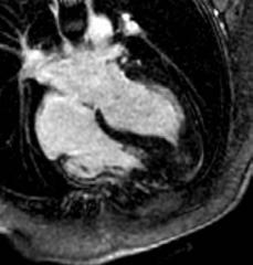
Figure 2. Example of a patient with an apical hypertrophic cardiomyopathy. Top cine image and bottom late gadolinium enhancement image in the 4-chamber view.
aortic stenosis on the left ventricular function and the prevalence, pattern, and expanse of left ventricular hypertrophy, myocardial stiffness, and myocardial fibrosis. The inclusion was completed in 2023. All patients underwent echocardiography with strain measurements and high frame rate echo to assess shear wave velocities and comprehensive CMR including parametric mapping. The results are expected in 2024.
Congenital Heart Disease
Erasmus MC is an expertise center for treatment of patients with congenital heart disease. Imaging plays an ever-increasing role in the diagnosis and follow-up of these patients. Even more so in the decision to re-intervene after initial correction.
Next to bicuspid valve pathology we investigate the role of CT and CMR in planning and follow-up of percutaneously implanted pulmonary valves. In the Cover study we included patients that underwent a percutaneous pulmonary valve implantation. These patients underwent a CMR, cardiac CT and echocardiography on the same day. The inclusion was completed in 2021 and the first results were published in 2023.
The Quality of Life study started in 2020 and scanning was finished in 2021. In this study the long term cardiological and psychosocial outcome in adults operated for congenital heart disease in early childhood are studied. Follow-up of this cohort is now more than 40 years and includes patients with a diverse spectrum of congenital heart disease from atrial septal defect to tetralogy of Fallot and transposition of the great arteries. The results of
189 a a a a a a a a a a a a a a a a a a a a a a a a a a a a a a a a a a a a a a a a a a a a a a a a a a a a a a a a a a a scientific report 2023 | CARDIAC IMAGING
the atrial septum defect cohort is already published and the results of the other cohorts are expected in 2024. Finally, we finished our exercise CMR study using a pushpull MR-compatible ergometer in patients with bronchopulmonary dysplasia (BPD). In total 60 participants were included: 20 premature born young adults with BPD and 20 premature born young adults without BPD. These were compared with 20 healthy age and gender-matched healthy subjects. The study aims to examine cardiorespiratory structure and function during (sub)maximal exercise to reveal dynamic abnormalities that are not apparent on conventional static tests at rest. Inclusion was completed in 2021. The validation of our exercise protocol was published in 2022 and final results were presented in 2023.
Non-ischemic cardiomyopathy
Several projects were continued and initiated in the field of non-ischemic cardiomyopathy including the value of CMR and or CT in non-compaction cardiomyopathy, hypertrophic cardiomyopathy, cardiac sarcoidosis, and cardio-oncology.
The CMR-substudy of the PROCARBI study was finished and published 2021. This study investigates the late cardiac toxicity induced by radiotherapy alone or combined with anthracycline chemotherapy in patients after Hodgkin lymphoma. In total 80 patients underwent CMR. The study showed that long-term lymphoma survivors are not exempt from cardiovascular disease, which can be detected by changes in left ventricular function and native myocardial T1 with CMR. A follow-up CMR was performed after 2 years. Results are expected in 2024.
We previously published a study exploring the role of CMR in patients with a pathogenic sarcomere gene variant without left ventricular hypertrophy (G+/LVH-). In this study, we assessed morphological, volumetric, and functional differences between a cohort of G+/LVH- subjects and healthy controls. The main findings were that the presence of multiple crypts and anterobasal hook only occurred in G+ subjects, and that a simple score system incorporating these and other CMR-derived myocardial morphological features could identify G+/LVH- subjects correctly. A follow-up study studying the use of artificial intelligence in this population is currently performed. Several other studies concerning the use of CMR in hypertrophic cardiomyopathy are currently being started.
In 2022 the covid@heart study was finalized. The objective of the study is to assess the presence and magnitude of myocardial injury, using a combination of transthoracic echocardiography and CMR among individuals with a known baseline cardiovascular health status who are recovered from Covid infection treated at home. In
short, participants of the Rotterdam Study who have undergone echocardiography in the past 5 years and had a confirmed diagnosis of Covid-19 are eligible to take part in the CMR substudy. The main results were presented at the late-breaking clinical trials session at the EACVI 2023 meeting in Barcelona.
Expectations & Directions
Coronary CT has shifted from anatomical to functional analysis. CT FFR and perfusion imaging will be further explored, and their role elucidated. The FUSION study is the first study to randomize patients with 50-90% stenosis to FFRct or routine care. It will provide important evidence in a randomized control trial set-up on the effect of using non-invasive FFRct to reduce the number of unnecessary invasive angiographies.
The introduction of PCCT in the clinical arena has led to important improvements in cardiac CT imaging. The technique is now firmly established in our daily clinical routine. And we anticipate the installation of a second PCCT scanner in Q1 2024 in Erasmus MC. However, PCCT development is still in its infancy and many aspects are expected to be improved even further. Ultimately, large scale patient studies will define the role of PCCT and the direction this research will take. But for now, we already see the improvements in cardiac imaging on a daily basis.The continuous annual follow-up of heart transplant patients with CCTA will provide the temporal results needed to identify the factors that are associated with (accelerated) coronary vasculopathy.
Data from the endocarditis database will shed more light on how to best implement PET/CT and CTA in patients suspected of endocarditis as well as identify important confounding factors. Also, longer term follow-up provides the information needed to evaluate how the diagnostic strategy is translated into quality of care. In 2024 the follow-up results about change in WSS from our bicuspid aortic valve study are expected. These results are important to explore changes in WSS over time. Furthermore, results from several other studies are expected including the cover study, quality of life study, and follow-up of Procabi study. Furthermore, several projects concerning imaging data from our hypertrophic cardiomyopathy database are expected.
Hands-on Cardiac CT Course
For many years already, we have organized the Handson Cardiac CT course in Erasmus MC. Ricardo Budde and Alexander Hirsch serve as course directors and are supported by an enthusiastic and experienced faculty including Marcel Dijkshoorn, CT technician at Erasmus MC.
190 a a a a a a a a a a a a a a a a a a a a a a a a a a a a a a a a a a a a a a a a a a a a a a a a a a a a a a a a a a a
During 5 consecutive days, the participants read over 150 CT scans on dedicated workstations fully equipped with the latest post-processing software. The sessions cover the entire spectrum of cardiac imaging from basic applications like calcium scoring to advanced applications like valve-in-valve TAVI planning as well as FFRct. The course is updated yearly to incorporate the latest developments including photon-counting CT. This year was the 17th edition, and the course was again a huge success and completely sold-out. We thank both Siemens and Bayer for their continuing support in organizing this course.
The next course will be June 10 th-14th 2024 and at the time of writing again already completely sold-out. We consider this a testimony to the continued high quality of the course. More info can be found on our dedicated course website: www.cardiovascularimaging.nl

Funding
Budde, Ricardo, Alexander Hirsch, and consortium partners Veelbelovende zorg ZonMW: ‘Addition of FFRct in the diagnostic pathway of patients with stable chest pain to reduce unnecessary invasive coronary angiography (FUSION Study)” to evaluate the role of FFRct in stable chest pain patients’. 2020-2025
Budde, Ricardo Vrienden van het Sophia: ‘Development of Photon Counting CT protocols for pediatric cardiovascular imaging’. 2023-2025
Invited Lectures
Ricardo Budde. ‘Photon Counting CT: all you need to know’. CVOI course, online. Dec 2023.
Ricardo Budde. ‘Imaging pitfalls in the diagnosis of endocarditis’. CVOI course, Utrecht, The Netherlands. Dec 2023.
Ricardo Budde. ‘Clinical applications of photon-counting CT: a review of pioneer studies and a glimpse into the future’. Bayer Cardiac Live Webinar, online. Nov 2023.
Ricardo Budde. ‘ Photon counting CT in carotid and coronary arteries’. Fill Rogue of Atherosclerosis, Cagliari, Sardinia. Oct 2023.
Ricardo Budde. ‘Photon Counting CT: a leap forward in Cardiovascular Imaging?’. Mayo Clinic, Rochester MN, USA. Oct 2023.
Ricardo Budde. ‘Imaging the dysfunctional prosthetic heart valve with CT and PET-CT'. Mayo Clinic, Rochester MN, USA. Oct 2023.
Ricardo Budde. ‘Updates in cardiovascular screening according to guidelines’. NASCI, Phoenix, AZ, USA. Sept 2023.
Ricardo Budde. ‘Photon-counting CT, how it is redefining cardiovascular diagnosis today’. Siemens Evening Event at ESC, Amsterdam, The Netherlands. Aug 2023.
Ricardo Budde. ‘Photon counting CT: understanding the principles & clinical applications combined’. SCCT, Boston, MA, USA. Jul 2023.
Ricardo Budde. ‘Photon-Counting CT: what and how or cardiovascular and MSK radiology’. Annual Meeting of the Dutch Society of Radiology, Ede, the Netherlands. May 2023.
Ricardo Budde. ‘Imaging prior to valve surgery: is the invasive coronary angiography obsolete?’ Hartfunctie dag, Thorax Academie, Breukelen, The Netherlands. Apr 2023.
Ricardo Budde. ‘ Endocarditis: a difficult diagnosis’. ECR, Vienna, Austria. Mar 2023.
Ricardo Budde. ‘Pushing the boundaries of CT imaging with photon-counting technology’. ECR, Vienna, Austria. Mar 2023.
Ricardo Budde. ‘ Spectral imaging: Cardiac’. ESCR, Berlin, Germany. Oct 2023.
Ricardo Budde. ‘ Cardiac application of AI’. Artificial Intelligence Course, Rotterdam, The Netherlands. Jun 2023.
Ricardo Budde. ‘Photon Counting CT’. Amsterdam Cardiac CT course, Utrecht, The Netherlands. Oct 2023.
Alexander Hirsch. ‘Mitochondrial cardiomyopathy: distinctive cardiac phenotype detected by cardiovascular magnetic resonance’. SCMR Scientific Sessions 2023, San Diego, USA. Jan 2023.
191 scientific report 2023 | CARDIAC IMAGING a a a a a a a a a a a a a a a a a a a a a a a a a a a a a a a a a a a a a a a a a a a a a a a a a a a a a a a a a a a
Figure 3. www.cardiovascularimaging.nl the website dedicated to our hands-on CT course.
Alexander Hirsch. ‘Hands-on CMR case session on cardiomyopathies with mapping and strain’. SCMR Scientific Sessions 2023, San Diego, USA. Jan 2023.
Alexander Hirsch. ‘Clinical application of cardiac MRI’. ISMRM British and Irish Chapter webinar, online. June 2023.
Alexander Hirsch. ‘MINOCA’. CVOI course Tidal changes in ACS, Utrecht, the Netherlands. March 2023.
Alexander Hirsch. ‘New development in the field of cardiac CT’. CAG symposium, Venlo, The Netherlands. March 2023.
Alexander Hirsch. ‘Current options for plaque characterization. MSCT state of the art’. Symposium Image guidance in PCI, Rotterdam, The Netherlands. Aug 2023.
Alexander Hirsch. ‘Hands-on CMR case session on how to interpret CMR parametric mapping in CMPs’. EACVI 2023, Barcelona, Spain. May 2023.
Alexander Hirsch. ‘How to evaluate left ventricular hypertrophy with cardiovascular magnetic resonance: in myocardial storage disease’. EACVI 2023, Barcelona, Spain. May 2023.
Alexander Hirsch. ‘How to session CMR: Late gadolinium enhancement imaging’. CVOI webinar, online. 2023.
Alexander Hirsch. ‘From raw data to beautiful pictures’. Rotterdam Radiology Artificial Intelligence Course, Rotterdam, The Netherlands. June 2023.
Alexander Hirsch. ‘New development in the field of cardiac CT’. NVVC Najaarscongres, Papendal, The Netherlands. Nov 2023.
Alexander Hirsch. ‘Cardiac Perfusion Imaging: stress MRI’. Sandwich Cursus Radiologie, Ede, The Netherlands, Feb 2023.
Alexander Hirsch. ‘Parametric mapping and infiltrative cardiomyopathy’. Amsterdam Cardiac MRI course, Amsterdam, The Netherlands. Oct 2023.
Highlights
Ricardo Budde was a visiting professor at Mayo Clinic, Rochester, MN, USA in September 2023.
Ricardo Budde served as course director for the “Sandwich course Cardiothoracic Radiology” organized by the Dutch Society of Radiology.
In 2023, Ricardo Budde and Jan-Jaap Visser organized a two-day course on artificial intelligence in Radiology. The Rotterdam Radiology AI course covered advanced topics within the practical implementation of artificial intelligence and discussed the added value of artificial intelligence in clinical radiological practice.
Savine Minderhoud won the Medical Delta Thesis Award for her thesis “Biomechanics in congenital heart disease: Using advanced imaging techniques”.
Additional Personnel
Willem A Helbing MD, PhD – Full Professor, Appointment in Pediatric Cardiology.
Mohamed Attrach, MD – cardiovascular radiologist Erasmus MC.
Britt van Dijk – PhD student
192 a a a a a a a a a a a a a a a a a a a a a a a a a a a a a a a a a a a a a a a a a a a a a a a a a a a a a a a a a a a
PhD Students

Eefje Dalebout, MSc

Advisors Jolien Roos – Hesselink, Ricardo Budde, Jolanda Kluin & Alexander Hirsch
Project Funding Departments of cardiology, radiology, cardiothoracic surgery
Email e.dalebout@erasmusmc.nl
Cardiac imaging in infective endocarditis
Imaging is one of the cornerstones in diagnosis and management of infective endocarditis. This PhD trajectory will focus on the role of imaging techniques such as PET-CT and advanced CT techniques in patients with (suspected) endocarditis and its additional value in diagnosis and clinical management.

Simran P. Sharma, MD

Advisors Ricardo Budde, Alexander Hirsch & Nicolas van Mieghem
Project Funding Program ‘Potentially Promising Care’ of Zorginstituut Nederland and ZonMw
Email s.sharma@erasmusmc.nl
FFRct and Coronary Artery Disease
Fractional Flow Reserve derived from coronary computed tomography (FFRct) analysis is a non-invasive technique that uses the CCTA images as a basis for complex software-based calculations and modelling to provide additional functional information based on the anatomical CCTA images. To investigate the impact of adding the FFRct analysis to the diagnostic pathway of stable chest pain patients, we have set up the FUSION study, a national, multicentre, randomised controlled trial

Judith van der Bie, MSc

Advisors Ricardo Budde, Daniel Bos & Marcel van Straten
Project Funding Siemens Healthineers
Email j.vanderbie@erasmusmc.nl LinkedIn www.linkedin.com/in/judithvdbie
Clinical applications of photon-counting computed tomography
Photon-counting CT (PCCT) is a novel imaging technique which enables higher spatial resolution and more advance spectral imaging compared to conventional CT. During my PhD I will investigate the clinical impact of PCCT on cardiovascular- and neuro-imaging. For example, the assessment of coronary in-stent restenosis with PCCT in patients with recurrent chest pain with invasive angiography as reference, to potentially minimalize number of patients undergoing this intervention.

Marguerite E. Faure, MD

Advisors Prof. dr. Ricardo Budde & Dr. Alexander Hirsch
Project Funding Erasmus MC Email m.faure@erasmusmc.nl
Ct and Mr Imaging of Prosthetic Heart Valves
This project focusses on imaging bioprosthetic valves with CT and MRI. In particular patients that underwent a percutaneous pulmonary valve implantation (PPVI). CT seems a reliable tool for risk assessment of coronary artery compression. There seems no relevant change in RVOT to coronary distance and coronary lumen diameter after PPVI. Conduit expansion does not seem to affect the relationship between the pulmonary trunk and coronary arteries after implantation. In a second part of the project we are now evaluating flow patterns after PPVI with 2D and 4D flow MRI.
193 scientific report 2023 | CARDIAC IMAGING a a a a a a a a a a a a a a a a a a a a a a a a a a a a a a a a a a a a a a a a a a a a a a a a a a a a a a a a a a a
LinkedIn LinkedIn LinkedIn LinkedIn

Ivo Schoots completed his professional radiologic training in 2012 at the Academic Medical Center, Amsterdam after a certified clinical fellowship in abdominal radiology. In 2004 he received his PhD on the ‘crosstalk of coagulation and inflammation in ischemia and reperfusion mechanism’ at the University of Amsterdam (Surgery & Internal Medicine). He performed a postdoctoral research fellowship at Harvard Medical School, Department of Molecular & Vascular Medicine, BIDMC, Boston, MA/USA. His research is now focused on oncologic abdominal imaging, in particular on developments in prostate cancer MR imaging and image-guided biopsies / interventions, imaging biomarkers, and clinical decision modeling. He participates in international and national working groups on prostate cancer imaging and image-guided targeted biopsies. As PI of the MR PROPER study he is involved in the clinical implementation of prostate MRI in combination with risk stratification. i.schoots@erasmusmc.nl
a a a a a a a a a a a a a a a a a a a a a a a a a a a a a a a a a a a a a a a a a a a a a a a a a a a a a a a a a a a
Ivo G S cho ots, MD, PhD associate professor ABDOMINAL IMAGING

Context
Our Abdominal Imaging research is focused on several organs, in the lower abdomen as well as in the upper abdomen. Some of our researchers have developed particular skills and interest in advanced MR imaging of the prostate or cervix within the clinical decision-making algorithms, others are more dedicated to liver and colorectal cancer imaging. We are very happy that the collaboration with our clinical partners in Surgery, Gastroenterology, Urology, Gynecology, Medical Oncology, and Radiotherapy are already starting to bear fruit by attracting funding and by creating some promising scientific output. We are convinced that in the coming years some of these areas of research and probably even other topics in abdominal imaging will gain more interest and become increasingly succesfull.
Top Publications 2023
Seyrek N, E Hollemans, IG Schoots, G van Leenders. ‘Association of quantifiable prostate MRI parameters with any and large cribriform pattern in prostate cancer patients undergoing radical prostatectomy’. European Journal of Radiology 2023; 166:110966.
Starmans MP, RL Miclea, V Vilgrain, M Ronot, Y Purcell, J Verbeek, WJ Niessen, JN IJzermans, RA de Man, M Doukas, S Klein, MG Thomeer. ‘Automated Assessment of T2-Weighted MRI to Differentiate Malignant and Benign Primary Solid Liver Lesions in Noncirrhotic Livers Using Radiomics’. Academic Radiology 2023; 1076-6332:00393-8.
Willemssen F, Q de Lussanet de la Sablonière, D Bos, R de Man, R Dwarkasing. ‘Potential of a Non-Contrast-Enhanced Abbreviated MRI Screening Protocol (NC-AMRI) in High-Risk Patients under Surveillance for HCC’. Cancers 2023; 14:3961.
a a a a a a a a a a a a a a a a a a a a a a a a a a a a a a a a a a a a a a a a a a a a a a a a a a a a a a a a a a a 195
Research Projects: Objectives & Achievements
Prostate cancer imaging
Prostate cancer is the most common malignancy and leading cause of cancer-related deaths in men. Optimization of diagnosis and treatment of prostate cancer is of great social importance. Prostate cancer, however, is a heterogeneous disease with clinically significant (aggressive) and insignificant (non - aggressive) tumors; only aggressive tumors need to be detected and treated. There is an urgent need for early tumor detection and characterization of tumor aggressiveness.
Imaging and image-guided strategies in diagnosis and treatment of prostate cancer are challenging. However, these strategies may improve diagnostic accuracy, needing less biopsy cores, and may navigate to better therapy choice with subsequent lower morbidity, increased quality of life and at lower costs. Projects on imaging and image-guided strategies of prostate cancer (Fig 1) address the need to reduce over-diagnosis, over-biopsy and overtreatment of prostate cancer, due to today’s less than ideal screening tests. Results of several trials with MRI (Erasmus MC) were published. This have led to influential guideline changes and may lead to the renewed question whether to screen for prostate cancer with MRI or not, as prostate cancer screening without MRI shows at least a relative cancer specific mortality reduction of 21% over time, however, with the harms of over-biopsy, over-diagnosis and over-treatment.
Liver imaging
As part of the largest liver center in the Netherlands, continuing efforts are done to improve the possibilities of non-invasive diagnostics of liver tumors.
First, regarding benign liver lesions, our study projects have focused on diagnosis of hepatocellular adenoma (HCA), focal nodular hyperplasia (FNH), and angiomyolipoma on MRI. One of our findings was the superiority of Gadoxetate disodium against Gadobenate dimeglumine for differentiation of HCAs from FNHs. Further research is focused on atypical presentations including imaging of atypical hemangiomata and atypical isointense lesions (AILs) in the hepatobiliary phase. The latter is one of the projects which are set up in close collaboration with all Dutch university medical centers (and is endorsed by the Dutch Benign Liver Tumor Group). Furthermore, we focus on cystic liver lesions and compare imaging modalities, Contrast Enhanced US (CEUS), CT and MRI. CEUS seems promising for clear enhancement of internal septations (Fig 2).
Second, regarding hepatocellular cancer (HCC), our study projects have focused on the use of ultrasound (US) and MRI for the detection of HCC. This tumor is the tenth most common malignancy and sixth cause of cancer-related deaths in men. Our referral center for hepatobiliary diseases is a screening center for HCC in hepatitis and cirrhosis. A review of our data concluded that abdominal US is inferior to MRI without contrast in HCC detection. Based on these findings, the SMS trial evaluates a shorter and more (cost) efficient MRI screening protocol compared to abdominal US (Fig 3).
Fig 1. Biochemical recurrence-free survival in patients with Grade group 2 prostate cancer, stratified for a) Gleason pattern 4 (logrank P=0.10), b) invasive cribriform and/or intraductal carcinoma (log-rank P<0.001), and c) tertiary pattern 5 (log-rank P=0.12) (Seyrek et al. Histopathology. 2022;80:558-65)
Third, together with the biomedical imaging group of our department, we developed a robust, multicentric radiomics platform for liver lesion phenotyping using MRI data from different liver centers around the world. This is named by the Liver Artificial Intelligence (LAI) consortium. Efforts are made to find generalizable applications for liver tumor classification and prognosis.

196 a a a a a a a a a a a a a a a a a a a a a a a a a a a a a a a a a a a a a a a a a a a a a a a a a a a a a a a a a a a

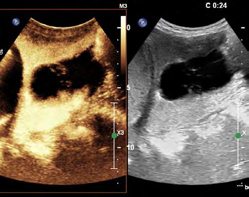
Fig 2. CEUS of a cystic liver lesion, with internal enhancement of septations at 0:24, indicating the neoplastic characteristics of this lesion. Internal septations and the enhancement are clearly demonstrated with CEUS.
Rectal cancer imaging
Colorectal cancer is the third most common malignancy and third cause of cancer-related deaths in men and women. With the Erasmus MC a few centers in the Netherlands are specialized in treatment of local recurrent rectal cancer. In our institution many of these patients will undergo surgery with curative intention. Dedicated MRI is an integral part of the work-up and important for clinical decision making. Our research aims to develop and validate modern MR techniques for better selection and follow up of complete responders after preoperative chemo-radiotherapy of (recurrent) rectal cancer. Furthermore, morphological and functional MRI characteristics based on multiparametric MR imaging and post-processing tumor texture analysis may help predict surgical outcome and long-term prognosis.

Fig 3. Fifty-two-year-old male patient with liver cirrhosis owing to chronic hepatitis C infection. NC-AMRI (a–d) demonstrates a small lesion (arrow (a–f)) in segment 4 that is hyperintense on axial T2W FS (a) and DWI (b), and hypointense on T1W in-phase (c) and opposed-phase (d) imaging, without signs of intracellular fat. CE-MRI (e,f) shows hyperenhancement in the arterial phase (e) with washout (arrow) in the delayed phase and capsular enhancement (f), confirming an HCC lesion (Li-RADS 5 lesion). The lesion was treated with radiofrequency ablation. (Ref. Willemssen et al. Cancers. 2023)
Cervical cancer imaging
Cervical cancer is the fourth most common cancer among females, and the fifth cause of cancer-related death in women. In daily practice clinical examination with or without anesthesia is until recently the principal investigation for assessment and therapy planning in cervical cancer. However, since our published systematic review comparing clinical examination vs MRI the new national guideline has given the latter a more prominent role in primary staging. Based on our findings the international guidelines of ESTRO, together with ESGO and ESP has shifted completely to MRI as the hallmark for diagnostic staging. In an Erasmus MC lead collaborative prospective multicenter study with UZ Leuven and AMC we investigated the value of optimized MR imaging, including diffusion weighted imaging (DWI) in the detection of early tumor residue in the cervix after radiotherapy. The main finding was that DWI forms an irreplaceable part of the protocol since it increases significantly the detection of residual tumor.
197 scientific report 2023 | ABDOMINAL IMAGING a a a a a a a a a a a a a a a a a a a a a a a a a a a a a a a a a a a a a a a a a a a a a a a a a a a a a a a a a a a
Expectations & Directions
Prostate cancer imaging: Means to detect prostate tumors at an early stage and to robustly characterize the aggressiveness are badly needed. MR imaging in prostate cancer is an important research line within the abdominal imaging section. Image-guided biopsy based on multi-parametric MR examinations has been established. MRI/US-fusion guided biopsies have been implemented in clinical trials and in patient care. Association of imaging traits with gene expression profiles of prostate cancer will certainly be an area of interest, in close correlation with the Erasmus MC departments of Urology, Pathology and Biomedical Imaging Group Rotterdam ( BIGR) . Developments on nuclear imaging techniques (o.a. PSMA PET CT) will be explored to correlate morphological and functional prostate MR images to PET CT images of prostate metabolism, most preferably with the new PET MRI scanner. We intend to explore strategies to improve diagnostic accuracy (Fig 4), reduce the number of biopsy cores needed, and improve therapeutic decision-making, thereby lowering morbidity, increasing quality of life, and reducing costs.

Fig 4. Proposal of membranous urethral length (MUL) measurement on midsagittal MR images. (Ref. Boellaard, Schoots et al. Membranous urethral length measurement on preoperative MRI to predict incontinence after radical prostatectomy: a literature review towards a proposal for measurement standardization. European radiology. 2023)
Hepatic cellular cancer imaging: MR imaging in hepatic cellular cancer (HCC) is expected to become a growing research line within the department, in close collaboration with the Erasmus MC departments of Hepatology/ Gastroenterology and hepatobiliairy Surgery. HCC screening in patients with hepatitis and cirrhosis is becoming centralized in academic centers. Collaborating networks such as the Dutch Hepatic Cancer Group (DHCG) are evolving, in which the Erasmus MC plays a dedicated and central role. Furthermore, interventional therapy plays an important role in minimal invasive liver cancer treatment. Critical for its success is an accurate target of the lesion. Research is focused on the development of real time image fusion techniques to guide interventional treatment and navigation surgery.
Radiomics: MR imaging biomarkers are being explored and validated for personalized decision-making and for prediction of treatment efficacy. A prerequisite for implementation of the multiparametric MR imaging modality is an accurate and fast post-processing method that can translate complex quantitative information into clinically useful directions. The development and validation of such methods is research in joint cooperation with industrial partners. ‘Radiomics’ entails the extraction of predictive features for diagnostic decision-making and treatment response from standard CT or MR image using mathematical models. We will further expand our imaging expertise and apply novel imaging methods to improve our understanding of the genetic and disease development in abdominal oncology. We will initiate new projects in the field of oncological imaging analysis, with machine learning and deep learning techniques (Fig. 5). We will continue to increase our research efforts into investigating clinically related oncological research questions.
Funding
Niessen, Wiro, Ivo Schoots, Jifke Veenland , and Chris Bangma (Urology) Erasmus MC-TKI-LSH: ‘Personalized Prostate Cancer Management using Multi-parametric MRI and Machine Learning (PPCM4)’. 2020 - 2024
Pensky, Bagdi Ulas, Wallace Bolan, Gonda Hecht, Marco Bruno, and Ivo Schoots NIH grant: ‘Deep learning methods for characterization of pancreatic cysts (Cyst-X project)’. 2021 - 2025
Schoots, Ivo , and Uulke Van der Heide (Radiation Oncology) KWF clinical implementation grant: ‘PROCESS study: PROstate Cancer - Expansion of Surveillance Selection criteria with MR imaging’. 2021 – 2025
198 a a a a a a a a a a a a a a a a a a a a a a a a a a a a a a a a a a a a a a a a a a a a a a a a a a a a a a a a a a a

Fig 5 Schematic overview of the radiomics approach. Input to the algorithm are the T2-weighted magnetic resonance imaging scans (1) and the lesion segmentations (2). Processing steps include feature extraction (3) and the creation of a machine learning decision model (5) using an ensemble of the best 100 workflows from 1000 candidate workflows (4), where the workflows are different combinations of the different analysis steps (e.g., the classifier used). (Starmans MPA, et al. Academic radiology. 2023).
Van den Bergh, Roderick, Rik Somford (Urology), and Ivo Schoots SKMS project/ZonMW: ‘Evaluatie en optimalisatie diagnostisch traject prostaatkanker middels MRI’ 2022 – 2026
Dwarkasing, Roy, Francois Willemssen , Rob de Man, Bart Takkenberg, and Carine Uyl-De Groot KWF implementation grant: ‘Validation of a Short and effective MRI Surveillance protocol for hepatocellular carcinoma screening in practice’ 2022 – 2026
Lambregts, Doenja, Ritse Mann, and Ivo Schoots KWF implementation grant: ‘Towards clinical implementation of novel diagnostic tools in oncologic imaging: an opensource web-based reviewing infrastructure for validation studies’. 2023 – 2027
Aben, Katja, Berdine Heesterman, Daniela Oprea, Pim van Leeuwen, and Ivo Schoots KWF implementation grant: ‘ADMINISTRATE (Advanced Diagnostic Modalities in ImagiNg Impacting on diagnosiS, TReatment And paTient outcomE) on prostate cancer’. 2023 – 2026
Thomeer, Maarten, et al. KWF clinical research Grant: effect of FAPI PET-CT on management in patients with potentially resectable biliary tract cancers: prospective multicenter study and cost-effectiveness analysis. 2023- 2027
Klein, Stefan, Maarten Thomeer NWO TTW open technology: 'The Liver Artificial Intelligence (LAI) consortium: a benchmark dataset and optimized machine learning methods for MRI-based diagnosis of solid appearing liver lesions'. 2023
Highlights
Ivo Schoots completed the 2-year Master of Business Administration (MBA) in healthcare (2021-2023) at the Amsterdam Business School, The Netherlands.
Ivo Schoots was invited to co-organize the upcoming 12th ESUR Prostate MRI Symposium (2024), Zeist, The Netherlands.
Roy Dwarkasing and Fokko Smiths are directors of the fellowship training program for Abdominal Radiology at the Erasmus MC.
In 2023 a total of three fellows completed their training at our department who are currently practicing abdominal radiologists abroad: Sandra Vennix , practicing in Bonaire, Renza van Gils currently employed in Willemstad, Curaçao and our international fellow Mohana Letchumanan, who returned to Kuala Lumpur, Malaysia.
Additional Personnel
Demi Huijgen – PhD student Petriatic Surgery Erasmus MC Angela Amirabile – Humanitas Research Hospital Italy
199 scientific report 2023 | ABDOMINAL IMAGING a a a a a a a a a a a a a a a a a a a a a a a a a a a a a a a a a a a a a a a a a a a a a a a a a a a a a a a a a a a

Roy S. Dwarkasing MD, PhD
Project Funding Validation of a Short and effective MRI Surveillance (SMS) protocol for hepatocellular carcinoma screening in practice (KWF 2021-2, project number 13803)
Email r.s.dwarkasing@erasmusmc.nl
Improved detection of early hepatocellular carcinoma in high risk patients with abbreviated (non-contrast MRI).
Hepatocellular carcinoma (HCC) is the sixth most common cancer worldwide and the third most common cause of cancer related death. The incidence of HCC has been rapidly rising in Western countries and is expected to continue to rise in the next decades. In general, prognosis of patients with HCC is poor, except for patients with early-stage HCC who are eligible for curative treatments. According to the Dutch HCC guideline, biannual US surveillance should be offered to high rsik patients (with cirrhosis and chronic hepatitis B or -C). However, the reliability of US is limited in these pa-

tients. Better surveillance tools are urgently needed. We have developed and validated a short MRI surveillance (SMS) protocol for HCC in high-risk patients. The SMS protocol consists of the following three sequences: a) Diffusion weighted imaging (DWI) with a minimum of 2 b-values (50 and 800 s/mm2); b) T2weighted (T2W) fast spin-echo (FSE) with fat saturation; and c) T1-weighted (T1W) in- and out-of-phase imaging. Evaluation of performance in daily practice and assessment of cost-effectiveness of the SMS are the next steps.
Maarten G Thomeer MD, PhD
Project Funding KWF clinical research: Effect of FAPI PET-CT on management in patients with potentially resectable biliary tract cancers: prospective multicenter study and cost-effectiveness analysis.
NWO-TTW Grant: A benchmark dataset and optimized machine learning methods for MRI-based diagnosis of solid appearing liver lesions
Email m.thomeer@erasmusmc.nl
Advances in abdominal imaging
One of the forefront topics in abdominal imaging is the integration of a novel tracer called FAPI for PET-CT/MRI imaging. In a recent systematic review, we highlighted the benefits of utilizing FAPI PET-CT/MRI in the pretreatment staging of pancreatic, gastric, and cholangiocarcinomas. Collaborating with Amsterdam University Medical Center and University Medical Center Utrecht, we are poised to embark on a prospective imaging study investigating the utility of FAPI PET-CT in pretreatment staging specifically for cholangiocarcinoma. This study, funded by the KWF funding agency, is spearheaded by Mara Veenstra, MD, who is undertaking her PhD within this research scope. Liver cancer stands as one of the most prevalent cancers globally. While magnetic resonance imaging (MRI) is pivotal in its diagnosis, the diverse nature of both malignant and benign liver lesions poses a significant challenge,
often leading to highly subjective interpretations. Recognizing this, the Liver Artificial Intelligence (LAI) consortium, recently awarded by the NWO, is dedicated to developing innovative MRI analysis techniques. These advancements aim to provide robust support in diagnosing liver lesions, ultimately enhancing treatment decisions tailored to each patient's needs.

Figure: Medical Institutions of the international Liver Artificial Intelligence (LAI) consortium
200 a a a a a a a a a a a a a a a a a a a a a a a a a a a a a a a a a a a a a a a a a a a a a a a a a a a a a a a a a a a
Post-docs
PhD Students

Mara Veenstra, MSc

Advisors Maarten Thomeer, Erik Verburg & Rutger-Jan Swijnenburg
Project Funding KWF Kankerbestrijding
Email m.m.k.veenstra@erasmusmc.nl
FAPI PET/CT for cholangiocarcinoma
We believe that additional preoperative [18F]F-FAPI PET/CT for patients suffering from proximal cholangiocarcinoma can detect metastases that cannot be found using CT and MRI, and that futile surgeries can be avoided using this new work-up.

Francois Willemssen, MD
Advisors Roy Dwarkasing & Daniel Bos
Email f.willemssen@erasmusmc.nl
Dr. Willemssen is responsible for the radiological contribution in the Multidisciplinary Liver Tumor Board, participates in the research committee of the Dutch Hepatocellular and Cholangiocellular Group (DHCG), is involved in the revision of two national guidelines, Cholangiocarcinoma and Hepatocellular carcinoma, and contributes to several research projects on liver imaging.
One of the projects is the use of Contrast-Enhanced US in cystic liver lesions.

Neslisah Seyrek, MD

Advisors Ivo Schoots & Arno van Leenders
Project Funding Jaap Schouten Foundation: “Significance of cribriform growth in prostate cancer risk stratification”
Email n.seyrek@erasmusmc.nl
The value of mpMRI on prediction of cribriform growth in prostate cancer
Assesment of the presence of cribriform growth plays a crucial role in risk stratification and active survellience eligibilty of prostate cancer patients, especially in intermediate risk group. Adverse imaging parameters derived from mpMRI may contribute in prediction of cribriform presence and enhance the presicion of risk assesment of men with intermediate risk prostate cancer.

Céline van de Braak, MSc

Advisors Aad van der Lugt & Rob de Man, Roy Dwarkasing & Daniel Bos
Project Funding KWF Grant
Email c.vandebraak@erasmusmc.nl
Validation of Short MRI Surveillance (SMS) protocol for hepatocellular carcinoma (HCC) screening in practice
An abbreviated MRI protocol for the screening of HCC will be validated in a high-risk patient population and compared to bi-annual ultrasound screening (current standard). To our knowledge, this will be the first study to perform this head-to-head comparison. This may provide improved detection of early stage HCC and consequently improved survival of HCC patients in a surveillance cohort.
201 scientific report 2023 | ABDOMINAL IMAGING a a a a a a a a a a a a a a a a a a a a a a a a a a a a a a a a a a a a a a a a a a a a a a a a a a a a a a a a a a a
LinkedIn LinkedIn LinkedIn

Edwin Oei is a Full Professor of Musculoskeletal Imaging, and Section Chief of musculoskeletal radiology in Erasmus MC’s Department of Radiology & Nuclear Medicine. He obtained his medical degree in 2004 and his PhD on MRI for traumatic knee injury in 2009, both from Erasmus University Rotterdam. He also holds an MSc in Clinical Epidemiology from the Netherlands Institute for Health Sciences. Dr Oei is the principal investigator of musculoskeletal imaging research and engages in many academic activities including supervising 14 PhD students, lecturing, board and committee memberships, and refereeing for various journals. He is the current President of the European Society
for Magnetic Resonance in Medicine and Biology (ESMRMB) and the Past-President of the Musculoskeletal MR Study Group of the International Society for Magnetic Resonance in Medicine. In 2013, Edwin Oei spent a one year research sabbatical as a Visiting Assistant Professor in the Joint and Osteoarthritis Imaging with Novel Techniques (JOINT) lab of the Department of Radiology of Stanford University, CA/USA. Dr. Oei is also the Clinical Research Focus Group leader in the Department of Radiology & Nuclear Medicine, and the principal coordinator of the Academic Center for Molecular and Cellular Imaging at Erasmus MC. e.oei@erasmusmc.nl
a a a a a a a a a a a a a a a a a a a a a a a a a a a a a a a a a a a a a a a a a a a a a a a a a a a a a a a a a a a a a a a a a a a a a a a a a a a a a a a a a a a a a a a a a a a a a a a a a a a a a a a a a a a a a a a a a a a a a
ADVANCED MUSCULOSKELETAL IMAGING RESEARCH ERASMUS MC (ADMIRE)
Edwin HG Oei, MD, PhD
full professor
Context
Imaging is key in the study of musculoskeletal diseases. More sensitive and accurate imaging techniques are needed to advance our understanding of the development, mechanisms, and subtypes of musculoskeletal disorders. This is especially important for disorders that are common and have large impact on patients and the society, such as osteoarthritis, osteoporosis, sports injuries, and chronic musculoskeletal pain. New imaging biomarkers based on CT, MRI, ultrasound and nuclear imaging can also facilitate early diagnosis and development of targeted therapies. Before they can be applied routinely, new imaging techniques need to be optimized and validated. When applied in clinical research studies, novel, quantitative, imaging tools can provide additional imaging biomarkers. In large population-based studies, imaging is essential to characterize the development and aging of the musculoskeletal system. To analyze large imaging datasets, efficient image analysis methods, incorporating radiomics and artificial intelligence, are needed.
Top Publications 2023
Wu T, J Yang-Huang, MW Vernooij, M Rodriguez-Ayllon, VW Jaddoe, H Raat, S Klein, EH Oei. Physical activity, screen time and body composition in 13-year-old adolescents: The Generation R Study. Pediatr Obes. 2023; 18:13076.
Mostert JM, NB Dur, X Li, JM Ellermann, R Hemke, L Hales, V Mazzoli, F Kogan, JF Griffith, EH Oei, RA van der Heijden. Advanced Magnetic Resonance Imaging and Molecular Imaging of the Painful Knee. Semin Musculoskelet Radiol. 2023; 27:618-631.
Booij R, NF Kämmerling, EH Oei, A Persson, E Tesselaar. Assessment of visibility of bone structures in the wrist using normal and half of the radiation dose with photoncounting detector CT. Eur J Radiol. 2023; 159:110662.
a a a a a a a a a a a a a a a a a a a a a a a a a a a a a a a a a a a a a a a a a a a a a a a a a a a a a a a a a a a 203
Research Projects: Objectives
& Achievements
Optimization and validation of advanced imaging techniques for common musculoskeletal diseases
This research line addresses the optimization and validation of novel imaging techniques based on CT, MRI, ultrasound and nuclear imaging. We successfully validated quantitative MRI techniques to assess altered processes or composition in musculoskeletal tissues such as cartilage, meniscus and tendon, against tissue references and clinical outcomes. We also validated advanced, contrastenhanced ultrasound and shear-wave elastography in osteoarthritis and tendinopathy. More recently, photon counting CT has become of great interest as it has shown promise to characterize bone disorders such as fractures and osteoporosis better than current methods because of its ultra-high resolution (Figure 1). Validation for routine clinical use also includes assessment of additional value for patients and clinicians. We are currently conducting a randomized controlled diagnostic trial to study the value of PET-MRI for chronic musculoskeletal pain (Figure 2). We collaborate closely with the MR Physics in Medicine group, Biomedical Imaging Group Rotterdam, Departments of Orthopedics and Sports Medicine and Pain Medicine, and external partners (University of Wisconsin, Stanford University, and GE Healthcare).
Current projects:
- Quantitative cartilage MRI and SPECT In knee osteoarthritis (Joost Verschueren)
- Photon counting CT (Ronald Booij, Post-doc Physics in CT group)
- PET-MRI for chronic musculoskeletal pain (Marijn Mostert)

Application of advanced imaging techniques in clinical studies
A strength of our research is the multidisciplinary embedding and collaboration with many clinical research groups, which creates the possibility to apply novel imaging techniques in clinical studies, providing additional imaging biomarkers. Previously, we successfully applied advanced quantitative MRI techniques for cartilage composition (dGEMRIC, T1rho-, and T2-mapping) in several clinical studies on knee osteoarthritis. In some studies we also applied perfusion imaging with dynamic contrast-enhanced MRI (DCE-MRI). We successfully implemented ultrashort echo time (UTE) MRI and shear-wave elastography (SWE) ultrasound in the largest randomized controlled trial of patients with patellar tendinopathy (study funded by General Electric and National Basketball Association). We recently also introduced a short MRI scan of both hands without the need for contrast agents in a cohort of patients with early clinically suspected arthralgia. (Figure 3). These projects are conducted in collaboration with the departments of Orthopedics and Sports Medicine (Duncan Meuffels, Robert-Jan de Vos, Koen Bos, Max Reijman, Denise Eygendaal, Sita Bierma-Zeinstra), Rheumatology (Annette van der Helm-van Mil, Pascal de Jong).
Current projects:
- Advanced MRI and ultrasound in a randomized controlled trial of patellar tendinopathy (Stephan Breda, Jie Deng)
- Short Dixon MRI for hand arthralgia (Sanne Boeren)


204 a a a a a a a a a a a a a a a a a a a a a a a a a a a a a a a a a a a a a a a a a a a a a a a a a a a a a a a a a a a
Figure 1. Ultra-high resolution photon counting CT image of the elbow displaying an ostochondral defect of the capitellum of the humerus.
Figure 2. PET-MRI image from the AMPHiBI trial, a randomized controlled diagnostic trial on the value of PET-MRI for chronic musculoskeletal pain. Increased FDG uptake is shown in a facet joint, consistent with degenerative change with inflammation.
Figure 3: Images from a rapid non-contrast bilateral hand MRI scan to evaluate patients with clinical suspect althralgia.
Musculoskeletal imaging in population studies
We are actively involved in the two large population studies in Erasmus MC – the Rotterdam Study and Generation R. In the Rotterdam Study among middle-aged and elderly, our research focus is on studying determinants and imaging aspects of osteoarthritis and osteoporosis, apart from our support in phenotyping these diseases on radiography (multiple joints and spine), MRI (knee) and, in the future, EOS imaging. In the Generation R cohort of children and adolescents we play a leading role in studying development, growth and abnormailities of the hip, knee (Figure 4), and spine on focused rapid MRI scans as well as body composition on whole-body MRI. For such large datasets, it is necessary to make use of automated image analysis tools, which we develop together with the Biomedical Imaging Group Rotterdam (Dr. Stefan Klein and Dr. Jukka Hirvasniemi). In both projects, we collaborate closely with researchers from the departments of General Practice (Sita Bierma-Zeinstra, Marienke van Middelkoop), Internal Medicine (Fernando Rivadeneira, Joyce van Meurs), Pediatrics (Vincent Jaddoe, Liesbeth Duijts), and Public Health (Hein Raat).
Current projects:
- Hip shape in children (Desirée de Vreede, Mirthe Kamphuis)
- Spine abnormalities in adolescents (Marleen van den Heuvel)
- Knee shape in adolescents (Rosemarijn van Paassen)
- Body composition in adolescents (Tong Wu)
- Osteoporosis and vertebral fractures (Fjorda Koromani)

Miscellaneous projects
We study various other topics in the field of musculoskeletal imaging. We are involved in many clinical studies on osteoarthritis using "conventional" radiographic or MR imaging with semi-quantitative osteoarthritis grading. Examples include studies on specific risk factors for osteoarthritis such as overweight, anterior cruciate ligament rupture, or menopause, in collaboration with the departments of Orthopedics and General Practice. We also lead a randomized sham-controlled clinical trial on the efficacy of genicular artery embolization with follow-up using advanced (DCE-)MRI. In another project, we assess hip and groin disorders in athletes, utilizing generated CT-like images from MRI. Together with the Value-based Imaging group (Jacob Visser) we conduct several clinical validation studies of artificial intelligence algorithms, e.g. for fracture detection or osteoarthritis grading on radiographs, collaborating with industrial partners, and with the BIGR group we work on more fundamental AI and radiomics projects, e.g. on musculoskeletal tumors. We also fulfil a consulting or supportive role in many other studies primarily conducted by other groups at Erasmus MC and internationally (e.g. Univerity of Queenland, LaTrobe, Lund, and Stanford Universities).
Current projects:
- Role of the meniscus in knee osteoarthritis (Jan van der Voet)
- Genicular artery embolization for knee osteoarthritis (Tijmen van Zadelhoff, with Adriaan Moelker )
- Imaging of hip and groin disorders in athletes (David Hanff)
- Artificial intelligence for musculoskeletal radiology (Huib Ruitenbeek)
- Trustworthy artificial intelligence for musculoskeletal tumors (Xinyi Wan)
205 scientific report 2023 | ADVANCED MUSCULOSKELETAL IMAGING RESEARCH ERASMUS MC (ADMIRE) a a a a a a a a a a a a a a a a a a a a a a a a a a a a a a a a a a a a a a a a a a a a a a a a a a a a a a a a a a a
Figure 4. 3D shape analysis of the patella based on automated segmentation of MRI scans in the Generation R cohort.
Expectations & Directions
An important new direction in our research is the integration of (PET-)MR imaging with biomechanical measurements in the new MOtionBiomechanics & Imaging (MOBI) lab, of which the construction started In 2023 (finished Spring 2024). This work will enable precision diagnosis of joint load in the context of osteoarthritis (Figure 5). This joint initiative with Jaap Harlaar from TU Delft marks one of several projects, largely conducted within the Flagship “Healthy Joints” as part of the “Health & Technology by Convergence” initiative between Erasmus MC Rotterdam, TU Delft and Erasmus University Rotterdam, in which our group plays a prominent role. In the area of image acquisition, we expect exciting new developments in photon counting CT for musculoskeletal applications which will need to be validated. In image analysis there will obviously be an increased activity in artificial intelligence, both in terms of validating and determining additional value of clinical AI algorithms as in the development and application of research oriented algorithms. Finally, we expect an growing role in Generation R with the availability of more MR imaging biomarkers across different joints and with regard to body composition.
Funding
Oei, Edwin General Electric Healthcare: ‘Pinpointing the source of chronic pain and therapy response with wholebody 18F FDG-PET/MRI’. 2021-2025
Oei, Edwin General Electric Healthcare: ‘RSNA QIBA MSK Profile Stage 3 and 4 Conformance Testing’. 2023-2024
Oei, Edwin (co-applicant) NWO Research along routes by Consortia (NWA-ORC): ‘Healthy Loading to combat osteoarthritis: Leveraging molecular variations in load bearing capacity for individualized movement aDvice: The LoaD project’. Main applicant: G.J.V.M. van Osch (Orthopedics). 2022-2030
Oei, Edwin (co-applicant) NWO ROBUST program and General Electric Healthcare: ‘Innovation Center for Artificial Intelligence (ICAI) lab Trustworthy AI for Magnetic Resonance Imaging’. 2023-2026
Oei, Edwin (co-applicant) Horizon 2020 EIC Accelerator: ‘AI algorithms in musculoskeletal radiography’. Main applicant: Radiobotics, Copenhagen, Denmark. 2020-2023

206 a a a a a a a a a a a a a a a a a a a a a a a a a a a a a a a a a a a a a a a a a a a a a a a a a a a a a a a a a a a
Figure 5. Envisioned workflow of the MOtionBiomechanics & Imaging (MOBI) lab
Oei, Edwin (co-applicant) Independent Research Fund Denmark: ‘Investigating pathology and tissue biomarkers of Osgood Schlatter to enhance treatment of children with growth-related pain’. Main applicant: M.Rathleff, University of Aalborg, Denmark. 2019-2023
Oei, Edwin (co-applicant) NWO Zon-MW Open Competition: ‘Biomechanical precision diagnostics in osteoarthritis’. Main applicant: S. Bierma-Zeinstra (General Practice/Orthopedics). 2020-2025
Oei, Edwin (co-applicant) NWO Zon-MW Goed Gebruik Geneesmiddelen: ‘Efficacy of antibiotic treatment for patients with chronic low back pain and Modic type I changes - randomised placebo controlled trial’. Main applicant: B.W. Koes (General Practice). 2023-2027
Oei, Edwin, Rianne van der Heijden, and Jukka Hirvasniemi (co-applicants) TU Delft-Erasmus MC Convergence Flagship: ‘Healthy Joints’. Main applicant: S.M.A. Bierma-Zeinstra (General Practice), J. Harlaar (TU Delft). 2022-2027
Oei, Edwin (co-applicant) ZonMw Gender en Gezondheid - Algemene onderzoeksronde: ‘The FOCUM human disease model for development of OA’. Main applicant: S.M.A. Bierma-Zeinstra (General Practice). 2019-2024
Oei, Edwin (co-applicant) ZonMw Gender en Gezondheid - Algemene onderzoeksronde: ‘Diagnosis, prevalence and associated factors of osteoarthritis in adults with intellectual disabilities’. Main applicant: D.A.M. Maes-Festen (AVG/ General Practice). 2020-2026
Oei, Edwin (co-applicant) National Health and Medical Research Council, Australia: ‘SUPER rehabilitation RCT for young people with old knees’. Main applicant: K. Crossley (La Trobe, Melbourne, Australia). 2018-2023
Oei, Edwin (project team member, WP leader) European Research Council (ERC) Advanced grant: ‘Biomechanical precision diagnostics in osteoarthritis Modelling trajectories and mechanisms of childhood hip dysplasia’. Main applicant: S.M.A. Bierma-Zeinstra (General Practice/Orthopedics). 2023-2028
Oei, Edwin (project team member) FOREUM Foundation for Research in Rheumatology, Preclinical Phases of Rheumatic and Musculoskeletal Diseases: ‘Novel Treatment Targets in Early-stage Osteoarthritis’. Main applicant: M. Englund (Lund University, Sweden). 2018-2023
Invited Lectures
Edwin Oei. ‘Oedema in bone and soft tissues of the knee’. ECR, Vienna, Austria. March 2023.
Edwin Oei. ‘Musculoskeletal photon counting CT cases, “An evening with NAEOTOM Alpha”’. ECR, Vienna, Austria. March 2023.
Rianne van der Heijden. ‘’Advanced Quantitative Imaging for Musculoskeletal Pain, with a special focus on DCE MRI and PET MRI’. Center of Magnetic Resonance Imaging University of Minnesota, Minneapolis, USA. March 2023.
Edwin Oei and Ricardo Budde. ‘Photon counting CT for cardiovascular and musculoskeletal imaging’. Radiological Society of the Netherlands (NVvR) Club Sandwich, Ede, The Netherlands. May 2023.
David Hanff. ‘Hands-on musculoskeletal ultrasound course’. Radiological Society of the Netherlands (NVvR) Club Sandwich, Ede, The Netherlands. May 2023.
Edwin Oei. ‘Imaging pain in osteoarthritis’. International Workshop on Osteoarthritis Imaging (IWOAI), Lausanne, Switzerland. June 2023.
Edwin Oei. ‘Advances in imaging of osteoarthritis’. University of Wisconsin-Madison, Grand Rounds of the Department of Radiology, Madison, USA. Dec 2023.
Stephan Breda. ‘PET-MRI for musculoskeletal applications’. Musculoskeletal Radiology section of the Dutch Society of Radiology (NVvR), Hilversum, The Netherlands. 8 Dec 2023.
Highlights
On 21 June 2023, Edwin Oei held his inaugural lecture, accepting his appointment as Full Professor of Musculoskeletal Imaging.
David Hanff became a board member of the Musculoskeletal Radiology section of the Dutch Society of Radiology (NVvR)
Edwin Oei became the President of the European Society for Magnetic Resonance in Medicine and Biology (ESMRMB).
Rianne van der Heijden continued her 2 year visiting assistant professorship in translational Body/MSK MRI at the University of Wisconsin-Madison, funded by Bracco Diagnostics.
207 scientific report 2023 | ADVANCED MUSCULOSKELETAL IMAGING RESEARCH ERASMUS MC (ADMIRE) a a a a a a a a a a a a a a a a a a a a a a a a a a a a a a a a a a a a a a a a a a a a a a a a a a a a a a a a a a a
David Hanff was awarded the Erasmus MC MORE Award of Master Teacher of the Year 2023.
Rianne van der Heijden became Junior Fellow of the International Society for Magnetic Resonance in Medicine (ISMRM)
On the occasion of his PhD defence, Stephan Breda's research on patellar tendinopathy was featured in national newspapers (Algemeen Dagblad and Telegraaf), on the national radio (NPO Radio 1), and in national radiological journals (Memorad and Imago).
Jie Deng was awarded the Best Poster Presentation award for students during the International Scientific Tendon Symposium (ISTS In, Valencia, Spain, from 9-11 November 2023.
Mariëlle Olsthoorn represented the Dutch Society of Radiology (NVvR) In developing the new national guideline on acute muscle injury of the lower extremity in athletes.
Edwin Oei served on the MSK Scientific Program Committee of the RSNA and the MSK Scientific Subcommittee for the ECR.
Additional Personnel
Galied Muradin, MD, PhD – Musculoskeletal radiologist. Mariëlle Olsthoorn, MD – Musculoskeletal radiologist.
Koen Willemsen, MD, PhD – Affiliated post-doc focusing on 3D imaging and printing.
Erin Macri, PhD – Affiliated post-doc from Dept. of Orthopedics focusing on biomechanics.
Guillaume Renaud, PhD – Affiliated post-doc from TU Delft focusing on ultrasound of bone.
Eveline Molendijk, MD – Affiliated researcher from Dept. of General Practice.
Núria Jansen, MSc – Affiliated PhD student from Dept. of General Practice.
Killian Zijlstra, MSc – Student Technical Medicine, TU Delft. Nov 2022-Feb 2023. Daily supervisor Marijn Mostert.
Maurijn Wieringa, MSc – Student Technical Medicine, TU Delft. Sep 2023-Nov 2023. Daily supervisor Marijn Mostert.
Max van Zijderveld, MSc – Student Technical Medicine, TU Delft. Nov 2022-Feb 2023. Daily supervisor Marijn Mostert.
Netanja Harlianto, MSc – Student Medicine, University of Utrecht. Throughout 2023. Daily supervisor Jukka Hirvaniemi.
Lucas Bronder, MSc – student NIHES, Nov 2023-Sep 2025.
208 a a a a a a a a a a a a a a a a a a a a a a a a a a a a a a a a a a a a a a a a a a a a a a a a a a a a a a a a a a a
Rianne van der Heijden, MD, PhD
Project Funding Visiting assistant professor University of Wisconsin-Madison, WI, USA
Email r.a.vanderheijden@erasmusmc.nl
Advanced quantitative (PET)-MRI for musculoskeletal pain
Chronic pain is an immense burden for patients and society with many patients getting sub-optimal treatment or no treatment at all. This is mainly due to the fact that the cause of pain cannot be accurately identified by current anatomy-based imaging methods. Innovative hybrid molecular imaging approaches have emerged targeting underlying pathophysiological changes occurring in the context of pain, such as (18) F-fluorodeoxyglucose (FDG) and Manganese (51Mn) PET-MRI. These diagnostic imaging methods can play a pivotal in achieving better-targeted treatments by accurately identifying the pain generators as shown in pilot studies. Moreover, perfusion imaging (e.g. dy-

namic contrast-enhanced MRI) can be of interest given it can act as a surrogate marker of inflammation. Dr. van der Heijden completed her PhD on patellofemoral pain in 2016 and is a board-certified radiologist with musculoskeletal differentiation and nuclear medicine experience. Her main goal is to translate advanced quantative (PET)-MRI approaches to the clinic in order to improve patient care. She is currently a visiting assistant professor at the University of Wisconsin-Madison, WI, USA where she expands her technical knowledge, focuses on clinical translation of advanced musculoskeletal and body MRI and sets-up a transatlantic collaboration. She also co-leads PET-MRI studies for pain imaging and knee osteoarthritis at Erasmus MC. She is actively involved in multiple international organisations, such as ISMRM, OSIPI and QIBA.
209 scientific report 2023 | ADVANCED MUSCULOSKELETAL IMAGING RESEARCH ERASMUS MC (ADMIRE) a a a a a a a a a a a a a a a a a a a a a a a a a a a a a a a a a a a a a a a a a a a a a a a a a a a a a a a a a a a
Post-doc
LinkedIn
PhD Students
 Sanne Boeren, MD, MSc

Advisors Annette van der Helm - van Mil, Edwin Oei & Pascal de Jong
Email a.boeren@erasmusmc.nl
Short MRI scan for clinically suspect arthralgia
Patients with Clinically Suspect Arthralgia are at risk for Rheumatoid Arthritis. MRI detected subclinical joint inflammation is an important predictor. A short modified Dixon sequence has been developed (in the LUMC) to reduce scantime and lower MRI-costs. We study its accuracy in the Rotterdam CSA cohort, the relation with ultrasound and we determine the cut-off for positivity in an "ATLAS-study". This project takes places in the Medical Delta. Ultimately, we envision that scans will be processed with AI and that a rapid scan is followed by a rapid answer.

Jie Deng, MD

Advisors Edwin Oei, Denise Eygendaal & Robert-Jan de Vos Project Funding Chinese Council Scholarship (CSC)
Email j.deng@erasmusmcmnl
The 5-year course of patellar tendinopathy after performing exercise therapy
This project is the continuation of the JUMPER study, a randomized controlled clinical trial evaluating two exercise therapies for patellar tendinopathy, evaluated with advanced ultrasound (including shearwave elastography) and MRI (including ultrashort echo time techniques). We evaluate the prognostic value of physical measurements and imaging on clinical outcomes, and conduct a 5-years follow-up of the trial that includes repeat ultrasound scans.

Joost Verschueren, MD, MSc

Advisors Edwin Oei, Max Reijman & Sita Bierma-Zeinstra
Project Funding Dutch Arthritis Association (Reumafonds) and Netherlands Orthopaedic Association (NOV): “Optimal timing for orthopaedic surgery in osteoarthritis”
Email j.verschueren@erasmusmc.nl
Brace versus Osteotomy trial
Multicenter RCT investigating clinical and structural effects of a surgical (high tibial osteotomy) and non-surgical (valgus unloader brace) treatment in patients with medial knee osteoarthritis and a varus knee malalignment. Changes in articular cartilage and subchondral bone are assessed using quantitative MRI and SPECT/CT.

David Hanff, MD, MSK radiologist

Advisors Edwin Oei, Adam Weir, Rintje Agricola & Joshua Heerey Email d.hanff@eramsusmc.nl
Hip and groin pain in athletes
In collaboration with the orthopedics department and the La Trobe University Melbourne. Investigating clinical and imaging findings in athletes with hip and groin pain. Main area Is the pubic symphysis and the hip conerning FAI (femoro acetabular imgipingement). Discovering new features of MR imaging technique called Ozteo giving CT like images on MRI.
210 a a a a a a a a a a a a a a a a a a a a a a a a a a a a a a a a a a a a a a a a a a a a a a a a a a a a a a a a a a a
LinkedIn LinkedIn LinkedIn LinkedIn

Tong Wu, MD, MSc

Advisors Edwin Oei, Stefan Klein & Liesbeth Duijts
Project Funding China Scholarship Council Email w.tong.1@erasmusmc.nl
Body composition imaging in adolescents
Our project (embedded in the Generation R Study, a population-based prospective cohort study) aimed to implement a deep learning-based method for the automated quantification of abdominal adipose tissue on whole-body Dixon MRI scans in adolescents. Subsequently, the associations of physical activity, body composition, and adolescent respiratory health were examined

Niels Dur, MD, MSc

Advisors Jaap Harlaar, Edwin Oei, Erin Macri & Rianne van der Heijden
Project Funding ZonMw Open Competition 'Biomechanical precision diagnostics in osteoarthritis' Email n.dur@erasmusmc.nl
The role of biomechanics in OA
By combining high-fidelity biomechanical measurements with multi-scale modeling, we aim to clarify the role of disturbances in joint biomechanics in early stages of osteoarthritis (OA). By correlating these measurements with findings on advanced PET-MR imaging, we hope to identify potential imaging-biomarkers for early-OA, which will aid in developing preventative or disease-modifying treatment strategies In future research.
 Marijn Mostert, MSc

Advisors Edwin Oei & Rianne van der Heijden Project Funding General Electric Healthcare: “Pinpointing the source of chronic pain and therapy response with whole-body 18F FDG-PET/MRI”. Email m.mostert@erasmusmc.nl
18F-FDG PET/MRI FOR CHRONIC PAIN
Chronic lower back pain and hip pain have a very large burden on patients and the healthcare system. Accurate identification of sources of pain in these patients is not straightforward, and conventional imaging techniques are insufficient. The AMPHiBI Trial aims to evaluate the value of 18F-FDG PET/ MRI for identifying chronic pain generators in a randomized controlled clinical trial. In addition, I work on technically oriented projects in the field of PET/MRI and DCE-MRI.

Jan van der Voet, MD
Advisors Sita Bierma-Zeinstra, Edwin Oei, Jos Runhaar & Dammis Vroegindeweij
Project Funding ZonMw, Reumafonds Email javandervoet@gmail.com
The role of the meniscus in knee OA, studied with MRI
To evaluate the association between meniscus extrusion, (change in) meniscus volume and the interplay between meniscus volume and extrusion with incident knee osteoarthritis. Factors associated with extrusion might be potential targets to prevent or delay the degenerative process. MRI data from the PROOF study and population-based Rotterdam Study are used.
211 scientific report 2023 | ADVANCED MUSCULOSKELETAL IMAGING RESEARCH ERASMUS MC (ADMIRE) a a a a a a a a a a a a a a a a a a a a a a a a a a a a a a a a a a a a a a a a a a a a a a a a a a a a a a a a a a a
LinkedIn LinkedIn LinkedIn

Stephan Breda, MD

Advisors Edwin Oei & Robert-Jan de Vos
Project Funding National Basketball Association (NBA) and GE Healthcare Orthopaedics and Sports Medicine Collaboration
Email s.breda@erasmusmc.nl
PhD Obtained 27-6-2023
Exercise therapy for patellar tendinopathy
evaluated with quantitative imaging
We compared eccentric exercise therapy (EET) with progressive tendon-loading exercises (PTLE) and found a better clinical outcome after 24 weeks for PTLE in athletes with patellar tendinopathy (PT). We also investigated advanced ultrasound and MRI-based imaging methods in the longitudinal assessment of structural changes that are associated with PT.

Fjorda Koromani, MSc

Advisors Edwin Oei, Ling Oei & Fernando Rivadeneira
Email f.koromani@erasmusmc.nl
Vertebral fracture risk a hallmark of osteoporosis
Only 30 % of vertebral fractures come to medical attention. One reason for underreporting is the lack of a gold standard definition to diagnose vertebral fractures in radiographic images. I aimed to compare quantitative morphometry (QM) which is based in direct measurement of vertebral body dimensions and shape, and the algorithm based qualitative (ABQ) method which diagnoses fractures based on endplate depression, using data from the Rotterdam Study.

Tijmen Alexander van Zadelhoff, MD

Advisors Edwin Oei, Koen Bos & Rianne van der Heijden
Project Funding Stichting Coolsingel, COOK
Medical, Erasmus MRace
Email t.vanzadelhoff@erasmusmc.nl
Genicular artery embolization for knee osteoarthritis
Genicular artery embolization (GAE) is a minimally Invasive treatment for knee osteoarthritis (KOA) with promising results from multiple cohort studies. This project is a 1 year RCT to determine the efficacy of GAE in symptomatic KOA patients resistant to conservative therapy. Advanced MRI is applied to assess tissue changes following embolization.

Marleen van den Heuvel, MD

Advisors Marienke van Middelkoop, Edwin Oei & Sita Bierma-Zeinstra
Project Funding EUR Fellowship 2017
Email m.m.vandenheuvel@erasmusmc.nl
Growing Up: lifestyle and jointh health
Structural spinal abnormalities and shape variations might play a role in the development of back pain in children and later in life, and have been shown in children on MRI. For this project data of children aged 9 years old from the Generation R Study is used to assess the prevalence of structural spinal abnormalities and shape variations, and to investigate associations with weight status and physical activity level of the children.
212 a a a a a a a a a a a a a a a a a a a a a a a a a a a a a a a a a a a a a a a a a a a a a a a a a a a a a a a a a a a

Rosemarijn van Paassen, MSc

Advisors Edwin Oei, Marienke van Middelkoop & Sita BiermaZeinstra
Project Funding ReumaNederland: “Identifying risk factors for knee osteoarthritis by understanding adolescent knee joint development”
Email r.vanpaassen@erasmusmc.nl
Semi-automatic segmentation of knee MRI
The developed semi-automatic segmentation algorithm, consisting of a spatial and an appearance component, is accurate and can be applied to large MRI datasets. This method enables us to explore the association between shape variation, development of the knee and the presence of knee pain in children in the population-based Generation R Study

Desirée de Vreede, MD
Advisors Edwin Oei & Aad van der Lugt
Email d.devreede@erasmusmc.nl
Development of the hip on MRI
In this project, embedded in the Generation R study, we analyze the shape and development of the hip joints. We developed an automated method to segment the proximal femur in 3D from MRI scans at the age of 9 years, and to extract morphometric features of the hip joint. These measures will be used to define normative values and to establish the relationship with other data available in Generation R, such as physical activity, pain, and genetics.
213 scientific report 2023 | ADVANCED MUSCULOSKELETAL IMAGING RESEARCH ERASMUS MC (ADMIRE) a a a a a a a a a a a a a a a a a a a a a a a a a a a a a a a a a a a a a a a a a a a a a a a a a a a a a a a a a a a
LinkedIn

a a a a a a a a a a a a a a a a a a a a a a a a a a a a a a a a a a a a a a a a a a a a a a a a a a a a a a a a a a a

Adriaan Moelker
In Memoriam
In July 2023 our dedicated colleague Adriaan Moelker passed away unexpectedly during his vacation in Curaçao.
Adriaan had been actively involved in the Radiology and Nuclear Medicine department for over 20 years. Initially, he worked as a radiology resident, and later, he specialized as an interventional radiologist.
Adriaan was the team-leader of a group of ten interventional radiologists, collaborating closely with interventional technologists and assistants. Their collective efforts significantly contributed to the complex patient care at Erasmus MC. Adriaan played a pivotal role, ensuring attention to each team member’s role. He excelled at motivating and connecting people, taking immense pride in the achievements of “his” team. His exceptional expertise made him a valuable partner for other specialists at Erasmus MC, often solving their patients’ challenging problems.
Adriaan remained up to date with the latest technical developments and promptly introduced them to Erasmus MC. He was involved in multiple research projects and co-authored over 170 peer-reviewed papers. Notably, he served as a prominent speaker at both national and international conferences. A highlight was his role in organizing the Dutch Vascular Days in 2022, where he successfully chaired the event.
It is surreal to realize that is no longer among us.
We miss him, but will carry on his legacy.
a a a a a a a a a a a a a a a a a a a a a a a a a a a a a a a a a a a a a a a a a a a a a a a a a a a a a a a a a a a
215

JOINT APPOINTMENT AT UNIVERSITY OF CAGLIARI (ITALY)
Pierluigi Ciet completed his radiology residency at University of Padova (Italy) in 2011. He obtained a PhD in chest magnetic resonance imaging (MRI) at Erasmus MC in 2016. During his PhD, he trained as thoracic radiologist at Beth Israel Deaconess Medical Center (BIDMC) and Boston Children’s Hospital (Harvard Medical School). After finalizing his PhD, he trained as pediatric radiologist at Erasmus MC Sophia Children’s hospital where since 2020 he works as staff pediatric and thoracic radiologist. Since 2020, he is also Associate professor of pediatrics and thoracic radiology at the University of Cagliari (Italy), which collaboration includes research and education activities. Since 2021, he is chair of the thoracic section of the cardiothoracic imaging taskforce of the European Society of Pediatric Radiology (ESPR). Since 2022, he is also member of the research committee of ESPR. His research line focuses on the development of chest MRI as new imaging tool for pediatric and adult pulmonary diseases. In collaboration with Dr. Daan Caudri, he is also involved in chest CT studies with advance post-processing tools for objective quantification of pulmonary disease in both pediatric and adult lung diseases. p.ciet@erasmusmc.nl
JOINT APPOINTMENT IN PEDIATRIC PULMONOLOGY
Daan Caudri received a MSc in Clinical Epidemiology in 2006, trained at Erasmus University Rotterdam and Harvard Medical School Boston. In 2007, he obtained his MD at the Erasmus MC. He received an NWO-Top talent grant for his PhD research on childhood asthma. He completed his training in Pediatrics in 2015, followed by a fellowship in Pediatric Respiratory and Sleep Medicine at the Perth Children's Hospital Western Australia from 2015-2018. He then joined the Pediatric Pulmonology and Allergology staff at the Sophia Children’s Hospital, and a joint appointment in Radiology since 2023. He also holds an Honorary position at the Telethon Kids Institute, Perth. He was member of the European Respiratory Society Guideline Working Group and is an active member of the European Board for Accreditation in Pneumology (EBAP).Since 2020, he is treasurer and secretary of the Netherlands Respiratory Society (NRS) and the Dutch Lung Congress (DLC). In 2022, he took over the lead from Prof. Harm Tiddens as Director of LungAnalysis, an image analysis core laboratory for lung images. In a collaboration with Dr. Pierluigi Ciet (Vice Director of LungAnalysis), he is working on continuously improving, standardizing, and validating lung imaging in a wide range of pediatric and adult lung diseases. d.caudri@erasmusmc.nl
a a a a a a a a a a a a a a a a a a a a a a a a a a a a a a a a a a a a a a a a a a a a a a a a a a a a a a a a a a a
ADVANCED THORACIC IMAGING RESEARCH
Daan Caudri, MD, PhD assistent professor & Pierluigi Ciet, MD, PhD assistent professor

Context
Chest CT has superior sensitivity to detect early structural changes compared to lung function tests and can be performed from infancy. Previously the role of chest CT in the clinic was limited, mainly due to the lack of quantitative outcome measures and sensitive and accurate image analysis tools. LungAnalysis group of the Erasmus MC has solved some of these issues in the past decade. Sensitive low-dose chest CT protocols were developed and implemented in most of the European CF centers. PRAGMA-CF has become the most accepted CT outcome in several International randomized clinical trials. However, the main drawback of using CT is exposure to ionizing radiation, which is especially concerning for children. While chest MRI can overcome this limitation by avoiding ionizing radiation, it is more complex than chest CT. Also, it typically does not match the image quality of CT, in particularly that of the latest Photon Counting Detector (PCD-CT) scanners, which also holds promises in further reducing the dose. On the other hand, chest MRI remains the optimal technique to obtain functional information related to ventilation, inflammation, perfusion, and structure (VIPS–MRI), all combined in a single examination.
Top Publications 2023
Pederiva F, SS Rothenberg, N Hall, H IJsselstijn, KK Wong, J von der Thüsen, P Ciet, R Achiron, A Pio d'Adamo, JM Schnater. Congenital lung malformations. Nature Review Disease Primers 2023; 9:60.
Lv Q, L Gallardo-Estrella, ER Andrinopoulou, Y Chen, JP Charbonnier, RM Sandvik, D Caudri, KG Nielsen, M de Bruijne, P Ciet, HA Tiddens. Automatic analysis of bronchus-artery dimensions to diagnose and monitor airways disease in cystic fibrosis. Thorax 2023; 79:13-22.
Chen Y, Q Lv, ER Andrinopoulou, L Gallardo-Estrella, JP Charbonnier, D Caudri, SD Davis, M Rosenfeld, F Ratjen, RA Kronmal, KDH Stukovsky, S Stick, HA Tiddens, on behalf the SHIP-CT study group. Automatic bronchus and artery analysis on chest computed tomography to evaluate the effect of inhaled hypertonic saline in children aged 3-6 years with cystic fibrosis in a randomized clinical trial . J Cyst Fibros. 2023; 22:916-925.
a a a a a a a a a a a a a a a a a a a a a a a a a a a a a a a a a a a a a a a a a a a a a a a a a a a a a a a a a a a
LungAnalysis group is dedicated to the standardization of imaging protocols for both CT and MRI in pediatric and adult thoracic/lung diseases, as well as the development and validation of new quantitative image analysis techniques for both CT and MRI. The current LungAnalysis portfolio encompasses quantitative outcomes for various conditions, such as airways disease, cystic fibrosis (CF), bronchopulmonary dysplasia (BPD), primary ciliary dyskinesia (PCD), non-CF bronchiectasis, congenital lung abnormalities (CLD), congenital diaphragmatic hernia (CDH), and neuromuscular diseases.
Research Projects: Objectives & Achievements
Chest CT outcome measures
Bronchiectasis and trapped air have been well validated as outcome measures in cystic fibrosis (CF), but traditional scoring systems were time-consuming and semi-quantitative. In close collaboration with scientists in Perth (PI: Prof. Stephen Stick), LungAnalysis has developed and validated a sensitive alternative image analysis method (PRAGMA-CF) to score chest CT in children and adults. PRAGMA-CF has now been successfully used as primary outcome measure in two large studies (RCT's in young children) and has proven to be substantially more sensitive to detect early CF lung changes than previously semiquantitative scoring methods. In 2023, after more than 30 research papers on PRAGMA-CF, we made an important step to fully automate this scoring method, through ongoing collaboration with Thirona B.V. (Nijmegen), a Dutch company developing cutting-edge artificial intelligence strategies for quantitative lung imaging. Abnormal widening (bronchiectasis) and/or thickening of the airways are important features of many lung diseases, but the (manual) assessment of all visible airways and arteries on a single chest CT can take up to 5 days. In another joint project, LungAnalysis and Thirona developed a fully automated sensitive system to measure bronchus-artery (BA)-dimensions of all visible BA-pairs on a chest CT. The algorithm has been integrated in Thirona certified software platform LungQTM. Furthermore, we built a local virtual platform at LungAnalysis group of the Erasmus MC where this software is installed. Current work on the validation of the automated PRAGMA-CF as well as BA methods in several different diseases such as CF (PhD student: Pranali Raut), severe asthma (PhD student: Tjeerd van der Veer), bronchiectasis (PhD student: Federico Mollica), COPD, BPD (PhD student: Ieva Aliukonyte), and PCD (PhD student: Federico Mollica) is ongoing.
Standardized chest CT
To make optimal use chest CT in clinical care & research and enable the use of automated scoring algorithm, standardized and optimized image acquisition is essential. Since 2020, LungAnalysis on behalf of the European CF Society (ECFS) has standardize 58 CF sites participating in the ECFS clinical trial network 2021/2022 (CTN). Standardization has been carried out through an interactive website developed by LungAnalysis staff (https:// lunganalysis.erasmusmc.nl/). LungAnalysis remains the official the CT-expert center of the ECFS-CTN, and still trains centers all over the world as part of new clinical studies using lung imaging outcome parameters.
Chest CTs in modeling studies and patient registries
In rare chest diseases, accurate imaging-based outcome measures are critical not only for clinical studies but also for patient registries. In 2022, a large grant was awarded by the US CF Foundation (CFF) to analyze 5000 to 10000 CTs to incorporate imaging-based outcome measures in the European CF Society Patient Registry (ECFS-PR) (PI: Daan Caudri and PhD student: Pranali Raut). A similar project is ongoing for the Bronchiectasis Registry (EMBARC) (PI: James Chalmers and PhD student: Angelina Pieters).
MR Imaging of pediatric pulmonary disease
LungAnalysis group has been actively involved in chest MRI research since 2006, initially focusing on CF. Currently, we are expanding our studies to include other respiratory conditions such as BPD, asthma, and interstitial lung disease (ILD). In 2023, an important new publication contributed to the validation of chest MRI as a radiation-free alternative to chest CT in children. Through a collabora -

Coronal reformats of A) 1 mm end-expiratory chest CT and B) 1.5 mm PD-w ZTE free-breathing MRI. Note tubular structure representing mucocele (thick arrows) in a surrounding area of hyperinflation (thin arrow) in a 10-year-old boy with bronchial atresia.
218 a a a a a a a a a a a a a a a a a a a a a a a a a a a a a a a a a a a a a a a a a a a a a a a a a a a a a a a a a a a
tion with the department of pediatric surgery (Dr. Marco Schnater), we have successfully validated the chest MRI protocol for assessing congenital lung malformations. This protocol has been implemented as a clinical follow-up procedure for children at the age of 8 years. The experience gained from working with both CT and MRI has also enabled us to contribute to the state-of-the-art review published in Nature Review Primer this year. We also prepared a chest CT protocol for the collaborative neonatal network involved in the first European Congenital Pulmonary Adenomatoid Malformation (CPAM) trial, known as the CONNECT trial. This trial aims to define the advantages and disadvantages of early interventions in patients with CPAM.
Previous studies conducted by PhD students Bernadette Elders and Laurike Harlaar have allowed us to implement chest MRI protocols for evaluating upper airway conditions in children and assessing diaphragmatic function in adults, respectively.
MRI for interstitial lung disease
In 2023, considerable efforts were devoted to finalizing the novel chest MRI protocol for adult ILD within the framework of the M-ILD study. This comprehensive study integrates chest MRI, PET-MRI, and photon counting Detector CT (PCD-CT) technologies to achieve precise differentiation between fibrotic and inflammatory ILD.
During the same year, a significant grant (Horizon Pathfinder) was awarded as part of the 3D Spiro MRI consortium. The consortium's primary objective is the development of innovative MRI methodologies for assessing pulmonary function and structure with low-field MRI. The newly devised 3D Spiro-MRI technique will undergo validation against PCD-CT. Additionally; it will be applied in pediatric pulmonary imaging, encompassing a substantial cohort of children diagnosed with CF, BPD, and asthma. Advanced post-processing tools will be utilized to compare spatial resolution and dosage enhancements with standard energy-integrating detector CT.
Expectations & Directions
LungAnalysis will continue to develop and validate automated systems to quantify image-related biomarkers in close collaboration with image analysis companies such as Thirona (Nijmegen). Professor emeritus Harm Tiddens has taken on a 0.4 FTE position as chief medical officer at Thirona as of April 1, 2022. This will facilitate the collaboration between LungAnalysis and Thirona. LungAnalysis continues to work closely with the Radiology Department of the Erasmus MC. In 2024, we will work to -
wards automation of manual scores currently performed in clinical care: PRAGMA-CF and PRAGMA-BPD. The roadmap and experience gained in the past decade with CF is currently applied to other lung diseases in both children and adults and may be applied to even more specific diseases in 2024, such as. Bronchiolitis Obliterans, Interstitial Lung Disease.
In the first quarter of 2024, we will enroll the initial patients for both the M-ILD and ADVANCER MRI studies. Additionally, our goal is to create a fully automated scoring system for assessing conditions such as CF, BPD, and asthma using MRI technology. Throughout 2024, we plan to collaborate on multicenter studies that specifically investigate the use of chest MRI in immunocompromised children, as well as pediatric and adult patients with diaphragm dysfunction.

Funding
Caudri, Daan, and Harm Tiddens American Cystic Fibrosis Foundation (CFF): ' ENRICH the ECFS Patient Registry with Structural Lung data from Chest CT scans'. 2022-2026
Kinghorn, BreAnna, Daan Caudri , and Federico Mollica PCD Foundation and GDMCC Early Career Investigator Award program: ' Structural lung disease in children with PCD: utilizing an automated airway-artery method to detect disease progression'. 2023
McNally, Paul, Harm Tiddens, and Daan Caudri CFF grant: 'Real world outcomes with novel modifier therapy combinations in children with CF (ENHANCE study)'. 2023-2026
Ciet, Pierluigi Work package 1 , pediatric MRI studies: 'Horizon Pathfinder 3D Spiro MRI project'. 2023
219 a a a a a a a a a a a a a a a a a a a a a a a a a a a a a a a a a a a a a a a a a a a a a a a a a a a a a a a a a a a scientific report 2023 | ADVANCED THORACIC IMAGING RESEARCH
Invited Lectures
Daan Caudri. 'Young lung: Novel CT-Techniques, Fast, Low and Ultra-low Dose', Scientific Meeting (ScieM) German CF Society, Montabaur, Germany. Sept 2023.
Pierluigi Ciet. 'Cystic fibrosis – Anything new out there?', European Society of Thoracic Imaging (ESTI), Berlin, Germany,Oct 2023.
Pierluigi Ciet. 'Imaging in paediatric respiratory disease: where are we now?', European Respiratory Society (ERS), Milan, Italy. Sept 2023.
Pierluigi Ciet. 'The role of MRI in evaluation of functional diaphragmatic disorders', and 'Radiological - pathological correlation in children with chILD: a structured approach to the diagnosis', European Society of Pediatric Radiology (ESPR) Post-graduate course 2023, Belgrade, Republic of Serbia. June 2023.
Highlights
Daan Caudri was awarded a CFF Out-of-Cycle Research Grant for the project entitled ENRICH the ECFS Patient Registry with Structural Lung data from Chest CT scans.
Pierluigi Ciet was awarded a Horizon Pathfinder 3D Spiro MRI project.
Professor Emeritus Harm Tiddens retired from clinical duties in September 2023. However, he will continue to oversee the progress of 8 PhD students until they finish their doctoral trajectories. Following his remarkable career and numerous accomplishments, he will offer support to both Dr. Daan Caudri and Dr. Pierluigi Ciet as they step into their new positions as Director and Vice Director of LungAnalysis.
Additional Personnel
Punit Makani – Lead Data Scientist
Jorien van de Puttelaar – Project Manager
Merlijn Bonte – Lead Image Analyst
Ahmad Taleb – Intern
Ashwin Jodenathmisier – Intern
Beyza Yagmur Ikiz – Intern
Hester de Klerk – Intern
Muhsen Al Sharad – Intern
Rosalie van Mechelen – Intern
220 a a a a a a a a a a a a a a a a a a a a a a a a a a a a a a a a a a a a a a a a a a a a a a a a a a a a a a a a a a a
PhD Students
Angelina van Beukering-Pieters,
MD
Advisors Harm Tiddens, Pierluigi Ciet, Menno van der Eerden & Daan Caudri
Project Funding Innovative Medicines Initiative, grant agreement n° 115721, FP7/2007-2013 and EFPI
Email a.l.p.pieters@erasmusmc.nl
Bronchiectasis: Quantification, characterization and clinical consequences
Bronchiectasis (BE) is characterized by permanently dilated airways. Chest CT scans are crucial for detecting and quantifying BE. Nonetheless, limited knowledge exists about the spectrum of structural lung changes (SLC) in BE patients. My PhD research seeks to explore SLC, correlating quantitative CT findings with etiology and severity in the European Bronchiectasis registry (EMBARC).

Federico Mollica, MD

Advisors Harm Tiddens, Pierluigi Ciet & Daan Caudri
Project Funding Research depot Harm Tiddens Email f.mollica@erasmusmc.nl
Airway Phenotyping of Chronic Lung Diseases with Chest Computed Tomography (CT): an Objective Quantification of Disease Progression
Chronic lung disease are characterized by progressive airway wall thickening and bronchiectasis (BE). Within this PhD project we use different BE scoring methods and artificial based algorithms for the analysis of chest CTs. These techniques are used in a large study in bronchiectasis, cystic fibrosis, primary ciliary dyskinesia, and patient infected with non tuberculous mycobacteria.

leva Aliukonyte, MSc

Advisors Ricardo Budde, Harm Tiddens, Pierluigi Ciet & Daan Caudri
Project Funding Horizon EIC Pathfinder 2023 Email I.aliukonyte@erasmusmc.nl
Advanced Imaging in Chronic Pediatric Pulmonary Disease (CPPD)
We will validate the new 3D Spiro MRI technique, which enables lung function measurements with MRI, to high resolution Photon Counting detector CT on pediatric patients with CPPD. 3D Spiro MRI could enable functional and structural imaging in a single examination.

Cristina Crețu

Advisors Juan A. Tamames, Pierluigi Ciet, Thomas Koudstaal & Marlies Wijsenbeek
Project Funding Erasmus MC Email c.cretu@erasmusmc.nl
Magnetic resonance imaging for phenotyping and accurate Monitoring of Interstitial Lung Disease: the M-ILD study
The M-ILD study aims to develop an innovative MRI protocol for effective phenotyping, patient-tailored treatment and monitoring of therapy response in ILD patients through the quantification of both fibrosis and inflammation using advanced chest MRI, PETMRI and Photon-Counting CT techniques.
221 a a a a a a a a a a a a a a a a a a a a a a a a a a a a a a a a a a a a a a a a a a a a a a a a a a a a a a a a a a a scientific report 2023 | ADVANCED THORACIC IMAGING RESEARCH
LinkedIn LinkedIn LinkedIn

Pranali Raut, MSc

Advisors Daan Caudri & Harm Tiddens
Project Funding Cystic Fibrosis Foundation (CFF), United States of America
Email p.raut@erasmusmc.nl
ENRICH: The ECFS Patient Registry with Structural Lung data from Chest CT scans
A retrospective multi-center cohort study making use of real-world evidence from European Cystic Fibrosis Society Patient Registry (ECFSPR). The aim of the project is to enrich the ECFSPR with longitudinal structural lung data by the collection and automated scoring of 5.000 to 10.000 chest CT scans and investigate real-life impacts of the newly developed CFTR modulator drug.

Wytse van den Bosch, MSc

Advisors Harm Tiddens, Hettie Janssens & Pierluigi Ciet
Project Funding Vectura Group PLC
Email w.b.vandenbosch@erasmusmc.nl
Imaging of small airways disease (SAD) in children with severe asthma
Structural and functional changes are present in the small airways of patients with severe asthma. These changes determine loss of asthma control and exacerbations. The ability to detect and monitor SAD is therefore highly important and relevant. My PhD focuses on the use of chest CT and MRI combined with advanced post-processing techniques to monitor SAD in children with severe asthma.

Yuxin
Chen, MSc, MD

Advisors Harm Tiddens, Pierluigi Ciet & Daan Caudri
Project Funding Cystic Fibrosis Foundation Therapeutics
Email y.chen.1@erasmusmc.nl
Chest CT: Sensitive outcome measures of early cystic fibrosis structural lung disease
Chest CT is the optimal imaging method to monitor cystic fibrosis (CF) structural lung disease. CF structural lung disease develops in a large proportion of young children with CF and has a negative impact on prognosis and quality of life. Therefore, sensitive outcome measures and effective therapies are needed to prevent and monitor progression of structural lung disease in young children with CF.

Tjeerd van der Veer, MSc, MD

Advisors Joachim Aerts, Gert-Jan Braunstahl & Harm Tiddens
Project Funding Erasmus MC
Email t.vanderveer@erasmusmc.nl
Clinical and radiological studies in bronchiectasis
My thesis explores various aspects of non-CF bronchiectasis, obstructive lung disease and their combinations. Studies include the impact of beclomethasone-formoterol on chronic cough, the efficacy of dupilumab in reducing exacerbations in ABPA, the relationship between structural lung disease and clinical phenotype in bronchiectasis, and automated analyses of bronchus dimensions, mucus plugs, and CT airway dimensions in different populations.
222 a a a a a a a a a a a a a a a a a a a a a a a a a a a a a a a a a a a a a a a a a a a a a a a a a a a a a a a a a a a
LinkedIn LinkedIn LinkedIn LinkedIn

Qianting Lv, MD
Advisors Harm Tiddens & Pierluigi Ciet
Project Funding Nederlandse Cystic Fibrosis
Stichting (NCFS) - Health Holland (PPS)
Email l.qianting@erasmusmc.nl
Computer-Aided Diagnosis for monitoring CF Airway Disease: The CAD-CAD method
By using an AI-driven algorithm, weautomatically measure bronchus and accompanying artery dimensions in bronchus-artery (BA) pairs on chest CT scans of CF patients. This method enables an objective assessment of bronchial wall thickening and bronchiectasis in CF, severe asthma, and bronchiectasis disease. Furthemore, this method is currently employed to establish the reference value of BA-ratios from the Normal Chest CT Group study.
223 a a a a a a a a a a a a a a a a a a a a a a a a a a a a a a a a a a a a a a a a a a a a a a a a a a a a a a a a a a a scientific report 2023 | ADVANCED THORACIC IMAGING RESEARCH


Focus Area
IMAGING IN HEALTH SCIENCES
The ultimate aims are to better understand typical and atypical development over the life course, from childhood to old age; to identify health-related factors that can improve public health and inform healthcare policy; to unravel the etiology of illnesses; and to improve disease prediction and decision-making in clinical practice.
225 a a a a a a a a a a a a a a a a a a a a a a a a a a a a a a a a a a a a a a a a a a a a a a a a a a a a a a a a a a a
JOINT APPOINTMENT IN EPIDEMIOLOGY
Meike Vernooij was trained in Radiology at Erasmus MC, followed by a clinical fellowship in Neuroradiology and Head & Neck Radiology. Meike combined her clinical work with a PhD in Neuroimaging/ Neuroepidemiology – completed cum laude in 2009 – and has been working as a clinician-researcher in the internationally renowned Rotterdam Study since 2005. Her main interest is the study of brain changes on imaging in aging, and in particular imaging markers for stroke and dementia. Ultimate aim is to improve insight into etiology and prediction of these diseases. She was awarded the Lourens Pen-

ning Prize by the Radiological Society of the Netherlands in 2008 and the Lucien Appel Prize in Neuroradiology by the European Society of Neuroradiology in 2014. Since 2010, she is principal investigator of Population Imaging of the Rotterdam Study and as such co-leading the Rotterdam Scan Study. In 2017, Meike was appointed as professor of Population Imaging, and in 2023 she was appointed as Medical Delta professor with an honorary appointment at TU Delft (Faculty of Applied Sciences). She has authored over 450 peer-reviewed publications and is currently supervising 10 PhD students, 3 postdocs and 1 assistant professor. m.vernooij@erasmusmc.nl
a a a a a a a a a a a a a a a a a a a a a a a a a a a a a a a a a a a a a a a a a a a a a a a a a a a a a a a a a a a
POPULATION IMAGING
Meike W Vernooij, MD, PhD
full
professor

Context
Once patients present with symptoms, in many cases irreversible damage is already present. This is true for many diseases that are common in the population, like cardiovascular disease and dementia. Clinical studies are usually limited to studying diagnosis, prognosis and treatment of disease. If we want to understand why disease develops and which factors drive its development, we need to study disease in its earliest forms, when symptoms are not yet present. This is the area in which population-based studies operate. Studies that start out with presumed healthy individuals, assess potential disease determinants and follow participants for occurrence of disease. Over the past decades, imaging is playing an increasingly important role in this study of associations between determinants and disease, by allowing us to non-invasively directly study the tissue at risk. Population Imaging, the large-scale acquisition of medical images in controlled population-based cohorts, allows to investigate structural and functional changes in the human body that may indicate early disease, can be used to identify persons at risk of developing disease, or may aid in disease prediction.
Top Publications 2023
van Arendonk J, J Neitzel, RM Steketee, DM van Assema, HA Vrooman, M Segbers, MA Ikram, MW Vernooij. Diabetes and hypertension are related to amyloid-beta burden in the population-based Rotterdam Study. Brain 2023; 146:337-348.
Rodriguez-Ayllon M, A Neumann, A Hofman, T Voortman, DR Lubans, J Yang-Huang, PW Jansen, H Raat, MW Vernooij, RL Muetzel. Neurobiological, Psychosocial, and Behavioral Mechanisms Mediating Associations Between Physical Activity and Psychiatric Symptoms in Youth in the Netherlands. JAMA Psychiatry 2023;80:451-458.
van der Velpen IF, V Vlasov, TE Evans, MK Ikram, BA Gutman, GV Roshchupkin, HH Adams, MW Vernooij, MA Ikram. Subcortical brain structures and the risk of dementia in the Rotterdam Study Alzheimers Dement 2023; 19:646-657.
a a a a a a a a a a a a a a a a a a a a a a a a a a a a a a a a a a a a a a a a a a a a a a a a a a a a a a a a a a a 227
Research
Projects:
Objectives & Achievements
Our Population Imaging studies at Erasmus MC primarily take place within two large cohorts. The Rotterdam Study is a prospective, population-based study aimed at investigating determinants of chronic and disabling diseases among nearly 15,000 persons aged 45 years and over. The Generation R Study is a prospective cohort study among 10,000 children who are followed from fetal life until young adulthood in a multi-ethnic urban population. Whereas the Rotterdam Study focuses at disease at old age, Generation R mainly aims to study child development, both physically and mentally.
Population imaging within the Rotterdam Study currently comprises brain MR imaging (more than 12,000 scans in over 8,000 individuals), CT-assessed arterial calcification (2,500 persons), carotid MR imaging (over 1,500 persons) and musculoskeletal imaging (knee MRI in over 800 subjects). Since 2018, we have also performed brain amyloid PET CT (with a florbetaben tracer) in 640 Rotterdam Study participants (data inclusion ended in December 2021). In 2020, we started in a subcohort of 200 participants high-field brain MRI (7T) to study cerebral small vessels in more detail. Data collection for this deep phenotyping study was completed in 2023.
Primary collaborators of the Department of Radiology within the population imaging research line in the Rotterdam Study are the Department of Epidemiology (department chair professor Arfan Ikram) , the Biomedical Imaging Group, the Intracranial Atherosclerosis research group (PI Dr. Daniel Bos) and the department of Neurology (PI Clinical Neuroepidemiology professor Kamran Ikram) Within Generation R, Dr. Ryan Muetzel and Dr. Tonya White (both with a joint appointment in Child Psychiatry) and Dr. Steven Kushner (Psychiatry) are primary collaborators.
Focus on Neuroimaging
A prime focus of research in population imaging is on brain imaging in ageing and neurological diseases. Main focus areas of the population neuroimaging research line within The Rotterdam Study are:
• Understanding of (patterns in) structural brain ageing.
• Determinants and outcomes of (quantitative) brain imaging markers, in particular in the context of cerebrovascular disease and neurodegeneration.
• Interplay between vascular and amyloid pathology in brain ageing and their impact on cognition.
Since 2005, all participants in the Rotterdam Study undergo MRI of the brain. The imaging protocol includes structural brain imaging for volumetric and shape analysis of various brain structures. This provides for assessing focal structural abnormalities—including brain infarcts and lacunes, white matter lesions, and microbleeds. In addition, diffusion tensor imaging yields quantitative information on the integrity of normal appearing white matter. Furthermore, we are using freely available software, such as Freesurfer, to obtain quantitative information on structural volumes, e.g. cortical thickness. Since 2012, resting-state functional MRI has been added to the imaging protocol, in order to assess measures of functional brain connectivity. Since 2020, the research dedicated MRI scanner was upgraded, allowing us to also acquire arterial spin labeling (ASL).
We apply automated computer algorithms to process all imaging data to extract relevant imaging features (e.g. volumetric assessments, but also more advanced measures such as white matter tractography or structure shape on brain MRI scans; or shear stress measurements on carotid MRI and calcification patterns on vascular CT).
Regarding methodology, we are applying increasingly data-driven techniques (e.g. machine learning, disease progression modelling, event based modeling) to understand underlying patterns in our data, which may help unravel etiology or disease risk.
Examples of results in recent years
Normal brain aging is still only sparsely understood, though it is an essential background to compare several age-related diseases against. We have written recently several landmark papers which provide basic insight into structural and functional brain aging in the general population, demonstrating for example the sequence with which brain changes occur during aging. In this line of research, we are continuously searching for ways to extract most meaningful (quantitative) information from the image data.
Using our recently acquired amyloid PET data, we estimated the impact of six vascular risk factors on the presence and severity of in vivo measured brain amyloid-beta (A β ) plaques. Findings suggest a contribution of diabetes, hypertension and hypercholesterolaemia to the pathophysiology of Alzheimer's disease in a general population of older non-demented adults.
We have demonstrated that subcortical brain structure volume of the amygdala, thalamus, hippocampus, and caudate is associated with risk of dementia in a population-based setting, and are currently studying this across
228 a a a a a a a a a a a a a a a a a a a a a a a a a a a a a a a a a a a a a a a a a a a a a a a a a a a a a a a a a a a

Figure 1. Dementia risk associated with volumes of subcortical structures, across various cohorts.
Legend Figure 1: Hazard ratio per standard deviation decrease of volume of subcortical structures; for two memory clinic populations (Amsterdam Dementia Cohort and NACC) and one population-based sample (Rotterdam Study).
various cohorts (Figure 1 ). We have furthermore shown that use of data-driven disease progression models can improve prediction of development of AD in a general population. We recently constructed data-driven clusters of white matter pathologies and demonstrated that these relate differently to risk of stroke, dementia and mortality (Figure 2 ).

Figure 2. White matter disease clusters.
Legend Figure 2: Radar plot showing median values of white matter imaging markers for each cluster. Outer rim values represent favorable values (i.e., smaller WMH volume, larger white matter volume, higher global FA and lower global MD). Abbreviations: FA = fractional anisotropy, MD = mean diffusivity, WMH = white matter hyperintensities.
Regarding risk factors for brain changes, we found that larger blood pressure (BP) variability was associated with a wide range of subclinical brain structural changes, including MRI markers of cerebral small vessel disease, smaller brain tissue volumes, and worse white matter microstructural integrity. These subclinical brain changes could be the underlying mechanisms linking BP variation to dementia and stroke. We are currently developing normative reference data for various brain and vascular imaging markers (Figure 3), to evaluate these as determinants or outcomes in ageing and disease development.
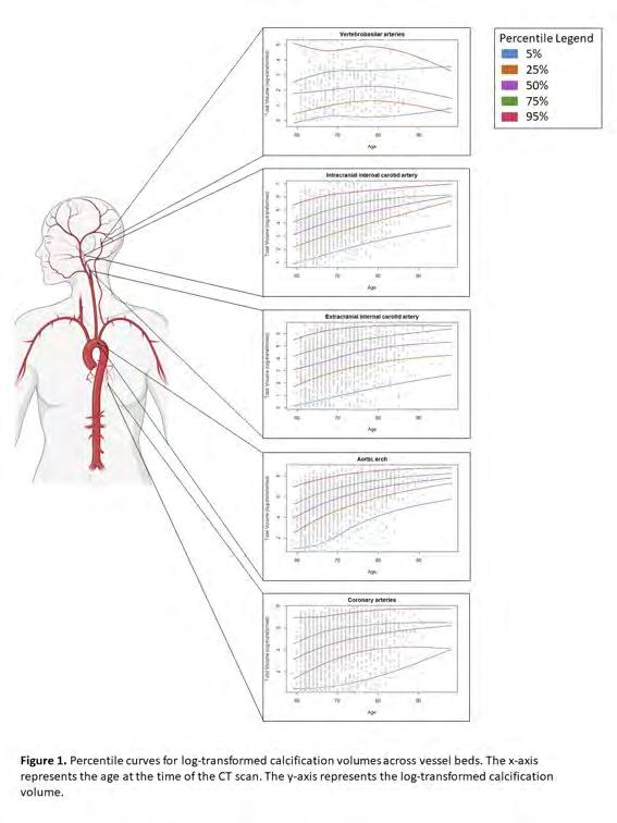
Expectations & Directions
Imaging in population-based studies is becoming ever more important in studying determinants of disease and in disease prediction. Non-invasive imaging techniques, such as MRI, enable us to detect increasingly subtle and early pathologic changes in asymptomatic individuals, tremendously enlarging our power and sensitivity to study common diseases, like stroke and dementia.
In the coming years, we expect that the amyloid PET data that were recently acquired will enable us to study the vascular and amyloid pathways towards dementia in depth, gaining more understanding in how these pathologies alone or in conjunction may lead to neurodegeneration and cognitive decline. Studies like the 7T small
229 scientific report 2023 | POPULATION IMAGING a a a a a a a a a a a a a a a a a a a a a a a a a a a a a a a a a a a a a a a a a a a a a a a a a a a a a a a a a a a
Figure 3. Vascular calcification percentile curves.
vessel disease study that is executed in collaboration with University Medical Centre Utrecht, will greatly add to this by allowing to directly image and measure microangiopathy, one of the most important contributors to both dementia and stroke. Furthermore, advances in image processing, yielding quantification of more and new markers and data-driven artificial intelligence research techniques (machine learning, deep learning) will bring the field of population imaging forward. Also, combining imaging with other high-dimensional data such as genomics, proteomics and metabolomics, is highly promising in unravelling pathways of disease and better understand disease pathophysiology.
Funding
Vernooij, Meike, Esther Bron , and Harro Seelaar ZonMW: ‘TAP-Dementia: timely, accurate and personalized dementia diagnosis’. 2023-2027
Vernooij, Meike, and Julia Neitzel Alzheimer’s Association Research Grant: ‘ Amyloid pathology and vascular disease: a tale of two pathways’. 2022-2025
Vernooij, Meike, Frank Wolters, and Arfan Ikram publicprivate partnership receiving funding from ZonMW and Health Holland: ‘A Personalized Medicine Approach for Alzheimer’s Disease (ABOARD)’. 2021-2025
Neitzel, Julia A global Marie Curie Fellowship: ‘DIVERTAD’. 2021-2025
Vernooij, Meike, Arfan Ikram, Danielle van Assema, Roelf Valkema , and Kamran Ikram Memorabel grant: ‘Amyloid pathology and vascular disease in focus: exploring interaction in two pathways towards neurodegeneration’. 2017-2023
Invited Lectures
Meike Vernooij. ‘ Imaging in neurodegeneration: towards an etiologic diagnosis’. RSNA, Chicago, USA.; Dec 2023.
Meike Vernooij. ‘ Role of neurodegenerative imaging biomarkers in clinical routine and trials’. RSNA, Chicago, USA. Dec 2023.
Meike Vernooij. ‘ White matter tract anatomy’’. Italian Neuroradiology Conference (AINR), Bergamo, Italy. Oct 2023.
Meike Vernooij.’Torgny Greitz Honorary Lecture: Imaging in dementia: the past, the present, the future’. Swedish Radiology Society conference, Uppsala, Sweden. Sept 2023.
Meike Vernooij. ‘ Preparing for Anti-Amyloid Treatment: the Radiologist’s Role’. ESNR, Vienna, Austria. Sept 2023.
Meike Vernooij . ‘Dementia imaging and SVD imaging’. ESNR summer School, Kusadasi, Turkey. June 2023.
Meike Vernooij. ‘ Imaging in neurodegeneration: towards an etiologic diagnosis’. ECR, Vienna, Austria. March 2023.
Meike Vernooij and Has Boulkhrif. ‘EDiR simulation session: Geriatric Radiology’. ECR, Vienna, Austria. March 2023.
Meike Vernooij. ‘Tackle twisted cases: Neuroradiology (EDiR)’. ECR, Vienna, Austria. March 2023.
Meike Vernooij. ‘European Diploma Prep Session: Neurovascular disorders and trauma of the brain’. ECR, Vienna, Austria. March 2023.
Meike Vernooij. ‘Specific versus nonspecific T2 hyperintensities: how to tell the difference?’. ECR, Vienna, Austria. March 2023.
Highlights
Eline Vinke en Isabelle van der Velpen successfully defended their PhD theses in February 2023 and November 2023, respectively.
Frank Wolters gave lectures for the general public at the prestigious AAIC (Alzheimer’s Association International Conference) and at the ABOARD consortium public-day meeting (organized by Jacqueline Claus)
Jacqueline Claus was awarded several travel grants from Alzheimer Nederland and Erasmus + for the ESOC, the European Alzheimer Academy Workshop and conference and a work visit to Colombia.
Jacqueline Claus appeared on national television (NPO1) in an interview on her research by Alzheimer Nederland.
Meike Vernooij was re-elected as Chair of the Diagnostic Committee of ESNR, as well as appointed as Subcommittee Chair for Neuroradiology at ECR 2024.
Meike Vernooij was appointed as Medical Delta professor at the TU Delft (Faculty of Applied Sciences).
Meike Vernooij coordinated an ESR-RSNA transatlantic course both at ECR 2023 and RSNA 2023.
Meike Vernooij was invited to give the Honorary Tony Greitz lecture of the Swedish Society of Neuroradiology (Sept 2023, Uppsala, Sweden).
230 a a a a a a a a a a a a a a a a a a a a a a a a a a a a a a a a a a a a a a a a a a a a a a a a a a a a a a a a a a a
The BIRD-NL -consortium on prevention of dementia, led by Frank Wolters and Arfan Ikram, had its kick-off meeting, just as the TAP-Dementia consortium, in which Meike Vernooij and Esther Bron are leading a work package.
Eline Vinke was awarded both a postdoc grant by the Dutch Heart Foundation (Dekker grant) for her project ‘Personalized MRI-based Cerebral Small Vessel Disease (SVD) burden quantification, for more accurate diagnosis and prognosis’, as well as an Alzheimer Nederland Early Career grant ‘Unraveling brain aging patterns predictive of AD or ADRD’.
Julia Neitzel was awarded an Erasmus MC Fellowship grant for her research project “Impact of Modifiable Factors on Dementia Risk across the Adult Lifespan” into the interplay between amyloid and vascular pathology in neurodegeneration.
Frank Wolters was awarded an Alzheimer Nederland Biomedical Research Grant, for his research into the APOEe2 paradox.
Additional Personnel
Anna Streiber – PhD student
Myrthe van Haaften – PhD student
Thuy Nguyen – research assistant
Rachida Hadouch – Radiology Assistant MRI Ommoord
Issrae Affani – Student Assistant MRI Ommoord
Michiel van den Akker – Student Assistant MRI Ommoord
Mehdi Badaoui – Student Assistant MRI Ommoord
Lucas de Groot – Student Assistant MRI Ommoord
Gaia Hermans – Student Assistant MRI Ommoord
Eileen Kikkert – Student Assistant MRI Ommoord
Levy Schimmel – Student Assistant MRI Ommoord, teamleader
Celine Tuik – Student Assistant MRI Ommoord
Suheda Yuce – Student Assistant MRI Ommoord
231 scientific report 2023 | POPULATION IMAGING a a a a a a a a a a a a a a a a a a a a a a a a a a a a a a a a a a a a a a a a a a a a a a a a a a a a a a a a a a a
Assistant Professors

Frank J. Wolters, MD PhD
Email f.j.wolters@erasmusmc.nl LinkedIn

Frank Wolters obtained his MD at Utrecht University and practiced for a while in clinical neurology, before specialising in neuro-epidemiological research with an MSc degree and PhD (cum laude) from Erasmus University Rotterdam. He completed research fellowships at the University of Oxford and the Harvard School of Public Health. At Erasmus MC, Frank applies various modalities of brain imaging to further prevention and timely diagnosis of cognitive disorders, both in population studies and clinical research. He leads the nationwide BIRD-NL dementia prevention consortium, and is active in various collaborations including the Netherlands Consortium of Dementia Cohorts (NCDC), Heart-Brain Connection group (HBCx), and neuro working group of the Cross-Cohort Collaboration (CCC).
Bridging clinical and population science
The core of my research aims at improving prevention of dementia, with a particular focus on vascular cognitive impairment. I strive to translate findings from population-based research to the clinic, and vice versa, in order to develop diagnostic tools and treatments that are broadly applicable in medical practice. A couple examples are highlighted below.
Covert brain infarcts
Covert brain infarcts –defined as infarcts on imaging that were not preceded by (recognised) stroke symptoms– are seen in 20% of elderly individuals on routine brain MRI, and increase the risk of subsequent cardiovascular disease and cognitive decline. Despite the large potential for secondary prevention, patients often go untreated, as optimal management is undetermined due in part to uncertainty about who is at highest risk. As part of my NWO Veni project, I combine data from various cohorts to find determinants of high risk, and ultimately improve diagnostic and therapeutic management in clinical patients, like those visiting the Alzheimer Centre outpatient clinic.
Heart-brain axis and post-stroke dementia
Heart disease is an acknowledged contributor to stroke, and emerging evidence indicates heart failure and atrial fibrillation also predispose to dementia. Within my group, we try to unravel how cardiac dysfunction relates to brain pathology on MRI, and how this translates into clinical sequelae. An important focus thereby is the overlap with Alzheimer's disease pathology, and the mediating role of vascular brain injury and clinical stroke. As such, we study in populationbased setting to what extent the occurrence of cognitive decline and dementia after TIA/stroke depends on

Figure 1. In collaboration with the bioinformatics group (Henri Vrooman and Hakim Achterberg), we developd a viewer to do side-by-side ratings of the Rotterdam Study scans, such that we can reliably assess changes over time and incident (covert) ischemia. This will allow more careful mapping of infarct patterns and identification of risk factors for developing brain ischemia over time in the population.
event severity, heart disease, arteriosclerosis, cerebral small-vessel disease, and concurrent brain atrophy and other Alzheimer biomarkers. By taking these findings to clinical studies, such as the Alzheimer Centre Imaging Cohort (n=1200) and the Erasmus Stroke Cohort, I aim to translate these findings into clinically applicable tools for diagnosis and prediction that facilitate patient information and personalized care.
Improving research methodology
Valid and sustainable science relies on adequate research methods, from basic science to machine learning and clinical trials. Epidemiology should play an important part in this process, and I advocate interaction across disciplines In Erasmus MC, with epidemiologists who speak the language of clinicians and biomedical researchers, to support them with knowledge on study design and causal inference.
232 a a a a a a a a a a a a a a a a a a a a a a a a a a a a a a a a a a a a a a a a a a a a a a a a a a a a a a a a a a a
Julia Neitzel, PhD
Email j.neitzel@erasmusmc.nl LinkedIn

POPULATION BRAIN IMAGING
Julia Neitzel’s research interest is to understand why some people are more resilient against age-related cognitive decline and dementia – important, yet missing knowledge which could inform better prevention programs. For addressing this question, she combines her knowledge on cognition and brain imaging gained during her underand postgraduate studies (MSc in Neuro-cognitive Psychology from Ludwig-Maximilians-Universität (LMU) Munich; PhD at the Neuroradiology of the Technical University Munich, Germany), with her postdoctoral work on genetic and modifiable risk factors of dementia (Institute for Stroke and Dementia Research at LMU; Depts of Radiology and Epidemiology at Erasmus MC). After her research visit at the Harvard School of Public Health in Boston, USA, she is excited to start her independent research group as an Erasmus Fellowship recipient.
Individualized dementia prevention
Prevention of chronic diseases is an ever-increasing forefront of medicine. Especially for dementia , once brain damage has occurred, treatment has been shown to be too late. My lab’s mission is to provide the knowledge for precision prevention strategies that promote brain health tailored to the demographic, genetic and clinical characteristics of each person. I support individualized dementia prevention through three key actions: (1) improving our understanding of the natural history of dementia and associated risk factors, (2) discovering new protective factors to pinpoint potential prevention targets, and (3) creating prediction algorithms that can identify high-risk individuals, enabling us to prioritize them in prevention approaches. Population brain imaging is the central component in my research, functioning as a powerful tool for early disease detection in asymptomatic individuals.
Poor sle ep, a new risk factor for Alzheimer’s disease?
Healthy sleep has been recognized as a key measure for maintaining cardiovascular health, as defined by the American Heart Association (Life’s Essential 8). Yet, it is unclear whether and which sleep patterns also contribute to the onset of Alzheimer’s disease. To address this question, we have measured 24-hour activity rhythms and sleep using actigraphy in 319 participants of the Rotterdam Study. All participants underwent 18F-florbetaben PET about 8 years later, on which we quantified their Alzheimer’s disease (amyloid-beta [Aβ] plaque) burden. We found that individuals with a more fragmented 24-hour activity rhythm, i.e. daytime napping and/or being wake at night, showed a higher disease burden at follow-up, also when accounting for disease

Figure 1. Association between a fragmented 24-hour activity rhythm and higher Aβ burden in APOE4 risk carrier compared to non-carrier
burden at baseline. Carriers of an Alzheimer’s risk allele (APOE4) appeared to be more susceptible than noncarriers (Fig. 1). Our results suggest that rhythm disturbances may be a modifiable risk factor for Alzheimer’s disease (Nguyen, … Neitzel, under review).
How well can we predict Alzheimer’s disease?
Models predicting A β plaques could become cost-efficient tools to identify individuals at risk of developing dementia. We showed that A β prediction models developed in a large sample of patients with A β-PET scans (A4 Study, n=4,119) were applicable to a population that was more representative of typical older nondemented adults (Rotterdam Study, n=500). Our best performing model in the A4 Study (AUC=0.73), including age, APOE4, family history of dementia, cognition, walking and sleep duration, predicted A β plaques with even higher accuracy in the Rotterdam Study (AUC=0.85; Nguyen, … Neitzel, 2023, Alz Dement). We currently investigate to what extent blood-based biomarkers can improve prediction accuracy.
233 scientific report 2023 | POPULATION IMAGING a a a a a a a a a a a a a a a a a a a a a a a a a a a a a a a a a a a a a a a a a a a a a a a a a a a a a a a a a a a

María Rodriguez-Ayllon, PhD
Project Funding Alicia Koplowitz Fellowship
Email m.rodriguez@erasmusmc.nl
ResearchGate www.researchgate.net/profile/Maria-Rodriguez-Ayllon
Physical activity and brain health
My research focused on understanding the role of physical activity in brain health across the lifespan. Physical activity has been suggested as a modifiable factor that might contribute to improving cognitive and brain function during aging. However, previous studies were mainly of cross-sectional design and did not consider the effects of time or potential reverse causality. In this context, I have explored the bidirectional longitudinal association between physical activity and brain structure in older people. Interestingly, I identified older adults with potentially advanced brain aging status as being at higher risk of physical inactivity over time, and therefore as a potential target group for prevention and novel intervention strategies.


In addition, I conducted an integrated and complex model to provide an overview of the mechanisms linking physical activity with mental health in youth. In brief, I found that among all examined neurobiological data, psychological constructs, and behaviors, self-esteem was identified as the only mediating factor through which physical activity relates to mental health in youth.
My short-term purpose is to continue my research at Erasmus MC by exploring the main paths (e.g., by reducing amyloid beta accumulation in the brain) that might explain why physical activity is a protective factor for brain health across the lifespan.
Eline Vinke, PhD
Project Funding Early Career Grant – Alzheimer Nederland; Postdoc Dekker Beurs – Hartstichting
Email e.vinke@erasmusmc.nl
Modelling (healthy) brain aging
My research is on the intersection of neuroimaging, aging, epidemiology and machine-learning. In my research I develop and apply machine-learning based technologies that help us understand where, when and how deviations from normal or healthy brain aging take place based on neuroimaging in combination with other biomarkers. With the ultimate aim to improve clinical diagnosis, prognosis and treatment or preventive strategies of neurodegenerative and neurological disease.
This year I received the Early Career Grant from Alzheimer Nederland and the Postdoc Dekker Beurs from the Hartstichting. With these grants I am starting my own research line focussing on modelling (healthy) brain aging based on neuroimaging.

In the coming years I will apply disease progression modelling in the context of brain aging, to Identify different brain aging patterns within the populationbased Rotterdam Study. I will study these patterns and Investigate the clinical value of the different patterns and the progression stage within these patterns, in relation to the risk of developing cognitive decline, dementia or stroke.
234 a a a a a a a a a a a a a a a a a a a a a a a a a a a a a a a a a a a a a a a a a a a a a a a a a a a a a a a a a a a
Post-docs LinkedIn LinkedIn
PhD Students

Joyce van Arendonk, MSc

Advisors Meike Vernooij, Arfan Ikram & Julia Nietzel
Project Funding ZonMW Memorabel grant
Email j.vanarendonk.1@erasmusmc.nl
Amyloid pathology and vascular disease in focus
To elucidate the interaction between the A β and vascular pathways in dementia etiology, participants of the Rotterdam Study aged 60 years and older underwent amyloid PET imaging. We studied the association between vascular risk factors and A β pathology. We observed that diabetes was associated with a higher prevalence and severity of A β pathology in both APOE4 carriers and noncarriers.

Isabelle van der Velpen, MD

Advisors Arfan Ikram, Meike Vernooij, René Melis & Marieke Perry
Project Funding ZonMW Memorabel: “Social factors in cognitive decline and dementia: towards an early intervention approach.”
Email i.vandervelpen@erasmusmc.nl
PhD Obtained 17-11-2023
Social factors in cognitive decline and dementia
Social health markers Including loneliness, perceived social support and marital status are associated with global brain volumes and white matter microstructural integrity in older adults. Subcortical brain structures, important for socioemotional functioning, are associated with increased risk of dementia.

Katrien Bracké, MD Camiel Box, MD

Advisors Meike Vernooij, Gwen Dieleman & Tonya White
Project Funding SSWO ( S15-13, S22-65)
Mrace Erasmus MC Grant
Email k.bracke@erasmusmc.nl
Unravelling the neurobiology of anorexia nervosa
We aim to unravel the neurobiological underpinnings of anorexia nervosa (AN), using non-Invasive Imaging techniques. We use data of the BRAVE study, a clinical case-control sample of adolescents with first-onset AN compared to gender-, age- and education-matched controls. A deeper understanding of the neurobiology may be a key element In developing novel therapeutic interventions.


Advisors Frank Wolters, Meike Vernooij & Kamran Ikram
Project Funding NWO Veni grant: "Securing brain Health by personalised treatment of INdividuals with cOvert Brain Infarcts (SHINOBI)"
Email c.box@erasmusmc.nl
Prognosis and Clinical Management of Covert Brain Infarcts
One in four elderly individuals are at high risk of stroke and dementia due to prior brain infarction that occurred unnoticed. In the Rotterdam Study we gather evidence on their prognosis and best clinical management in order to create clear guidelines to help clinicians deal with these covert brain infarcts.
235 a a a a a a a a a a a a a a a a a a a a a a a a a a a a a a a a a a a a a a a a a a a a a a a a a a a a a a a a a a a scientific report 2023 | POPULATION IMAGING
LinkedIn LinkedIn LinkedIn
LinkedIn
PhD Students

Jacqueline Claus, MD

Advisors Arfan Ikram, Meike Vernooij & Frank Wolters
Project Funding Trustfonds and ABOARD project (ZonMW/HealthHolland)
Email j.claus@erasmusmc.nl
Prevention of dementia: from theory to clinical practice
Identification of modifiable risk factors for dementia, with a focus on early recognition and treatment of stroke and estimation of post-stroke dementia risk. Identifying those eligible for disease-modifying treatments against b-amyloid. Improvement and implementation of the dementia prediction tool in a clinical setting.

Duygu Kilinc, MD MSc

Advisors Meike Vernooij & Geert Jan Biessels
Project Funding TAP-dementia, project TAP-VaMP:
“Timely Accurate and Personalized diagnosis of dementia: Validation of Multimodal diagnostic biomarkers for application in clinical Practice”
Email d.kilinc@erasmusmc.nl
Improving diagnosis of SVD based on MRI scans: what is considered normal?
To bridge the translational gap between small vessel disease (SVD) imaging research and clinical practice , I will focus on creating age- and genderspecific normative data of the SVD markers using the Rotterdam Study data. Ultimately, I want to develop a tool that estimates the potential cognitive impact of SVD , based on lesion burden and distribution, in individual patients.

Marjolein Dremmen, MD
Advisors Meike Vernooij & Tonya White
Project Funding Mrace Erasmus MC Grant
Email m.dremmen@erasmusmc.nl
BRain development, Imaging trajectories and Deviations in brain morpholoGy in the pEdiatric population; BRIDGing thE gap
The goal is to construct ‘neuroimaging growth reference curves’ for total and regional brain volumes in children and adolescents. To be able to detect early deviations in brain development in patients groups; crucial for better understanding the neurobiology of these clinical conditions. To gain knowledge on variations in brain development and evaluate the consequences of incidental findings on brain imaging

Mathijs Rosbergen, MSc

Advisors Frank Wolters, Meike Vernooij & Arfan Ikram
Project Funding ZonMW/TKI Health Holland: ABOARD consortium
Email m.rosbergen@erasmusmc.nl
Unraveling MRI markers for dementia prediction
When an individual is diagnosed with Alzheimer's disease, severe brain damage has already occurred. Treatment in an early stage could prevent brain damage and dementia. Therefore, my research focuses on unraveling new MRI predictors to improve existing dementia prediction models, so that we are able to identify individuals with high risk of dementia.
236 a a a a a a a a a a a a a a a a a a a a a a a a a a a a a a a a a a a a a a a a a a a a a a a a a a a a a a a a a a a
LinkedIn LinkedIn LinkedIn

Merel de Vries, BSc

Eline Vinke, MSc
Advisors Meike Vernooij & Steven Kushner
Project Funding Donation funds psychiatry department
Email m.w.devries@erasmusmc.nl
Decoding the maternal brain
My research focuses on how pregnancy changes the brain. In collaboration with the population-based Generation R Next study, we collect brain imaging data in women who are trying to become pregnant. We scan our study subjects before pregnancy and after pregnancy. The aim of this study is to better understand the remarkable impact that pregnancy has on the human brain, with a secondary focus on maternal mental health.
Advisors Meike Vernooij & Arfan Ikram
Project Funding Horizon 2020, EuroPOND: “Data-driven models for Progression Of Neurological
Disease
Email e.vinke@erasmusmc.nl
PhD Obtained 3-2-2023
Disentangling ‘normal’ brain aging to help us understand neurodegenerative disease
My research focuses on quantification of structural brain changes with aging, using longitudinal data from the population-based Rotterdam Study. The large variation in structural brain changes in aging overlaps with changes resulting from neurodegenerative diseases. Unraveling the normal brain aging spectrum is therefore essential to better understand disease.
237 a a a a a a a a a a a a a a a a a a a a a a a a a a a a a a a a a a a a a a a a a a a a a a a a a a a a a a a a a a a
scientific report 2023 | POPULATION IMAGING
JOINT APPOINTMENT IN EPIDEMIOLOGY
Daniel Bos was trained as a medical doctor and epidemiologist at Erasmus MC. He obtained his PhD in the field of vascular imaging and vascular epidemiology in 2013. The main focus of his work is on the elucidation of the etiology and pathophysiology of arteriosclerosis, and the clarification of the contribution of arteriosclerosis to clinical neurovascular and neurodegenerative diseases. For his research he leverages the strengths of population-based studies and combines this with clinical patient-

studies in order to be able to make a direct clinical impact with his work. Over the years, he has been awarded the Best Scientific Paper Award by the Radiological Society of the Netherlands and the Lourens Penning Prize by the Radiological Society of the Netherlands. He holds a position as Adjunct Associate Professor at the Harvard School of Public Health and a position as Professor at KU Leuven. Daniel has authored over 150 peer-reviewed publications.
d.bos@erasmusmc.nl
a a a a a a a a a a a a a a a a a a a a a a a a a a a a a a a a a a a a a a a a a a a a a a a a a a a a a a a a a a a
IMAGING OF ARTERIOSCLEROSIS: FROM POPULATION TO CLINICAL PRACTICE
Daniel Bos, MD, PHD associate professor

Context
Arteriosclerosis is a highly frequent vascular disease causing stiffening of arteries throughout the whole arterial system. Major clinical consequences of arteriosclerosis include myocardial infarction and stroke, which are top causes of morbidity and mortality in middle-aged and elderly persons worldwide. Due to the aging of the population, the global burden of arteriosclerosis, and thereby of these clinical conditions, will continue to rise in the coming decades.
The research in “Imaging of Arteriosclerosis: from Population to Clinical Practice” is focused on the elucidation of the etiology and pathophysiology of arteriosclerosis, and on the clarification of the contribution of arteriosclerosis to clinical neurovascular and neurodegenerative diseases, with a strong emphasis on the use of state-of-the-art medical imaging. Within this unique research-field on the interface of arteriosclerosis, neurovascular, and neurodegenerative diseases, the research group specifically aims at obtaining relevant imaging-based biomarkers for improved understanding of arteriosclerosis and improved prediction of abovementioned diseases, and at identifying potential targets for intervention strategies.
Top Publications 2023
Van Den Beukel T, FJ Wolters, U Siebert, W Spiering, M Ikram, M Vernooij, P de Jong, D Bos. Intracranial arteriosclerosis and the risk of dementia: A populationbased cohort study. Alzheimers & Dementia 2023; 10.1002/alz.13496.
van Dam-Nolen H, N van Egmond, P Koudstaal, A van der Lugt, D Bos. Sex Differences in Carotid Ahterosclerosis: A Systematic Review and Meta-Analysis. Stroke 2023; 54:315-326.
Melgarejo J, M Vernooij, M Ikram, Z Zhang, D Bos. Intracranial Carotid Arteriosclerosis Mediates the Association Between Blood Pressure and Cerebral Small Vessel Disease. Hypertension 2023; 80: 618–628.
a a a a a a a a a a a a a a a a a a a a a a a a a a a a a a a a a a a a a a a a a a a a a a a a a a a a a a a a a a a 239
Research Projects: Objectives & Achievements
Intracranial Arteriosclerosis
This line of research is specifically focused on the causes and consequences of intracranial arteriosclerosis. In this field, important pioneering-work on the identification and quantification of calcification in the intracranial carotid arteries and the vertebrobasilar system has been performed by our research group. By applying nonenhanced computed tomography to participants of the population-based Rotterdam Study, we demonstrated that the presence and amount of intracranial carotid artery calcification is the strongest risk factor for developing a first-ever stroke, and that the amount of calcification at this location even contributes to the development of dementia, including Alzheimer’s Disease. This work still has a considerable global impact and has also led to an extension of the research horizon towards clinical studies on the influence of intracranial arteriosclerosis in stroke patients. In these patients, the amount of intracranial arteriosclerosis at time of admission to the hospital directly influences the prognosis after stroke treatment.

Previous work in the field of pathology and imaging uncovered the existence of two morphological patterns of intracranial arteriosclerotic calcification. These include intimal, atherosclerotic calcification and circular calcification of the internal elastic lamina (IEL). Work from this research group showed that these two patterns contribute differentially to stroke, and influence stroke treatment, given the divergent effects of calcification on the arterial wall structure and the accompanying structural stresses within the artery. In this light, the research horizon on the consequences of these calcifications has been broadened towards neurodegenerative brain disease, including dementia. The first work into this direction indeed showed that primarily the circular IEL calcification seems to contribute to neurodegenerative changes. In the near future, we will delve into the underlying mechanisms of this association by combining information from different imaging modalities, including CT, PET, and MRI.
Advanced Imaging of Arteriosclerosis
This line of research is devoted to more advanced imaging of arteriosclerosis, with a specific focus on its most common subtype, namely atherosclerosis. Atherosclerosis is characterized by thickening of the intimal layer of the arterial vessel wall due to lipid-accumulation, neo-vascularization, and calcification. Especially when located in the carotid artery bifurcation, atherosclerosis is known to substantially increase the risk of ischemic stroke. The work in this research line is targeted at the optimal identification and evaluation of atherosclerosis in the carotid artery bifurcation. Using advanced, stateof-the-art imaging modalities (CT, MRI, etc.), the aim of the research group is to further understand the development of this disease and the value of assessing plaque composition and plaque morphology for the prediction of neurovascular and neurodegenerative diseases. Recent accomplishments of the research group in this field have shown that the presence of intraplaque hemorrhage in carotid artery atherosclerosis is the single most important risk factor for a first-ever stroke and stroke recurrence. Moreover, our work showed that the composition of these plaques substantially differ between men and women, which is one of the topic we aim to further elucidate in coming years.
Within this line of research, an important novel focus is on photon-counting CT (PCCT) for imaging of arteriosclerosis. Specifically in the cerebral vessels, the resolution of conventional CT is too limited and PCCT may represent an important novel modality in this field. Current studies that are being performed within the research group revolve around comparisons of PCCT with conventional CT on calcification detection, calcification quantification and image quality, mainly using autopsy specimen.
240 a a a a a a a a a a a a a a a a a a a a a a a a a a a a a a a a a a a a a a a a a a a a a a a a a a a a a a a a a a a
Two subtypes of intracranial arteriosclerosis; calcified atherosclerosis (yellow) and calcification of the internal elastic membrane (green). Left: in-vivo CT-images; Right: corresponding autopsy specimen.
Optimization of Cardiovascular Image Analysis
This research line revolves around the early detection of vascular disease, by maximally leveraging existing medical imaging examinations. By using novel methods for image-analysis (e.g. machine-learning algorithms), we aim to obtain more sensitive and more accurate imaging markers of vascular disease. My group performed important pioneering-work on the quantification of epicardial fat as emerging markers of vascular risk. Over the last year, this novel marker of vascular risk has gained rapid attention, which has led to a strong collaboration on its role in heart-transplantation patients with the Dept. of Cardiology. This collaboration was further extended with additional measurements of imaging-based markers of vascular risk (liver density) and overall-health status (bone density), which can all be readily assessed using conventional CT. Additionally, we have set up a strong collaboration with the department of Vascular Internal Medicine of the Amsterdam UMC in the field of early identification of aortic valve stenosis through CT-based assessment of calcification on the aortic valve.

Expectations & Directions
The field of research into arteriosclerosis is rapidly evolving and substantial developments at many levels will occur in which the research of this group will play an important role. First, through the contributions of research of this group, intracranial arteriosclerosis is increasingly being recognized as an important risk factor for stroke, but may also play a key role in the development of dementia. In order to advance these insights, detailed visualization of arteriosclerotic disease in the cerebral vessels (not only the large afferent arteries such as the carotids and the vertebral arteries, but particularly in the smaller arteries) using combinations of different advanced imaging modalities is the way forward. An important focus should be on the difference between ‘pure’ atherosclerotic changes in the arteries (intimal thickening, plaque formation) and hardening/stiffening of the arteries through calcifications in the medial layer of the artery.
Subsequently, using population-based data, etiological differences between these types of arteriosclerosis can be determined. Similarly, these features of arteriosclerosis may be added to existing risk-prediction models for stroke and even dementia, further contributing to their early identification and to the development of therapeutic and preventative strategies. One field in which rapid developments are made is that of image-post-processing (using machine learning algorithms), which now allows for the possibility to investigate arteriosclerotic disease in much more detail. In addition to ‘conventional’ measurements such as the Agatston score, or volumetric assessments of calcification or plaque-size, it is becoming possible to also assess shape characteristics of the disease, make vessel-stress calculations, link specific configurations of arteriosclerotic disease (including combinations of shape, volume and other properties) with outcomes.
Third, an important achievement of this group was the completion of a large-scale follow-up imaging study (10-15 years of follow-up) targeted at the visualization of change in the amount of vascular calcification across multiple arteries. These data are unique in the world and will provide valuable insight into the natural course of arteriosclerosis
241 scientific report 2023 | IMAGING OF ARTERIOSCLEROSIS a a a a a a a a a a a a a a a a a a a a a a a a a a a a a a a a a a a a a a a a a a a a a a a a a a a a a a a a a a a
Imaging markers on non-contrast coronary calcium scans. Four slices of a coronary calcium scan of a single patient showing the heart at different levels with, in colour, the different tissues from which the potential imaging markers may be obtained
Funding
Bos, Daniel, Kamran Ikram, Ali Akyildiz, and Jolande Wentzel Erasmus MC2 Research Synergy Grant: ‘Unraveling the vascular biomechanics of stroke and dementia: the navier-stokes study’. 2023-2027
Bos, Daniel, Frank Wolters, Meike Vernooij, Julia Neitzel, Maarten Leening, Mohsen Ghanbari, and Arfan Ikram Alzheimer Nederland Research Grant: ‘Trajectories of Vascular Disease in Aging to Predict Dementia.’ 2023-2026
Bos, Daniel, and consortium partners World Cancer Research Fund: ‘Treatment tolerance and prognosis in survivors with non-metastatic colorectal cancer: a matter of liver fa(c)t?.’ 2023-2027
Bos, Daniel, Theo van Walsum, and consortium partners ‘ICAI Stroke Lab: From 112 to Rehabilitation.’ 2023-2027
Bos, Daniel, Ricardo Budde, and consortium partners Maarten van der Weijden Foundation: ‘Statins to Prevent Immune checkpoint inhibitor-induced pRogression of AtheroscLerosis: the SPIRAL study’. 2023-2026
Invited Lectures
Daniel Bos. ‘Research Aims in Clinical Medicine’. Grand Round Department of Cardiovascular Sciences KU Leuven, Leuven, Belgium. March 2023.
Daniel Bos. ‘AI in het kader van Epidemiologie: de waarde van AI in wetenschappelijk onderzoek’. Rotterdam Radiology AI, Rotterdam, The Netherlands. June 2023.
Daniel Bos. ‘Similarities and Differences of extra- and intracranial arteriosclerosis’. Fil Rouge of Atherosclerosis, Cagliari, Italy. Oct 2023.
Daniel Bos. ‘Intracranial Arteriosclerosis: etiology and clinical consequences’. Grand Round at Scripps Medical, San Diego, USA. Oct 2023.
Highlights
Tim van den Beukel successfully defended his thesis entitled “Intracranial arteriosclerosis: determinants and clinical consequences” (co-promotor: Daniel Bos)
Daniel Bos set up a new collaboration with the Scripps Health System in San Diego (US) with Prof. Alois Zaune r to extend his research on intracranial arteriosclerosis in Hispanic populations.
The paper ‘Sex Differences in Carotid Atherosclerosis: A Systematic Review and Meta-Analysis' by Dianne van Dam-Nolen (senior author: Daniel Bos ) was chosen as paper of the month by the European Stroke Organization.
Daniel Bos was appointed as expert in the Coalition for Lifestyle and Healthcare (Leeftstijl in de Zorg) and coauthored the Knowledge Agenda on this topic for the Ministy of Healthcare (VWS).
Daniel Bos successfully finished the leadership programme of the Dutch CardioVascular Alliance (DCVA) and became DCVA-ambassador.
Additional Personnel
Robin Camarasa – PhD student Department of Radiology & Nuclear Medicine (PI: Marleen de Bruijne)
Céline van der Braak – PhD student Department of Radiology & Nuclear Medicine (PI: Ivo Schoots)
Hyunho Mo – Postdoc Department of Radiology & Nuclear Medicine (PI: Esther Bron)
Judith van der Bie – PhD Student Department Radiology & Nuclear Medicine (PI: Ricardo Budde & Alexander Hirsch)
Cevdet Acarsoy – PhD student Department of Epidemiology
Brian Berghout – PhD student Department of Epidemiology
Luoshiyuan Zuo – PhD student Department of Epidemiology
Mitra Nekouei – PhD student Department of Epidemiology
Peter van Hulst – PhD student Department of Neurology
Xi Li – PhD student Department of Public Health
Kostas Stoitsas – Postdoc Department of Epidemiology
242 a a a a a a a a a a a a a a a a a a a a a a a a a a a a a a a a a a a a a a a a a a a a a a a a a a a a a a a a a a a
Assistant Professor

Maarten J.G. Leening, MD, MSc, PhD
Email m.leening@erasmusmc.nl

JOINT APPOINTMENT IN CARDIOLOGY AND EPIDEMIOLOGY
Maarten Leening obtained his medical degree (cum laude) and MSc in clinical epidemiology at the Erasmus MC. He received his PhD (cum laude) at the Erasmus MC (Depts of Cardiology and Epidemiology). In 2014-2015, he worked as a post-doc at the Division of Preventive Medicine at the Harvard Brigham and Women's Hospital (Nancy Cook and Paul Ridker). Subsequently, he trained as a cardiologist with a focus on non-invasive imaging (EACVI certified in cardiac CT and echocardiography), CVD prevention (EAPC certified), and lipids/atherosclerosis (EAS certified). By the end of 2023, Maarten Leening joined the vascular imaging research group (Daniel Bos).
Preventive Cardiology
Atherosclerotic cardiovascular disease (ASCVD) events are preventable in the vast majority of individuals. My research aims at early life identification and treatment of cardiovascular risk factors and imaging of subclinical atherosclerosis in presymptomatic individuals.
Specific interests Includes the biology and treatment of atherogenic lipids and chronic inflammation.
On the side of imaging, areas of interest include noninvasive assessment of coronary atherosclerosis and aortic valve disease, and the role of automated software and AI in cardiovascular imaging.
243 scientific report 2023 | IMAGING OF ARTERIOSCLEROSIS a a a a a a a a a a a a a a a a a a a a a a a a a a a a a a a a a a a a a a a a a a a a a a a a a a a a a a a a a a a
PhD Students
 Taihra Zadi, MSc

Britt van Dijk, BSc

Advisors Aad van der Lugt & Daniel Bos
Project Funding GE Healthcare Email t.zadi@erasmusmc.nl
Atherosclerotic plaque morphology of the carotid artery
Atherosclerotic plaque composition in the carotid artery has been an important topic of research for the last few decades. Different components harbor different risks of plaque ruputure and a (recurrent) stroke. Particularly the presence of a lipid-rich necrotic core, thin fibrous cap or intraplaque hemorrhage are components that are vulnerable. Conversely, plaques with more calcification seem to be less prone to rupture.

Sven Luijten, MSc,
MD

Advisors Aad van der Lugt, Diederik Dippel, Daniel Bos & Bob Roozenbeek
Project Funding Collaboration for New treatments of Acute Stroke (CONTRAST)
Email s.luijten@erasmusmc.nl
Imaging-based prediction of outcome in ischemic stroke
The overarching aim of my research is to assess and identify imaging markers that affect patient prognosis and efficacy of endovascular thrombectomy (EVT) for ischemic stroke, with the ultimate goal of imaging-based prediction of functional outcome and benefit of EVT in individual patients.
Advisors Ricardo Budde, Olivier Manintveld, Daniel Bos & Rudolf de Boer
Project Funding Departments of Cardiology, Radiology & Nuclear Medicine, Erasmus MC
Email b.vandijk@erasmusmc.nl
Cardiac Allograft Vasculopathy after Heart Transplantation
The aim of this PhD trajectory is to gain insight into causes and consequences of Cardiac Allograft Vasculopathy (CAV) after a heart transplant (HTx) using cardiac imaging. With these insights we want to predict CAV, discover risk factors for CAV and possibly prevent CAV after HTx.

Dianne van DamNolen, MSc, MD

Advisors Aad van der Lugt, Peter J Koudstaal & Daniel Bos
Project Funding The PARISK study is supported by the Center for Translational Molecular Medicine (CTMM) and the Dutch Heart Foundation
Email h.nolen@erasmusmc.nl
Imaging of high-risk carotid plaques
Carotid atherosclerosis is a main cause of ischemic stroke. The Plaque At RISK study shows that imaging of carotid atherosclerosis by CTA and MRI improves the prediction of recurrent ischemic strokes. Carotid imaging can identify high-risk carotid plaques based on specific plaque characteristics such as intraplaque hemorrhage.
244 a a a a a a a a a a a a a a a a a a a a a a a a a a a a a a a a a a a a a a a a a a a a a a a a a a a a a a a a a a a
LinkedIn
 Anna Streiber, MSc

Advisors Meike Vernooij, Julia Neitzel & Daniel Bos
Project Funding Cure Alzheimer’s Fund
Email a.streiber@erasmusmc.nl
Disentangling the Role of Arteriosclerosis in Alzheimer’s Disease
The etiology underlying Alzheimer’s disease is multifactorial and may be associated with cerebrovascular changes. Within the Roterdam study, we investigate the relationship between arteriosclerosis and various markers for Alzheimer’s diseas. These markers are measured in blood plasma, on (f)MRI, or PET-CT.
 Aikaterini Tziotziou, MSc

LinkedIn LinkedIn
Advisors Ali Cagdas Akyildiz, Daniel Bos, Ton van der Steen & Aad van der Lugt
Project Funding PhD-project Erasmus MC-Grants (MRace) 2019
Email a.tziotziou@erasmusmc.nl
Mechanical wall stress is associated with atherosclerosis development. Atherosclerotic plaque onset and progression are known to be affected by local biomechanical factors.
We investigated the association of mechanical wall stress (MWS) and wall shear stress (WSS) to atherosclerosis development in human coronary (IMPACT Study) and carotid (PARISK Study) arteries.
245 scientific report 2023 | IMAGING OF ARTERIOSCLEROSIS a a a a a a a a a a a a a a a a a a a a a a a a a a a a a a a a a a a a a a a a a a a a a a a a a a a a a a a a a a a
Jacob Visser is a musculoskeletal radiologist, health economist and epidemiologist. As of July 2020, he is appointed as assistant professor in value-based imaging and working on issues such as structured reporting, decision support software, and integrated diagnostics. Furthermore, he is involved in setting up further collaboration with radiologists at the Radiology department of the Massachusetts General Hos-

pital (MGH) in Boston, MA, USA. In addition, he is a member of the Value-based Radiology Committee of the ESR and the RSNA Working Group for Common Data Elements. He has served in the ESR eHealth and Informatics Subcommittee. As of January 1, 2020, he was appointed as Chief Medical Information Officer at the Erasmus MC. j.j.visser@erasmusmc.nl
a a a a a a a a a a a a a a a a a a a a a a a a a a a a a a a a a a a a a a a a a a a a a a a a a a a a a a a a a a a
VALUE-BASED IMAGING
Jacob J Visser, MD, PhD, MSc assistant professor
Context
As health care rapidly changes from volume to value-based, there is an urgent need for radiologists to position themselves from a valuebased perspective.
Therefore, the Radiology Department at the Erasmus MC has started the value-based imaging program. This means that all activities in the Radiology Department are evaluated in the light of value-based imaging.
The value-based imaging program is closely related to the IT department. Most topics that are being covered by this program require expertise in the field of information technology.
Top Publications 2023
Topff L, ER Ranschaert, A Bartels-Rutten, A Negoita, R Menezes, RG Beets-Tan, JJ Visser. Artificial Intelligence Tool for Detection and Worklist Prioritization Reduces Time to Diagnosis of Incidental Pulmonary Embolism at CT. Radiol Cardiothorac Imaging 2023; 5:220163.
Topff L, KB Groot Lipman, F Guffens, R Wittenberg, A Bartels-Rutten, G van Veenendaal, M Hess, K Lamerigts, J Wakkie, E Ranschaert, S Trebeschi, JJ Visser, RG Beets-Tan; ICOVAI, International Consortium for COVID-19 Imaging AI. Is the generalizability of a developed artificial intelligence algorithm for COVID-19 on chest CT sufficient for clinical use? Results from the International Consortium for COVID-19 Imaging AI (ICOVAI). Eur Radiol . 2023; 33:4249-4258.
a a a a a a a a a a a a a a a a a a a a a a a a a a a a a a a a a a a a a a a a a a a a a a a a a a a a a a a a a a a 247
Research Projects: Objectives & Achievements
Artificial intelligence
Currently, several projects are under investigation in the value-based imaging program. Firstly, as good quality of radiology reports is a core element in the era of valuebased imaging, strategies are developed to investigate the quality of these reports. As manual review to identify sufficient cases for retrospective analysis is often infeasible, a stepwise natural language processing (NLP)method for case identification was developed and applied for the identification of critical finding cases.
Quantitative and radiogenomics imaging is becoming more important. It turned out that visual assessment alone was not able to generate all available information. Instead, state-of-the-art computer techniques like machine and deep learning are able to extract quantitative features that are able to play an important role in the diagnostics of musculoskeletal and other diseases. A model was developed and is to be validated that is able to predict the MDM2-amplification in lipoid tumors.
Implementation of artificial intelligence algorithms in the radiology workflow is becoming a hot topic. Although several algorithms have been developed of which some are CE-marked / FDA approved and only a few validated in clinical practice, it is important to make the right decisions about implementation of those algorithms. Depending on the type of the algorithm, automatic measurements, case prioritization, pathology detection, a certain amount of research is needed before usage in the clinic can start.

Artificial intelligence algorithm automatically detects fractures.
Data-driven workflow
More than 200,000 exams are being performed in our department annually. This requires good logistics and insights in the workflow. In order to get the information about the processes, it is needed to identify relevant data sources. Modern software packages allow for visual representation of process and quality indicators. The research focuses on how to involve people and use dash boarding tools in order to improve efficiency and quality metrics.
Integrated diagnostics
Integrated diagnostics will be one of the key steps forward in the coming decade. In this respect, the work-up for patients with adrenal incidentaloma was evaluated. Furthermore, the diagnostic work-up for cervix carcinoma, carcinoma of unknown primary, and chest pain was evaluated. Currently, a workflow integration of radiology and pathology findings is investigated in lung cancer patients. Also for liver and brain tumors, further integration of radiology and pathology can increase efficiency and improve diagnosis. In addition, it is to foreseen that image oriented specialties like dermatology can be involved in the integrated diagnostics approach.
Expectations & Directions
In the coming years, we aim to further expand our research in the domains of artificial intelligence, data-driven workflow, and integrated diagnostics. An important challenge will be to bring these together and establish reference architectures and pipelines how to structurally evaluate new workflows and technologies.
Funding
Visser, Jacob , Martijn Starmans , Ken Redekop, and Frans Vos TKI: ‘Assessing the effect on the clinical workflow and a cost-effectiveness analysis of a comprehensive AI tool for chest CT reading - on the use case of incidental pulmonary embolism’. 2023-2027
Visser, Jacob Radiology Research Fund: ‘Enabling Value impact assessment for Artificial Intelligence Tools in radiology (eVAIT)’. 2022-2027
Klein, Stefan, Martijn Starmans, Jacob Visser, Kees Verhoef, and Dirk Grunhagen Hanarth Fonds: ‘Automatic grading and phenotyping of soft-tissue tumors through machine learning to guide personalized cancer treatment’. 2021-2025
248 a a a a a a a a a a a a a a a a a a a a a a a a a a a a a a a a a a a a a a a a a a a a a a a a a a a a a a a a a a a
Invited Lectures
Jacob Visser. ‘ Masterclass on AI in Radiology’. EUR, Rotterdam, The Netherlands. Dec 2023.
Jacob Visser. ‘ EuSoMII RoundTable Publishing’. Annual Meeting, Pisa, Italy. Oct 2023.
Highlights
The group of Jacob Visser organized on 22 nd and 23 rd of June 2023 the Rotterdam Radiology AI course.
Additional Personnel
Mart Rentmeester, PhD – ICT
Post-docs

Jan-Willem Groen, MD, PhD
Email j.groen@erasmusmc.nl
Artificial Intelligence in Systematic Review formation; Efficiency and time reduction, in search, screening, and data extraction through Natural Language Processing.
A systematic review (SR) is considered one of the most esteemed forms of scientific research. A Living Systematic Review (LSR) is an evolving SR that continually integrates new evidence as it emerges. The process of conducting an (L)SR has become increasingly demanding due to the expanding body of published research.
Artificial Intelligence (AI), particularly Natural Language Processing (NLP) tools, can assist researchers in identifying crucial terms and sentences during title/ abstract screening and in extracting pertinent data during full-text appraisal.
Our objective is to assess the current utilization of these tools, employing an online survey. We aim to highlight user demographics and explore the barriers and facilitators influencing their usage. Additionally, we seek to gain hands-on experience by conducting our own SR on AI in Lung Nodule Detection. Subsequently, we plan to transform this SR into a "Living" review by leveraging AI for monthly updates based on newly available literature post-publication.
249 a a a a a a a a a a a a a a a a a a a a a a a a a a a a a a a a a a a a a a a a a a a a a a a a a a a a a a a a a a a scientific report 2023 | VALUE-BASED IMAGING

Ties Mulders, PhD
Project Funding PINPOINT
Email t.mulders@erasmusmc.nl
PINPOINT
(Pulmonary Incidental Nodules: Improve Detection and Follow-up by integrating Artificial Intelligence)
PINPOINT is a one-of-a-kind programme implementing Artificial Intelligence (AI) in clinical practice at scale. It aims to develop insights about incidental pulmonary nodules (IPNs) detection and management in participating hospitals. The project will also identify best practices for IPN management and early lung cancer diagnosis. With the help of AI, pulmonary nodules will be automatically detected, measured, classified and tracked the growth of pulmonary nodules.

In nodule clinics, a multidisciplinary team evaluates the pulmonary nodules detected in routine practice.They are a practical approach to organising a streamlined follow-up for patients, so no early-stage lung cancer is missed. PINPOINT aims to implement nodule clinics in participating hospitals and establish them as the standard of care. PINPOINT aims to analyse 300.000 to 600.000 scans for early detection within two years. After a pilot stage, the programme will expand to a planned 20 European hospitals across up to ten countries. The project is expected to develop new best practices and change the standard of care for IPN detecting and follow-up.
Arlette Odink, MD, PhD
Project Funding PINPOINT 2
Email a.odink@erasmusmc.nl
Pulmonary Incidental Nodules:
Improve Detection and
Follow-up
by integrating Artificial Intelligence in a prospective real-world cohort.
To assess the impact of the implementation of CAD combined with dedicated incidental nodule clinics with patient tracking, a prospective observational study will be performed in multiple European medical centers. The rate of stage I and II lung cancer diagnosis with curative intent treatment, health care usage for nodules as well as perform a cost and benefit analysis will be determined. This is de project of a PhD student, and I will be one of the post docs involved.
250 a a a a a a a a a a a a a a a a a a a a a a a a a a a a a a a a a a a a a a a a a a a a a a a a a a a a a a a a a a a
PhD Students

Bart-Jan Boverhof, MSc

Advisors Ken Redekop, Maureen Ruttem, Carin Uyl & Jan-Jaap Visser
Email boverhof@eshpm.eur.nl
Health technology assessment of artificial intelligence
Articial intelligence (AI) are complex health technologies that call for a novel approach to health technology assessment (HTA), since various AI-specific issues remain unadressed in the current HTA methodology. In my research I aim to create and apply a suitable methodology for HTA of medical AI.

Jasika Paramasamy, MSc

Advisors Jan-Jaap Visser, Aad van der Lugt & Joachim Aerts
Project Funding Kansen voor West
Email j.paramasamy@erasmusmc.nl
Lung Lesion detection in Chest Imaging using Artificial Intelligence
In my PhD research, I focus on investigating the value of AI in detecting lung nodules on CT scans. As AI gains prominence in medicine, numerous tools exist for nodule detection. Yet, the necessity of AI alongside skilled radiologists remains uncertain. During my research I will examine the impact of AI on radiological practices and, ultimately, patient outcomes.

Huib Ruitenbeek, MSc

Advisors Jan-Jaap Visser & Edwin Oei
Project Funding Radiobotics ApS Qure.ai
Email h.ruitenbeek@erasmusmc.nl
Clinical implementation of Artificial Intelligence solutions for Muskuloskeletal Radiology
Artificial Intelligence (AI) solutions are increasingly vital in radiology, aiding or automating tasks to lessen workload. However, clinical evaluation of these algorithms in radiology is scarce. Collaboration between AI vendors and ErasmusMC's scientific expertise can yield new insights.

Sanne Steltenpool, MSc

Advisors Jan-Jaap Visser, Meike Vernooij & Ken Redekop
Project Funding Enabling Value impact assessment for Artificial Intelligence Tools in radiology (eVAIT).
Email s.steltenpool@erasmusmc.nl
Comparison AI software packages
Output comparison of AI software packages RAPID AI Software Perfusion Module (iSchemaView, Inc) and Strokeviewer (Nicolab B.V.) for CT perfusion and ASPECTS.
It is important to investigate whether there is a difference in outcomes when applying these AI tools in stroke population.
251 a a a a a a a a a a a a a a a a a a a a a a a a a a a a a a a a a a a a a a a a a a a a a a a a a a a a a a a a a a a scientific report 2023 | VALUE-BASED IMAGING
LinkedIn LinkedIn LinkedIn LinkedIn

Jamie Verwey, MSc

Advisors Maarten Ijzerman, Sandra Sulz & Jan-Jaap Visser boverhof@eshpm.eur.nl
Project Funding ROBUST consortium
Email Verwey@eshpm.eur.nl / j.r.verwey@erasmusmc.nl
User-acceptance of AI for radiology in a clinical setting
Develop a framework outlining how factors and actors reinforce each other, the conditions for user acceptance, and the interactions between users and the technology. We will evaluate the interactions between users and diagnostic AI technology to gain insights into the users, their needs, and how it changes the decision-making process.

Asabi Leliveld, MD

Advisors Jan-Jaap Visser & Joachim Aerts
Project Funding AstraZeneca
Email a.leliveld@erasmusmc.nl
Improving incidental pulmonary nodule detection and follow-up through the implementation of AI-based software
We aim to assess the clinical benefit of implementing computer assisted detection software for nodule detection, combined with a dedicated nodule clinic and patient tracking software. The assessment includes the effect on nodule detection rate, lung cancer stage distribution at diagnosis, healthcare resource use, cost-benefit ratio and psychological burden of follow-up.

Julie Hamm, MSc

Advisors Pieter Tanis, Dirk Grunhagen, Kees Verhoef & Jan-Jaap Visser
Project Funding KWF: PREDICT_LN
Email j.hamm@erasmusmc.nl
PREDICT_LN: a diagnostic tool with deeplearning model to evaluate thoracic CT scans of colorectal cancer patients for detection and classification of lung nodules
Pulmonary nodules in patients with CRC are common and often create diagnostic and therapeutic dilemmas. Improvement of diagnostic tools and classification of pulmonary nodules are necessary. Deep-learning models will be used to evaluate thoracic CT scans.

Erik Kemper, MSc
Advisors Jan-Jaap Visser, Martijn Starmans, Ken Redekop & Frans Vos
Project Funding Erasmus MC TKI-LSH: “An artificial intelligence (AI)-based model for detection of incidental pulmonary embolism in chest CTs”
Email e h.m.kemper@erasmusmc.nl
Value driven AI model for detection of incidental pulmonary embolism in CTs
The incidental finding of pulmonary embolisms (IPE) on CTs are quite often missed. The goal is to develop, deploy, and evaluate an IPE detection AI model for the radiologic workflow. The deployed model will be part of a comprehensive tool to increase the detection rate for IPE.
252 a a a a a a a a a a a a a a a a a a a a a a a a a a a a a a a a a a a a a a a a a a a a a a a a a a a a a a a a a a a
LinkedIn LinkedIn LinkedIn

Laurens Topff, MD

Advisors Jan-Jaap Visser, Erik Ranschaert & Regina Beets-Tan
Email l.topff@nki.nl
Implementation of artificial intelligence in radiology practice
A data-centric approach to the development and validation of deep learning-based applications for detection and segmentation in chest imaging and neuroradiology. LinkedIn

Gigi Vissers, MSc

Advisors Iris Wallenburg, Jan-Jaap Visser, Petra Porte & Rik Wehrens
Project Funding Convergence Health & Technology Flagship "Consultation Room 2030"
Email g.c.w.p.vissers@eshpm.eur.nl
An ethnographic exploration of the digital transformation of healthcare: case studies on digital innovations in healthcare and the evolving sociotechnical infrastructure that comprises the social, organisational, technical and ethical aspects of the digitization of care. LinkedIn
253 a a a a a a a a a a a a a a a a a a a a a a a a a a a a a a a a a a a a a a a a a a a a a a a a a a a a a a a a a a a scientific report 2023 | VALUE-BASED IMAGING
JOINT APPOINTMENTS IN EPIDEMIOLOGY AND HARVARD TH CHAN SCHOOL OF PUBLIC HEALTH
Dr. Hunink completed a BSc in applied mathematics, an MD degree, and a PhD in health decision sciences. She trained as a radiologist in Amsterdam (VUMC), did sub-specialty training in interventional and cardiovascular radiology in Boston (BWH/Harvard), and did a research fellowship at Harvard (HSPH/BWH/HMS).
She currently directs the Assessment of Radiological Technology (ART) program and the division of Clinical Epidemiology at the Erasmus MC and she is adjunct

professor of Health Decision Science at the Harvard TH Chan School of Public Health, Boston. In addition, she is the Program Director of the Master in Health Sciences at Erasmus MC. Her research interests include technology assessment of imaging biomarkers and image-guided therapies, computerized decision support for evidence-based use of imaging tests, optimizing the design of RCTs, and evaluating the effects of lifestyle interventions that foster resilience and wellbeing among healthcare professionals and students.
m.hunink@erasmusmc.nl
a a a a a a a a a a a a a a a a a a a a a a a a a a a a a a a a a a a a a a a a a a a a a a a a a a a a a a a a a a a
ASSESSMENT OF RADIOLOGICAL TECHNOLOGY (ART)
MG Myriam Hunink, MD, PhD
full professor
Context
Medical imaging tests and imaging biomarkers play an important role in diagnostic and prognostic prediction, which in turn are crucial in making wise therapeutic decisions. Whereas precision medicine aims to provide such predictions at the subgroup level, personalized medicine combines these predictions with individual patient-centered outcomes and values, as well as setting-specific considerations, all with the goal of making smart choices for individual patients. The continual development of novel imaging technologies for diagnosis, prognosis, and treatment requires comparative effectiveness research and health technology assessment to justify its use. The results from such research lead to value-based imaging.
Top Publications 2023
Dijk SW, T Kroencke , C Wollny, J Barkhausen, O Jansen, MC Halfmann, D Rizopoulos, MG Hunink. Medical Imaging Decision And Support (MIDAS): Study protocol for a multi-centre cluster randomized trial evaluating the ESR iGuide. Contemp Clin Trials 2023; 135:107384.
Caulley L, SW Dijk, E Krijkamp, SX Dong, F Alkherayf, L Amrani, MA Doyle, A Eid, S Johnson-Obaseki, M Khoury, J Malcolm, D Mavedatnia, N Sahlollbey, D Schramm, J Whelan, K Thavorn, S Kilty, MG Hunink. Cost-effectiveness of postoperative imaging surveillance strategies for nonfunctional pituitary adenomas after resection with curative intent. Journal of Neurosurgery 2023; 139:1207-1215.
Caulley L, J Whelan, M Khoury, D Mavedatnia, N Sahlollbey, L Amrani, A Eid, MA Doyle, J Malcolm, F Alkherayf, T Ramsay, D Moher, S Johnson-Obaseki, D Schramm, MG Hunink, SJ Kilty. Post-operative surveillance for somatotroph, lactotroph and non-functional pituitary adenomas after curative resection: a systematic review. Pituitary 2023; 26:73-93.
a a a a a a a a a a a a a a a a a a a a a a a a a a a a a a a a a a a a a a a a a a a a a a a a a a a a a a a a a a a 255
The main objectives of this research program are to (1) assess the added value of imaging tests and imaging-guided therapies for personalized decision making ( Evidencebased and Value-based Radiology) ; (2) develop and assess the value of computerized decision support systems that guide imaging referrals for the appropriate and justified use of imaging tests; (3) develop methods to optimize study design for the evaluation of pharmaceutical and non-pharmaceutical interventions; and (4) evaluate the effects of lifestyle interventions that foster resilience and wellbeing. The methods we use include systematic reviews, meta-analyses, prediction modeling, randomized controlled trials, computer simulation of randomized trials, comparative effectiveness research, and health technology assessments. Our research focuses on optimizing health care decisions by combining the best-available quantitative evidence on risks and benefits from diverse sources and integrating patient-centered outcome measures, preferences, quality of life, and costs. Ultimately, we intend to incorporate the results in clinical guidelines and computerized decision support systems to facilitate patient-physician shared decision making and guide the optimal use of imaging technology.
Our work included the design, performance, and supervision of randomized controlled trials (RCTs) and computer simulation models to evaluate diagnostic imaging tests and image-guided therapies. Some of our studies have led to practical web-based prediction models and smartphone tools that can help guide evidence-based and justified use of imaging tests and therapeutic interventions.
Evidence-based and Value-Based Radiology
Imaging is used for screening, early diagnosis, clinical diagnosis, treatment planning, image-guided therapy, and follow-up after treatment. This research line encompasses diverse projects in radiology that aim to determine what the optimal diagnostic and therapeutic algorithm is for patients based on the added value to the patient and society. The projects in this line of research cover a wide range of topics, including diagnostic imaging, image-guided therapy, and imaging surveillance strategies in cardiovascular, neurological, musculoskeletal, and oncological disease.
Symptomatic cardiovascular disease
We support the FUSION study ( Ricardo Budde and Alexander Hirsch of the Cardiac Imaging group ), an RCT which evaluates the value of CT Fractional Flow Reserve in the workup and treatment planning of patients with chest pain suspected of having coronary artery disease (see
contribution Simran Sharma, Cardiac Imaging ) Furthermore, we evaluat e the cost-effectiveness of triage strategies for chest pain patients that include pre-test probability prediction models, CTCA, CT FFR, and PET ( Olivier Clerc, Thom Korthals, master students ).
Asymptomatic cardiovascular disease
In another cardiovascular project, we use decision modeling and computer simulation studies to integrate the best-available evidence to assess imaging markers for screening and prevention of CVD. With a modeling approach we evaluated the role of CT coronary artery calcium to guide preventive therapy in diabetes mellitus type II patients and to compare novel therapeutic drugs for such patients. This is a collaborative effort with researchers at UCSF in San Francisco ( Kirsten Fleischmann, Umesh Masharani, Wendy Max ) and at Mt Sinai in New York ( Bart Ferket ) and a master student ( Luuk Avezaat ).
Neuroimaging and neurointerventional
We contribute to the PERISCOPE study which analyzes the value of perfusion MRI in the surveillance after treatment of a brain tumor ( Jeremy Labrecque ). The PERISCOPE project is a collaborative effort with the Physiological Neuroimaging group, led by Marion Smits , and we work with Wouter Teunissen on this project. Currently, we are collaborating on the cost-effectiveness analysis of this project. With Peter van Hulst, Bob Roozenbeek, and Diederik Dippel from the department of Neurology we are working on a cost-effectiveness analysis of the Mobile Stroke Unit – an ambulance with a CT that facilitates early imaging and treatment of stroke patients. Furthermore, we completed a project with Lisa Caulley on evaluating MR imaging surveillance strategies of non-functioning pituitary adenoma’s following curative resection. (Figure 1).
Musculoskeletal imaging
We support the musculoskeletal imaging group, led by Edwin Oei , with their AMPHiBI randomized controlled trial evaluating the value of PET-MRI in patients with chronic hip pain and patients with lower back pain.
Decision support for imaging referrals
To ensure evidence-based choices for the use of imaging technology and evidence-based management of imaging findings in day-to-day clinical practice requires imple -
256 a a a a a a a a a a a a a a a a a a a a a a a a a a a a a a a a a a a a a a a a a a a a a a a a a a a a a a a a a a a

Figure 1. Cost-effectiveness acceptability curves of three strategies for MR imaging surveillance of non-functioning pituitary adenoma’s following curative resection. At currently acceptable willingness-to-pay thresholds of 50000-100000 $/QALY a personalized MR imaging strategy is the most likely to be costeffective.
mentation of a computerized decision support system which is available at the point of care and is adaptable to the individual patient. Our vision is to provide the most recent evidence on test performance together with prediction models that are dynamically updated and revised in an easy-to-use format to support decision-making with respect to diagnostic imaging at the point of care.
A step to achieve this goal is to provide imaging referral guidelines in the form of a computerized decision support system (CDSS). In this line of research, we have initiated a clinical trial to evaluate the effect of introducing a CDSS in the hospital setting that guides imaging requests, the ESR iGuide. The Medical Imaging Decision and Support (MIDAS) study is being performed as a multicenter cluster randomized trial with departments acting as clusters combined with a before-after-revert design. Three hospitals with a total of 26 departments (clusters) have been recruited for the study and are collecting data. Departments have been randomly assigned to the active intervention or the control condition. In the revert condition, decision support is removed to evaluate the sustainable educational effect of temporary system use. PhD student Stijntje Dijk is working on this project. This is a collaborative effort with Thomas Kröncke in Augsburg,

Jörg Barkhausen in Lübeck, Olav Janssen in Kiel, Peter Mildenberger in Mainz, and Florian Demuth and Katherine Anikina of the ESR iGuide.
Methods to optimize study design
The randomized controlled trial (RCT) has been the standard study design for evaluating health-care interventions for decades. Such trials involve millions of patients and cost billions of Euro’s. Furthermore, the trials are timeconsuming which leads to delayed implementation of potentially beneficial interventions. In this research line we aim to reduce the cost, time, number of patients needed, and burden to patients participating in trials evaluating health-care interventions by optimizing the design of such trials.
Rather than designing trials with the focus on minimizing statistical error, we advocate using a value-driven approach. Using decision modeling of health benefits, patient values, and costs, considering the uncertainty around the input parameters, we can subsequently determine the uncertainty around the outcomes and calculate the value of reducing this uncertainty by doing further research. These analyses help prioritize research and guide study design. In addition, we are utilizing causal inference methods to analyze observational data using target trial designs, that is, emulating a randomized trial design within the data considering confounding factors ( Jeremy Labrecque ). In addition, we are describing the potential role of causal inference methods in performing systematic reviews, meta-analyses, and decision modeling.
In this research line we focus on non-pharmacological interventions including diagnostic and prognostic imaging biomarkers, medical devices for image-guided therapy, lifestyle interventions, and organizational interventions. Nevertheless, during the pandemic we expanded this line of research to also guide trial design for emerging pharmacological therapies for COVID-19 ( Stijntje Dijk, Eline Krijkamp, Aradhana Pandit ). Prioritization of research is of particular interest during a pandemic because performing more trials of an unproven therapy that appears to be beneficial has an opportunity cost: by delaying implementation of the drug many lives could potentially be lost. Since we had the expertise to tackle this policy issue, we felt it was our responsibility to contribute to research efforts related to the pandemic with this analysis and we are currently expanding on this line of research to help prepare for future pandemics.
257 scientific report 2023 | ASSESSMENT OF RADIOLOGICAL TECHNOLOGY (ART) a a a a a a a a a a a a a a a a a a a a a a a a a a a a a a a a a a a a a a a a a a a a a a a a a a a a a a a a a a a
The effect of lifestyle interventions
Chronic stress and burnout have become an epidemic among health care professionals and form a threat to the sustainability of health care. Radiologists (in-training) and radiological technologists are among those at the highest risk due to the push for productivity and efficiency, the increased interaction with computers rather than human beings, the administrative burden, medico-legal issues, 24/7 connectivity, and long hours in dark rooms. To maintain our professionalism, health, and happiness, preventive measures at both the individual and organizational level are necessary.
In this research line we evaluate lifestyle interventions such as physical exercise, yoga, mindfulness-based stress reduction, martial arts, and music therapy to reduce chronic stress, prevent burnout, and increase resilience and wellbeing among health care professionals and those in training for healthcare professions. We perform systematic reviews, observational studies, RCTs, and modeling studies of interventions. In a clinical trial among medical students, research master students and PhD students, we evaluated the effects of lifestyle interventions. The study had a hybrid design combining a longitudinal cohort, a nested RCT, a preference design, sequential multiple assignment, and adaptive design ( Stijntje Dijk, Larissa Setzer, Kirsty Huininga ). We are currently evaluating the results of this trial.
In a commentary in European Radiology this year we wrote about the challenges faced by medical imaging professionals and how these affect health and well-being. Key points that we make are:
• Prioritizing health, resilience, and well-being among healthcare professionals is not a luxury but a necessity for a sustainable health care system
• Numerous studies have shown mindfulness practice to be beneficial and safe in enhancing mental health and well-being among healthcare professionals
• Advocating self-care strategies among healthcare professionals is beneficial but insufficient: organizational interventions are necessary and have a larger effect
Expectations & Directions
Our future projects will integrate various study designs to maximize the return on research investment. A typical project would integrate simulation studies, a before-after study, a randomized controlled trial (RCT), a prospective longitudinal observational study and value of information analyses. Ideally, we would simulate the study before performing it. The results of our research will in-
form patients, physicians, insurers, industry, and healthcare policy makers and will guide future research. In the research line concerning professional well-being and resilience we intend to evaluate interventions that reduce experienced stress among radiologists (in-training) and radiological technologists. Our intention is to ensure that Radiology remains an attractive specialty to pursue, and that professionalism will be maintained by ensuring that our employees are happy, healthy, resilient, and engaged.
Funding
Kroencke, Thomas, Myriam Hunink, and Stijntje Dijk German Innovation Fund: 'Medical Imaging Decision and Support (MIDAS)'. 2019-2024
Dijk, Stijntje, and Eline Krijkamp Gordon and Betty Moore Foundation: 'SMDM COVID-19 Decision Modeling Initiative'. 2020-2023
Budde, Ricardo, Alexander Hirsch, and Myriam Hunink ZonMW Health Care Efficiency Research: 'Addition of CT Fractional flow reserve in the diagnostic pathway of patients with stable chest pain to reduce unnecessary invasive coronary angiography (FUSION Study)'. 2021-2024
Ferket, Bart, Kirsten Fleischmann, Umesh Masharani, Wendy Max, and Myriam Hunink RO1, National Institutes of Health, USA: 'Novel antidiabetic medications to reduce cardiovascular events in patients with diabetes mellitus type 2 – a modelling study'. 2021-2024
Invited Lectures
Myriam Hunink. ' Prioritization and design of clinical trials using value of information analysis'. Seminar series, Department of Epidemiology, Harvard Chan School of Public Health, Boston, USA. March 2023.
Highlights
Stijntje Dijk received the Lambers Student Excellence Award, Erasmus University Rotterdam. Awarded during the Dies Natalis in November 2023.
Myriam Hunink received the Lifetime Achievement Award, Society for Epidemiology. Awarded during the annual meeting in June 2023, Rotterdam.
258 a a a a a a a a a a a a a a a a a a a a a a a a a a a a a a a a a a a a a a a a a a a a a a a a a a a a a a a a a a a
Additional personnel
Thom Korthals – MSc student Clinical Epidemiology Health Sciences. Supervisor Myriam Hunink.
Olivier Clerc – MD, MPH student Harvard Chan School of Public Health. Supervisor Myriam Hunink.
Kirsty Huininga – MSc student Clinical Epidemiology Health Sciences. Supervisors Stijntje Dijk and Myriam Hunink.
Larissa Setzer – MSc student Clinical Epidemiology Health Sciences. Supervisors Stijntje Dijk and Myriam Hunink
Jeremy Labrecque – department of Epidemiology, Erasmus MC
Dimitris Rizopoulos – department of Biostatistics, Erasmus MC
Nicole Erle – department of Biostatistics, Erasmus MC
Peter van Hulst – department of Neurology, Erasmus MC
Bob Roozenbeek – department of Neurology, Erasmus MC
Diederik Dippel – department of Neurology, Erasmus MC
Kirsten Fleischmann – UCSF, San Francisco, USA
Umesh Masharani – UCSF, San Francisco, USA
Wendy Max – UCSF, San Francisco, USA
Bart Ferket – Mt Sinai, New York, USA
Thomas Kröncke – University Hospital Augsburg, DE
Jörg Barkhausen – UKSH, Lübeck, DE
Olav Janssen – UKSH, Kiel. DE
Peter Mildenberger – University of Mainz, Mainz, DE
Florian Demuth – ESR, Vienna, AT
Katherine Anikina – ESR, Vienna, AT
John Wong – New England Medical Center, Boston, USA
Natalia Kunst – University of York, UK
Lisa Caulley – University of Ottawa, Canada
PhD Students

Stijntje W Dijk, MD, MSc

Advisors Myriam Hunink, Jeremy Labrecque & John Wong
Project Funding German Innovation Fund: MIDAS Gordon and Betty Moore Foundation: COVID-19 research Studievoorschotmiddelen: DESTRESS Email s.dijk@erasmusmc.nl
Medical Imaging Decision And Support
In a Multi-Center Cluster Randomized Trial we evaluate the effect of implementing active decision support (ESR iGuide) versus control. The decision support is implemented in a physician order entry system. Outcomes are the appropriate use of imaging tests, radiation, and costs. The results will be analyzed early 2024.
259 scientific report 2023 | ASSESSMENT OF RADIOLOGICAL TECHNOLOGY (ART) a a a a a a a a a a a a a a a a a a a a a a a a a a a a a a a a a a a a a a a a a a a a a a a a a a a a a a a a a a a
LinkedIn
JOINT APPOINTMENT IN CHILD AND ADOLESCENT PSYCHIATRY/ PSYCHOLOGY
Ryan Muetzel obtained his BA in Psychology and Biology from the University of Minnesota. After learning neuroimaging at the University of Minnesota’s Center for Magnetic Resonance Research, he joined the Erasmus MC in 2012 for an Msc in Epidemiology and a PhD in population neuroscience, where he also helped build the Generation R Neuroimaging program.

After completing his PhD in 2016, he conducted a postdoctoral fellowship, continued to develop the Generation R Neuroimaging program, and in 2018 became an assistant professor. Ryan now leads the Child and Adolescent Psychiatry department’s pediatric population neuroimaging line and the Generation R Neuroimaging program. Together with his team, the Integrative and Precision Neuroimaging lab, they continue to collect large-scale neuroimaging data through the Generation R Study. r.muetzel@erasmusmc.nl
a a a a a a a a a a a a a a a a a a a a a a a a a a a a a a a a a a a a a a a a a a a a a a a a a a a a a a a a a a a
PEDIATRIC POPULATION NEUROIMAGING
Ryan Muetzel, PhD assistant professor
Context
The importance and utility of large-scale pediatric neuroimaging initiatives has become increasingly clear over the last decade. Operating at the intersection of epidemiology child psychiatry, and neuroimaging, the pediatric population neuroimaging program at Erasmus MC has been helping to shine light on the importance of this field, representing one of the first and largest neuroimaging initiatives of its kind in the world. Rich, repeated-measures neuroimaging data collected in tandem with prospective deep-phenotyping data during the prenatal, postnatal, and childhood periods provides unprecedented opportunities to understand neurodevelopment across a multitude of disciplines including neuroscience, psychology, psychiatry, pediatrics, public health, nutrition, and more. My team focuses on understanding typical and atypical neurodevelopment, primarily in the context of emergent psychopathology.
Top Publications 2023
Rodriguez-Ayllon M, A Neumann, A Hofman, T Voortman, DR Lubans, J Yang-Huang, PW Jansen, H Raat, MW Vernooij, RL Muetzel. Neurobiological, Psychosocial, and Behavioral Mechanisms Mediating Associations Between Physical Activity and Psychiatric Symptoms in Youth in the Netherlands. JAMA Psychiatry 2023; 80:451-458.
Dall'Aglio L, B Xu, H Tiemeier, RL Muetzel. Longitudinal Associations Between White Matter Microstructure and Psychiatric Symptoms in Youth. J Am Acad Child Adolesc Psychiatry 2023; 62:1326-1339.
Estévez-López F, HH Kim, M López-Vicente, JS Legerstee, MH Hillegers, H Tiemeier, RL Muetzel. Physical symptoms and brain morphology: a population neuroimaging study in 12,286 pre-adolescents. Transl Psychiatry 2023; 13:254.
a a a a a a a a a a a a a a a a a a a a a a a a a a a a a a a a a a a a a a a a a a a a a a a a a a a a a a a a a a a 261
Research Projects: Objectives & Achievements
My team focuses on 4 key themes. First, typical and atypical neurodevelopment. We use repeated neuroimaging assessments to characterize structural and functional brain changes over time, and examine how this relates various exposures (e.g., early life stress, pollution) as well as outcomes (e.g., psychopathology, substance use initiation). Second, we employ multivariate methods (e.g., machine learning) to identify complex patterns in neuroimaging data, primarily focusing on those which are related to mental health outcomes. These methods allow for the use of high dimensional neuroimaging data in prediction, as well as etiological (explainable) work. Third, we focus on etiological work which examines how various risk and resilience factors relate to the brain and mental health. For example, factors such as physical activity and how it is related to (a)typical brain features. Fourth, my team focuses on developing novel techniques which integrate epidemiological concepts into neuroimaging, or population neuroimaging methods. For example, taking into consideration confounding factors in high dimensional data analysis, how selection bias can be accounted for in large-scale neuroimaging studies, and also applying novel methods such as target trial emulation for research questions which would be challenging to address within an RCT framework.
Understanding Typical and Atypical neurodevelopment
Neurodevelopment is a protracted process which begins prenatally and extends into young adulthood. While the last three decades of research have provided an immense amount of information about neurodevelopment from childhood, adolescence and young adulthood, much of the work is based on relatively modest sample sizes and substantial investigation remains a high priority. We have been using the Generation R Study data to better understand typical development in several ways. First, we explored how brain structural connectivity develops over time, and particularly how childhood and pre-adolescent psychiatric problems were related (Dall’Aglio et al.). Using data from both Generation R and the ABCD cohort (Nscans=11,400), we did not demonstrate any robust links between white matter and psychopathology. Given we have identified associations at younger ages, this suggests that earlier periods of development may be more sensitive, or that delayed neurodevelopment can ‘catch up’ with time. Lastly, we developed a normative model of both the anatomical and functional development of the cerebellum (Gaiser et al.). We demonstrated that the cerebellum follows a gradient of development similar to what is observed in the cortex (e.g., sensorimotor areas develop first). Further, we demonstrated that the norma-

1. A.) The Multidomain Task Battery Functional Parcellation of the cerebellum (adapted with permission from J. Diedrichsen). B.) Standardized beta coefficients for age for each functional parcel for males and females separately, and for sex differences. Volume represents the Jacobian determinant, gray/white densities are average tissue class probabilities within a parcel. Data are from 3 time points, over 7,500 scan sessions from 5,000 individuals.
tive estimates of the cerebellum show robust links with neurodevelopmental disorders (i.e., Autism) at the individual level (i.e., rather than group level).
Risk and Resilience factors of brain and mental health
While several forms of psychopathology have demonstrated considerable heritability, individual-level genomic risk profiles explain only a modest amount of variability in mental health problems. Thus, it remains crucial to explore how various environmental factors related to risk and resilience for mental health. We have been focusing on several key areas, including physical activity, early life adversity, and various prenatal factors such as maternal immune activation. For example, we found that the number of hours of physical activity per week was related to both white matter microstructure cross-sectionally, but also predicted larger neurodevelopmental changes over time in limbic structures known to be both involved in psychopathology and show considerable plasticity. In terms of risk, recent work from our team has shown that early life adversity, both prenatal and during childhood, is related to intracortical myelin. Importantly, contextual risk factors, such as financial difficulties, were a prominent factor in these associations.
262 a a a a a a a a a a a a a a a a a a a a a a a a a a a a a a a a a a a a a a a a a a a a a a a a a a a a a a a a a a a
Figure

Figure 2. Canonical correlation analysis of psychiatric symptoms and brain structure in 10 year-olds. A.) Psychiatric Canonical variate with high Attention Problems loading, B.) Brain-Based Canonical variate, colors depicting brain areas highly correlated to attenion problems.
Multivariate techniques for neuroimaging
Neuroimaging in child psychiatric has elucidated several key features of emerging mental illness over the years. However, we still lack robust ‘biomarkers’ which aid in providing families with better information on for example treatment response prediction and longer-term prognosis. Multivariate techniques, such as machine learning and deep learning, may help to uncover more robust, clinically-relevant patterns in neuroimaging data. My group has been applying several “AI” methods in order to better understand whether such features do meaningfully predict clinical information in children and adolescents with, or at risk for, mental illness. Using nearly 13,000 MRI scans from Generation R and ABCD, we found that with both functional MRI data and with structural MRI data, we could detect patterns which suggested a multivariate ‘biotype’ for attention problems in children. However, while functional MRI data from both cohorts produced similar results when run separately, results trained in one cohort did not translate well to the other. Conversely, results from structural MRI data was generalizable across cohorts, suggesting either more robust signal in sMRI or less issues with harmonization of this modality across cohorts. Figure 2 shows canonical variate (psychiatric symptoms with structural brain data) which was robust across cohorts, and model estimates could be transferred from one cohort to the other.
Population neuroimaging methods development
In addition to a broad range of descriptive, etiological, and prediction work, my team also develops open-source software to help facilitate population neuroscience research. The clearest example is our implementation of a vertex-wise analysis framework for structural MRI data. We developed the QDECR package (github.com/slamballais/QDECR) in order to address substantial gaps in current techniques which are particularly relevant for the field of population neuroscience. For example, many current tools have been implemented for much smaller datasets, and do not run properly when the sample size is too large. Further, most packages do not allow for missing data, which can lead to substantial selection bias in statistical analysis if ignored. Finally, and perhaps most importantly, we ensured the tool was developed in the R statistical software using familiar R code in order to ensure it was user friendly and approachable to neuroimagers without strong backgrounds in statistics and programming. This work has recently led to funding which will allow us to expand the software package to additional statistical methods, and implement new features such as flexibility with data type inputs (e.g., voxels or connectivity matrices instead of vertices)
Expectations & Directions
Within the Pediatric Population Neuroimaging program, we expect to wrap up the Focus at 17 MRI assessment in March 2024 with over 2,500 individuals scanned. These data open countless opportunities to better understand, for example, age-related trajectories of neurodevelopment and the corresponding deviations related to agespecific psychopathology (depression, anxiety, and psychosis). We further aim to continue to characterize how substance use initiation/abuse are related to neurodevelopment. With the closing of the Focus at 17 chapter, we will also initiate the next round of MRI scanning at age 22. As part of Focus at age 22, we will be expanding cerebellar research line, most notably through newly acquired funding for understanding the role of the cerebellum in autism spectrum disorders (NWO NWA). This will include high-field imaging at the Spinoza Center, and also functional MRI at the Erasmus MC. We will also be undertaking several other cerebellar projects, including understanding the role of the cerebellum in cortical maturation (Synergy) and building a voxel-wise atlas of cerebellar changes across the lifespan (Raynor Foundation). Lastly, the group with be embarking on several key methods areas, including integrating multivariate methods into neuropsychiatric risk predictions and also developing open-source tools for population neuroscience research.
263 scientific report 2023 | PEDIATRIC POPULATION NEUROIMAGING a a a a a a a a a a a a a a a a a a a a a a a a a a a a a a a a a a a a a a a a a a a a a a a a a a a a a a a a a a a
Funding
Muetzel, Ryan Erasmus MC Fellowship: ‘Disentangling brain-behavior temporality, and multivariate brain-based dimensions of psychopathology’. 2020-2024
Muetzel, Ryan NIH / NIMH: IMPACT Study: ‘Understanding Maternal Immune Activation and child neurodevelopment’. 2021-2026
Muetzel, Ryan, Maarten Frens, Alexandra Badura, Rick van der Vliet, Jeremy Labrecque, and Monique Bernsen Erasmus MC Synergy: ‘Understanding Cortico-cerebellar maturation trajectories’. 2023-2026
Muetzel, Ryan , and Henning Tiemeier. EU KA171: ‘Population neuroimaging mobility program, Erasmus MC, Harvard, University of Minnesota’. 2023-2025
Invited Lectures
Ryan Muetzel. ‘ Population Neuroimaging: Lessons Learned in the Generation R Study’. COPSYCH Symposium, Copenhagen, Denmark. Sept 2023.
Highlights
Bing Xu was awarded a prestigious Ter Meulen Grant to visit dr. Phil Lee at the Mass General Research Insitute and Harvard and learn about incorporating cutt-edge geneomic analyses into her population neuroimaging work. Specifically, to incorporate transdiagnostic polygenic risk profiles into a high dimensional MRI analysis framework.
Carolin Gaiser visited prof. dr. Opher Donchin at Ben-Gurion University in order to learn about Bayesian methods and how to implement them into her cerebellar neuroimaging work.
Additional Personnel
Jonathan Krikeb – Data manager
Mascha Joustra – Project manager
264 a a a a a a a a a a a a a a a a a a a a a a a a a a a a a a a a a a a a a a a a a a a a a a a a a a a a a a a a a a a
PhD Students

Bing Xu, MSc
Advisors Ryan Muetzel & Henning Tiemeier
Project Funding ZonMw Vici: Adolescent Depression (Tiemeier)
Email b.xu@erasmusmc.nl
Bing is a PhD student with a background in developmental and computational psychology. She is interested in applying advanced statistical techniques in neuroimaging to address the heterogeneity in child psychopathology. She has been working on identifying ‘biotypes’ of children’s ADHD via brain connectivity and morphology using machine learning.

Lorenza Dall'Aglio, MSc

Advisors Ryan Muetzel & Henning Tiemeier
Project Funding ZonMw Vici: Adolescent Depression (Tiemeier)
Email l.dallaglio@erasmusmc.nl
Lorenza investigates the neurobiology of anxiety and depression across adolescence, in the Generation R and ABCD Studies. In particular, she focuses on bi-directionality of psychiatric neuroimaging and risk factors in brain maturation. She also founded the Rotterdam site of the R.I.O.T. Science Club (riotsciencenl.com), an open research training initiative.

Michelle Kusters, MD, MSc

Advisors Ryan Muetzel, Henning Tiemeier & Monica Guxens
Project Funding ISGlobal
Email m.kusters@erasmusmc.nl
Michelle works on a collaborative PhD project between ISGlobal in Barcelona and the Erasmus MC, which focusses on the influence of environmental pollutants on neurodevelopment in children. Her project has focused specifically on understanding the potential consequences of air pollution on behavior, cognition, and brain development.

Advisors Ryan Muetzel, Danielle Remmerswaal, Ruth van der Hallen & Pauline Jansen
Project Funding Convergence: Healthy Start. Email borkhuis@essb.eur.nl
Eva is a PhD student working within the convergence initiative, namely within Healthy Start. Her project is situated within the mental well-being ambition of healthy start, and focuses on understanding risk and resilience factors of mental wellbeing using a novel, youth-centered approach.
265 scientific report 2023 | PEDIATRIC POPULATION NEUROIMAGING a a a a a a a a a a a a a a a a a a a a a a a a a a a a a a a a a a a a a a a a a a a a a a a a a a a a a a a a a a a
LinkedIn
LinkedIn
Eva Borkhuis, MSc

Caro Gaiser, MSc

Advisors Ryan Muetzel, Rick van der Vliet & Maarten Frens
Project Funding Deptartment of Neuroscience: Cerebellar Development
Email c.gaiser@erasmusmc.nl
Caro is a PhD student investigating the cerebellum, motor control and motor learning at the Department of Neuroscience and in the Generation R study. Her research focuses on describing typical cerebellar development and finding factors that can explain individual differences in motor learning using behavioral, electrophysiological, neurostimulation, and neuroimaging methods.

Annet Dijkzeul, MSc
Advisors Ryan Muetzel, Jeremy Labrecque & Henning Tiemeier
Project Funding Sophia Foundation: Target Trial Emulation in ADHD and Neurodevelopment.
Email j.dijkzeul@erasmusmc.nl
Annet is a PhD student with a background in Neuropsychology and Neurosciences. Her research focuses on combining neuroimaging and epidemiological techniques to better understand neurodevelopment and ADHD. Specifically, she will utilize target trial emulation to make causal inferences on the impact of ADHD medication on brain development.

Anna Suleri, MSc
Advisors Charlotte Cecil, Tonya White & Veerle Bergink
Project Funding NIH Impact Study
Email a.suleri@erasmusmc.nl
Anna is a PhD student working on an NIH-funded R01 grant to better understand how prenatal exposure to maternal infection and immune activation are related to neurodevelopment. She is focusing on self-reported measures of infection status during pregnancy and a unique cytokine assay of samples collected during early pregnancy.
266 a a a a a a a a a a a a a a a a a a a a a a a a a a a a a a a a a a a a a a a a a a a a a a a a a a a a a a a a a a a
LinkedIn

267 scientific report 2023 | PEDIATRIC POPULATION NEUROIMAGING a a a a a a a a a a a a a a a a a a a a a a a a a a a a a a a a a a a a a a a a a a a a a a a a a a a a a a a a a a a


INPUT & OUTPUT
269 a a a a a a a a a a a a a a a a a a a a a a a a a a a a a a a a a a a a a a a a a a a a a a a a a a a a a a a a a a aa
CONFERENCE CONTRIBUTIONS 2023 (SELECTION)
This section provides a selection of the 2023 conference contributions from the Erasmus MC Department of Radiology & Nuclear Medicine. Given the large number of conferences to which we contribute, this overview is necessarily incomplete, but it gives some idea of the magnitude of our conference participation.
270 a a a a a a a a a a a a a a a a a a a a a a a a a a a a a a a a a a a a a a a a a a a a a a a a a a a a a a a a a a a
20 countries 80 cities 127 total attended USA Chicago 7 Boston 1 Washington 1 Nashville 1 New York 1 Washington DC 1 Honolulu 1 Chicago 1 Honolulu 2 Canada Toronto 4 Vancouver 4 Chile Santiago 1 Iceland Reykjavik 1
The Netherlands
Rotterdam 12
Amsterdam 8
Utrecht 3
Maastricht 1
Noordwijk 1
Groningen 2
Nijmegen 4
Delft 2
Ede 1
Belgium
Brussels 4
Spain A Coruna 1
Valencia 1
Madrid 2
Palma de Mallorca 1
Switzerland
Basel 4
Lausanne 3
Ireland
2
Malta Saint Julians 1
Austria Vienna 13
Salzburg 1
Republic of Serbia Belgrade 1
271 a a a a a a a a a a a a a a a a a a a a a a a a a a a a a a a a a a a a a a a a a a a a a a a a a a a a a a a a a a a
Germany Mannheim 2 Munich 3 Berlin 4 Heidelberg 1 United Kingdom London 1 Bristol 1 Online 1 Liverpool 1 Edinburgh 1 Manchester 1 France Paris 2 Italy Cagliari 1 Pisa 1 Torino 1 Milan 2
Dublin
South Africa Cape Town 4 Australia Syndey 1 Perth 1 China Shanghai 1 South-Korea Seoul 1
PUBLICATIONS 2023
External publication partners for the Department of Radiology & Nuclear Medicine, 2019-2023

Publicationlist 2023
To see complete publicationlist of all our staffmembers and external publication partners, please scan QR code below.

272 a a a a a a a a a a a a a a a a a a a a a a a a a a a a a a a a a a a a a a a a a a a a a a a a a a a a a a a a a a a
PhD dissertations
1. Yao Yao. 2023, January 31. Deep Learning for Intracellular Particle Tracking and Motion Analysis. EUR Prom./coprom.: Prof. Dr. Niessen/ Prof. dr. ir. Meijering.

2. Jendé Zijlmans. 2023, January 31. Cognitive and Brain Reserve in Middle-aged and Elderly Persons. EUR Prom./coprom.: Prof. dr. Ikram/ Prof. dr. Vernooij/ Dr. Luik.

3. Eline Vinke. 2023, february 3. The Aging Brain: A tale as old as time. EUR Prom./coprom.: Prof. dr. Ikram/ Prof. dr. Vernooij.

4. Adriaan Coenen. 2023, february 7. CT Derived FFR and CT Myocardial Perfusion Imaging. EUR Prom./coprom.: Prof. Dr. Budde/ Prof. dr. Zijstra/ Prof. dr. Nieman.

5. Wiebe Knol. 2023, february 14. Risk Reduction in Cardiac Surgery Using Computed Tomography. EUR Prom./coprom.: Prof. dr. Bogers/ Prof. dr. Budde.

6. Eline Ruigrok. 2023, March 21. Preclinical studies to improve targeted radionuclide therapy for prostate cancer. EUR Prom./ coprom.: Prof. dr. Verburg/ Prof. dr. ir. de Jong†/ Dr. ir. van Weerden/ Dr. Nonnekens.

7. Vicky Chalos-Andreou. 2023, May 2. Endovascular treatment for ischemic stroke: predicting and improving outcome. EUR Prom./ coprom.: Prof. dr. Dippel/ Prof. dr. van der Lugt/ Prof. dr. Lingsma/ Dr. Roozenbeek.
8. Stephan Breda. 2023, Juny 27. Exercise Therapy for Patellar Tendinopathy Evaluated with Quantitative Imaging. EUR Prom./coprom.: Prof. Dr. Oei/ Dr. de Vos.

9. Wouter van der Steen. 2023, Juny 28. Improving Outcomes After Endovascular Stroke Treatment. EUR Prom./coprom.: Prof. Dr. van der Lugt/ Prof. dr. Dippel/ dr. Roozenbeek.

273 a a a a a a a a a a a a a a a a a a a a a a a a a a a a a a a a a a a a a a a a a a a a a a a a a a a a a a a a a a a
10. Maryana Handula. 2023, October 17. Strategies Based on Peptide Antagonists to Improve Imaging and Treatment of Prostate Cancer and Neuroendocrine Tumors. EUR Prom./coprom.: Prof. Dr. Verburg/ Dr. Seimbille/ Dr. ir. Denkova.

11. Jiahang Su. 2023, October 25. Development and Assessment of Learningbases Vessel Biomarkers from CTA in Ischemic Stroke. EUR Prom./coprom.: Prof. dr. Niessen/ Prof. dr. van der Lugt/ Dr. ir. van Walsum.

12. Raluca Chelu. 2023, November 7. 4D FLOW CMR in Congenital Heart Disease. EUR Prom./coprom.: Prof. dr. Budde/ Prof. dr. Roos-Hesselink/ Prof. dr. Nieman.

13. Maria Ilva Klomp. 2023, November 10. Epigenetic Regulation of Somatostatin Receptors in Neuroendocrine Tumors. EUR Prom./coprom.: Prof. dr. Lowik/ Prof. dr. Hofland/ Prof. dr. ir. de Jong†/ Dr. Dalm.

14. Noémie Minczeles. 2023, November 14. Peptide Receptor Radionuclide Therapy: new therapeutic perspectives and potential pitfalls. EUR Prom./coprom.: Prof. dr. de Herder/ Dr. Hofland/ Dr. Brabander.

15. Isabelle van der Velpen. 2023, November 17. Connecting social health and the brain in older age: a population perspective to biological mechanisms in dementia etiology. EUR Prom./coprom.: Prof. dr. Ikram/ Prof. dr. Vernooij/ Dr. Melis/ Dr. Perry.

16. Wouter Teunissen. 2023, November 21. Perfusion MRI for Brain Tumour Surveillance. EUR Prom./coprom.: Prof. Dr. Smits/ Dr. van der Hoorn/ Dr. Dirven.

17. Savine Minderhoud. 2023, December 15. Biomechanics in Congenital Heart Disease – Using advanced imaging techniques. EUR Prom./coprom.: Prof. Dr. Roos-Hesselink/ Dr. ir. Wentzel/ Dr. Hirsch

274 a a a a a a a a a a a a a a a a a a a a a a a a a a a a a a a a a a a a a a a a a a a a a a a a a a a a a a a a a a a

275 a a a a a a a a a a a a a a a a a a a a a a a a a a a a a a a a a a a a a a a a a a a a a a a a a a a a a a a a a a a scientific report 2023 | PUBLICATIONS 2023
INDEX
276 a a a a a a a a a a a a a a a a a a a a a a a a a a a a a a a a a a a a a a a a a a a a a a a a a a a a a a a a a a a
A Abo Seada 46 Alafandi 172 Ali ................................................................. 141 Alim 97 Aluikonyte 221 Andrade de Jesus 73 Arendonk .................................................... 235 Arif 107 Arzanforoosh 179 B Bastiaansen 82 Becx 116 Benmahdjoub 74 Beukering-Pieters ..................................... 221 Bie 193 Blois 153 Boeren 210 Bokhout ....................................................... 126 Booij 53 Boodt 164 Borkhuis 265 Bos ................................................................ 239 Bosch 222 Boverhof 251 Box 235 Braak ............................................................ 201 Brabander 143 Bracke 235 Breda 212 Bron 85 Bruijne 61 Budde 187 Byanju 57 C Camarasa 65 Campeiro 124 Caudri 217 Chapeau 154 Chawda 141 Chen, Y 222 Chen, S 64 Ciet 217 Claus 236 Coerts 131 Cox................................................................ 146 Cretu 221 D Dalebout ..................................................... 193 Dall'Aglio 265 Dalm 119 Dam-Nolen 244 Damiana ...................................................... 126 Deng 210 Derks 147 Dijk, B 244 Dijk, S ........................................................... 259 Dijkzeul 266 Dille 172 Dremmen 236 Droogers ..................................................... 185 Dudurych 65 Dur 211 Dwarkasing 200 E Ende 164 Engbers 116 F Faure 193 Feddersen 48 Feijtel 117 G Gahrmann 171 Gaiser 266 Gama Franceschi 141 Garderen 172 Graaf 163 Groen 249 H Haaften 91 Hamm 252 Handula 154 Hanff 210 Harrison 47 Heide ........................................................... 126 Heijden 209 Heuvel 212 Hilten 97 Hirsch ........................................................... 187 Hirvasniemi 82 Hooijman 154 Hunink 255 J Jong, A 147 Jong, J 185 K Kamphuis 83 Kang 91 Katz ................................................................. 97 Kayal 65 Kemper, E. 252 Kemper, L. 179 Kervadec ........................................................ 64 Kilinc 236 Klein 77 Klomp 125, 127 Koromani..................................................... 212 Kotek 45 Kusters 265 L Leening 243 Leeuwen 173 Leliveld 252 Li, B. 90 Li, L. 154 Li, Z. 97 Ling 147 Liu 96 Lowik 133 Lugt 5, 159 Luijten 244 Lv 223
277 a a a a a a a a a a a a a a a a a a a a a a a a a a a a a a a a a a a a a a a a a a a a a a a a a a a a a a a a a a a M Ma 153 Marzetti 103 McMorrow ................................................... 135 Mezzanotte 137 Minczeles 146 Mo 90 Moelker ....................................................... 215 Mollica 221 Mostert 211 Mouheb 91 Muetzel........................................................ 261 Mulders 250 Murce Silva 155 N Neitzel 233 Nijenhuis 74 Nonnekens 111 Ntihanose.................................................... 155 Nunez Gonzalez 46 O Odink ........................................................... 250 Oei 203 Overdevest 185 Ozdamar 146 P Paassen 212 Papp 48 Paraiso 155 Paramasamy 251 Perrin 115 Poot 55 Pruis 185 R Raut 222 Reuvers 117 Rodriguez-Ayllon 234 Rojas 83 Rosbergen 236 Roshchupkin 93 Ruigrok 117, 124 Ruitenbeek 251 S Sabatella 115 Sabidussi 57 Samadifardheris 57 Samuels ....................................................... 163 Sanchez Brea 73 Sanden 131 Schoon 164 Schoots ........................................................ 195 Seimbille 149 Seyrek 201 Sharma 193 Sijtsema.......................................................... 48 Sluijs 75 Smits 167 Spaanderman 82 Starmans ........................................................ 99 Steltenpool 251 Straten 51 Streiber 245 Su ..................................................................... 74 Suleri 266 T Tamames ........................................................ 40 Tamborino 116 Tang 179 Teunissen 172 Thabit 75 Thomeer 200 Topff 253 Tziotzio 245 V Veenland 105 Veenstra 201 Veer 222 Vegt 131 Veldhuijzen van Zanten 181 Veldt 143 Velpen 235 Venugopal 48 Verburg 129 Verhoeven 126 Vernooij ....................................................... 227 Verschueren 210 Verwey 252 Vinke 234, 237 Visser ........................................................... 247 Vissers 253 Voet 211 Vreede 212 Vries ............................................................. 237 Vrooman 89 W Wakker ............................................................ 74 Walsum 67 Wan 103 Wang 58 Warnert ....................................................... 175 Werff 57 Wijnen 65 Willemssen 201 Wolff, L. ........................................................... 75 Wolff, T. 117 Wolters 232 Wu, T. 211 Wu, Y. ........................................................... 179 X Xu 265 Y Yu 96 Z Zadelhoff 212 Zadi 244 Zambito 140 Zijta 83 Zoetelief 146