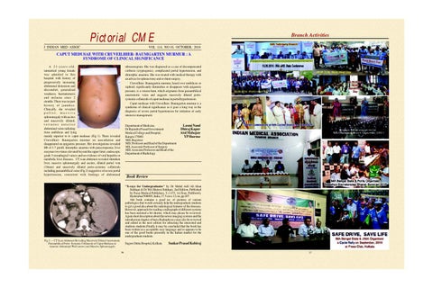
Branch Activities
Pictorial CME J INDIAN MED ASSOC
VOL 114, NO 10, OCTOBER, 2016
CAPUT MEDUSAE WITH CRUVEILHIER- BAUMGARTEN MURMUR : A SYNDROME OF CLINICAL SIGNIFICANCE A 21-years-old, unmarried young female was admitted to this hospital with history of progressively increasing abdominal distension and discomfort, generalised weakness, haematemesis and melaena since 2 months. There was no past history of jaundice. Clinically, she revealed pallor, massive splenomegaly with ascites and massively dilated, tortuous anterior abdominal veins radiating from umbilicus and lying mainly superior to it- caput medusae (Fig 1). There revealed Cruveilhier- Baumgarten murmur on auscultation and disappeared on epigastric pressure. Her investigations revealed Hb of 3.7 gm/dl, dimorphic anaemia with pancytopaenia, liver enzymes two-times elevated beyond the upper limit, endoscopic grade 3 oesophageal varices and no evidence of viral hepatitis or metabolic liver diseases. CT scan abdomen revealed shrunken liver, massive splenomegaly and ascites, dilated portal vein (18mm) and massively dilated porto-systemic collaterals including paraumbilical veins (Fig 2) suggestive of severe portal hypertension, consistent with findings of abdominal
ultrasonogram. She was diagnosed as a case of decompensated cirrhosis (cryptogenic), complicated portal hypertension, and dimorphic anaemia. She was treated with medical therapy with an advice for splenectomy and or shunt surgery. Cruveilhier- Baumgarten murmur, heard over umbilicus or xiphoid, significantly diminishes or disappears with epigastric pressure, is a venous hum, which originates from paraumbilical anastomotic veins and suggests massively dilated portosystemic collaterals of caput medusae in portal hypertension. Caput medusae with Cruveilhier- Baumgarten murmur is a syndrome of clinical significance as it goes a long way in the diagnosis of severe portal hypertension for initiation of early intensive management.
Department of Medicine, Dr Rajendra Prasad Government Medical College and Hospital, Kangra 176001 MD, Registrar MD, Professor and Head of the Department MS, Associate Professor of Surgery MD, Associate Professor and Head of the Department of Radiology
Laxmi Nand Dhiraj Kapur Atul Mahajan YP Sharma
2
3
4
1
2
3
4
Book Review "X-rays for Undergraduates" by Dr Mohd Adil Ali khan Siddiqui & Dr MA Mateen Siddiqui, 2nd Edition, Published by Paras Medical Publishers, 5-1-475, 1st floor, Putlibowli, Hyderabad 500095, India, 17.5 cm x 12 cm, pp 207. THE book contains a good no. of pictures of various pathologies that would certainly help the undergraduate students to get a good idea about the radiological features of the diseases. However, approach for reading a radiograph of different systems has been narrated a bit shorter, which may please be reviewed. Again short description about the newer imaging systems and the introduction chapter of basic Radiophysics may also be reviewed and edited in the next edition for attracting the interested and studious students.Finally it may be concluded that the book has been written in a acceptable easy language and so appears to be one of the good books presently in the Indian market for the undergraduate students.
Fig 2 — CT Scan Abdomen Revealing Massively Dilated Anastomotic Paraumbilical Porto- Systemic Collaterals of Caput Medusae in Anterior Abdominal Wall (arrow) and Massive Splenomegaly
Sagore Dutta Hospital, Kolkata 36
Sankar Prasad Kabiraj 37