


PROF. TAMONAS CHAUDHURI Hony. Editor
MBBS,
MS, FAIS, FMAS, FACS, FACRSI (Hony)

PROF. TAMONAS CHAUDHURI Hony. Editor
MBBS,
MS, FAIS, FMAS, FACS, FACRSI (Hony)
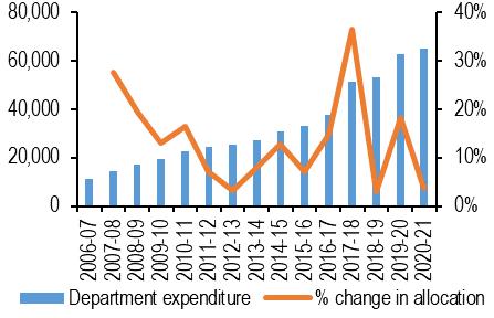
The development of a nation was once measured by the per capita income. Its growth suggested that the country is progressing. The per capita income of a nation is calculated by dividing the Gross Domestic Product (GDP) of a country with population. But in a developing country like India where inequitable distribution of wealth is almost an axiom, the growth in GDP is only enjoyed by the meager percentage of the population. Thus the present school of economics has introduced the Quality of Life Index as the measure of wellbeing of all the citizens of a country from the capitalist to the proletariat class. The definition of the Physical Quality of Life Index (PQLI) is the measure of the quality of life or well-being of a country. The value is the average of three statistics: basic literacy rate, infant mortality rate, and life expectancy at age one. Now if we consider the QOL of various countries in the year 2021, Canada stands with 159.99. US stands with 166.98, Australia stands with 181.52 where as India stands with 104.52, a very discouraging and a staggering figure. The Niti Aayag Member (Health) has confessed that “India’s overall spending on the health sector is low and the situation must be corrected”1. Government in a democratic setup is for the people of the people and by the people. The statement however may seem to be a myth if we focus on the percentage of GDP spent by the government of India on health expenditure. India stands at 170 out of 188 countries in domestic general government health expenditure as a percentage of GDP, according to the Global Health Expenditure database 2016 of World Health Organization 2. The public expenditure on health as a percentage of GDP for 2017-18 was a mere 1.28 of GDP3, 4. (Fig-1) The government aims to raise the public health expenditure to 2.5 per cent of GDP at around 20255 . Let us compare the figure with the developed nations—According to the statistics presented on health care expenditure as part of GDP was for US 8.5 per cent, China 3.2 per cent, Germany 9.4 per cent6. This shows that even the targeted percentage in future falls far short of the expectations. The expenditure budget on medical research has not increased significantly over the past few years. The expenditure budget for research was Rs 1727 crore in financial year 2019, which has grown to Rs 2100 crore in financial year 2021, according to the Budget documents6
My point of discussion however is not to lay down statistical facts and figures but to get into the shoes of the common man and Health Care Professional and feel their pulse of urgency and thereafter voice their needs of the time. As we are well aware, India is still dominated by the proletariat class whose meager income scarcely enable them to meet their two ends meet leave alone expenditure on health, thus their lives need more to be ensured and not insured. Insurance companies are mere business players trying to reap the maximum profit out of their investments then how could they be entrusted with the responsibility of massive development of health care in the secondary and tertiary health strata. Let me place before you a statistic which will certainly leave us petrified. In 2017, Indians
Vol 119, No 2, February 2021Journal
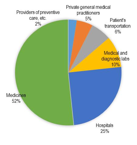
expended around sixty-two percent of their total health expenditure as out-of-pocket expenditure being paid directly to the service providers7, 8 . (Fig-2) India ranks as one of the poorest in terms of this indicator and common people incurred heavy expenses as a reason of health care. Out of pocket expenditure on health is one of the biggest reasons for people falling into poverty in India. It is only the government which can intervene in this matter with the sole service motive to develop a healthy India in future where nobody is deprived of health services. To me, the government needs to take care of other avenues of development also as education, nutrition, clean potable drinking water, housing, sanitization, clean air et al. thus providing mere health cards is not enough. If the resources of the government get diverted as payments in the form of insurance to the private health care then they would be left with less funds to develop the primary, secondary and tertiary health care networks. The recent pandemic has made us realize the importance of a super and actively functioning synchronized network of the primary secondary and tertiary network along with people’s participation. Private Insurance companies have no role in here to set up such a network as they are mere business houses thriving on the failing health of their clients.
The nexus between private healthcare institutions and the insurance companies is also satanic. Following the PPN (Preferred Provider Network) model the tie up between the two is malicious. And Doctors have no role to play except to accept the burden. Under pressure of the Insurance Company and Private Health Care management to reduce the cost of treatment there is possibility of substandard care which is deterrent to the interest of the patients. Again the doctors are losing their medical autonomy to serve the patients ethically. Thus in spite of expending high out of pocket expenditure the patients are not getting best benefits at times when they need them the most. It can thus the inferred that doctors voice must be recorded at the time of creation of such understandings.
Again being driven solely by profit motives the insurance houses are least interested in setting up research and development wings for further advancement of medical care. This can only be achieved with active government intervention.
As a consequence of the liberalization and privatization of health care system, the health care expenses have also sky rocketed since the ending phase of the
20 th century. Health expenditures have surged at 14% and this growth is higher for in-patient care. Here the government must step in as the savior.
The recommendation is to spend at least two third of Government’s health expenditure on primary healthcare, in addition to setting a target to reduce the proportion of households facing catastrophic health expenditure from the current levels by 25 percent by 20255. These efforts on behalf of the Government have enough reasons for us to be optimistic.
Now the certain policies of the government become questionable here. Do the citizens of India demand from the government only health coverage schemes to tide over financial challenges during health emergencies or do they look up to the government for better medical infrastructure and facilities and quality insurance packages from the public sector at affordable premiums? The doctor-population ratio in India is 1:1456 against the WHO recommendation of 1:1000. According to the data of 2018 there has been a short fall of doctors by 46% and specialists by 82%9. Poor working conditions, poor remuneration and procedural delay in recruitments are prime cause of shortfall in government run medical centres be it secondary or tertiary. Oodles of such data can be produced to show that lot remains to be done for up gradation of the health sector and mere insurance policies are not the solutions.
However, till now the public sector has major players in Insurance like the Life Insurance Corporation (LIC), The General Insurance Corporation of India which has four subsidiary companies as: National Insurance Company Limited (NIC); New India Assurance Company Limited (NIA); Oriental Insurance Company Limited (OIC); and United India Insurance Company Limited (UIIC). They have their wide array of policies to cover the potential health hazards. Why don’t we revamp and widen the facilities of these
sectors so that the common man finds solace in investing in these sectors. This, along with sustained strengthening of primary healthcare system and steady growth in medical infrastructure which includes better trained medical staffs in adequate numbers with
state of art technology will certainly bring about a health renaissance in India. Let us all hope that India will rapidly achieve this health revolution with a little bit more of cognitive and pragmatic efforts from the Health Policy makers.
1(2020). ‘In 2018-19, India’s spending on health sector was 1.5% of GDP’, The Hindu, 19 November.
2https://www.financialexpress.com/economy/india-spending-more-onhealthcare-now-but-yet-not-as-much-as-others-heres-how-much-uschina-spend/1922253/
3Economic Survey, 2019-20, Ministry of Finance, https:// www.indiabudget.gov.in/economicsurvey/doc/vol2chapter/ echap10_vol2.pdf
4Domestic General Government Health Expenditure (% of General Government Expenditure) World Health Organization Global Health Expenditure Database (https://apps.who.int/nha/database)
5National Health Policy 2017, Ministry of Health and Family Welfare, Government of India. https://www.nhp.gov.in/nhpfiles/ national_health_policy_2017.pdf
6Demand for Grants 2020-21 Analysis: Health and Family Welfare, Government of India. https://www.prsindia.org/parliamenttrack/budgets/ demand-grants-2020-21-analysis-health-and-family-welfare
7Share of Out of Pocket Health Expenditure India2001-2017, Statista Research Department, October 16th, 2020. https://www.statista.com/ statistics/1080141/india-out-of-pocket-expenditure-share-in-totalhealthcare-expenditure/
8Out of pocket expenditure (% of current health expenditure). World Health organization Global Health Expenditure Database (apps.who.int/ nha/database)
9Rural Health Statistics 2018, Health Management Information Systems, Ministry of Health and Family Welfare. main.nohfw.gov.in
Sukanta Dutta1, Partha Pratim Chakraborty2, Sugata Narayan Biswas3, Krishnendu Roy4
Purpose of The Study : A plethora of conditions are associated with multiple pituitary hormone deficiency (MPHD). Aetiologies and clinical spectrum of MPHD in the developing countries are varied and quite different from that in the West. Tumours of the hypothalamo-pituitary (HP) region are the dominant cause of MPHD in the tertiary care referral centres, not only in the Western World, but also in India. There is real paucity of information regarding clinical profile and aetiology of MPHD from rural India.
Methods : We analysed the presenting manifestations, hormonal parameters and imaging abnormalities in all patients of MPHD (n=53), evaluated in the Department of Medicine, Midnapore Medical College & Hospital between January 2016 to December 2018.
Findings : Hypogonadism was the most common (54.7%) clinical manifestation of hypopituitarism in this study. Repeated hospitalization with spontaneous hypoglycaemia, recurrent hyponatremia and refractory anaemia were also common in this cohort. Vasculotoxic snake (viper) bite (VSB) was the commonest aetiology (30.2%) of MPHD overall, while Sheehan’s syndrome (SS) dominated in females. Patients of VSB were exclusively male, with the youngest one being 13-year old. Majority of these patients (87.5%) underwent several sessions of haemodialysis following the bite. 61.5% of SS delivered at home, and almost 92% had had severe post-partum haemorrhage requiring transfusion support. All patients of VSB had hypogonadism and hypoadrenalism, while all but one case of SS had cortis ol deficiency. Agalactia was universal in SS. Empty sella on magnetic resonance imaging (MRI) was the dominant abnormality (52.8%) encountered, while 3 patients had normal MRI findings.
Implications : Hypopituitarism is often unrecognized; hence remains untreated, as primary care physicians are unaware of the varying clinical manifestations of MPHD. They need to be sensitised to have a high index of suspicion in appropriate clinical settings.
[J Indian Med Assoc 2021; 119(2): 13-8]
Key words :Multiple pituitary hormone deficiency, Hypopituitarism, Vasculotoxic snake (viper) bite, Sheehan’s syndrome, Hypophysitis
Multiple pituitary hormone deficiency (MPHD), also termed combined pituitary hormone deficiency (CPHD), is used to describe the condition associated with deficiency of more than one hormone, produced from the pituitary gland. Impaired pituitary hormone production, often described as hypopituitarism, results from a plethora of acquired and inherited conditions, and has an approximate global prevalence of 1 in 22001. The aetiology of MPHD varies greatly not only between countries, but also among centres within a country, due to referral bias, age of the participants and sex-ratio of the study population. Majority of studies, particularly those from the Western World documented an unequivocal predominance of tumours in the hypothalamo-pituitary (HP) region
1MBBS, Junior Resident, Department of Medicine, Midnapore Medical College, Midnapore 721102
2MBBS, MD, DM, DNB, MNAMS, FACE, FICP, Clinical Tutor, Department of Endocrinology & Metabolism, Medical College, Kolkata 700073 and Corresponding Author
3MBBS, MD, Senior Resident, Department of Gastroenterology, Sanjay Gandhi Postgraduate Institute of Medical Sciences, Lucknow 222001
4MBBS, MD, Professor, Department of Medicine, Midnapore Medical College, Paschim Medinipur 721102
Received on : 03/11/2020
Accepted on : 05/01/2021
Editor's Comment :
In rural West Bengal, VSB and SS are commonest cause of hypopituitarism in males and females, respectively.
Hypogonadism, spontaneous hypoglycaemia, recurrent hyponatremia, and refractory anaemia are the dominant clinical manifestations in these patients.
Hypoadrenalism due to secondary adrenal insufficiency is almost universal in hypopituitarism following VSB or SS.
Acute kidney injury necessitating haemodialysis and post-partum haemorrhage are the predictors of future hypopituitarism in VSB and SS, respectively.
Majority of the primary care physicians are unaware of this entity, and the condition remains undiagnosed with resultant morbidity, poor quality of life and excess mortality.
as the underlying cause of MPHD. In the Dutch National Registry, one of the largest databases of adult onset growth hormone deficiency (GHD), the dominant causes of MPHD were pituitary tumours and/or their treatment and craniopharyngiomas, involving ~60% of patients 2. Similar observation was noted in adult onset hypopituitarism database from Serbia (n=512; tumours: 61%) and Spain (n=209; tumours: 55%) 3,4 . There are only a handful of population-based studies related to the aetiology of MPHD from our country. A single-centre study (n=113) from North India has also found tumours in overwhelming majority (84%) of patients with
119, No 2, February 2021Journal
hypopituitarism attending a tertiary clinic5. In contrast, a study conducted at a tertiary care hospital in Eastern India noted tumours responsible for 45% cases of adult onset hypopituitarism, highlighting the centre-specific differences in aetiology6
Clinical manifestations of MPHD depend upon several factors like underlying aetiology, number of pituitary cell lines affected, magnitude of hormone deficiency, and rapidity of onset and disease progression. Symptoms and signs of hypopituitarism, particularly early in the course of disease are non-specific and often subtle, resulting in delay in diagnosis and appropriate management. Adrenocorticotrophic hormone (ACTH) deficiency makes them vulnerable, particularly during acute stress and intercurrent illnesses7. Patients with undiagnosed, hence untreated MPHD, have a poor quality of life and are at increased risk of mortality both in short term and due to cardiovascular disease in the long run. Untreated gonadotropin deficiency has also been identified as an independent risk factor for excess mortality8,9. This is particularly important in countries like India, where Sheehan’s syndrome (SS) and vasculotoxic snake (viper) bite (VSB), conditions often associated with combined ACTH and gonadotropin deficiencies, contribute to a sizable portion of cases with hypopituitarism6. The adverse consequences of hypopituitarism can be mitigated to a large extent by early diagnosis and appropriate management. We conducted this study to look for clinical manifestations and underlying aetiologies of MPHD in a group of population from relatively poor socio-economic stratum, residing in a region of India with limited access to tertiary health care facility.
MATERIALS AND METHODS
In this hospital record based retrospective study, data of 53 patients with a confirmed diagnosis of MPHD, either attending the Endocrinology clinic or discharged from the in-patient department (IPD) of Department of Medicine, Midnapore Medical College & Hospital between January 2016 to December 2018 were analysed. The study was approved by the Institutional Ethical Committee (Memo No. MMC/IEC-2019/193 dated 28/01/2019). We looked into the different aetiologies of hypopituitarism and characterized the clinical presentation and biochemical profile of these patients. Clinical presentations, relevant information including number of prior hospitalization, traumatic brain injury (TBI), history of VSB, acute kidney injury (AKI) after VSB, number of haemodialysis (HD) sessions, history of cranial surgery and/or radiation, history of stroke were noted for all. Female participants were also evaluated for menstrual history, childbirth, post-partum haemorrhage (PPH), blood transfusion (BT) and agalactia.
Cortisol, free thyroxine (FT4), thyroid stimulating hormone (TSH), prolactin, and insulin like growth factor 1 (IGF1) were measured in all patients from serum samples collected between 8:00-9:00 AM after an overnight fast. Patients with AM cortisol between 5-14.5 µg/dl underwent ACTH stimulation. Due to nonavailability of Synacthen (tetracosactide acetate), we used twentyfive units of Acton Prolongatum® (Ferring pharmaceuticals, available as 5 ml vial with concentration of 60 units/ml) injected intramuscularly,
and sample was collected after 60 minutes; a cortisol concentration of less than 18 µg/dl was considered as hypocortisolism10. Serum testosterone was measured only in males after the age of 14 years from samples collected in fasting state between 8:00-9:00 AM. FSH was measured in males with low testosterone, and in females after 13 years of age with history of oligo-amenorrhoea. Polyethylene glycol (PEG) precipitation test was performed in cases with high prolactin.
All hormonal assays were performed by chemiluminescent immunoassay (CLIA). Cortisol, FT4 and TSH assays were performed in ADVIA Centaur CP system (SIEMENS). Testosterone, IGF1, FSH, and prolactin were measured in cobas e 411 analyser (Roche Diagnostics). Magnetic resonance imaging (MRI) of the HP region was performed using SIGNA Explorer, 60 cm 1.5T system (GE Healthcare).
Hypophysitis was diagnosed based on the radiological score proposed by Gutenberg et. al. that showed a sensitivity of 92%, a specificity of 99%, a positive predictive value of 97%, and a negative predictive value of 97% for the diagnosis of autoimmune hypophysitis11. All such patients underwent routine cerebrospinal fluid (CSF) analysis, and estimation of adenosine deaminase (ADA), angiotensin-converting enzyme (ACE), β -human chorionic gonadotropin (hCG) and α-fetoprotein (AFP) in CSF in addition to measurement of serum immunoglobulin G4 (IgG4) level. Idiopathic intracranial hypertension (IIH) was suspected in presence of empty sella (ES), dilated optic nerve sheaths, protrusion of optic nerve papilla into the posterior globes, and compressed ventricles and CSF cisterns on MRI 12
OBSERVATIONS
The study cohort consisted of 53 patients with male (n=31) to female (n=22) ratio of 1.4. The mean age of the study population was 40.5 (±14.6) years (range: 7-65 years). 6 of them (11.3%) belonged to an age group below 18 years, and 47 subjects (88.7%) were of more than 18 years of age. The clinical presentations have been summarized in Table 1.
The most common clinical feature in this population was loss of secondary sexual characters (54.7%), elicited from history and/or clinical examination, performed at presentation. Symptoms, which were potentially life-threating, like severe hypoglycaemia (second in order) and recurrent hyponatremia (fifth in order), requiring repeated hospitalisation were observed in 34% and 22.6% of the cohort, respectively. Another 4 (7.5%) patients had history of frequent hospitalisation only for BT. Overall, 31 patients (58.5%) had previous episodes of hospitalization (<5 times: 18 patients; 510 times: 10 patients; >10 times: 3 patients); however, the diagnosis of MPHD had been missed during those occasions.
In our study, 16 subjects (30.2%) (all of them were male, youngest one was of 13 years of age) had history of VSB in the past, and 14 out of these 16 subjects (87.5%) required HD due to AKI (Table 2). 17 female subjects (32% of the entire cohort and 77.3% of the total female population) had secondary amenorrhoea, while another 12 (54.5% of the female population) had agalactia
with secondary amenorrhea since last childbirth. Only one lady had agalactia, but with preserved menstrual cycles. Out of 13 patients of SS, definite diagnosis was made within 5 years of the responsible obstetric event in 1 (7.7%), between 5-10 years in 4 (30.8%), 10-20 years in 4 (30.8%) and more than 20 years in another 4 (30.8%) patients.
The patient with acromegaly had haemoglobin (Hb) level of 20.1 gm/dl secondary to polycythaemia. The mean Hb concentration in the rest of the cohort was 9.9 (±1.4) gm/dl. One patient had pancytopenia and another one had β -thalassemia major. Hyponatremia (<135 mmol/L) was observed in 24 (45.3%) and hypokalemia (<3.5 mmol/L) in 3 subjects (5.7%). Low FT4 (<0.8 ng/dl) was encountered in 32 patients (60.4%), and 13 of them (40.6% of those with low FT4) had TSH below reference range (<0.5 µIU/ml). Low AM cortisol (<5 µg/dl) was seen in 44 patients (83%). Peak cortisol of more than 18 µg/dl following ACTH injection was noticed only in 7 patients. All patients of VSB, hypophysitis, and all but one patient of SS had ACTH deficiency, either low AM cortisol or failed ACTH stimulation test. The hormonal parameters have been summarized in Tables 3&4. Among 29 male patients, 28 (96.6%) had low testosterone (<300 ng/dl). The median value of testosterone was 43 ng/dl. The median value of FSH was 2.65 mIU/ml, consistent with the diagnosis of hypogonadotropic hypogonadism. Only 6 patients had (11.3%) elevated monomeric prolactin and the median value of serum prolactin was 5.18 ng/ml. IGF1 was below age and sex specific reference ranges in 39 patients (73.6%) while one patient had elevated IGF1. Median value of serum IGF1 was 73.85 ng/ml.
Majority of the patients had abnormal appearances of the HP region on MRI (Table 5) (Figs 1&2). Empty sella with varying thickness of the remaining pituitary tissue was the commonest abnormality noted (28 patients, 52.8% of the cohort). Only 3 patients (5.66%) had apparently normal looking HP region. Hypophysitis was diagnosed retrospectively in 1 lady with ES, whose previous MRI was consistent with hypophysitis11. One male with hypophysitis
presented with diabetes insipidus. Pituitary transcription factor defect was considered in 3 subjects with MPHD presenting during childhood and adolescence periods. The commonest aetiology of MPHD in our study was VSB (Table 6).
DISCUSSION
Table 2 — Characteristic of patients with viper bite
History of haemodialysis14 Interval between bite and diagnosis of MPHD (n=16) <1 year2 (12.5) 1-5 years3(18.8) 5-10 years9 (56.3) >10 years2 (12.5)
Tumours dominated over other causes like cranial irradiation, traumatic brain injury, SS and lymphocytic hypophysitis in 7708 patients of adult onset GHD (as a component of MPHD) in the KIMS database13. However, a difference in aetiology was noted amongst the six largest contributing countries. The Pituitary Study Group of the Society of Endocrinology and Metabolism of Turkey (SEMT) looked into the aetiology of hypopituitarism in 773 patients aged between 16 to 84 years, in nineteen tertiary care institutions. Though non-tumoral causes dominated overall (49.2%), a significant gender deference was noted, and SS contributed to majority of females14. The dominant causes of MPHD in our study were VSB (30.2%), tumours (de novo and post-treatment) (26.4%), and SS (24.5%). VSB and SS were encountered exclusively in males and females, respectively. Tumours contributed to ~26% cases (n=14) of MPHD (de novo pituitary macroadenoma:9; post-operative cases of pituitary macroadenoma: 3; craniopharyn-gioma: 1; hypothalamic mass: 1). Of the 9 newly diagnosed cases of pituitary macroadenoma, seven patients had non-functional pituitary adenoma, one had acromegaly and the other one had macroprolactinoma. A study from our part of the country reported SS in 27% and VSB in 14.6% cases of adult with MPHD6
Number of haemodialysis sessions (n=14) <54 (28.6) 5-107 (50%)
Table 4 — Pituitary hormone deficiency in different aetiologies of MPHD (n=53) LowLowLowLow cortisolFT4 IGF1 testosterone (n=46)* (n=32)(n=39) (n=28)**
Viper bite (n=16)16121116
Sheehan’s syndrome (n=13)121010
Pituitary macroadenoma (n=9)6478
Sequel of pituitary surgery (n=3)3221
Craniopharyngioma (n=1)1011
Hypothalamic mass (n=1)111
Hypophysitis (n=4)4211
Transcription factor defect (n=3)2131
Idiopathic intra-cranial hypertension (n=1)001
Idiopathic hypopituitarism (n=1)101
β-thalassemia major (n=1)001
*Low cortisol includes AM cortisol <5 µg/dl or post-ACTH cortisol <18 µg/dl
**Only in males above 14 years of age
Viper bite (n=16) Partial empty sella: 14; hypoplastic pituitary: 1; normal HP region: 1
Sheehan’s syndrome (n=13) Partial empty sella: 12; hypoplastic pituitary: 1
Pituitary macroadenoma (n=9) Sellar mass with/without supra & parasellar extension and heterogenous contrast enhancement
Sequel of pituitary Enlarged sella with residual surgery (n=3) tissue and fibrotic changes with deviation of stalk: 3
Craniopharyngioma (n=1) Sellar and suprasellar mass with mixed solid and cystic components and calcification
Hypothalamic mass (n=1) Isointense mass in T1 weighted MRI with intense contrast enhancement
Hypophysitis (n=4)
Transcription factor
Diffuse enlargement of the pituitary with stalk thickening and rapid, intense, and homogenous contrast enhancement:3; partial empty sella: 1
Hypoplastic pituitary: 3; Absent defect (n=3)pituitary stalk & ectopic posterior pituitary bright spot: 1
Idiopathic intra-cranial Partial empty sella with protruded hypertension (n=1) optic nerve head inside orbit and dilated optic nerve
Idiopathic hypopituitarism (n=1) Normal HP region
β-thalassemia major (n=1) Normal HP region
Aetiology of MPHD varies depending on age of the study population. In those 6 patients of our study, aged less than 18 years of age, 3 had transcription factor defects, and craniopharyngioma (n=1), hypothalamic mass (n=1) and VSB contributed to MPHD in the remaining 3. Conditions, which rarely contribute to MPHD in the western world, often are the dominant causes of pituitary dysfunction in the developing countries. The list includes VSB, SS and central nervous system (CNS) infections15. Hypopituitarism following VSB is common (61%) in patients who develop AKI requiring HD. Increasing number of sessions of HD confers higher risk and cortisol deficiency is the commonest abnormality encountered. Hypopituitarism has been reported as early as 7 days following snake bite 16. Our findings were consistent with this study as majority of our patients required more than 5 sessions of HD. One of our patients developed severe hypoglycaemia due to hypopituitarism on 5th day after viper bite. SS is quite prevalent in rural India. The prevalence of SS is about 3% in women above 20 years of age residing in Kashmir valley; and almost two-third of them delivered at home17 . The diagnosis is often delayed, and the average time lag between the culpable obstetric event and diagnosis was found to be 13 years18. Definitive diagnosis of SS was reached after 10 years in 8 (61.5%) of our patients. 12 out of 13 patients of SS (92.3%) had history of PPH, and all of them required BT suggesting severe blood loss. 8 (61.5%) of these ladies had delivered at home. PPH has been reported in 82-100% of patients with SS18,19. Frequency of agalactia
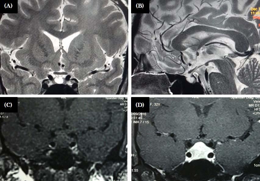
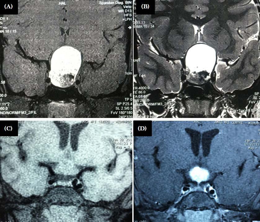
Fig 2 — Upper panel: MRI of patients with craniopharyngioma. Mixed solid-cystic lesion with cystic component being hyperintense in both T1 (A) and T2 (B) weighted sequences
Lower panel: Patient with hypothalamic mass, that is isointense in T1 (C) with intense contrast enhancement (D)
in SS has variably been reported in about 40-67% in studies outside India18,20. However, agalactia is much higher in our country and, was seen in 100% of our patients with SS, a finding consistent with another study published from the same geographical region19 One patient of SS had preserved gonadotroph functions, as evidenced by cyclic predictable menses, and she conceived spontaneously during follow-up. Spontaneous pregnancy in SS, though rare, has been reported in literature21
Hypopituitarism is a frequently overlooked cause of severe hyponatremia (<130 mmol/L). The prevalence of hypopituitarism in patients with “euvolemic severe hyponatremia” is 20% in the
6 — Aetiology of MPHD in the study population (n=53)
Idiopathic intra-cranial hypertension11.9
Western World, and much higher in India22. 22.6% of our patients had been admitted with recurrent severe hyponatremia, and the diagnosis of MPHD was never considered. Another interesting observation we came across was refractory anaemia (in 4 patients) and pancytopenia (in 1 patient) in our cohort. Deficiency of thyroid hormone, testosterone, IGF1, and perhaps cortisol either in isolation or in combination have been found to impair haematopoiesis23 Refractory anaemia and pancytopenia have been reported in MPHD, SS in particular and are completely reversible after supplementing the deficient hormone(s)24,25
The patient with pancytopenia in our study had SS and the 4 patients with refractory anaemia had hypopituitarism due to VSB. We came across one patient with polycythaemia secondary to GH secreting pituitary macroadenoma26.
In rare patients with MPHD, no obvious aetiology of hypopituitarism could be identified and such patients have normal morphology of the HP region on MRI. In one of the retrospective analyses (n=230), 21 patients (9%) with hypopituitarism had normal pituitary imaging27 . Despite extensive work-up definitive diagnosis remained elusive in 2. We came across one such patient in our study, and we used the term idiopathic hypopituitarism for that patient.
To conclude, aetiology of MPHD demonstrate a strong geographical variation and referral bias. In regions with poor obstetric care, SS undoubtedly is the leading cause of hypopituitarism in females, while in rural West Bengal, VSB contributes significantly to MPHD in males. Recurrent hyponatremia, unexplained hypoglycaemia, frequent BT are often the presenting manifestations of MPHD in non-tertiary care centres. Many of these patients remain unrecognized, due to lack of awareness among primary care physicians, rendering them to repeated hospitalization, poor quality of life and high risk of mortality.
Funding : None
Conflict of Interest : None REFERENCES
1Matsumoto AM, Anawalt BD — Testicular disorders. In Melmed S, Auchus RJ, Goldfine AB, Koenig RJ, Rosen CJ eds William Textbook of Endocrinology 14th edn. Elsevier, Philadelphia; 2020: 668-755.
2van Nieuwpoort IC, van Bunderen CC, Arwert LI, Franken AA, Koppeschaar HP, van der Lelij AJ, et al — Dutch National Registry of GH Treatment in Adults: patient characteristics and diagnostic test procedures. Eur J Endocrinol 2011; 164(4): 491-7.
3Fernandez-Rodriguez E, Lopez-Raton M, Andujar P, Martinez-Silva IM, Cadarso-Suarez C, Casanueva FF, et al — Epidemiology, mortality
Vol 119, No 2, February 2021Journal of the Indian Medical Association
rate and survival in a homogeneous population of hypopituitary patients. Clin Endocrinol (Oxf) 2013; 78(2): 278-84.
4Dokniæ M, Pekiæ S, Miljiæ D, Soldatoviæ I, Popoviæ V, Stojanoviæ M, et al — Etiology of Hypopituitarism in Adult Patients: The Experience of a Single Center Database in the Serbian Population. Int J Endocrinol 2017; 2017: 6969286.
5Gundgurthi A, Garg MK, Bhardwaj R, Brar KS, Kharb S, Pandit A — Clinical spectrum of hypopituitarism in India: A single center experience. Indian J Endocrinol Metab 2012; 16(5): 803-8.
6Chatterjee P, Mukhopadhyay P, Pandit K, Roychowdhury B, Sarkar D, Mukherjee S, et al — Profile of hypopituitarism in a tertiary care hospital of eastern India—is quality of life different in patients with growth hormone deficiency? J Indian Med Assoc 2008; 106(6): 384-5, 388.
7Burman P, Mattsson AF, Johannsson G, Höybye C, Holmer H, Dahlqvist P, et al — Deaths among adult patients with hypopituitarism: hypocortisolism during acute stress, and de novo malignant brain tumors contribute to an increased mortality. J Clin Endocrinol Metab 2013; 98(4): 1466-75.
8Tomlinson JW, Holden N, Hills RK, Wheatley K, Clayton RN, Bates AS, et al — Association between premature mortality and hypopituitarism. West Midlands Prospective Hypopituitary Study Group. Lancet 2001; 357(9254): 425-31.
9Sherlock M, Ayuk J, Tomlinson JW, Toogood AA, Aragon-Alonso A, Sheppard MC, et al — Mortality in patients with pituitary disease. Endocr Rev 2010; 31(3): 301-42.
10Gundgurthi A, Garg MK, Dutta MK, Pakhetra R— Intramuscular ACTH stimulation test for assessment of adrenal function. J Assoc Physicians India 2013; 61(5): 320-4.
11Gutenberg A, Larsen J, Lupi I, Rohde V, Caturegli P — A radiologic score to distinguish autoimmune hypophysitis from nonsecreting pituitary adenoma preoperatively. Am J Neuroradiol 2009; 30(9): 1766-72.
12Salzman KL, Osborn AG — Sellar Neoplasma and Tumor-like Lesions. In Osborn AG, Hedlund GL, Salzman KL, Concannon KE, Gelsinger AG, Marmorstone JJ et al. eds Osborn’s Brain Imaging, Pathology and Anatomy. 2nd edn. Elsevier, Philadelphia; 2018: 771-818.
13Brabant G, Poll EM, Jönsson P, Polydorou D, Kreitschmann-Andermahr I — Etiology, baseline characteristics, and biochemical diagnosis of GH deficiency in the adult: are there regional variations? Eur J Endocrinol 2009; 161 Suppl 1: S25-31.
14Tanriverdi F, Dokmetas HS, Kebapcý N, Kilicli F, Atmaca H, Yarman S, et al — Etiology of hypopituitarism in tertiary care institutions in Turkish population: analysis of 773 patients from Pituitary Study Group database. Endocrine 2014; 47(1): 198-205.
15Beatrice AM, Selvan C, Mukhopadhyay S — Pituitary dysfunction in infective brain diseases. Indian J Endocrinol Metab 2013; 17(Suppl 3): S608-S611.
16Bhat S, Mukhopadhyay P, Raychaudhury A, Chowdhury S, Ghosh S — Predictors of hypopituitarism due to vasculotoxic snake bite with acute kidney injury. Pituitary 2019; 22(6): 594-600.
17Zargar AH, Singh B, Laway BA, Masoodi SR, Wani AI, Bashir MI — Fertil Steril 2005; 84(2): 523-8.
18Gei-Guardia O, Soto-Herrera E, Gei-Brealey A, Chen-Ku CH — Sheehan syndrome in Costa Rica: clinical experience with 60 cases. Endocr Pract 2011; 17(3): 337-44.
19Mandal S, Mukhopadhyay P, Banerjee M, Ghosh S — Clinical, endocrine, metabolic profile, and bone health in Sheehan’s syndrome. Indian J Endocr Metab 2020; 24: 338-42.
20Diri H, Tanriverdi F, Karaca Z, Senol S, Unluhizarci K, Durak AC, Atmaca H, Kelestimur F — Extensive investigation of 114 patients with Sheehan’s syndrome: a continuing disorder. Eur J Endocrinol 2014; 171(3): 311-8.
21Zargar AH, Masoodi SR, Laway BA, Sofi FA, Wani AI — Pregnancy in Sheehan’s syndrome: a report of three cases. J Assoc Physicians India 1998; 46(5): 476-8.
22Diederich S, Franzen NF, Bähr V, Oelkers W — Severe hyponatremia due to hypopituitarism with adrenal insufficiency: report on 28 cases. Eur J Endocrinol 2003; 148(6): 609-17.
23Laway BA, Bhat JR, Mir SA, Khan RS, Lone MI, Zargar AH — Sheehan’s syndrome with pancytopenia—complete recovery after hormone replacement (case series with review). Ann Hematol 2010; 89(3): 3058.
24Smith WH — Hypopituitarism a cause of refractory anaemia. Med J Aust 1960; 47(1): 1022-5.
25Lang D, Mead JS, Sykes DB — Hormones and the bone marrow: panhypopituitarism and pancytopenia in a man with a pituitary adenoma. J Gen Intern Med. 2015; 30(5): 692-96.
26Patra S, Biswas SN, Datta J, Chakraborty PP — Hypersomatotropism induced secondary polycythaemia leading to spontaneous pituitary apoplexy resulting in cure of acromegaly and remission of polycythaemia: ‘The virtuous circle’. BMJ Case Rep 2017 Dec 7; 2017:bcr2017222669.
27Wilson V, Mallipedhi A, Stephens JW, Redfern RM, Price DE — The causes of hypopituitarism in the absence of abnormal pituitary imaging. QJM 2014; 107(1): 21-4.
Santanu Saha1, Arijit Singha2, Arindam Mitra3
Background : Gamma Glutamyl Transpeptidase (GGT) is an enzyme of transferases family. Serum GGT is a faithful indicator of alcohol consumption. Studies have suggested that serum GGT is an independent risk factor of stroke.
Method : We undertook a case-control study, enrolling patients admitted to a tertiary care facility to assess the correlation between GGT levels and stroke status.
Result : Serum GGT was significantly higher in the age group 40-60 years (p value <0.05). Similarly, serum GGT level was significantly higher in males (p value <0.05). This was true for 24 hr, 48 hr and 72 hr samples. There was no statistically significant correlation between serum GGT levels and hypertension and dylipidemia status, though interestingly, we observed a significant increase in the level of serum GGT in non-diabetic stroke patients (p value <0.05). There was also no significant correlation observed between serum GGT levels and different types of stroke though there was slightly higher mean GGT value in patients suffering from ischaemic stroke. Logistic regression models suggest a significant correlation between stroke status and GGT levels. The current study establishes the association between elevated GGT levels and stroke status, a finding which needs to b e confirmed through longitudinal investigations.
[ J Indian Med Assoc 2021; 119(2): 19-21]
Key words : Gamma glutamyl transpeptidase, Stroke, Ischemic stroke, Hemorrhagic stroke.
Gamma Glutamyl Transpeptidase (GGT), an enzyme of transferases family, catalyzes the transfer of a gammaglutamyl group of a gamma-glumatyl peptide(such as glutathione) to a peptide or amines acid type acceptor1. Being widely distributed in human organs, it is frequently localized to the plasma membrane with its active site directed into the extracellular space and is mostly concentrated in tubular epithelial cells of kidney2,3. Discovered by Hanes in 19504, it took several years to enter the clinical background when reports of the MALMO study (Sweden, 1975) came to light. Several reports have shown that serum GGT levels are associated with an increased risk of myocardial infarction as an independent predictor and is not merely a marker of alcohol consumption5,6
Meanwhile the association of GGT with diabetes mellitus, obesity, hypertension, atherosclerosis have been established7-9 and colocalization of GGT activity with Low-density lipoprotein (LDL) in human atherosclerotic plaques further established the role of GGT in oxidative stress10
Subsequently, several studies revealed the association between GGT and stroke. In particular, the study byJousilathi P et al concluded that serum GGT was an independent risk factor of stroke;they also explained that answers given by patients to questionnaires concerning their lifestyles are unreliable and serum
1MBBS, MD (Medicine), Associate Professor, Department of Medicine, NRS Medical College, Kolkata 700014 and Corresponding
Author
2MBBS, MD (Medicine), Residential Medical Officer cum Clinical Tutor, Department of Endocrinology & Metabolism, IPGME&R, Kolkata 700020
3MBBS, MD, Postgraduate Trainee, Department of Medicine, NRS Medical College, Kolkata 700014
Received on : 18/04/2018
Accepted on : 19/04/2018
Editor's Comment :
Serum Gamma Glutamyl Transferase (GGT) is an indicator of alcohol consumption.
Studies have suggested that serum GGT is an independent risk factor of stroke.
In our study serum GGT level was significantly higher in stroke patients.There was no significant correlation observed between serum GGT levels and different types of stroke though there was slightly higher mean GGT value in patients suffering from ischemic stroke.
Looking forward for further study, we can say that increased serum GGT level is an independent risk factor of stroke particularly in nonalcoholic male patients.
GGT is a more faithful indicator of alcohol consumption,i.e.the actual determinant of the occurrence of stroke11
As there is a probability of under-reporting of the drinking habits among heavy drinkers, we have investigated the correlation between the stroke status and serum GGT levels inself-reported non-alcoholic stroke patients and their non-strokecontrol counterparts, in both and younger and older age group, while comparing them with members of the control group who did not suffer from an episode of stroke. Further our study evaluated any differences in serum GGT level in acute stroke between the younger and older age group and also between men and women. Lastly we looked into correlation of serum GGT level in acute stroke with the other known risk factors.
This is a cross-sectional, observational, study conducted over one-year (January 2011- January 2012) and incudes stroke and non-stroke patients who were admitted ata tertiary hospital in Kolkata.Stroke was defined according to the WHO stroke manual. We recruited 107 cases (patients suffering an episode of stroke)
and 93 controls (patients admitted with complains other than a stroke) – so a total of 200 individuals were studied.
After matching age and sex we divided the patients into two age group -40-60 yrs (52 stroke and 55 non-stroke) and 60 years and above (48 stroke and 45 non stroke). Patients with a past history of stroke, alcohol consumption, heart failure or drugs that can increase serum GGT level were excluded. All patients underwent the following investigations except computed tomography & carotid doppler which were done only in stroke patients:
•History taking- smoking, DM, hypertension, any drug intake
•Systemic Examination - particularly Central Nervous System (CNS), Cardio Vascular System (CVS) or any hepatomegaly
•Blood pressure is measured according to standard guideline
•Complete haemogram
•Blood sugar- Fasting and 2 hour post-prandial
•Serum cholesterol level - Triglyceride, High-density lipoprotein (HDL), LDL by standard auto analyzer recommended by International Federation of Clinical Chemistry and Laboratory Medicine (IFCC)
•Serum GGT level by kinetic spectrophotometric method in automated analyzer
•Standard LFT, PT
•Serum Na+, K+, Urea, Creatinine
•USG abdomen,Echocardiography and ECG.
We tested serum GGT level after 24 hours,48 hours and 72 hours of admission in stroke patients whereas non-stroke patients had a single serum GGT level after their admission (Tables 1-3).
There were 52 male and 48 female stroke patients and 50 male and 50 female non-stroke patients.There were 52 case and 55 control patients in 40-60 years age group and 48 case and 45 control patients are in 60-80 years age group. Of the 100 stroke patients there were 44 were known to be suffering from diabetes; there are 34 controls who were diabetic. All 78 patients were suffering from type 2 diabetes mellitus, taking oral hypoglycaemic, either regularly or irregularly. Both fasting and post-prandial blood sugars were within normal range in non-diabetic participants. There were 134 hypertensive participants, of whom, 76 were stroke patients and 58 were controls. Of the remaining 66 non-hypertensive patients there are 24 stroke and 42 controls.Of all the participants in the study, only 9 patients, all of whom were in the stroke group,were dyslipidemic. We defined serum triglyceride level >150 mg/dl or serum HDL<40 mg/dl in male and <50 mg/dl in female as dyslipidemia12
Among the 100 stroke patients 53 were diagnosed to be suffering from haemorrhagic strokes, 39 from ischaemic strokes and 8 from subarachnoid haemorrhage.
Serum GGT was significantly higher in the age group 40-60 years (p value<0.05). Similarly, serum GGT level was significantly higher in males (p value <0.05). This was true for 24 hr, 48hr, and
Table 1 — Demographic Profile of Study Participants
Characteristics Case ControlTotal (200) (stroke) non-stroke) (N=100)(N=100)
>60484593
Gender Male5250102 Female485098
SmokerYes423678 No5864122
HypertensiveYes7658134 No244266
DyslipidemicYes909 No91100191
Diabetes mellitusYes443478 No5666122
Table 2 — Type of strokes identified in the study
Type of Stroke Frequency Percent Haemorrhagic stroke5353 Ischaemic stroke3939 Subarachnoid Haemorrhage88
Table 3 — 24-hour GGT levels in case and control participants 24H GGT(U/L)
72 hr samples. There was no statistically significant correlation between serum GGT levels and hypertension and dylipidemia status, though interestingly,we observed a significant increase in the level of serum GGT in non-diabetic stroke patients (p value <0.05). There was also no significant correlation observed between serum GGT levels and different types of stroke though there was slightly higher mean GGT value in patients suffering from ischaemic stroke (Table 4).
According to logistic regression model as there is significant odds ratio and p value is<0.001 it can be concluded that increased serum GGT can independently cause stroke in spite of the presence of other known risk factors (Table 5)
DISCUSSION
From the results of the current study it is obvious that serum GGT level is significantly higher in stroke patients. This observation supports the results of the EUROSTROKE PROJECT by M L Bots et al which has shown an increased GGT is associated with an increased risk of stroke13. As discussed earlier, alcohol consumption
Table 4 — Serum GGT levels in stroke patients
Predictor variables 24H GGT48H GGT72 H GGT Categories (U/L) (U/L) (U/L)
Age in years :
40-60 years63.47+56.3458.88+42.0 357.85+42.44
>60 years 46.21+16.2845.15+16.13 43.75+16.11
P Value 0.0440.0360.033
Gender :
Female
42.03+14.4541.52+13.7939.94+13.64
Male67.33+55.3162.23+41.3961.37+41.73
P Value 0.0030.0010.001
Diabetes mellitus :
No63.93+53.2659.05+39.7757.93+40.09
Yes44.06+19.3243.68+18.1642.36+18.47 P Value 0.0210.0200.019
Hypertension :
No45.21+16.7144.33+16.8743.17+16.50
Yes58.33+47.9254.80+36.2353.58+36.66
P Value : P Value 0.1920.1750.182
Dyslipidemia :
Yes68.44+24.8766.00+22.6164.56+24.07
No53.87+44.1150.93+33.5149.75+33.77 P Value 0.3330.1910.203
Stroke type :
can cause stroke and alcohol itself can increase serum GGT levels; we excluded any alcoholics, both from case and control groups to avoid this confounding factor.
The current results are also consistent with the findings of the study by Yuji Shimizu et al14 , which found that serum GGT levels appear to be associated with risk of total and ischaemic strokes for Japanese women, especially in never-drinkers,though they have no significant association amongst teetotaller men. Probably this result difference is due to different socioeconomic status and proportion of smoking in both these studies, where most of the male patients were smokers and all female participants were nonsmokers.Though there was no statistically significant correlation between serum GGT and hypertension and dyslipidemia, a slightly higher level of mean serum GGT level was seen in hypertensive and dyslipidemic patients. Interestingly there were increased GGT
levels observed in non-diabetic patients, which is contrary to expected trends. One explanation of this peculiar observation may be that most diabetic patients in the recruited stroke population had well-controlled blood sugar levels15. There was also no significant correlation observed between GGT and type of stroke.
The current study is hamstrung by some limitations.This is a cross sectional study and hence does not allow us to contemplate on causation. Since we recruited controls from the admitted patients, the results are prone to suffer from Berkesonian bias as well. Further, there are concerns that serum GGT levels may rise in post-stroke patients. Hypertensive and diabetic stroke patients were included in study; they are known to have organic pathology which may independently increase GGT levels in the serum. However in looking forward for further study we can certainly say, as suggested by the logistic regression model,increased serum GGTlevel is and independent risk factor of stroke particularly in non-alcoholic male patients.
1Whittfield JB — Gamma glutamyl transferase”.Crit 2001; 3798-804.
2Gibinski K — La gamma-glutamyl-transpeptideI’etatphysiologique et pathologoque. Rev In D Hepatologic 1996; 16: 1249-68.
3Renner EL — Liver function test,In :Hayes PC, Editor. Investigations in hepatology. Londress:Balliare Tindall; 1995: 664.
4Hanes CS, Hird F — Synthesis of peptides in enzymatic reactions involving glutathione. Nature (Londres) 1950; 166: 288-92.
5Kristenson H, Ohrn J, Hood B — Convictions for drunkenness or drunken driving, sick absenteeism, and morbidity in middle-aged males with different levels of serum gammaglutamyltransferase. Prev Med 1982; 11: 403-16
6Conigrave KM, Saunders JB, Reznik RB — Prediction of alcoholrelated harm by laboratory test results. Clinical Chemistry 1993; 39: 2266-70.
7Perry IJ, Wannamethee SG, Shaper AG — Prospective study of serum gamma-glutamyltransferase and risk of NIDDM. Diabetes Care 1998; 21: 732-7.
8Miura K, Nakagawa H, Nakamura H — Serum gammaglutamyltransferase level in predicting hypertension among male drinkers. J Hum Hypertens 1994; 8: 445-9
9Whitfield JB, Allen JK, Adena MA — Effect of drinking on correlations between biochemical variables. Ann ClinBiochem 1981; 18 : 143-5.
10Emdin M, Passino C, Michelassi C, Titta F, L’Abbate A, Donato L, PompellaA, Paolicchi A — Prognostic value of serum gammaglutamyltransferase activity in patients with ischaemic heart disease. Eur Heart J 2001; 22: 1802-7.
11Jousilahti P, Rastenyte D, Tuomilehto J — Serum Gamma glutamyl transferase, self-reported alcohol drinking, and the risk of stroke.Stroke 2000; 31: 1851-5.
12Harrison’s Principles of Internal Medicine.NCEP:ATP III 2001 and IDF criteria for the Metabolic Syndrome.18th edition; vol 2: p-1992.
13Bots ML, Salonen JT, Elwood PC — Gamma glutamyltransferase and risk of stroke:the EUROSTROKE project.JEpidemiol Community Health 2002; 56(Suppl 1): i 25-i29.
14Yuji S, Hironori I Tetsuya O — Gamma Glutamyl transpeptidase and Incident Stroke Among Japanese Men and Women: The Circulatory Risk in Communities Study (CIRCS). Stroke 2010, 41: 385-8.
15McLennan SV, Heffernan S, Wright L — Changes in hepatic glutathione metabolism in diabetes. Diabetes 1991; 40: 344-8.
Sandip Saha1, Pasang Lahmu Sherpa2, Nilanjana Ghosh3, Biplab Mandal4
Background : Precautionary behaviour is important for prevention of disease spread. Preparedness for pandemic requires understanding and monitoring of disease-related perceptions and psychological responses in the general public and can be assessed by Health Behavioural Model (HBM).
Objectives : This study aims to assess the COVID-19 related precautionary behaviour among population in hills of West Bengal, India conforming to the health belief models.
Methods: A descriptive cross-sectional study was conducted among 351 participants with purposive sampling. The questions were formed in simple way to make it easier for the general population to understand and answer respectively. Based on Health Belief Model with its 6 constructs answers were rated on 5-point Likert scale with 5 being highest score and 1 the lowest. Data was analysed using principles of descriptive and inferential statistics.
Result : Majority of subjects were educated and males. Risk perception and vaccination intent was high. Majority study subjects agreed that perceived severity and susceptibility was high and disagree that perceived benefits were high. Majority stated that they were not sure how they will respond to others in times of need.
Interpretation and Conclusion : Study concludes that risk perception is high and perceived preventive behaviours were higher among majority of subjects. However, a larger study is recommended.
[J Indian Med Assoc 2021; 119(2): 22-5]
CKey words : Hills, Precautionary behaviour, Health Belief Model, Covid pandemic.
oronavirus disease 2019 (COVID 19) is an infectious disease caused by an RNA virus, Severe Acute Respiratory Syndrome coronavirus 2, SARS CoV -2 1 . The outbreak of Coronavirus was first identified in Wuhan, China, in December 20192. It spread rapidly, posing threat to the health care system all over the world. The most common presenting clinical symptoms are fever and cough, in addition to other non-specific symptomatology, such as fatigue, dyspnoea, headache, muscle soreness, diarrhoea and loss of smell and taste sensation can occur3. Majority of the patients experience mild symptoms. Some have moderate respiratory illness and do not require any specific treatment. The disease is more severe in high risk groups comprising of older people more than 60yrs of age, and those with other ailments like diabetes, cardiovascular disease, chronic respiratory illness, cancer and other major illness. This disease mainly spreads through droplets of saliva and discharge from the nose when an infected person coughs or sneezes. The best way to prevent and slow down transmission of COVID-19 virus is to have a knowledge regarding the modes of transmission of the disease and ensure preventive measures to protect and prevent further transmission of the virus. WHO declared COVID-19 as Global pandemic on 11th
1MD, Assistant Professor, Department of Medicine, North Bengal Medical College, Sushrutanagar 734012 and Corresponding Author
2MD, Associate Professor, Department of Medicine, North Bengal Medical College, Sushrutanagar 734012
3MD, Assistant Professor, Department of Community Medicine, North Bengal Medical College, Sushrutanagar 734012
4MD, Assistant Professor, Department of Medicine, North Bengal Medical College, Sushrutanagar 734012
Received on : 13/01/2021
Accepted on : 29/01/2021
Editor's Comment :
To understand sociodemographic profile of the study subjects To determine risk perception and vaccination intent among study participants
To ascertain the precautionary behaviours conforming to health belief model among the study participants
March and recommended all countries to increase their level of preparedness and identify, manage and care for new cases of COVID-19. Guidelines were issued for all individuals to take care of their own health and take certain precautionary measures like washing hands frequently with soap and water or using hand sanitizer, maintaining social distancing of at least 1 metre, following cough and sneeze etiquette and avoid touching eyes, nose and mouth.
Perception about the disease and related risks will increase preparedness and psychosocial response in the general public during any pandemic4. This is turn will automatically bring some behavioural changes in a person so as to avoid or reduce the chances of acquiring the disease. Such behavioural changes to perceived risks have been earlier seen during Influenza pandemic and outbreak of SARS in 2003.This precautionary behaviour can be assessed using health belief model (HBM) which predicts a person’s belief about health related problems, perceived susceptibility, perceived severity, perceived benefit, barrier of action, self-efficacy and cue to action 6,7. All these components of HBM vary between individuals and they help us to predict health related behaviours and uptake of health services7,8
Perceived susceptibility is the subjective assessment of risk of developing a health related problem and Perceived severity is assessment of the severity of a health problem and its potential
119, No 2, February 2021Journal
consequences6,7,10. The individuals with high perceived susceptibility and perceived risk will engage in positive behavioural practices so as to reduce the risk of developing the disease. The individuals with low perceived susceptibility and perceived risk will deny the risk of developing the disease and will not have a positive behavioural pattern.Perceived benefits refer to an individual’s assessment of the value or efficacy of engaging in a health-promoting behaviour to decrease risk of disease6. Perceived barriers refer to an individual’s assessment of the obstacles to behavioural changes and it may prevent engaging in health promoting behaviour. The cue to action or trigger, is necessary for prompting engagement in health-promoting behaviours9. Self-efficacy refers to an individual’s perception of his or her competence to successfully perform a behaviour10
The first case of COVID-19 in India was reported on 30 th January 2020. Due to its high infectivity rate, the health bodies around the world, including the Ministry of Health and Family Welfare (MOHFW), Government of India announced total lockdown to restrict movement, initiate social distancing and facemask wearing regulations to curb down the transmission of COVID-19 in India11-13.
As there is no study in the hills on these issues, this study was conducted to assess the COVID-19 related behavioural changes amongst the people in hills and the outcome of those precautionary behaviours in the containment of the disease outbreak. The hills of West Bengal includes districts of Darjeeling and Kalimpong. Darjeeling district further includes Kurseong and Mirik as subdivision. These hills comprises of rough difficult terrains with harsh climate and has its own logistic constraints. Health care availability and accessibility is compromised and hence though density of population is less than plains the other contributing factors make it mandatory to assess their precautionary behaviour as these are hard to reach areas catering vulnerable and marginalized population. Moreover, Kalimpong, district first reported a positive patient in hills on 24th March 20202 and all the 11 primary contacts had tested positive which was a ringing bell for the district administration of all the hilly areas. Moreover these are tourist locales with lots of homestays and foreign tourists favourite destination is Darjeeling the queen of hills. Since International travels were mainly implicated as the source of transmission hence it is prerogative to understand risk perception and modify their precautionary behaviour if needed since lodge owners are suffering from huge financial losses due to lockdown in peak season.
A descriptive cross-sectional study was conducted with data collection for two months, June and July 2020. The study population was selected by convenience sampling. Their phone numbers were collected from various associations like Hotel Owner’s Association, Himalayan transport coordination committee and local municipalities. Permission was taken from the respective concerned authority. To minimise personal contact during outbreak, a mixed method study design was executed after pre sensitisation with
telephonic interviews to elicit various themes identified for in-depth interview. Soft copy of the questionnaire / Google form Application prepared in English and local language Nepali was shared in various on line platforms like WhatsApp Messenger, Electronic mail and Facebook Messenger. Based on Health Belief Model with its 6 constructs answers was rated on 5 point Likert scale with 5 being highest score and 1 the lowest 7. Data was analysed using principles of descriptive and inferential statistics. The questionnaire included information on following points and data was collected by online method after taking consent. Study variables were social demographic profile of the study population, their risk perception and vaccination intent and questions pertaining Health Belief Model (HBM) which includes Perceived susceptibility, Perceived Severity, Benefit of Action, Barrier to Action, Cue to Action and Self-Efficacy. The study inclusion criterion were willing, healthy adults who are permanent residents living in hills of Darjeeling and whose livelihood were mainly dependent on tourism like people running hotels, homestays and lodges, drivers plying tourist vehicles.
A total of 351 responses were recorded as they complied initially. However 327 responses could be recorded in entirety and 24 were non responders.
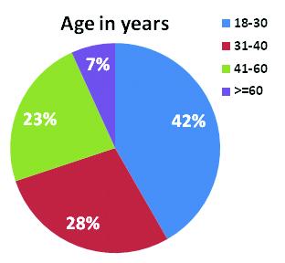
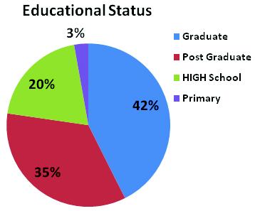
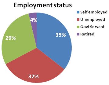
(a)Sociodemographic Profile: Majority of study subjects belonged to Darjeeling district (72.5%), of age range 18-30 years (41.7%). 42.6% study participants were graduates, 62.4% males and 35.5% were selfemployed as depicted in Figs 1,2 and 3 respectively. 50% were married and 59.3% resided in nuclear families and lived in pukka houses. 77.1% were suffering from chronic disease for which they were under treatment.
(b)
Vaccination intent was found to be considerably high. 81.1% opined in favour. Interestingly risk perception was found to be very high among 87.2% of study subjects.
association was found to exist among risk perception and employment status of study subjects (p < 0.001) with those salaried fearing more of the disease due to inadvertent exposure and compulsory attendance at office.
(c)Six domains Health Belief Model was applied to understand the pattern of precautionary behaviour among the study subjects. The results have been expressed verbatim and quantitative estimated have also been given, being a mixed method study (Fig 4).
stringently followed was opined by 92.2% cases and the rest few opined they were indifferent to it as they knew they would be attacked by COVID sooner or later. On assessing on a 5 point Likert’s scale 56.1% agreed masks were an absolutely essential, 68.1% strongly agreed to benefits of handwashing, 42.6% strongly agreed to maintaining cough etiquettes. Necessity of lockdown was strongly agreed by 88.7%. These are indicative of the positive responses of the preventive strategies despite the odds and promise a similar enforcement of precautionary behaviour in a sustaining fashion with similar geopolitical distribution. Maintaining social distancing was strongly agreed upon by 73.9%
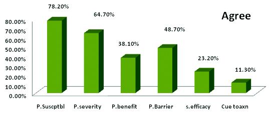
1.Perceived susceptibility of disease was estimated by assessing knowledge of study subjects. As high as 91.2% had correct knowledge on the transmission dynamics and agent causing the pandemic. 73.9% opined social media was responsible for information dissemination. Risk perception was higher among those who were educated and 34.3% opined those in hills were more at risk due to touristsvisiting their place and it being their main source of livelihood. 91.5% were aware of the symptoms.
2.Perceived severity of disease was assessed and it was seen that majority opined disease was self-limiting and risky only for elderly or immunocompromised. As study subjects were literate, the response was anticipated. However, they avoided hospitals and only 13.1% had visited health care facility while 50.7% had resorted to alternative therapy. Interestingly 38% opined in favour of safe homes if tested positive and only 45.2% felt severe symptoms can be managed in a hospital better. However, 53.2% opined migrant labourers played a pivotal role in disease transmission dynamics.
3.Perceived benefits were assessed and almost all study participants agreed that the pandemic was better avoided and had incurred huge harm. However the importance of precautionary behaviours was stressed by 54.2% cases and needed to be
4.Cue to action was however not responded well as majority opined neither agree nor disagree to any proactive steps to be taken by community to prevent the pandemic spread. However 86.4% opined they would contact nearby health facility for taking care of a tested positive patient in their community and prefer they stay in safe homes rather than in community. The fear and stigma of the disease could be felt.
5.Perceived Barrier was high among the study subjects as majority strongly agreed that the pandemic and the subsequent lockdown had a huge impact on their economy as majority sustained on tourism. 67.2% opined Government should have been applying a phased approach and thought of alternative ways of livelihood for those thriving on tourism industry and tea export business. Many tea gardens faced acute crisis and study subjects strongly disagreed to the sudden lockdown enforcements. 47.1% often worried about COVID-19 and 57.5% felt lockdown affecting general well-being . 40.1% expressed their anguish over having no idea when and where the pandemic ends and death toll stops.
6.Self-efficacy was not understood by majority of study subjects. However 56.4% opined they could tie over the pandemic by following the Government directives. This is an encouraging finding as 40.7% also opined they understood the meaning and methods of asymptomatic transmission hence stayed back home.
The country have never faced such a pandemic where prevention remains the mainstay. HBM is perceived to not only predict future behaviour of residents of the area but also anticipate behavioural dynamics of similar population in such remote inaccessible settings where internet connectivity is also an issue. Source of livelihood being tourism, the industry is hit hard. Yet, positive response to lockdown and in-depth understanding of situation by residents is encouraging. Policies needs to be drafted in accordance for sustaining the population though. Interestingly 24.6% population asked on sequel of the Covid pandemic in terms of their pulmonary functions, morbidity and mortality rates.
16.2% asked about the Government strategies to combat stigma, ostracization of health care workers and means to fight unemployment, social pathology and dwindling economic losses. Since law enforcements are not self-sustaining behavioural models hence applying HBM to understand the precautionary behaviours and reasons behind is anticipated to go a long way in fighting the most dreaded pandemic which hit face of mother earth.
Health behaviour model reflected the perceived severity and susceptibility to be higher than the perceived benefits. Not many studies have been conducted regarding the precautionary behaviour among hilly population. HBM indicates that diffusion of sustainable behaviour and its persistence depends on human perceptions rather than law enforcements. Population in hills, have their unique set of challenges due to remote geographical terrain and thriving mainly on tourism and tea industry for livelihood gets badly affected due to lockdown. Hence the benefits needs to be weighed along with the perceived barriers.
Similar studies on HBM concluded that people’s intent is the main driving force regarding behavioural dynamics in combating a disease. As COVID is new and not many diseases had prevention as mainstay hence the risk of not adhering to precautionary behaviours is not felt earlier. Other studies found literate and employed people to be more compliant to the rules. They also found those who were self employed were however prone to break the protocols of preventive behaviour as business was the sole source of livelihood. However, HBM applied to Avian Influenza and SARS cases clearly showed like in present study higher perceived severity and susceptibility than other diseases. Risk perception was also high as was vaccination intent. However perceived threats and self-efficacy was low in contrast to present study. However more studies on the area are in the making and recent spikes of reinfection may alter the course of preventive therapies altogether14-17
Present study conducted in hilly areas for the first time concluded that risk perception and vaccination intent was high among the study subjects. Majority of study subjects were educated and employed. However perceived susceptibility and severity was very high as compared to perceived benefits and cue to action. This shows that COVID-19 pandemic has had a huge impact on precautionary behaviour as perceived by applying the health behaviour model and hence can be inferred that diffusion of sustainable behaviour change among the hilly population will be more if and when implemented.
Online method of data collection has its own restrictions and elicitation of information may remain suboptimal. Connectivity remains an issue in hills. Moreover, precautionary behaviours elicited in hilly areas will have its own set of unique challenges given the difficult geographic terrains and hence the results are to be interpreted in accordance viz. accessibility of health services still being lower they already follow preventive strategies as mainstay for majority of cases and conditions.
1Jiang F, Deng L, Zhang L, Cai Y, Cheung CW, Xia Z — Review of the clinical characteristics of coronavirus disease 2019 (COVID-19). J Gen Intern Med 2020; 1-5.
2Chinazzi M, Davis JT, Ajelli M, Gioannini C, Litvinova M, Merler S, et al — The effect of travel restrictions on the spread of the 2019 novel coronavirus (COVID-19) outbreak. Science 2020. doi: 10.1126/ science.aba9757
3Guan WJ, Ni ZY, Hu Y, Liang WH, Ou CQ, He JX, et al — Clinical characteristics of coronavirus disease 2019 in China. N Engl J Med 2020.
4Lau JT, Kim JH, Tsui H, Griffiths S — Perceptions related to human avian influenza and their associations with anticipated psychological and behavioral responses at the onset of outbreak in the Hong Kong Chinese general population. American Journal of Infection Control 2007; 35: 38-49. doi: 10.1016/j.ajic.2006.07.010. [PMC free article] [PubMed] [CrossRef] [Google Scholar]
5Brug J, Aro AR, Richardus JH — Risk perceptions and behavior: Towards pandemic control of emerging infectious diseases. International Journal Behavioral Medicine 2009; 16: 3-6. doi: 10.1007/s12529-0089000-x. [PMC free article] [PubMed] [CrossRef] [Google Scholar]
6Janz Nancy K, Marshall H Becker — The Health Belief Model: A Decade Later. Health Education & Behavior 1984; 11(1): 47. doi:10.1177/ 109019818401100101. hdl:2027.42/66877. PMID 6392204
7Rosenstock Irwin — Historical Origins of the Health Belief Model”. Health Education & Behavior 1974; 2 (4): 328-35. doi:10.1177/ 109019817400200403.
8Siddiqui Taranum Ruba, Ghazal Saima, Bibi Safia, Ahmed Waquaruddin, Sajjad Shaimuna Fareeha — (2016-11-10). ”Use of the Health Belief Model for the Assessment of Public Knowledge and Household Preventive Practices in Karachi, Pakistan, a Dengue-Endemic City”. PLOS Neglected Tropical Diseases . 10 (11): e0005129. doi:10.1371/ journal.pntd.0005129. ISSN 1935-2735. PMC 5104346. PMID 27832074.
9Carpenter Christopher J — A meta-analysis of the effectiveness of health belief model variables in predicting behavior”. Health Communication 2010; 25(8): 661-9. doi:10.1080/10410236. 2010.521906. PMID 21153982.
10Glanz Karen, Barbara K Rimer, K Viswanath — Health behavior and health education: theory, research, and practice (PDF) (4th ed.). 2008; San Francisco, CA: Jossey-Bass. pp. 45–51. ISBN 978-0787996147
11Zhao S, Lin Q, Ran J — Preliminary estimation of the basic reproduction number of the novel Coronavirus(2019 n-CoV) in China from 2019 to 2020: a data-driven analysis in the early phase of outbreak. International Journal of Infectious disease 2020; 92: 214-7.
12Zaka A, Shamloo SE, Fiorente P — CoOVID-19 pandemic as a watershed moment: A call for systematic psychological healthcare for frontline medical staff. Journal of Health Psychology 2020 Epub ahead of print 30 March
13Biscayart C, Angeleri P, Lloveras S — The next big threat to global health ? 2019 novel corena virus (2019-n CoV) :: What advise can we give to travellers ?- Interim recommendations January 2020 from Latin American Society for Travel Medicine (SLAM VI) Travel Medicine Infectious Disease 2020; 101567
14Najimi A — Knowledge, beliefs and preventive behaviors regarding Influenza A in students: a test of the health belief model. J Educ Health Promot 2013; 2: 23
15Chan JF-W, Yuan S, Kok K-H, To KK-W, Chu H, Yang J, et al — A familial cluster of pneumonia associated with the 2019 novel coronavirus indicating person-to-person transmission: a study of a family cluster. The Lancet 2020; 395(10223): 514-23.
16Rosenstock IM, Strecher VJ, Becker MH — Social learning theory and the health belief model. Health education quarterly 1988; 15(2): 175-83
17Zhang L-L, Dalal K, Wang S-M — Injury Related Risk Behaviour: A Health Belief Model-Based Study of Primary School Students in a Safe Community in Shanghai. 2013; PLoS ONE 8(8): e70563. https://doi.org/ 10.1371/journal.pone.007056.
Tanvi Jha1, Keashav Mohan Jha2
Introduction : Educational environment is a major factor that determines curriculum effectiveness.
Aims: This study aimed to assess the perception of a single batch of MBBS students of a North Indian Medical College, towards the existing education environment in clinical postings and the changes in this perception at different points of exposure.
Methods : It was a longitudinal, descriptive study using DREEM questionnaires to assess the education environment and its domains. Questionnaires were provided to 197 students of the MBBS batch of 2015 at the end of their clinical postings in 4th/5 th, 6th/7th , 8th and 9th . Responses were assessed for each domain using one-way ANOVA test. Mean item scores, domain scores, and global scores were calculated and compared.
Results : The average global score was 133.24 ± 6.79, i.e. more positive than negative.There was, however, a highly significant decline (P<0.001) of total scores from 4th to 9th semester with the lowest domain score for the students’ social self-perception. The major problem areas uncovered include poor support system for stressed students, angry and authoritarian teachers and factual nature of the curriculum. Further, students in their 9th semester felt that teachers were more likely to get angry during teaching sessions as compared to 4th.
Conclusion : There is a need for improvement across all the domains of the education environment. However, particular attention must be given to the students’ perception of teachers and social self-perception.
[ J Indian Med Assoc 2021; 119(2): 26-30]
Key words : Education Environment, DREEM, Clinical Subjects, Medical Education.
The current Bachelor of Medicine, Bachelor of Surgery course in India is 4.5 years and 9 semesters. First two semesters focus on preclinical subjects, in semesters 3, 4 and 5 students learn para-clinical subjects while being initiated into clinical subjects and semester 6 onwards is clinical subjects. This traditional framework lacks a structured mechanism for student feedback.
Over time, reports from developing countries including India, indicate medical students’ dissatisfaction with current curriculum, teaching methods and educational environment1,2. A need exists to produce more innovative and sustainable model.
Educational environment is one of the major factors that determine effectiveness of a curriculum 3. Assessment of this environment is thus a good measure of curriculum effectiveness and students’ acceptance.
Over time, methodologies have been developed to assess education environment, including qualitative methods like questionnaires4-7. Dundee Ready Education Environment Measure (DREEM) developed by International Delphi Panel (1997) is one such standardized, culturally nonspecific, widely accepted questionnaire for gauging student attitude towards learning environment in healthcare coursework8-10. Several studies have been conducted in India, especially as the need for improving the curriculum was felt11,12, highlighting students’ negative perception towards various domains. There remains a need for studies with
1MBBS, PG Resident, Department of Pathology, University College of Medical Sciences and G T B Hospital, New Delhi 110095 and Corresponding Author
2MCh, Professor and Head, Department of Neurosurgery, Indira Gandhi Institute of Medical Sciences, Patna 800014
Received on : 12/09/2020
Accepted on : 13/10/2020
Editor's Comment :
Educational environment is a major factor that determines curriculum effectiveness.
Our longitudinal, descriptive study aimed to assess the perception of a single batch of MBBS students of a North Indian Medical College, towards the existing education environment in clinical postings and the changes in this perception at different points of exposure using standardized DREEM questionnaires.
The average global score was 133.24 ± 6.79, ie, more positive than negative.
There was, however, a highly significant decline (P<0.001) of total scores from 4th to 9th semester and several problem areas were identified.
There is a need for improvement across all the domains of the education environment, with particular attention to be given to the students’ perception of teachers and social selfperception.
Also, regular feedback must be taken from students in order to improve the effectiveness of the education environment.
larger group of students and longer time periods to accurately assess the current Indian curriculum.
Additionally, keeping in mind the long duration of medical curriculum, an overall improvement in education environment can be achieved when each subject studied over the course’s duration is assessed and its shortcomings addressed.
Clinical postings are introduced in earnest in 4th semester, till the final semester with final examinations taking place after 3.5 years of study. Clinical subjects include Medicine, Surgery, Pediatrics and Obstaetrics and Gynecology. The Medical Council of India provides a broad outline for curriculum planners and how to achieve this is left to individual institutions.
In our institution, the undergraduate curriculum aims at imparting
cognitive, psychomotor and communication skills by lectures, tutorials and clinical sessions. Postings in the clinical set up start in the 4th semester aiming at providing experiential learning opportunities. These postings are structured comprehensively: initial clinical postings in 4th/5th and 6th/7th semesters sensitize students towards the subjects, patient population and common medical problems faced by them (Table 1). Informal evaluation is done in 4 th semester, while formative assessment with grading is done in later semesters, each followed by individual and collective feedback sessions.
This study aimed to assess the education environment of clinical postings of the existing medical curriculum in a specific batch of students, and the changes in this perception at different points of exposure throughout their course. It also aimed to identify the problem areas, hence allowing course organizers to address them
Methodology :
This was a longitudinal, descriptive, student on student study conducted in a North Indian Medical College. Based on convenience sampling, all students of MBBS batch of 2015, comprising of 197 at the beginning of their 4th semester were recruited. The study was conducted over a period of three years, from January, 2017 to December, 2019, till the said batch appeared for their final professional examinations. Out of 197 subjects, 2 left the study (1 was held back and 1 failed to maintain regular attendance in the posting).
Students were enrolled after obtaining informed consent, with the freedom to withdraw at any time during the course of study, without it being reflected in their evaluation.
The students were provided with DREEM questionnaires after they had finished all the postings in clinical departments for the semester during their 4th/5th, 6th/7th, 8th and 9th semesters. The questionnaire, consisting of 50 statements, was responded to using a 5-point Likert scale. Incomplete questionnaires and students who failed to return the forms were excluded from the study. All questionnaires were anonymous and complete privacy was maintained by using a student specific unique identification number.
The quantitative data was entered in Microsoft Excel spreadsheet (Microsoft Co., Redmond, WA, USA) and SPSS ver. 20.0 (SPSS Inc, Chicago, IL, USA) was used for statistical analysis. Total scores for each domain and overall score (out of 200) was calculated on the basis of the DREEM score card for each semester, individually and combined.
To identify drawbacks in the education environment, items with a mean score below 2 were taken as problem areas requiring improvement, items with a mean score between 2 and 3 were considered as those that could possibly be improved on, and items with a mean score of 3 and above were considered as positive. One-way ANOVA test was used to compare global scores and mean domain scores amongst the semesters. P values < 0.05 were at 95% confidence interval was considered as significant.
Results :
The mean global DREEM score over all the semesters was 133.24 ± 6.79. Table 2 shows global DREEM score for individual semesters, individual domain wise scores in each semester, their average overall score and domain and interdomain trends over the course of the study.
Fig 1 shows the most highly rated positive and negative statements and their difference between 4th/5th and 9th semesters. In addition, there was a significant decline in students’ perception that the course was well timetabled from 4th/5th (3.39 ± 0.28) to 9th semester (2.52 ± 0.20), (P<0.05). Students consistently marked the statement “teachers overemphasize on factual learning” below 2 and there was no significant change in this perception throughout. Fig 2 shows changes in perception for individual domains between the 4th/5thand the 9th semesters.
Discussion :
Students’ perception of education environment is indicative of its effectiveness and also influences their academic performance3. It may also help identify lacunae and hence, help in modification and improvement of the curriculum. A positive educational environment is essential for a positive learning outcome 13. We determined undergraduate medical students’ perception of education environment in their clinical postings in the existing curriculum and changes in this perception over a period of 3 years using the DREEM questionnaire.
Students, with a global score of 133.24 ± 6.79, rated the education environment as being “more positive than negative” (Table 2). The total average global score was highest after 4th/5th semester, following which there was a progressive fall in the scores up to 8th semester, suggesting a decline in the students’ perception of education environment. This could be indicative of increasing work load and stress amongst the students. It was, however, followed
119, No 2, February 2021Journal
Table 2 — Average Dundee Ready Education Environment Measure (DREEM) score for the different domains expressed as scores and percentages for different semesters and total average score for each domain over the period of the entire Pediatrics curriculum for the purpose of comparison. P is comparison with the baseline, i.e. 4th semester scores. P# is when compared to the scores of the previous semester. * P<0.05 and ** P<0.001. $P<0.05 when domains U, V, W and X were compared with domain Y, ie, U vs Y (P = 0.04), V vs Y (P = 0.02), W vs Y (P =0.08) and X vs Y (P = 0.35) Areas
U48n34.11
V44n29.19
W32n22.33
X48n34.89
Y28n17.97
Z200 141.14 ±15.55133.06 ±19.31127.65 ± 20.54131 ± 10.80133.24 ± 6.79 P 0.0004**0.0003**0.0003**
0.0004**0.008* 0.12
Note : Here, U = Students’ Perception of Learning, V = Students’ Perception of the Course Organizers, W = Students’ Academic Self Perception, X = Students’ Perception of Atmosphere, Y = Students’ Social Self Perception and Z = Total average DREEM Score. The interpretation of total average score is done based on the guidelines provided by Roff et al, where a score of 0-50 is interpreted as very poor, 51-100 as having plenty of problems, 101-150 as more positive than negative and 151-200 as excellent.
by a non-significant rise in the score at the end of 9 th semester. This could be explained by an increase in study hours put in by the students as a result of approaching examinations leading to an improvement in perception of the learning environment.
Important problem areas identified included dissatisfaction with timetabling of postings, absence of a support system, angry and authoritarian teachers, inability to memorize and concentrate, factual nature of coursework and the absence of a support system. Students, however, also reported that teachers were knowledgeable and had good communication skills with patients and that teaching was patient centered. Table 3 shows the interpretation of the individual
domain scores as per DREEM guidelines.
Total average global score observed in this study is higher than those of other similar Indian studies11,12,14-16 and those conducted in other developing countries6,7,17-19. It was, however, found to be similar to that reported by a study done in New Zealand20 Similar global scores were observed by Medical Colleges with teacher centered, factual curriculum 21,22 , whereas, centers with student centered, problem-based learning showed higher scores23-25
A cross sectional study conducted in a similar North Indian medical college on students studying in various semesters, had an average global score of 101. It, similarly, observed lowest scores for the support system available to students and found that students had a negative perception of teaching and found the stress on factual learning and the course content to be
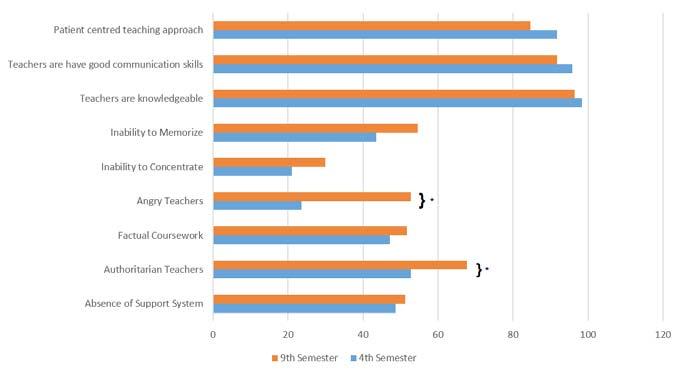
Fig 1 — Graph showing statements with the highest and the lowest scores and the changes in their perception between 4th and 9th semesters. There was a significant increase in the perception among students that teachers were authoritarian and were more likely to get angry during teaching sessions. *P<0.05
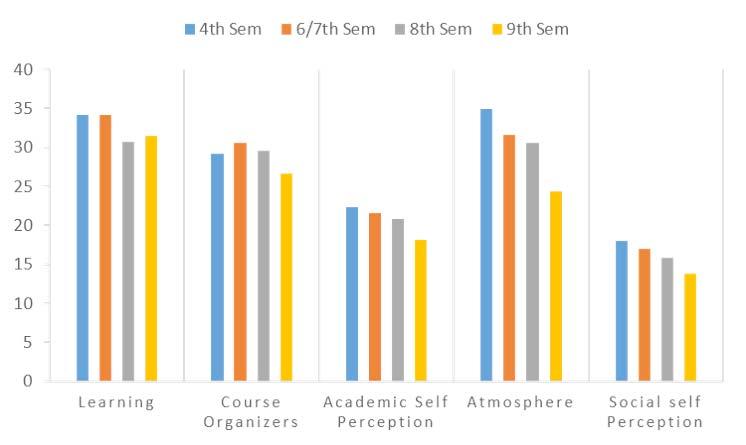
bothersome14
Sunkad et al reported that their students, as ours, found the education environment to be more positive than negative15 Abraham et al found that early semesters seemed to find education environment more positive than later semester students22
Another Indian study reported that the social atmosphere for students was not congenial and they were stressed26 . Similar findings were also seen in other states16,27 . The students further complained that they were unable to memorize all that was needed of them27. Naser et al found the learning environment was perceived positively by students but they felt that the course emphasized on factual learning and was not well timetabled and that the atmosphere during clinical teaching was not relaxed28
Gupta et al performed a cross sectional study on medical students of 2 nd to 4 th semesters and unlike our study, observed that teaching was teacher-centric, burdensome and boring. Another study done specifically for education environment in the subject Community Medicine, reported that students were too bored to enjoy their course12
Based on the results of the present study, the lacunae that were identified can be addressed by introducing innovative teaching techniques including problem-based learning29 and computer based cognitive representation30, better timetabling of clinical postings and integration of teaching amongst various Departments2. Introduction of a strong support system is essential for the students considering the stressful nature of the
curriculum. Institution of a mentor-mentee system and inclusion of extracurricular activities into the academic framework may provide a possible solution 31 . Regular student feedback and participation allows them to become stakeholders in their education and further, may help strengthen the curriculum as well its effectiveness.
Limitations :
Our study, having been performed on the same cohort, helped assess the overall perception to education environment and the changes in it with each passing semester. However, being limited to clinical postings in a single batch and a single institution in a single city of North India, is not representative for the entire country. A multicentric study is hence, desirable. Further, the DREEM questionnaire has a fixed number of statements that cannot be modified. Some students also complained that the questionnaire was too long and bothersome to fill.
Conclusion :
This study showed that the education environment in clinical postings in our institution is more positive than negative. It identified several positive areas; however, a few problem areas were also identified across all domains. Addressing these issues and suitably modifying the curriculum based on students’ feedback may help
Students’ 0-12Very PoorA more positive perception Perception of 13-24Teaching is viewed negatively Learning 25-36A more positive perception 37-48Teaching highly thought of
Students’0-11 Abysmal Moving in the right direction Perception of 12-22In need of some retraining Course 23-33 Moving in the right direction Organizers 34-44 Model course organizers
Students’0-8Feelings of total failureFeeling more on the Academic 9-16Many negative aspectspositive side Self17-24 Feeling more on the Perception positive side 25-32 Confident
Students’ 0-12A terrible environmentA more positive attitude Perception of 13-24 There are many issues Atmosphere which need changing 25-36A more positive attitude 37-48A good feeling overall
Students’0-8Feelings of total FailureMany negative aspects Social Self 9-16Many negative aspects Perception 17-24 Feeling on the more positive side 25-32 Confident
Vol 119, No 2, February 2021Journal of
improve the education environment and hence, help improve its effectiveness. Further, routine feedbacks at fixed periods of time, may help identify improvements as well as new problem areas as and when they develop.
Financial Support and Sponsorship : None. Conflicts of Interest : There are no conflicts of interest.
1Nabilou B, Khorasani-Zavareh D — The bridge between real and ideal: Students perception on quality gap in reality and their educational expectations. Iran Red Crescent Med J 2014; 16(9): e14254.
2Adlakha V, Jha T, Sahoo P, Muralidharan A, Bachani D — Students’ perception of lacunae in medical education in India, and suggestions for reforms. Natl Med J India 2018; 31(1): 29-31.
3Bassaw B, Roff S, McAleer S, Roopnarinesingh S, de Lisle J, Teelucksingh S, et al — Students’ perspectives on the educational environment, Faculty of Medical Sciences, Trinidad. Med Teach 2003; 25: 522-6.
4Seabrook MA — Clinical students’ initial reports of the educational climate in a single medical school. Med Educ 2004; 38(6): 659-69.
5Audin K, Davy J, Barkham M — University quality of life and learning (UNIQoLL): An approach to student well-being, satisfaction and institutional change. J Furth High Educ 2003; 27(4): 365-82.
6Sobral DT — Medical students’ self-appraisal of first-year learning outcomes: Use of the course valuing inventory. Med Teach 2004; 26(3): 234-8.
7Al Rukban MO, Khalil MS, Al-Zalabani A — Learning environment in medical schools adopting different educational strategies. Educ Res Rev 2010; 5(3): 126-9.
8Roff S, McAleer S, Harden RM, Al-Qahtani M, Ahmed AU, Deza H, et al — Development and validation of the Dundee Ready Education Environment Measure (DREEM). Med Teach. 1997; 19(4): 295-9.
9Roff S — The Dundee Ready Educational Environment Measure (DREEM) - A generic instrument for measuring students’ perceptions of undergraduate health professions curricula. Med Teach 2005; 27: 3225.
10Pimparyon P, Roff S, McAleer S, Poonchai B, Pemba S — Educational environment, student approaches to learning and academic achievement in a Thai nursing school. Med Teach 2000; 22(4): 359-65.
11Pai PG, Menezes V, Srikanth, Subramanian AM, Shenoy JP — Medical students’ perception of their educational environment. J Clin Diagnostic Res 2014; 8(1): 103-7.
12Gupta S, Gupta A, Verma M, Kaur H, Kaur A, Singh K — The attitudes and perceptions of medical students towards basic science subjects during their clinical years: A cross-sectional survey. Int J Appl Basic Med Res 2014; 4(1): 16-9.
13Hutchinson L — ABC of learning and teaching: Educational environment. BMJ 2003; 326(7393): 810-2.
14Kohli V, Dhaliwal U — Medical students’ perception of the educational environment in a medical college in India: a cross-sectional study using the Dundee Ready Education Environment questionnaire. J Educ Eval Health Prof 2013; 10: 5.
15Sunkad MA, Javali S, Shivapur Y, Wantamutte A — Health sciences students’ perception of the educational environment of KLE University, India as measured with the Dundee Ready Educational Environment
Measure (DREEM). J Educ Eval Health Prof 2015; 12: 37.
16Bhosale U — Medical students’ perception about the educational environment in western Maharashtra in medical college using DREEM scale. J Clin Diagnostic Res 2015; 9(11): JC01-JC04.
17Sundus A, Nadir HM, Faisal IM, Nida Y, Talha FM, Fatiha I, et al — Medical students perception of their medical environment-expected versus actual perceptions - a cross sectional study. J Pak Med Assoc 2014; 64(2): 230-6.
18Kim H, Jeong H, Jeon P, Kim S, Park YB, Kang Y — Perception Study of Traditional Korean Medical Students on the Medical Education Using the Dundee Ready Educational Environment Measure. Evidence-based Complement Altern Med 2016; 2016: 6042961.doi: 10.1155/2016/ 6042967.
19Roff S, McAleer S, Ifere OS, Bhattacharya S — A global diagnostic tool for measuring educational environment: Comparing Nigeria and Nepal. Med Teach 2001; 23(4): 378-82.
20Foster PLA, Kang I, Anderson VR, Thomson WM, Meldrum AM, Moffat SM — Changes in Bachelor of Oral Health students’ perceptions of their dental education environment. N Z Dent J 2013; 109(4): 134-40.
21Al-Ayed IH, Sheik SA — Assessment of the educational environment at the College of Medicine of King Saud University, Riyadh. East Mediterr Heal J 2008; 14: 953-59.
22Abraham R, Ramnarayan K, Vinod P, Torke S — Students’ perceptions of learning environment in an Indian medical school. BMC Med Educ 2008; 8: 20.doi:10.1186/1472-6920-8-20.
23Demirören M. Perceptions of Students in Different Phases of Medical Education of Educational Environment: Ankara University Faculty of Medicine. Med Educ Online 2008; 13: 8.doi: 10.3885/ meo.2008.Res00267.
24Zawawi AH, Elzubeir M — Using DREEM to compare graduating students2 perceptions of learning environments at medical schools adopting contrasting educational strategies. Med Teach 2012; 34(Suppl1): S25-31.
25Edgren G, Haffling AC, Jakobsson U, McAleer S, Danielsen N — Comparing the educational environment (as measured by DREEM) at two different stages of curriculum reform. Med Teach 2010; 32: e233e238.
26Thomas BS, Abraham RR, Alexander M, Ramnarayan K — Students’ perceptions regarding educational environment in an Indian dental school. Med Teach 2009; 31: e185-e186.
27Patil AA, Chaudhari VL. Students’ perception of the educational environment in medical college: a study based on DREEM questionnaire. Korean J Med Educ 2016; 28(3): 281-8.
28Naser SM, Biswas A, Nandy M, Niyogi S, Biswas G, Das AK — Perception of students regarding educational environment in a medical college in eastern region of India. J Indian Med Assoc 2012; 110(11): 800-802.
29Imanieh MH, Dehghani SM, Sobhani AR, Haghighat M — Evaluation of problem-based learning in medical students’ education. J Adv Med Educ Prof 2014; 2(1): 1-5.
30Wu B, Wang M, Johnson JM, Grotzer TA — Improving the learning of clinical reasoning through computer-based cognitive representation. Med Educ Online 2014; 19: 25940.. doi:0.3402/meo.v19.25940.
31Bhatia A, Singh N, Dhaliwal U. Mentoring for first year medical students: humanising medical education. Indian J Med Ethics. 2013; 10: 100-3.
Kaushik Bhattacharya1, Neela Bhattacharya2
The coronavirus disease 2019 (COVID-19) pandemic has brought about unprecedented adaptations in healthcare management, be it at a local or global level. Surgeons are facing a lot of dilemmas in the ongoing pandemic regarding the practice. On one side there are compulsions to manage the surgical patients without any hesitation in the operation theatre , there is another side of surgeons where he must return back home safely without self getting infected with the virus. Till the curve flattens, its going to be a surgical challenge and a herculean task for the surgeons at large to fight the COVID-19.
[J Indian Med Assoc 2021; 119(2): 31-2]
TKey words : COVID19, Pandemic, Laparoscopy, Chemoprophylaxis.
he Doctor’s Dilemma’ is a play by George Bernard Shaw, which was first staged in 1906 and highlighted doctor’s dilemmas created by scarce medical resources in a different context.The ongoing outbreak of novel corona virus [COVID-19] has created a global crisis of unprecedented dimensions. It is wreaking havoc across healthcare services and has crashed economies across the globe. It has disrupted human society on a scale that most living people have never witnessed. Surgeons are witnessing a lot of unprecedented dilemmas in the surgical practice due to this pandemic.
Dilemma in Surgical Practice :
The private clinics of surgeons which continue to provide services are often viewed as opportunists and have to face the resistance from society. In contrast, if the private clinics stop their services, they are viewed as cowards by the society and noncompliant by the government agencies. Surgeons are facing an exceedingly difficult task providing outpatient and inpatient services. During the COVID-19 pandemic, face-to-face outpatient appointments are avoided wherever possible and this is causing a communication gap and trust deficit between surgeon and the patient.
Dilemma due to Shortage of Personal Protective Equipment :
Shortage of Personal Protective Equipment (PPE) is another problem for the surgeons.There are emerging reports of medical staff complaining and protesting about the paucity of protective equipment. If the problem is not addressed at the earliest, such discontent may adversely impact our fight right at the frontline. Few hospitals provide N95 masks and PPE only to the chief surgeon and ordinary mask and normal OT dress to the rest in the operating team. Such biased attitude makes everyone vulnerable to COVID-19 .
Dilemma due to Lack of Test :
As of June 1, 2020, India had conducted approximately 3.8
1MS, DNB, MNAMS, FAIS, FACS, FRCS (Glasgow), Specialist Surgery, CAPFs Composite Hospital BSF Kadamtala, Siliguri 734010
2MS, DNB, MNAMS, M Ch (Plastic Surgery), Consultant Plastic and Reconstructive Surgeon, Anandaloke Multispeciality Hospital, Siliguri 734001
Received on : 28/06/2020
Accepted on : 30/07/2020
Editor's Comment :
Surgeons are facing a challenging time during COVID-19. They must not only choose the fittest candidate for surgery during this pandemic, but they have to keep themselves fit too to face the challenge. There are several dilemmas which each surgeon are facing in their career
million tests since it began testing in February, but many experts have noted that testing capacity is still drastically insufficient for the needs of the population1. Daily COVID-19 tests per 1,000 people are only 0.08 in India compared with 1.16 in the United States and 1.02 in Italy. Lack of test facilities make surgeon highly at risk when dealing with any emergency Surgery or Oncosurgery during this pandemic.
Dilemma in Laparoscopy :
In our opinion there is no reason to abandon laparoscopic surgery over open surgery. But all the literature warns to not underestimate the risks and perform laparoscopic surgery on COVID-19 positive patients only when really necessary and use logical and common sense to protect the surgeon and others by performing surgery in a safe and protected environment.During laparoscopy smoke and aerosols are generated, not only for cauterization of blood vessels, but also for dissection. This smoke can contain viral DNA and/or RNA and is sometimes evacuated straight into the over-pressured Operating room by opening a valve on a trocar2
Dilemma in Wearing PPE :
A pulse survey of 675 GPs has revealed that one in four have seen Covid-19 patients face-to-face without PPE, while more than half feel unsafe as a result of the lack of PPE. The very act of donning PPE with a N95 mask and face shield, operating in conditions where the surgeon feels hot, suffocated and dehydrated, with fogged inadequate vision, always with a niggle that such uncomfortable circumstances may lead to a major surgical error, all adds to the extreme stress that a surgeon faces in these times3
Dilemma of Postoperative Course :
Surgeons are also stressed during surgery due to the findings of a retrospective cohort study of 34 operative patients with confirmed COVID-19, 44.1% of patients required ICU care in the post-
119, No 2, February 2021Journal
operative period and the mortality rate was 20.5%4. Even after taking informed consent and prognosticating all the postoperative catastrophes which might happen to the patient party, there is always a fear psychosis in the back of the mind of every surgeon entering the operation theatre in this pandemic.
Dilemma of Choosing Surgical Patients :
As the elective surgeries are now stopped, surgeons have been asked to prioritize surgery that are both medically necessary and time sensitive to perform. Although no surgeon likes cancelling surgery, the necessity to choose which operation to proceed with and which can wait is an unusual circumstance for most of the surgeons. Another unique surgical challenge is the personal risk from intraoperative infection during prioritization of who receives the limited available surgical care. The backlog of postponed surgical proceduresis another headache for the surgeons5
Dilemma of Getting Burn Out :
Doctors and health care workers are putting in long hours at work but don’t have proper quarantine facilities near hospitals.Health care professionals are responding with an astounding display of selflessness, caring for patients despite the risk of profound personal harm. There is a risk of burn out of the surgeons with the daily routine of performing long hours of surgery wearing the PPE. There is also the tension of daily rigorous donning and doffing of PPE and going home at the end of the day safely and virus free tonot infect the family members.Tectonic shifts are at work as hospitals and clinics suffer grave financial losses and the workforce is diminished by illness and exhaustion.In a cross sectional study from China involving 1257 health care workers in China during the coronavirus pandemic, 50.4% had symptoms of depression, 34.0% reported insomnia, 44.6% reported symptoms of anxiety and 71.5% reported distress6
Dilemma of Work or Death for Surgeons :
Reading about intensive care admission or death of doctors due to COVID-19 regularly on print media and social media brings about certain fear psychosis among all the surgeons. The biggest thing happening is uncertainty of what is going to happen in quarantine and the social distance from family and friends. Also, there is a strong fear of stigma as people feel they will face social ostracism from the community.
Dilemma of Increasing Cost versus Facing Blame:
Surgical expenses in the hospital is going to rise due to the cost of PPE, the expenses of COVID-19 tests, the cost of special sterilization of the operation theatre and for maintaining the social distancing and due to less workforce . All these expenses are to be borne by the patients in a Corporate Hospital and Surgeons may be facing the blame for the expenses. There is an apprehension of overcharging or overuse of personal protective equipment.
Dilemma of Chemoprophylaxis :
Surgeons also are facing the dilemma of chemoprophylaxis with hydroxychloroquine on a regular basis without a clear guideline
from the regulating authorities. Although pre clinical results are promising, to date there is a dearth of evidence to support the efficacy of hydroxychloroquine in preventing COVID-197. Surgical community is at present confused about the role of chemoprophylaxis in this pandemic due to conflicting report emerging daily.
Dilemma in Resuming Elective Surgery :
There is confusion regarding when the elective surgery can be resumed as the cases of COVID -19 cases are rising daily everywhere. There should be a sustained reduction in rate of new COVID-19 cases in the relevant geographic area for at least 14 days before resumption of elective surgical procedures and the resumption should be authorized by the appropriate Municipal or State Health Authorities. This makes everything uncertain and unpredictable for the surgeons.
Limitation to study :
The article only concerns principally the dilemmas of the general surgeons which they are facing in performing their duties daily during COVID-19. The other super speciality Surgical Departments must be having some other surgical dilemmas specific to their speciality but has been kept out of purview of this study.
Conclusion :
“Life does not cease to be funny when people die any more than it ceases to be serious when people laugh .”
— George Bernard Shaw, The Doctor’s Dilemma
The most challenging field during the COVID-19 pandemic is surgery. Surgeons are facing a lot of dilemmas in the line of duty but still fighting the pandemic head on till now. A lot of support and encouragement is required from the masses, administration, hospital authorities and all quarter of society so that the surgeon can deliver till the curve is flattened without getting physically or mentally fatigued. With no clear picture emerging still about when the COVID -19 will be bought under control, the surgeon must wait and struggle with pandemic till the light at the end of the tunnel is seen.
Funding : None
Conflict of Interest : None REFERENCES
1https://www.icmr.gov.in/pdf/covid/update/ICMR_testing_update
2de Leeuw RA, Ceccaroni M, Zhang J, Tuynman J, Mabrouk M, Barri P, et al — COVID-19 and laparoscopic surgery; are we underestimating the risks? A narrative review of current literature and local expertise.JMIR Preprints. 27/03/2020:18928DOI: 10.2196/preprints.18928
3Bhattacharya K— Surgeon’s COVID-19 Traumatic Stress Disorder. Ind J Surg 2020; https://doi.org/10.1007/s12262-020-02504-4
4Lei S, Jiang F, Su W, Chen C, Chen J, Mei W, et al — Clinical characteristics and outcomes of patients undergoing surgeries during the incubation period of COVID-19 infection. E Clinical Medicine 2020; 21: 100331. doi: 10.1016/j.eclinm.2020.100331
5Angelos P — Surgeons, Ethics, and COVID-19: Early Lessons Learned. J Am Coll Surg 2020; 230(6): 1119-20.
6Lai J, Ma S, Wang — Factors Associated With Mental Health Outcomes Among Health Care Workers Exposed to Coronavirus Disease 2019. JAMA Netw Open 2020; 3(3): e203976. doi:10.1001/ jamanetworkopen. 2020. 3976
7Shah S, Das S, Jain A, Misra DP, Negi VS. A systematic review of the prophylactic role of chloroquine and hydroxychloroquine in coronavirus disease-19 (COVID-19). Int J Rheum Dis 2020; 23(5): 613-619. doi:10.1111/1756-185X.13842
Usha B Saraiya1, Nidhi Shah2
The role of folic acid in the prevention of congenital anomalies in the new born is known to all Doctors since the last 93 year s. However, what is perhaps not known is that the subject is very closely connected to India.
Two of the pioneers responsible for the scientific discovery of folic acid – Dr. Lucy Wills, an English physician and Dr. Yellapragada Subbarao of Andhra Pradesh were almost contemporaries and global citizens who worked in various parts of the world.
Dr. Wills conducted clinical research work on macrocytic anaemia of pregnancy in the late 1920’s in the slums of Mumbai. She called it “Pernicious anaemia of pregnancy". Her article was published in the British Medical Journal way back in 1931.
Dr. Subbarao was the second scientist from India who further contributed to this subject. He developed a method to synthesize folic acid and Vitamin B9. He is credited with initial Research on Folic Acid antagonists and tetracyclines.
There after the progress in folic acid was rapid with contributions from many Researchers : Bryan Hibbert in 1964, Chanarin in 1969, Richard Smithells in 1983, Scott & Weir in 1995, 1970 - Ultrasound revolutionised thediagnosis of early birth defects.
This presentation will chronicle the life histories of Dr. Lucy Wills of UK and Dr. Yellapragada Subbarao of India who played a monumental role in the understanding of the role of folic acid in health and disease. The authors have also reviewed the recent developments in the synthesis of folic acid.
[J Indian Med Assoc 2021; 119(2): 33-5]
TKey words : Folic Acid, Lucy Wills, Dr Y Subbarao, Antenatal Use of Folic Acid.
he role of folic acid in the prevention of congenital anomalies in the new born and the associated foetal wastage is well known to all the Obstetricians. Further all Doctors & Physicians know that it is used in the prevention & treatment of Megaloblastic Anaemia. However, what is perhaps not known is that this subject is very closely connected to India. Two of the pioneers who were responsible for the scientific discovery have a connection with India. We should take pride in this and let the new generation of Indian Doctors be inspired by their life and works.
This presentation will chronicle the life histories of Dr Lucy Wills1 of UK & Dr Yellapragada Subbarao2 of India who played a monumental role in the understanding of the role of folic acid in health & disease. The authors have also reviewed the recent developments in the synthesis of folic acid.
Dr Lucy Wills, an English lady Doctor, worked in Mumbai at Haffkine Institute from 1928 to 1933. Dr Yellapragada Subbarao was born in Andhra Pradesh. They were almost contemporaries and were born in 1888 and 1895 respectively. They were both global citizens and worked in various parts of the world including India. It is not known whether they ever met or communicated with each other. However, both have left an unforgettable stamp on the story of folic acid and pregnancy.
The First Most Important Indian Connection is Dr Lucy Wills :
Dr. Lucy Wills was born in 1898 into a learned family which lived in Birmingham area3. She studied at Cheltenham College for
1MD, DGO, FIAC, FICOG, FRCOG (UK), Panel Consultant, Breach Candy Hospital and Reaearch Centre,Mumbai 400026
2MBBS, DGO, DNB, OBGYN, Gynaecologist Jaslok Hospital, Mumbai 400036
Received on : 28/06/2020
Accepted on : 30/07/2020
Editor's Comment :
Periconceptional supplementation of folic acid is recommended for all pregnant women. Whenever Medical discoveries made my Indian Doctors, or have occurred in India, it is important to ensure that Indian doctors are aware of it.
There is a continual Research going on even in a simple drug like FOLIC ACID. All doctors need to be informed. Improvements in the health of pregnant women will ensure Good Health of Future Generations.
girls and was known as a brilliant student.
Dr Lucy Wills was encouraged by her family to become a Doctor. In that era, opportunities for women to enter Medical profession were few. The London School of Medicine for Women was established in 1874 and had the distinction of being the first Medical School in England to enrol women 3
In January 1915, she joined the London University as a Medical Student. She graduated in May 1920 with a degree of LRCP (Licentiate of the Royal College of Physicians). In December same year, she was conferred MBBS (Medical Bachelor and Bachelor of Science) from London University.
There were a few Indian Students at the Royal Free Hospital & there were some academic interactions with India. One of the well-known &well-respected students was Dr Jerusha Jhirad4 who graduated with a degree in Obstetrics and Gynaecology in 1919, the year before Lucy Wills graduated.
On qualifying, Lucy Wills took a surprising decision not to practice as a physician, but to teach and do research. She decided to go to India and arrived in Mumbai. She joined Haffkine Institute as a Researcher in 1928.
Dr Margaret Balfour of the Indian Medical Service had asked her to join the Maternal Mortality Enquiry sponsored by the Indian Research Fund Association4 at the Haffkine Institute in Bombay
119, No 2, February 2021Journal
(now Mumbai). The Lady Tata Foundation also provided financial support.
Lucy Wills was in India between 1928 and 1933, mostly based at the Haffkine Institute in Bombay. She conducted Clinical Research work in India on Macrocytic Anaemia of Pregnancy5.
She saw many pregnant young women with classical Macrocytic Anaemia who were working in the cotton mills. It was identical to Pernicious Anaemia described earlier. However, that anaemia occurred in the elderly, was progressive and sometimes fatal. The anaemia she encountered was limited to pregnancy and there was spontaneous cure after delivery. Therefore, she called it Pernicious Anaemia of Pregnancy.
Dr. Lucy Wills found some striking differences in the anaemia in Mill workers, who had poor nutritional status from true pernicious anaemia5 . Her patients did not have deficiency of gastric acid in the stomach known as achlorhydria. True pernicious anaemia patients responded to “pure” liver extracts (Vitamin B12) whereas her patients responded only to “crude” liver extract. She came to the conclusion that there must be another nutritional factor other than Vitamin B12 deficiency. For many years this was known as the “Wills factor”. It was only in 1940 that it was discovered that this was a “natural folate” which was found in yeast extracts & a supplement called “Marmite”. Much later it was found that “Natural Folates” was a precursor of synthetic Folic Acid.
Her article entitled “Treatment of Pernicious Anaemia of Pregnancy and Tropical Anaemia” with special reference to yeast extract as curative agent was published in the British Medical journal way back in 19316
Dr. Wills report was based on clinical observations and she treated her patients with a yeast extract called “Marmite” which was a rich source of folates. Hence, she is credited with the discovery of “folates” in the treatment of anaemia in pregnant women.
Dr. Wills was keen to do further Research. She was able to get an opportunity to work in world famous Pasteur Institute which was in Coonoor in The Nilgiris. In the year 1929, she moved to Coonoor. Luckily Sir Robert McCarrison was the Director of Nutrition & he was very supportive. She was able to work with Indian Researchers, so was able to publish 2 Scientific papers on her experiments on Albino Rats7,8
She undertook experimental work on dietary manipulations on Albino Rats. Rats when fed on diet similar to the Muslim women working in the Mills developed this type of anaemia & some of them even died during pregnancy. This anaemia could be prevented by nutritional supplement of yeast extract without the addition of Vitamin B12. Later she repeated these experiments on Macaque monkeys. After Coonoor, she returned back to Bombay & carried on with the Clinical Research proving that megaloblastic anaemia of pregnancy could be prevented & treated with yeast extract9
She returned to UK to work at Royal Free Hospital during the summer months. In 1933, she went back as a full time Pathologist at Royal Free but still was able to come back to Haffekine Institute & continue her Research. In early January 1938, for the first time, Lucy Wills travelled by air to Karachi and onwards by sea9
Before retiring from Royal Free Hospital, she set up a fullfledged Haematology Department. In 1947, she retired after a long & distinguishing career. During her lifetime, she travelled extensively to South Africa, Fiji & Jamaica continuing her interest in the subject.
Lucy Wills proved that hard & sincere Research work brings lasting benefits to Mankind. Ever after retirement she continued to stay in touch with people & Institute, she served all her life. Her virtues were recognised by her colleagues & she received much honour, respect & adulation from them.
Her last few years she spent with her lifelong friend Margot Hume in a Cottage in Surrey where they nurtured a Botanical Garden.
When she passed away on 26 th April 1964, she was deeply mourned by many friends from all over the world10
The Second Most Important Indian Contributor is Dr Yellapragada Subbarao :
Dr Subbarao developed a method to synthesise folic acid and vitamin B9. His work on synthesising Folic Acid to treat megaloblastic anaemia followed that of Lucy Wills earlier work11. He did this while he was appointed at Lederle laboratories, a division of American Cyanamid. It is now a division of Wyeth Ltd. which has recently merged with Pfizer Ltd.
Dr Y Subbarao was born on 12/1/1895 in a village in Andhra Pradesh. He had a difficult childhood but was known for being a bright student. He entered Madras Medical College. His father in law supported his education. Those were turbulent years of Indian National Congress and freedom struggle. He started wearing khadi clothes and took interest in Indian Systems of Medicine12. He was encouraged to go to USA by his friends and well-wishers for further education. He joined Harvard. He did monumental work in the role of ATP (adenosine triphosphate) in muscular activity and obtained a PhD.
On joining Lederle Laboratories, in addition to folic acid he worked on methotrexate along with Dr. Sidney Farber. He also discovered Hetrazan which WHO used against filariasis in World War II. Aureomycin, world’s first tetracycline was developed by him in 1945 with Benjamin Duggar.
His journey into the development of cancer chemotherapy was a logical sequence of his knowledge of folic acid and then the role of folic acid antagonists.
Through all the professional successes he achieved, he remained a very simple man, a staunch vegetarian and a private person. His achievements should have been enough to guarantee him a professorship at Harvard so says Mukherjee in “Emperor of all Maladies”13. Tribute was also paid to him by his colleague George Hitchings who shared the 1988 Nobel Prize in Physiology with Gertrude Elion. Many believed that he too, should have been included among the group who received the 1988, Nobel prize14
The only lasting tribute was paid by American Cyanamid who named a fungus “SUBBAROMYCES SPLENDUS” in his honour.
Writing in the April 1950 issue of ARGOSY, D K Antrim observed “you’ve probably never heard of Dr. Yellapragada Subbarao. Yet
Vol 119, No 2, February 2021Journal
because he lived, you may be alive and are well today. Because he lived, you may live longer” 14
He died at the young age of 53 years in New York on 9th august 1948. But he lived long enough to see his beloved India gain independence. Till the end he maintained his Indian passport and did not seek a green card though unhesitatingly he contributed to some of America’s most important Medical Research during World War II.
Conclusion:
It took another quarter of a century for further progress in the use of folic acid in pregnancy. It was Bryan Hibbard15 in 1964 who reported the importance of folates in the embryonic development. Chanarin16 in 1979 explained the genesis of megaloblastic anaemia.Another important research was the work of Richard Smithells17 in 1980 when he stated that the early birth defects could be prevented by the use of folic acid. This applied especially to neural tube defects. It was only in 1995 that Daly, Scott and Weir categorically showed that folic acid therapy prevents Neural Tube defects18,19
Fortunately, today in the 21 st century, ultrasound can pick up neural tube defects in early pregnancy and in most countries termination of pregnancy is permitted. However, it is a great personal tragedy for the parents. What is needed is awareness and education of the public that peri-conceptional therapy with folic acid is required to prevent this foetal wastage. There are some countries where fortification of food is done with folic acid as a preventive measure.
Progress has continued in the 21st Century. Newer formulations of Folic Acid have been introduced in practice.
The understanding of several enzymatic processes has unravelled the role of a biologically active L-Methyl Folate20. It can overcome the deficiency of MTHER which is a genetic polymorphism.
Further L-Methyl Folate if combined with DOCOSAHEXAENOIC ACID (DHA)21 improves brain & eye development in the 3rd trimester of pregnancy. Hence this combination is highly recommended from 24th week of gestation to lactation period to ensure maximum brain development in the foetus.
We can look forwards to further developments in the Story of Folic Acid in promoting Health of Future Generations.
So, we hope that when all the Medical Practitioners who write prescriptions of folic acid will at that moment feel proud of the fact that India has made a significant contribution to the knowledge of this great product which has saved so many lives.
We would also like to reiterate that Science knows no boundaries,
neither geographic nor racial. Finally, Research is a never-ending ongoing process which brings much benefit to Mankind.
Funding : None
Conflict of Interest : None REFERENCES
1Historical Review — The History of Folic Acid. 2001 Blackwell Science Ltd, British Journal of Haematology , 113: 579-589.
2http://www.ysubbarow.info
3Roe D A, 1978 — The Journal of Nutrition , Volume 108, Issue 9, September 1978, Pages 1377-1383 https://academic.oup.com/jn/ articleabstract/108/9/1377/4770512?redirectedFrom=fulltext
4Purandare CN, Patel MA, Balsarkar GD —Indian Contribution to Obstetric &GynaecologicalJournal of Obstetrics& Gynaecology of India, 2012.
5https://scientificwomen.net/women/wills-lucy-138
6Wills L — Treatment of pernicious anaemia of pregnancy & tropical anaemia to special reference to yeast extract as curative agent. British Medical Journal 1931; 1: 1059-64.
7Wills L, Mehta M M — Studies in ‘pernicious anaemia’ of pregnancy. Part I preliminary report. Indian Journal of Medical Research 1930; 17: 777-92.
8Wills L, Talpade SN — Studies in ‘pernicious anaemia’ of pregnancy Part II a Survey of Diabetic & Hygienic Conditions of Women in Bombay. India Journal of Medical Research 1930; 18: 283-306.
9https://en.wikipedia.org/wiki/Lucy_Wills
10Lucy Wills (Obituary). Lancet 1964; 283: 1225-6.
11Bhargava PM — History of Medicine: Dr. Yellapragada SubbaRow (1895-1948) - He Transformed Science; Changed Lives.” Journal of the Indian Academy of Clinical Medicine 2001; 2 (1,2): 96-100.
12Kapur S, Gupta SP — Dr Yellapragada SubbaRow (1895-1948): the man and the method. Indian J Exp Biol 1998; 36(11): 1087-92. PMID: 10085777.
13Mukherjee, Siddhartha (16 November 2010). The Emperor of All Maladies: A Biography of Cancer. Simon and Schuster. pg. 31. ISBN 9781-4391-0795-9.
14https://www.researchgate.net/publication/ 330284166_Unknown_facets_of_Not_so_wellknown_scientist_Dr_Y_Subbarow_A_great_scientist_who_did_ not_receive_the_Nobel_Prize
15Hibbard BM — The role of folic acid in pregnancy with particular reference to anaemia, abruption and abortion. Journal of Obstetrics and Gynaecology British Communication 1964; 71: 529-42.
16Chanarin I — The Megaloblastic Anemias, 3rd Edition Blackwell Scientific, Oxford 1990.
17Smithells RW, Sheppard S, Schorah CJ — Vitamin deficiencies and neural tube defects. Archives of Diseases of Childhood 1976; 51: 94450.
18Scott JM, Weir DG, Kirke PN — Folate and neural tube defects. In: Folate in Health and Disease (ed. By L.B. Bailey) 1995; 12: 329-60. Marcel Dekker, New York.
19Scott JM, Weir DG — Role of folic acid/ folate in pregnancy: Prevention is better than cure. In: Recent Advances in Obstetrics and Gynaecology 1998; (ed By J Bonnar) Ch 1, pp. 1-20. Churchill Livingstone, Edinburgh. 20Greenberg JA, Bell SJ — Rev Obstet Gynecol 2011; 4(3-4): 126-7. https://www.ncbi.nlm.nih.gov/pmc/articles/PMC3250974/
21Arellanes I C, Choe N, Solomon V — Brain Delivery of DHA. 2020. https://www.thelancet.com/journals/ebiom/article/PIIS23523964(20)30258-9/fulltext
Kausik Ray1, Samantha Greenhouse2
Healthcare sector is an ever-evolving area which has been challenged heavily with unprecedented crisis recently. The leaders have to change their stance and shuffle their policies to adapt with the changing circumstances for immediate crisis-management and safeguarding the future from unforeseen catastrophe. Knowing the theory and sharing the experience of healthcare leadership is imperative for all the interested professionals who wish to thrive through the challenges. As it is a fact that satisfied and engaged employees in a conducive environment can produce better performance, the leadership should bear that responsibility to create and safeguard a conducive environment for the employees at the care-givers end. This article aims to throw light to those areas and suggests the importance of collective leadership with different styles and the steps to bring about a positive change in the system.
Key words : Healthcare-related crisis, Leadership, Change management.
‘ You may delay, time will not .’ — Benjamin Franklin ‘Time’ is the biggest treasure and its call is the ultimate call. When a particular period of time or ‘Situation’ demands something, we need to respond accordingly.On the other hand, ‘Trends’ or cultures are the rituals or habits in a system which are followed repeatedly to achieve a reasonable outcome. The validity of the trends needs to be checked regularly with logical questions according to the demand of the situation. Those who can adapt to the change, survive. The effectivity of ‘Situational response’ determines the immediate survival and it influences the future course of action for any organization. Even a small change in the trends can result in a ‘Paradigm shift’. For example, this ongoing pandemic has necessitated the change by introducing mandatory ‘Maskgloves’ culture.
What is ‘Change’ in Healthcare Leadership ?
“Change” is the only constant in this ever-evolving world and the human race could not have reached the height it has achieved so far without the constant effort to change for the better. The word “Change” brings lots of emotional, physical and functional challenges in a system including excitement, apprehension, adaptability, unlearning and relearning. These challenges create a lot of resistance in a system when the word “Change” comes to the fore1,8
According to Kurt Lewin’s theory of ‘Change model’, there are three steps for a change —Unfreezing, Changing and Refreezing1. To inculcate an idea that a system needs change among the subjects of the system is the initiating point of any reform. It creates huge turmoil and revolt for the aforementioned factors and needs great deal of positive energy to ‘Unfreeze’ the established culture, attitude and so-called ‘comfort zone’ in the system. The main challenge lies
1MS, DNB, MRCS (Ed), FRCS (Gen Surgery), Leadership and Education Fellow, Brighton and Sussex University Hospitals NHS Trust, Brighton, UK
2Lecturer and Course Convener, Healthcare Leadership and Commissioning, Brighton and Sussex Medical School, Brighton, UK
Received on : 17/01/2021
Accepted on : 29/01/2021
[J Indian Med Assoc 2021; 119(2): 36-8]
Editor's Comment :
The Healthcare sector is an ever-evolving area that has been challenged heavily with unprecedented crisis recently. The leaders have to change their stance and shuffle their policies to adapt to the changing circumstances for immediate crisismanagement and safeguarding the future from unforeseen catastrophe.
Knowing the theory and sharing the experience of healthcare leadership is imperative for all the interested professionals who wish to thrive through the challenges.
As it is a fact that Satisfied and engaged employees in a conducive environment can produce better performance, the leadership should bear that responsibility to create and safeguard a conducive environment for the employees at the care-givers end.
This article aims to throw light on those areas and suggests the importance of collective leadership with different styles and the steps to bring about a positive change in the system.
in the grinding psycho-dynamic process that involves painful unlearning without loss of ego identity and difficult relearning as people cognitively attempt to restructure their thoughts, perceptions, feelings, and attitude1,2. As we need heat-energy to melt a chunk of ice, the ‘unfreezing’ also needs lots of energy and positive force which may be eaten up as waste at the early stage like ‘latent heat’ and becomes frustrating for the initiators. Resistance to the change is an automatic response from the subjects in the system 8. To overcome the resistance, employee involvement is the oldest and most effective strategy. For promoting employee involvement four elements, namely- power, information, knowledge and skill, and rewards are involved in formulating effective plan and implementing change smoothly2,3 . Employees should realize that the change is not an ‘option’ but a ‘necessity’.
‘Changing’ comes next which needs meticulous planning for reframing, setting up objectives to achieve maximum effectiveness and diligent execution. Identifying the inherent strength of the existing system is a major step in this process. Ignoring it can bring disaster as the majority of the basic components of the system should not be made redundant. They can only be reframed for better productivity
119, No 2, February 2021Journal
and effectiveness by means of knowledge sharing, acquisition of newer skills including technological expertise and empowerment3-5
‘Bridges Transition Model’ can throw light here by mentioning that ‘Change’ is an external process framed by the leaders and ‘Transition’ is the internal repercussion inside the employees which has three phases—‘Saying goodbye’(endings), ‘the Neutral zone’(explorations) and ‘Moving forwards’(New beginnings)4
According to Bridges et al, the ‘The Neutral Zone’ is the most uncomfortable zone for the followers or employees where they either retreat to the old base or embark on a new voyage. Ideally, the leadership and the employees should spend some time at this phase to make the real transition happen. Understanding ‘Transition’ is very important for the leadership as their ‘Change’ seems undoable because ‘Transition’ holds people back. ‘Coaching’ and ‘Mentoring’ are very much essential to overcome the transition phase for both the leaders and followers4,5
‘Refreezing’ is the step where the changes get established and solidified by positive reinforcement and reassurance. People should feel rewarded and can see the development of the organization. The change in attitude and culture should be cemented for the benefit of the system.
The role of ‘Leaders’—‘Gardener’ and ‘Firefighter’ concept :
Leadership is defined as “any activities tied to the core work of the organization that are designed by organizational members to influence the motivation, knowledge, affect, or practices of other organizational members”1,2. Thus, the persons who engage in these activities should be considered as Leaders for the activities, not for the position. According to Bridges et al, the leaders take the followers through the transition phase by actively communicating with them and reiterating regarding 4 ‘P’s - the Purpose: why it needs to be done; the Picture: what it will look or feel like; the Plan: steps to reach there; and, the Part: what role the individual is expected to play4. As different kind of qualities are displayed by the leaders, the health care system needs a combination of different variants like the transactional, transformational, servant, autocratic and democratic leadership according to the need of the situations5-8,19
The word ‘Situation’ is brought again into the fore to stress the importance of the adaptability of a leader to the changing circumstances for leading a group of skilled and intelligent people towards the goal of achieving excellence in patient care. Contingency (situational) theory says that there is no single right way to lead because the internal and external dimensions of the environment require the leaders to adapt to that particular situation7,18. It is not necessarily to be displayed by a single person as a leader or ‘hero’. It can be displayed by a group of different people working cohesively from the ‘Blunt or managerial end’ of the organization with an ‘Inclusive’ intent11,13
To author’s own understanding, the need for the ‘Firefighters’ for solving the day-to-day crisis is immensely important for the smooth running of the system. Equally important is the role of the ‘Gardeners’ that can plan for the unknown future by sowing seeds
of newer possibilities for the further growth and development of it. As extinguishing ‘Burning’ problems necessarily help the immediate survival of an organization, the ability to invest in the future and long-term planning determines its prosperity.
As a minuscule unit of the health-care system from the ‘Provider’s end’, the author feels the urge to create some extra ripples of any form towards the betterment of the existing system.
Quality Improvement Activity (QIA) : Audit, P-D-S-A, Gap-analysis, G-R-O-W
As we all have the potential to become a leader in our own way, Quality Improvement Activity is the easiest way to participate in individual capacity for the betterment of the existing healthcare system.According to Jones et al.,the improvement journey starts with — (1) Assessing readiness of the organization, (2) Securing board support, (3) Securing wider organizational involvement, (4) Developing improvement of skills and infrastructure, (5) Aligning activity of clinical, managerial and corporate teams towards the same goal, and (6) sustaining an organization wise approach14. In reality, this kind of activity needs broad based involvement of timemoney-infrastructure-logistics of the organisation. An audit on ‘infection control’ or ‘Fluid-electrolyte balance in acute admissions’ can be good examples of simple but effective QIAs which are doable and easy to draw inferences for change the system for improvement.The completion of audit cycle is imperative for QI.Application of PDSA (Plan-Do-Study-Act) model in the second and third cycle of the audit will bring about the actual improvement11,13,25
‘Gap analysis’ is another example to get involved ‘easily‘in QIA. It is defined as an instrument used to recognize and define the inconsistency between current reality in health care and the desired or optimal health care situation 21,22.The steps to carry on a Gap analysis are step 1 – Identify/clarify what is currently happening with patients or health care professionals in a specific therapeutic area step 2.– Define the “Gold Standard”, step 3.– Clarify the gap/ discrepancy between Step 1 and Step 2 and, whether the gaps related to knowledge, skill, attitude or practices 21-24. In step 4 –decide on the learning goals for your anticipated QI endeavor from the identified gaps21
To start with, a ‘G-R-O-W’ (Goals-Reality check-Options available-Will) model can be discussed with the other team members for getting a solution for road-blocks9,10
DISCUSSION
The fundamental intention of Medical Leadership is to engage doctors in improving services across the Health-care system. Doctors can play the role as a bridge between the ‘blunt’ or managerial end and ‘sharp’ or providers’ end of the “Service arrow” 11. Rigid or protocolized pathways for Quality Improvement (QI) activities are useful but innovation and flexibility often play an important role as a strenuous learning curve and limited resources become major impediments in the way to achieve a goal 13 Resilience, patience and tenacity are three qualities often required among the key-players to surpass those hurdles. A conducive
119, No 2, February 2021Journal
‘working environment ‘is essential for the cohesive functioning of both the ends of the system for the common goal of improved ‘Patient Care’. The synthesis of experiences to create an ‘Evidence base’ is essential as well, which can allow many visions into the challenges of reliably safe, superior-quality care and promote important learning about how improvement can best be secured across the Healthcare system11-13,15. In favor of an ‘Evidence base’, Dixon-woods et al mentioned, ’Patients may be deprived of benefit, resources, and energy may be wasted on ineffective QI interventions or on interventions that distribute risks unfairly, and organizations are left unable to make good decisions about trade-offs given their many competing priorities. The study of improvement has an important role in developing an evidence-base and in exploring questions beyond effectiveness alone, and in particular, showing the need to establish improvement as a collective endeavor that can benefit from professional leadership11’.

Management 2016; 16:
7Cummings GG — “Leadership styles and outcome patterns for the nursing workforce and work environment: A systematic review,” Int J Nurs Stud 2010; 47(3): 363-85.
8Appelbaum S — “Organizational outcomes of leadership style and resistance to change (Part One). Ind Commer Train 2015; 47(2): 73-80. 9https://www.mindtools.com/pages/article/newLDR_89.htm 10https://www.performanceconsultants.com/grow-model
11Dixon-Woods, M. How to improve healthcare improvement—an essay by Mary Dixon-Woods. BMJ 2019; 366.
In the context of the present environmental and political situation across the country, the uncertainty can definitely tell upon the financial support from the ‘Health budget’ to the different aspects of health care due to changing priorities. Small scale QI projects would be a prudent step instead of bigger leaps. Setting up clear ‘Goals’ is important. Robust ‘Evidence base’ can help the ‘Reality check’ and exploration of ‘Options’. Limited financial resource needs innovation and innovation needs ‘resourceful ‘minds who can think out of the box. A far-sighted leadership of ‘distributive ‘and ‘Inclusive’ nature can involve more human-resources by influencing the workforce from different strata with different skill sets12,18-19 . Satisfied and engaged employees in a conducive environment can produce better performance16,18 (Fig 1).
Only then, a tiny ripple of change can be converted into a bigger wave with wider range of participation.
Funding : None
Conflict of Interest : None
1Schein E H — Kurt Lewin’s change theory in the field and in the classroom: Notes toward a model of managed learning. Systems practice 1996; 9(1): 27-47.
2Ambrosini V, Bowman C —Tacit knowledge: Some suggestions for Operationalization. Journal of Management Studies 2001; 38(6): 81129.
3 Hussain ST, Lei S, Akram T, Haider MJ, Hussain SH, Ali M — Kurt Lewin’s change model: A critical review of the role of leadership and employee involvement in organizational change. Journal of Innovation & Knowledge 2018; 3(3): 123-7.
4Bridges,W, Mitchell, S — Leading Transition: A new model for change. Leader to Leader 16(3): 30-6.
5Kaleem, Y., Asad, S.,Khan, H. Leadership styles & using appropriate styles in different circumstances (https://www.researchgate.net/ publication/323797001_Leadership_Styles_Using_Appropriate_ Styles_in_Different_Circumstances)
6Khan ZA, Nawaz A, Khan I — Leadership Theories and Styles: A Literature Review. Journal of Resources Development and
12Spillane, James P — Distributed leadership (1st ed.). San Francisco: Jossey-Bass 2006; 11-2. ISBN 978-0-7879-6538-9.
13Dixon-Woods M, Baker R, CharlesK, DawsonJ, Jerzembek G, Martin G,1 McCarthy I, McKee L, l Minion J, Ozieranski P, Willars J, Wilkie P, West M — Culture and behaviour in the English National Health Service: overview of lessons from a large multimethod study. BMJ Qual Saf 2014; 2(3): 106-15. doi:10.1136/bmjqs-2013-001947
14Jones B, Horton T, Warburton W — The improvement journey :Why organisation-wide improvement in health care matters, and how to get started. The Health Foundation, May 2019.
15Smith I, Hicks C, McGovern T — Adapting Lean methods to facilitate stakeholder engagement and co-design in healthcare Business, Medicine BMJ 2020.
16West MA, Dawson JF, Admasachew L — NHS staff management and health service quality: results from the NHS staff survey and related data. London: Department of Health, 2011
17RN Amanchukwu, GJ Stanley, NP Ololube — A Review of Leadership Theories, Principles and Styles and Their Relevance to Educational Management. Management 2015; 5(1): 6-14.
18Nielsen k, Munir f — How do transformational leaders influence followers affective well-being? Exploring the mediating role of self-efficacy, Work Stress 2009; 23(4): 313-29.
19Sow M, Murphy J, Osuoha R — The Relationship between Leadership Style , Organizational Culture , and Job Satisfaction in the US Healthcare Industry, Manag. Econ Res J 2017; 3 1-10.
20Sarathchandra K — Implementation challenges and Research Gaps of Electronic Medical Records in Public Sector Hospitals of Sri Lanka, Int J Sci Res Publ 2019; 9(7): 174-80.
21https://www.cpd.utoronto.ca/newsletter/2009_fall/ Quick_Tips_initiating_a_learning_needs_assesment__Gap_Analysis_ of_health_care_issues.pdf
22Silvestro R — Applying gap analysis in the health service to inform the service improvement agenda. International Journal of Quality & Reliability Management 2005; 22(3): 215-33.
23Abili K, Thani F N, Afarinandehbin M — Measuring university servicequality by means of SERVQUAL method. Asian Journal on Quality 2012; 13(3): 204-11.
24Anderson E — High tech v high touch: a case study of TQM implementation in higher Education.Managing Service Quality: An International Journal 1995; 5(2): 48-56.
25https://improvement.nhs.uk/resources/pdsa-cycles/
Sujan Kumar Mohanty1, Virendra Kumar2, Jaffar Hussain AP3, V Bhuvan4
Cardiac tamponade due to free cardiac wall rupture is a medical emergency with almost 100% mortality. Sudden death can occur in a patient after myocardial infarction due to rupture of myocardium. But in certain cases, the patient dies suddenly without any specific cardiac symptoms. Only at the autopsy, the cause of death is found to be cardiac tamponade due to rupture of free wall, which has undergone ischemic necrosis. We are describing two cases of sudden death where death occurs due to free cardiac wall rupture after few non-specific symptoms. So the general physician should have high index of suspicion of cardiac diseases from nonspecific symptoms and refer the patient for further intervention.
[ J Indian Med Assoc 2021; 119(2): 39-40]
CKey words : Autopsy, Cardiac tamponade, Free cardiac wall rupture.
ardiac tamponade is defined as collection of blood or fluid in the pericardium leading to compression of the heart caused by increased intra pericardial pressure1 The extra collection of fluid in pericardial space (Normally 15-50 ml)2 is called pericardial effusion. When the collection is a chronic process, the pericardial sac can accommodate around 2000 ml of fluid due to adaptive stretching of pericardium but sudden accumulation of 200 ml of fluid can produce a critical state, called cardiac tamponade2
The causes of cardiac tamponade can be traumatic or non-traumatic. The traumatic causes may be either blunt or penetrating injuries to the heart, iatrogenic, post cardiac biopsy and transseptal rupture at cardiac catheterization. The non-traumatic causes include myocardial rupture after myocardial infarction, myocardial aneurysm, pericarditis, cancer, uremia and aortic dissection3
The myocardial wall usually fibroses & thinned out due to scarification after single or repeated episode of ischemia. At this weaker point, the myocardium ruptures leading to tamponade & sudden death which is many times diagnosed only at autopsy.
Case Report - 1
A 47 year old male school bus driver was found dead in the morning hour in his bathroom. The body was found in a prone position with his head leaned on the bathtub. He was healthy without known history of any disease except complain of intermittent epigastric discomfort, which was
treated in line of gastritis but there was no history of hematemesis or melena. He was a chronic smoker but nonalcoholic. His wife said that he was apparently healthy last night. His elder brother and his father died at young age but she did not know the cause of death.
The deceased was well-nourished, muscular (168cm, 72kg). Postmortem lividity was fixed on back and rigor was well developed without evidence of any external injury.
The pericardium was intact containing about 600 ml of fresh blood & clot (Fig 1a). The heart (470 g) was hypertrophic with extensive transmural infarction of left ventricle. A perforation of 1.5 cm diameter was found at about 2 cm from the apex (Fig 1b). The left anterior descending artery show severe atherosclerotic change with focal area of thrombosis and complete luminal occlusion measuring about 0.5 cm in length and situated 2.5 cm from the coronary ostium. The left ventricular wall thickness was 18 mm. The infarcted area was ulcerated & necrosed measuring around 10mm in thickness. The left circumflex and right coronary artery also showed

1MBBS, MD, Professor and Head, Department of Forensic Medicine, HI-TECH Medical College & Hospital, Bhubaneswar 751025 and Corresponding Author
2MBBS,MD, Dean, Mayo Institute of Medical Sciences, Barabanki, Uttar Pradesh 225001
3MBBS,MD, Assistant Professor, Department of Forensic Medicine, Guntur Medical College, Andhra Pradesh 522002
4MSc, Tutor, Department of Forensic Medicine, Meenakshi Medical College Hospital & Research Institute, Tamilnadu 631552
Received on : 05/12/2015
Accepted on : 30/01/2021
evidence of atherosclerosis with 50-60% occlusion. The aorta showed multiple atheromatous plaques that mainly affected the abdominal segment, which was mildly ulcerated. Stomach was intact and empty without any characteristic odour, mucosa congested.
On histopathological study of heart, focal areas of hemorrhage, degeneration and necrosis of myocardium was found with mild infiltration by lymphocytes and neutrophils. Left anterior descending artery revealed severe atherosclerosis with complete occlusion and evidence of organized thrombus.
A 43 year old unmarried female had history of fever for 2 days prior to her death. She also complained of chest pain and dizziness and sought treatment at a local GP clinic. At about 4 pm on the same day, she was woken up from sleep by her mother, but almost immediately collapsed upon getting up from the bed. As per the history narrated by her mother, the deceased had a similar episode 3 to 4 years ago, but cured after treatment. The deceased was unmarried and doing odd jobs. Due to financial issues, she was not able to continue the treatment. She was unable to tell either the kind of treatment given or produce any medical records.
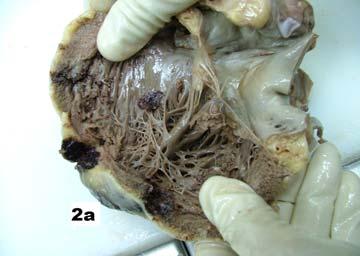
2a — Transmural infarction, haemorrhage in the myocardium
She was obese (80kg), measuring 157cm in length. Postmortem lividity was fixed on back and rigor mortis was well developed. No external injuries were detected.
The pericardium was intact and contained about 200ml of clotted blood. The heart (465 g) was hypertrophic. The pericardial fat and epicardium showed an area of hematoma measuring 4cm x 3cm at the apex. Cut section shows a transmural area of pallor and hemorrhage (infarction) of the left ventricular myocardium near apical region extending from the endocardium (Fig 2a). The left ventricular wall (Fig 2b) was slightly thickened (1.8cm thick). The left anterior descending artery showed moderate atherosclerosis with total occlusion of proximal 1/3rd by thrombosis. The right coronary artery showed mild atherosclerosis but was patent. Stomach was intact and empty.
On histopathological study of heart, focal areas of hemorrhage, degeneration and necrosis of myocardium was found with mild infiltration by lymphocytes and neutrophils. There was diffuse extravasation of blood into the pericardial fat. Left coronary artery showed atheromatous plaques comprising of histiocytes and lymphocytes with cholesterol clefts and micro calcifications on the intimal layer of the artery, with thrombosis causing significant to total occlusion of the artery.
In both cases, the cause of death was opined as cardiac tamponade due to myocardial rupture post myocardial infarction.
DISCUSSION
Risk factors for free cardiac wall rupture include age >60 years, female gender, pre-existing hypertension and lack of left ventricular wall hypertrophy7 The lateral wall, at midventricular level, is the most common site for post-infarction free-wall rupture7
The most common cause of myocardial rupture is a recent myocardial infarction, with the rupture occurring three to five days after infarction4 Left ventricle free wall rupture occurs in 2% to 4% of patients following acute Myocardial Infarction (MI)5. In the modern era of early revascularization and intensive pharmacotherapy for MI, the incidence of myocardial rupture is about 1% of all MI6
The free wall rupture is associated with immediate hemodynamic collapse and death secondary to acute cardiac tamponade. The overall risk of death depends on the speed of diagnosis, treatment provided and the underlying cause of the tamponade. Though in one case series, it was described that the mortality rate was 100% if myocardial rupture involved the free wall of Left Ventricle (LV)6
But another study8 describes, in some patients who survive LV free-
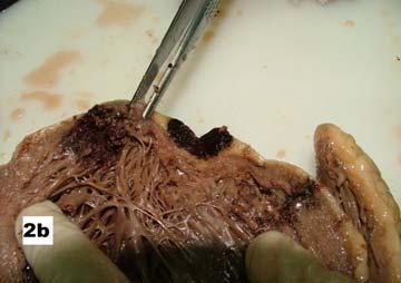
wall rupture following Acute Myocardial Infarction (AMI), the rupture can be sealed by epicardium (visceral pericardium) or by a hematoma on the epicardial surface of the heart. This has been referred to as LV diverticulum or contained myocardial rupture. Pericardiocentesis and surgical drainage of hemopericardium may be indicated9 or referred for urgent cardiac surgery (infarctectomy and Teflon patching) 10 .
But sometimes, sudden death due to (LV free-wall rupture may be the first manifestation of Coronary Artery Disease (CAD) in a small percentage of patients with AMI9. The cases described here were presented with very nonspecific symptoms. The autopsy & histopathological findings indicate repeated ischemic episodes of myocardium. Though none of the deceased ever complained of severe chest pain, which could had led to diagnosis of acute myocardial infarction.
So the physicians should have to suspect for myocardial ischemia from nonspecific symptoms like recurrent epigastric distress in a hypertensive (first deceased) and chest discomfort with dizziness and history of syncope as in the second case.
1Dorland’s Pocket Medical Dictionary. 26th ed. Philadelphia: WB Saunders Co, 2001
2Braunwald E — Pericardial Disease. In: Fauci AS, Braunwald E, Kasper DL, Hauser SL, Longo DL, Jameson JL, et al editors. Harrison’s Principles of Internal Medicine. Vol II. 17th ed. New York: Mc Graw-Hill companies, 2008: 1490.
3Oxford Handbook of Clinical Medicine. 5 th ed. New York: Oxford University Press ; 2002
4Figueras J, Alcalde O, Barrabes JA, Serra V, Alguersuari J, Cortadellas J, et al — Changes in Hospital Mortality Rates in 425 Patients with Acute ST-Elevation Myocardial Infarction and Cardiac Rupure Over a 30-Year Period. Circulation 2008; 118: 2783-9.
5Sakaguchi G, Komiya T, Tamura N, Kobayashi T — Surgical treatment for postinfarction left ventricular free wall rupture. Ann Thorac Surg 2008; 85(4): 1344-6.
6Yip HK, Wu CJ, Chang HW, Wang CP, Cheng CI, Chua S, Chen MC — Cardiac rupture complicating acute myocardial infarction in the direct percutaneous coronary intervention reperfusion era. Chest 2003; 124(2): 565-71.
7Schoen FJ — The heart. In: Kumar V, Abbas AK, Fausto N, editor. Robbins and Cotran Pathologic Basis of Disease. 7th. Philadelphia, Elsevier Saunders; 1999. 555-618.
8Helmy TA, Nicholson WJ, Lick S, Uretsky BF — Contained myocardial rupture: a variant linking complete and incomplete rupture. Heart 2005 91(2): e13.
9Myocardial Rupture — http://emedicine.medscape.com/article/156455 (accessed Sept 06, 2015)
10Matangi MF, Norris RM, Hill DG — Subacute cardiac rupture: a surgical emergency. N Z Med J 1983; 96(745): 1003-5.
J B Sharma1, S Gupta2, Alka Kriplani3, Sudheer Arava4, Swati Tomar5
Vulval tuberculosis is a rare form of female genital tuberculosis causing less than 1.1% of genital tuberculosis cases. It can manifest as hypertrophic lesion or ulcerative lesion and may simulate vulval cancer necessitating vulval biopsy and histopathological examination to confirm the disease. A case of a 23 years old female presenting with large hypertrophic vulval tuberculosis confirmed on histopathology and treated by antitubercular therapy is presented.
[J Indian Med Assoc 2021; 119(2): 41-2]
Key words : Female genital tuberculosis, vulval tuberculosis, biopsy, antitubercular therapy, hypertrophic lesion.
According to WHO Global TB Report 2015, out of total 9.6 million tuberculosis (TB) cases in the world in 2014 there were 3.2 million cases in women1. Out of 1.5 million deaths which occurred due to tuberculosis in 2014 - 4,80,000 deaths occurred in women1
Female genital tuberculosis is an important cause of significant morbidity especially infertility in India2. It mainly involves fallopian tubes (90-100% cases), endometrium (50-80% cases), ovaries (20-30% cases), cervix (5-15% cases)2,3. Involvement of vulva and vagina is rare2,3. Lesions on vulva and vagina may present as hypertrophic lesions resembling malignancy, less often non healing ulcers in the vulva may be seen4
Mrs X 23 years old P1L1 female presented to the Department of dermatology and Gynaecology, All India Institute of Medical Sciences (AIIMS) with complaints of vaginal discharge, swelling and fissuring of genitalia since last 5 years. The patient developed the symptoms while she was pregnant. She underwent Lower segment Caesarean section (LSCS) for obstetric indication and developed vulval swelling in the post operative period. She was operated in a Private Hospital in Jaipur but recurrence occurred within a month. On examination her general condition was fair. Systemic examination was unremarkable. Local examination revealed involvement of vulva extending into adjacent perineal, gluteal and inguinal areas in the form tender bosselated firm to soft swelling 1520 cm in size with irregular ulcers of 5 cm over both inguinal creases
1MD, FRCOG, Professor, Department of Obstetrics and Gynaecology, All India Institute of Medical Sciences, New Delhi 110029
2MD, Professor, Department of Dermatology, All India Institute of Medical Sciences, New Delhi 110029
3MD, FRCOG, FAMS, Professor and Head, Department of Obstetrics and Gynaecology, All India Institute of Medical Sciences, New Delhi 110029
4MD, Assistant Professor, Department of Pathology, All India Institute of Medical Sciences, New Delhi 110029
5 MD, Junior Resident, Department of Obstetrics and Gynaecology, All India Institute of Medical Sciences, New Delhi 110029
Received on : 16/03/2017
Accepted on : 09/08/2017
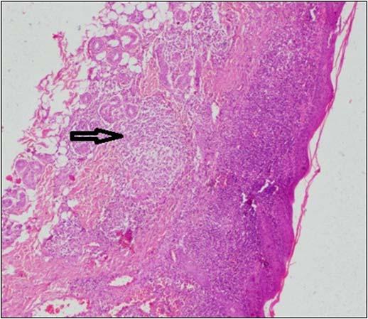
Fig 1 — Photomicrography shows (A) mild irregular acanthotic epidermis with dense band like chronic inflammation in the upper dermis along with a granuloma (Arrow)
(Figs 1-3). No inguinal lymphadenopathy. She was prescribed local antibiotic and steroid treatment which gave her no relief. Vulval biopsy was taken which showed epithelioid cell granulomas. Patient was started on Antii Tubercular Therapy (ATT) with rifampicin 450 mg, Isoniazid 300 mg, Ethambutol 1200mg and pyrazinamide 1500 mg. The patient is on follow up and showing improvement.
DISCUSSION
Tuberculosis of vulva is rare around 1% of all the female genital tuberculosis cases. Vulval lesions usually arise by direct extension from lesions in genital tract or from exogenous infection5,6. Exogenous infection can rarely be through sexual intercourse with a male partner suffering from either tuberculosis epididymitis, renal tuberculosis or tuberculosis of seminal vesicles7,8. Tuberculosis of Bartholin gland has also been described9
In the present case, the woman presented with hypertrophic lesions on vulva with significant edema of labia majora and elephantiasis like appearance of labia minora. The histopathology of vulval biopsy was
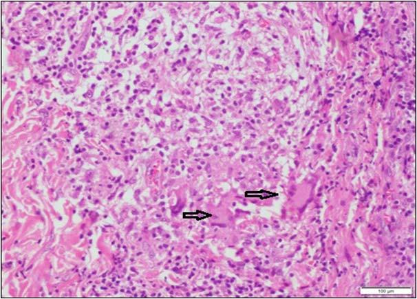
Fig 2 — (B) Higher magnification shows well-formed non necrotizing epithelioid cell granuloma with Langhans type of giant cell (Arrow). Stain for Acid-fast bacillus was negative
suggestive of tuberculous granuloma and was put on anti tuberculous therapy.
Kumar et al12 reported a large vulval tumor which was not responding to medical treatment. Surgical excision of this tumor was done and histopathological examination was suggestive of tuberculosis. Patient received ATT in post operative phase and recovered well. So a large tumor and vulval lesion may not respond to anti tubercular therapy and may require surgical removal followed by anti tubercular therapy for complete relief.
Tuberculosis of vulva should be considered in differential diagnosis of a hypertrophic lesion of vulva necessitating vulval biopsy to rule out malignancy.
Compliance with ethical requirements and conflict of Interest: The patient gave informed consent before inclusion in the case study. The author does not have any conflict of interest.
Acknowledgements : Author is thankful to Dr. Sandeep Mathur, Department of Pathology for performing Histopathology, Dr Somesh Gupta, Department of Dermatology.
1Global TB report 2015 — World Health Organization. WHO/HTM/TB/ 2015, Geneva, WHO,2015.
2Sharma JB — Current Diagnosis and Management of Female Genital Tuberculosis. J Obstet Gynaecol Ind 2015; 65(6): 362-71.
3 — A case showing vulval tuberculosis
3Sharma JB — Tuberculosis and obstetric and gynaecological practice In Studd J, Tan SL, Chervenak FA, (editors), Progress in Obstetrics and Gynaecology Philadephia: Elsevier 2008; 18: 395-427.
4Kumar S — Female genital tuberculosis. In: Sharma SK, Mohan A, editors tuberculosis 3rd edition Delhi: Jaypee Brothers Medical Publisher Ltd.; 2015. P 311-24.
5Tripathi SN — Tuberculosis Manual for Obstetrics and GynaecologistsJaypee Brothers Medical Publisher (P) Ltd. New Delhi. First Edition,2015.
6Schaefer G — Female genital tuberculosis. Clin Obstet Gynecol 1976; 19(1): 223-39.
7Lattimer JK, Colmore HP, Sanger G, Robertson DH, McLellan FC — Transmission of genital tuberculosis from husband to wife via the semen. Am Rev Tuberc 1954; 69(4): 618-24.
8Sutherland AM, Glen ES, MacFarlane JR — Transmission of genitourinary tuberculosis. Health Bull 1982; 40(2): 87-91.
9Dhall K, Das SS, Dey P — Tuberculosis of Bartholin’s gland. Int J Gynecol Obstet 1995; 48(2): 223-4.
10Tiwari P, Pal DK, Moulik D — Hypertrophic Tuberculosis of Vulva- a rare presentation of Tuberculosis. Indian J Tuberc 2010; 57: 95-97.
11Wong S, Rizvi H, Cerio R, O’Toole EA — An unusual case of vulvalpapulonecrotictuberculid. Clin Exp Dermatol 2011; 36(3): 277-80.
12Kumar S, Kameshwarachari P, Ray R — Giant vulval tumor due to tuberculosis. Int J Gynaecol Obstet 2010 Jul; 110(1): 69-70.
Q1. What is the pre-requisite for enrolling in residency programme?
In India as of date, the rank obtained in the NEET examination is the major criteria for admission to any medical course be it undergraduate or post graduate courses. Some institutions in the past like CMC Vellore, Ludhiana etc were giving emphasis on an interview which was looking at an aptitude of the candidate in the subject but largely was aimed at assessing the ability to fit into the cultural fabrics of that institution. However by law today the ranking in the NEET is the sole criterion. But by default of the system, the choice on subject and place given by the candidate also influence the admission. The greatest draw back in the present system is that the speciality based knowledge, aptitude or skill is NOT tested but is confined to global knowledge on the whole of medical subjects. It also does not permit anyone to change the speciality during the tenure of his course.
Q2. What should be the Ideal Duration of Residency Programme ?
In India, MD/MS/DNB is considered as the end of training programme and we do not have a structured post-doctoral supervised training programme. The three years of training, currently followed is grossly inadequate even in accrual of knowledge particularly with exponential growth in medical science. Unscientific increase in postgraduate medical seats has a detrimental effect on skill development in most of the procedure based specialties. To start with the number of medical seats is to be kept based on national population based needs. After basic speciality exposure on a common platform for a period of three years, further defined specialty based training for five years again based on demographic needs will be the ideal system. But the concept of true residency as followed in the West needs to be followed where, the system should
understand the residency is NOT just equivalent to the other non-medical courses but is an in house job training where the resident carries out certain amount of the work. This concept should hence provide a reasonable pay for each year of residency replacing the meagre stipend given for a trainee. The subspecialty division should also be judicious with a practical clinical outlook.

Dr Santhosh John Abraham MS, DipNB(Surg), FRCS(Eng), FRCS(Edin), FRCS(Glas), FACS, Head, Department of Surgery, Lourdes Hospital, Kochi, Kerala
Procedure based specialities need more comprehensive skill development programmes. Hence it may be beneficial for a longer period of speciality training than the current three year duration.
Q3. Do we need to have Separate Programmes / Schedules for Surgical & Non-surgical Residencies?
The basic ethos of training can be the same in both types. I would rather be tempted to mention that when there is a procedure to be learned and performed in any speciality, there needs to be a structured training and a graded autonomy which needs to be constantly audited, watched and interfered by a competent professional body. Currently the system in India only looks at the completion of the prescribed duration of the course for purpose of legal licensing to practice that specialty. We have no system that ensures certain mandatory hands on training for procedures.
Q4. What is the present status of various residencies in India?
All residency programme India today is governed only by a stipulated time frame and is based on curriculum defined by the State Medical University or National Board or any other University. There is no interim assessment and there is no vigilant system to ensure quality of training. The completion is tested on the basis of just one exit examination. A continuous objective and skill based assessment can substantially improve the standard of training which will also make the trainers more accountable. The trainers also need to be brought under constant scrutiny to improve the entire system.
Intermittent assessment of both trainees and trainers may lead to a holistic system of education
Q5. How is it different from other developed countries like USA or UK?
In India, the post-graduation is still considered as a “course” involving only a process of admission and an exit examination. But in most other countries, it is called a specialty based training programme well-structured by a specialty based professional body which is on the constant watch on the quality of both the trainer and the trainees. Unless the trainers are made accountable, the training system will have lots of bias.
Q6. What changes are necessary for betterment of the programme? How to fill in the lacunae?
The concept of “Course” needs a change to “in house training programme”. Both trainer and trainee need to come under the constant scrutiny of a “professional” body which should be held responsible for constant in house training, assessment and exit examination. Licensing can on still legal grounds be done by a non-professional agency like Medical Council or its equivalent. Concept of state wise Medical Universities granting the degrees
leads to variation in standards and should be made uniform all across the country with independent bodies like National Board of Examinations.
A standardized training programme under the guidance of an independent professional body all over the country is desirable for the improvement of the current system of residency.
Q7. Should we regulate the working hours of the residency programme?
Certainly it should be regulated to prevent early burn out. The concept of cheap labour needs to be changed into a proud professional training,by the managements of private institutions. The time of professional work should be kept reasonable.
Q8. Does the present condition affect the mental condition of the student?
It is dependent now on the practices adopted in the institutions and the methodology followed by the trainer and is not uniform. Both these should be made uniform to ensure national uniformity. The pressure on workload can affect the mental status and systems should be in place to avoid this and to render help if such a situation arises.
Q9. What steps can be taken to ensure standardization of programme in different institutes?
I think I have addressed this in the previous questions
Q10. Is there any residency exchange programme?.
Presently we don’t have any exchange programmes. It is only an institution based. It is a good idea to expose trainees at different levels to different institutions to understand the various methodologies adopted to address the same diseases or for subspecialty exposure.
Thank you Dr Santhosh John Abraham for your answers. We appreciate the time taken by you and we are sure that our readers will be benefited immensely.
Ghanshyam Goyal1, Rekha Srivastava1, Shammi Kapoor1

A 57 years old, female having type 2 diabetes presented to the diabetic foot clinic with multiple ulcers on planter surface of both feet (Figs 1&2).
Patient had a history of walking bare foot to the temple every morning. She had developed blisters and now the ulcers were non healing since last five months.
Editor's Comment :
Offloading plays an important role in the management of neuropathic planter surface Diabetic Foot Ulcer. In this patient, S K Offloading , an indigenous offloading technique based on the principals of Samadhan System and Modified Posterior Slab (synthetic splinting system made from polyurethane coated fibreglass covered by polypropylene padding) were the offloading techniques used.
On examination both Dorsalis Pedis and Posterior Tibial were palpable. Ankle / Brachial Index was normal. Vibration Perception Threshold was severely impaired. X-Ray of both feet were normal. A diagnosis of non healing neuropathic ulcers in insensate feet caused by thermal injury and maybe minor trauma due to barefoot walking was made.
Debridement of ulcers was done in out patient setting (Figs 3 & 4).
After debridement the patient was put on a modified posterior slab in right foot (Fig 5) and S K Offloading in the left foot ( Fig 6).
Family members were trained to take care of the cast and dress the wound at home. The patient was followed up after every 2 weeks and complete wound closure in both feet was achieved in 15 weeks (Figs 7 & 8).
1https://iwgdfguidelines.org/guidelines
REFERENCES
2IDF Clinical Practice Recommendations on the Diabetic Foot – 2017
Uttam Biswas1, Pallavi Mahato2, Milan Chakraborty3
Fever is one the most common presenting symptoms in our daily practice both in OPD or IPD but sometimes it becomes the diagnostic challenge for most clinicians with differentials running in hundreds. Pyrexia of Unknown Origin (PUO) reserved for those febrile illness in which no diagnosis could be reached in spite of extensive investigations. In most of the time it is very challenging to diagnose a case of true PUO, specially in a resource limited country like India. Thorough history and clinical examination is very important to find out potentially diagnostic clues (PDCs) and it is very useful for further evaluation .In this article we have outlined the latest approach to diagnosed a case of pyrexia of unknown origin (PUO). So this article will be very useful for the physicians to evaluate a case of Pyrexia of unknown origin. (PUO).
[J Indian Med Assoc 2021; 119(2): 46-9]
FKey words : Pyrexia of Unknown Origin (PUO) , Approach,Evaluation.
ever may be the most common presentation in OPD/IPD but is the most intriguing diagnostic challenge for most clinicians with differentials running in hundreds. Most fevers usually resolve even prior to a proper diagnosis can be reached or develop additional features that help localise the cause. Pyrexia of Unknown Origin (PUO) also known as Fever of Unknown Origin (FUO), reserved for those febrile illness in which no diagnosis could be reached in spite of extensive investigations. In a resource limited country like India it difficult to diagnose a case of true PUO, thorough history and clinical examination is very useful for further work up. So this article will be very useful for the evaluation of a case of Pyrexia of unknown origin (PUO).
Definition :
In 1961, Petersdorf and Beeson first coined the term “fever of unknown origin” defined it as temperature more than 38.3°C (101°F) on two occasions and duration of fever more than 3 weeks but failure to reach to diagnosis despite 1 week of inpatient investigation.1 Later on inpatient management was provided only to patients requiring hospitalisation, hence the latter part of the definition had to be excluded.
Later on, In 1991,Durack and Street 2 classified PUO into 4 types:
•Classical
•Nosocomial
•Neutropenic
•HIV
Minimum 3 OPD visits or 3 days of in hospital investigation is needed before classifying a case as PUO.
Nowadays, PUO is defined as: 3
•Fever more than 38.3°C (101°F) on atleasttwo occasions
•Illness duration of more than 3 weeks
•No known immunocompromised state
1MD (Medicine), MRCP(UK), Assistant Professor, Department of Medicine, Burdwan Medical College and Hospital, Purba Burdwan 713104
2MD (Medicine PGT), Junior Resident, Department of Medicine, Burdwan Medical College and Hospital, Purba Burdwan 713104
3MD (Medicine), Professor, Department of Medicine, NRS Medical College and Hospital, Kolkata 700014
Received on : 05/02/2021
Accepted on : 08/02/2021
•Diagnosis that remains uncertain after a thorough history taking, physical examination, and the following obligatory investigationdetermination of ESR, CRP, platelet count, leucocyte count, differential count, haemoglobin level, electrolytes, creatinine, total protein, alkaline phosphatase, alanine aminotransferase, aspartate aminotransferase, lactate dehydrogenase, creatine kinase, ferritin, antinuclear antibody and rheumatoid factor; protein electrophoresis; urine analysis; blood culture(n=3); urine culture; chest xray; abdominal usg; and tuberculin skin test or interferon gamma release assay.
Causes of PUO :
The possibilities can be extensive and with the various studies it has been postulated that PUO is more often caused by an atypical presentation of common diseases rather than by a very rare disease. 4 The original categories for the diseases that cause classic PUO are still the same. These are:
•Infectious
•non-infectious inflammatory disease (NIID)
•neoplastic
•miscellaneous (fraudulent fever, factitious fever, drug fever, etc)
The most common causes of each category are described in the table here:5 Infection is much more common cause of PUO in India as compared to western world (43% vs.17%) and tuberculosis accounts for 50% of those infections.6
As because a wide variety of etiology may cause PUO, a stepwise approach consisting of two phases is followed. PUO related mortality has decreased immensely to around 10% as most cases are due to some treatable cause. Malignancy is usually responsible for most mortality especially NHL.
First Stage Diagnostic Test :
The most important step is to search for potentially diagnostic clues (PDCs) that will point towards a diagnosis. PDCs are defined as all localising signs, symptoms and abnormalities through repeated history taking, thorough clinical examination and obligatory investigations listed below.
History : should include information about •Fever pattern and duration
Infectious Non Infectious Neoplastic Miscellaneous Inflammatory Disease
Bacterial
• Occult abscess
• Complicated urinary
• Giant cell arteritis
• RA
• Non-Hodgkin lymphoma
• Drugs (antibiotics,
• Leukaemiaantiepileptic, tract infection
• Culture negative
• endocarditis
• Sarcoidosis
• Adult Still’s disease
• Systemic lupus
• Renal cell carcinoma NSAID, antriarrthymic)
• Hepatocellular carcinoma
• Metastatic lesions
• Cirrhosis
• Pulmonary embolism
• Osteomyelitis erthymatosus (commonly hepaticand DVT
• Tuberculosis
• Coxiellaburnetti
• Polyarteritisnodosa metastasisfrom
• Inflammatory bowel
• Granulomatosis adenocarcinomas disease (Q fever) with polyangitis
• Rickettsial infections (Wegener granulomatosis)
• Enteric fever
• Familial Mediterranean
• Brucellosis fever
Viral
• Epstein–Barr virus
• Cytomegalovirus
• Human immunodeficiency virus
Parasitic
• Malaria
• Visceral leishmaniasis
• amebiasis
• Toxoplasmosis
Fungal
• Histoplasmosis
• Aspergillosis
• Crytococcosis
• Endocrine disease
• Hyperthyroidism
• Factitious fever
Source : Wassimabdelwahab E Pyrexia of Unknown Origin: current perspective. IJBR 2019; 10(1): e4987.
•Previous medical and surgical history
•Recent drug history
•Family and sexual history
•Recent or remote travel
•Unusual environmental exposures associated with travel or hobbies and animal contacts
Physical Examination : Complete and repeated physical examination to search for the PDCs must include-
•Examination of eye and fundoscopy
•Skin
•Lymph nodes
•Temporal arteries
•Liver and spleen
1st stage investigation : Investigations discussed in the new definition are obligatory , however further biochemical testing may lead to a definitive diagnosis.6 Before diagnostic tests are initiated all drugs must be stopped including steroids or antibiotics which can mask may diseases as well as rule out drug fever. PDCs are a diagnostic suspicion which represent after an initial workup (Fig 1).With a directed personalized approach, few additional investigations are to be done to confirm the suspicion according to the Fig 1.
When PDCs have been obtained, a list of differential diagnosis have to be formed and specific tests are performed to confirm them. If PDCs are absent or guided tests come out to be negative, following investigations.
•Cryoglobulin
•Fundoscopy
•FDG-PET/ gallium scan/ labelled leucocyte scintigraphy .If used
at an early stage FDG-PET/CT can be cost effective diagnostic tool in establishing an early diagnosis, reducing hospital stay and decreasing the use of unnecessary testing. 7If fever persists after discontinuation of drugs for at least 72 hours ,scintigraphy or FDG-PET/CT should be performed.3
Potential diagnostic clues and possible etiologies:8
Later stage diagnostic tests :
Scintigraphicor FDG PET / CT abnormalities need to be confirmed by pathology or culture biopsy of specimen.6If no diagnosis can be reached, reconsidering history and physical examination to search for PDCs followed by invasive guided testing to be done. If still nothing can be found, the following 3 tests to be done.
•Chest CT
•Abdominal CT
•Temporal artery biopsy (>55 years)
Biopsy is considered to be an important diagnostic modalities in the second stage diagnostic testing. It includes temporal artery biopsy liver biopsy, lymph node biopsy, pleural biopsy, pericardial biopsy, bone marrow biopsy.9,10 If no diagnosis can be reached till now then further follow up to be done for new PDCs if patient is stable or consider for therapeutic trial if patient is deteriorating.
PDCs
Possible Etiology
•Unintentional wt loss•Neoplastic process
•Tuberculosis
•Brucellosis
•Drenching night sweats•Haematological malignancy
•tuberculosis
•joint pain (early morning)• inflammatory arthritis
•unilateral retroorbital headache, •giant cell arthritis
jaw claudication,
•history of steroid use, surgeries,• occult abscesses
prosthetic material
•history of smoking•malignancy
•prolonged immobility, OCP• thromboembolic disease
•recent change of medication• drug fever
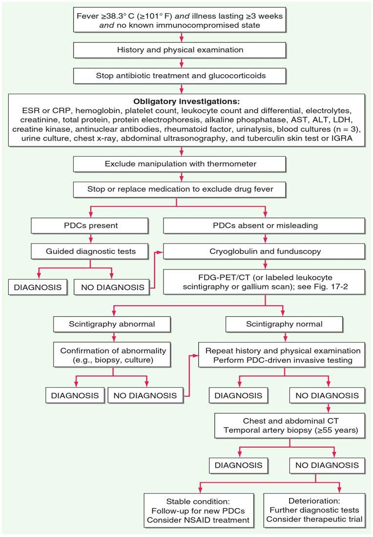
119, No 2, February 2021Journal
Treatment :
As a norm empirical therapy is to be avoided as that may change the clinical picture and definite diagnosis can never be reached. Emperical therapy with antiobiotic, ATD or glucocorticoid should be started only in certain conditions as given below:
• Antibiotics : hemodynamic instability and neutropenia, empirical antibiotic to be started.
• Antitubercular drugs : If TST or IGRA is positive or if granulomatous disease is present but sarcoid seems unlikely; a trial of ATD may be done. If fever doesn’t resolve after 6 weeks of ATD, alternate diagnosis to be considered.
• Colchicine : may be tried in patients with symptoms compatible with familial mediterranena fever, especially those residing in high prevalence regions.
• NSAIDS: when dramatic response to NSAID is seen, adult onset stills disease may be a diagnostic possibility.
• Glucocorticoids: similar dramatic response is seen in giant cell arteritis and polymyalgia rheumatica.
• Anakinra: therapeutic trial of this IL1 antagonist may be considered in patients whose PUO has not been diagnosed after later stage diagnostic testing. Anakinra remains free from adverse effects associated with steroid but with a better control.
The ability of immunosupressants to be able to mask fever yet allowing the spread of infection or lymphoma enforces that their use to be avoided until infectious disease/ malignancy is ruled out and inflammatory disease is a strong suspicion and is organ/life threatening.
Conclusion :
With the greater understanding of the PUO and easy accessibility tothe wide range of investigations both non invasive and invasive, now a days majority of PUO cases can be diagnosed but still it is a diagnostic challenge.It is highly recommended to search for uncommon manifestation of a common disease rather than common manifestations of an uncommon disease. Despite promising newer technologies, detailed and repeated history, thorough clinical examination and baseline investigations still remain the cornerstone to reach at a definitive diagnosis.
1Petersdorf RG, Beeson PB — Fever of Unexplained Origin: Report on 100 cases. Medicine 1961; 401-30.
2Durack DT — Fever of unknown origin -re-examined and redefined.Curr Clin Top Infect Dis 1991; 11: 35-51
3Patro S — Approach to a case of pyrexia of unknown origin: What’s New in 2020.Medicine Update 2020. Japee brothers medical publishers Vol. 30; Chapter 203; 871-74.
4Bleeker-Rovers CP, Vos FJ, De Kleijn EM — A prospective multicenter study on Fever of Unknown Origin: the yield of a structured diagnostic protocol Medicine 2007; 86(1): 26-38.
5Wassimabdelwahab E — Pyrexia of Unknown Origin: current perspective. IJBR 2019; 10(1): e4987.
6Fauci AS, Jamesson JL, Kasper DL — Harrison’s principles of internal medicine,20 th edition New York : McGraw Hill, Health Professions Divission 2018. 114-22.
7Nakayo EB, Vicente AG, Castrejon AS — Analysis of cost-effectiveness in the diagnosis of fever of unknown origin and the role of 18F-FDG PETCT: A proposal of diagnostic algorithm.Revista Espanola de medicina nuclear eimagen molecular(English edition) 2012; 31(4): 178-86.
8Beresford RW, Gosbell IB — Pyrexia of unknown origin: causes, investigation and management. Int Med J 2016; 46(9): 1011-6
9Rupali P, Garg D, Viggweswarupu S — Etiology of Classic Fever of unknown origin (PUO)among immunocompetent Indian adult. JAPI 2019; 67: 21.
10Jha A, Sarda R —Value of bone marrow examination in pyrexia of unknown origin. J Pathol Nepal 2013; 3(6): 447-51.
The information and opinions presented in the Journal reflect the views of the authors and not of the Journal or its Editorial Board or the Publisher. Publication does not constitute endorsement by the journal.
JIMA assumes no responsibility for the authenticity or reliability of any product, equipment, gadget or any claim by medical establishments/institutions/ manufacturers or any training programme in the form of advertisements appearing in JIMA and also does not endorse or give any guarantee to such products or training programme or promote any such thing or claims made so after.
— Hony Editor
[We are publishing this Special Correspondence to commemotare Word Cancer Day on 4th February]
Arnab Gupta1
As we observe World Cancer Day, it is important that we remember of the basics of Cancer prevention and management for a better future. Incidence of Cancer is rapidly increasing. It is likely to affect every 4 th person by 2030 according to WHO. Even now, awareness of this deadly disease is hardly there especially in rural India. Late-stage presentation and inaccessibility to healthcare services are still common. There are several myths which make things even more complicated. Although atleast 50% of cancer is preventable and mostly lifestyle related, not much is done about these preventable factors. Cancer is curable if detected early and treated properly. Hence stress should be put on prevention and early detection through awareness and screeing programmes. Access to healthcare should also be there for early and proper treatment. Only 1 in 5 low and middle income countries have the necessary data to drive cancer policy 5 [J Indian Med Assoc 2021; 119(2): 50-5]
Key words : Cancer, incidence, lifestyle, screening, awareness, prevention.
Cancer is the second leading cause of death globally, and is responsible for an estimated 9.6 million deaths in 2018. Globally, about 1 in 6 deaths is due to cancer. The incidence of Cancer in India is 94.1 & 103.6/ 100,000 population in males and females respectively. Approximately 70% of deaths from cancer occur in low and middle income countries. The economic impact of cancer is significant and is increasing. The total annual economic cost of cancer in 2010 was estimated at approximately US$ 1.16 trillion4 Cancer is as old as mankind. It was found in the mummy of Egypt from 5000 years back. Unfortunately, even now, many people are unaware about its nature, its cause, its prevention and most importantly there are several misconcepts which make the problem even more complicated. The incidence of Cancer is increasing. WHO is predicting that in another 10 yrs, almost every family will have a member who is likely to get cancer1. The only way to escape this ordeal is to know what causes cancer and how we can prevent them. Also, one must seek early medical help so that it can be detected at a very early or even pre-cancerous stage and the cure rate can be very high.
What is Cancer ?
Cancer arises from the transformation of normal cells into tumour cells in a multistage process that generally progresses from a precancerous lesion to a malignant tumour. These changes are the result of the interaction between a person’s genetic factors and 3 categories of external agents, including:
•physical carcinogens, such as ultraviolet and ionizing radiation;
•chemical carcinogens, such as asbestos, components of
1MBBS, PLAB, FRCS (Edinburgh), ATLS, FIAGES, FRCS, Medical Director & Professor, Department of Surgical Oncology, Saroj Gupta Cancer Centre & Research Institute, Mahatma Gandhi Road, Thakurpukur, Kolkata 700063
Received on : 30/01/2021
Accepted on : 01/02/2021
tobacco smoke, aflatoxin (a food contaminant from mouldsx), and arsenic (a drinking water contaminant); and
•biological carcinogens, such as infections from certain viruses, bacteria, or parasites.
WHO, through its cancer research agency, International Agency for Research on Cancer (IARC), maintains a classification of cancercausing agents.
Ageing is another fundamental factor for the development of cancer. The incidence of cancer rises dramatically with age, most likely due to a build-up of risks for specific cancers that increase with age. The overall risk accumulation is combined with the tendency for cellular repair mechanisms to be less effective as a person grows older.
What are the Common Types of Cancer ?
In male, where tobacco addiction is a common problem, the common cancers are that of oral cavity, lung and prostate. In females, in rural population the common cancer is that of uterine cervix, whereas that of urban women it is breast cancer.
The most common cancers are:
•Lung (2.09 million cases)
•Breast (2.09 million cases)
•Colorectal (1.80 million cases)
•Prostate (1.28 million cases)
•Skin cancer (non-melanoma) (1.04 million cases)
•Stomach (1.03 million cases)
The most common causes of cancer death are cancers of:
•Lung (1.76 million deaths)
•Colorectal (862 000 deaths)
•Stomach (783 000 deaths)
•Liver (782 000 deaths)
•Breast (627 000 deaths)
What are the symptoms of cancer?
1.General symptoms :
Loss of appetite and body weight, weakness, anaemia (as the rapidly multiplying cancer cells take away all the nutrition and also for decreased appetite and recurrent bleeding).
2.Local symptoms:
A new lump which suddenly appears and rapidly increases in size.
An ulcer which is not healing, increasing and bleeding. Symptoms will also depend on the site of cancer.
Cancer in the lung : will cause persistent cough which may be associated with heamoptysis
That of the Larynx : persistent hoarseness, stridor
Oesophagus : increasing difficulty in swallowing solids and later liquids also.
Breast : Lump (initially painless), skin changes, bleeding from the nipple
Stomach : anorexia, anaemia, asthenia, haemoptysis, melaena
Uterus : intermenstrual bleeding, bleeding after coitus, postmenopausal bleeding
Urinary bladder or kidney- haematuria
Colo-rectum- bleeding with stool (fresh/ altered), change in bowel habit.
119, No 2, February 2021Journal
Leukaemia- common symptoms are recurrent fever, anaemia, bruising and bleeding that fail to stop even after minor injuries. What are the Causes of Cancer ? How We Can Prevent Them?
Unfortunately, the causes in 50% of all cancers are still not clear, and several researches are going on worldwide to determine this. In nearly half of the cases, the causes are proved beyond doubt- of which the most important is tobacco in any form- smoking (Bidi, cigarette, hukka etc.) and smokeless (paan, paan masala, ghutka, khaini, jarda etc.)2.
Around one third of deaths from cancer are due to the 5 leading behavioral and dietary risks: high body mass index, low fruit and vegetable intake, lack of physical activity, tobacco use, and alcohol use.
Tobacco use is the most important risk factor for cancer and is responsible for approximately 50% of cancer deaths in men & 25% in women.Tobacco has nearly 4800 chemicals of which atleast 69 are known to be carcinogenic. It causes 1 death every 6 seconds and kills 1 million people every year globally and WHO predicts that the number is going to cross 2 millions by 2030. Not only the people who smoke actively are at risk but also the people around, through passive smoking. It is known to cause Heart attacks, stroke, bronchitis, different cancers, depression along with many other medical ailments.
The common cancers that are caused by tobacco are that of Oral cavity, Larynx, Oesophagus, Lungs, Stomach, Pancreas, Urinary bladder, Uterine cervix, Prostate and kidney. Active & Passive smoking are equally dangerous.
The Tobacco addicts should have strong determination to quit tobacco. The addiction is mostly because of one of its thousands of toxic components called Nicotine for which a person becomes dependent both mentally and physically. Nicotine chewing gums/ patch3 which are available should be used when there is craving for tobacco. It alone is much less toxic and this can also be gradually stopped over a few weeks. Fortunately the risks of a tobacco addict comes down significantly within 10 yrs after stoppage of tobacco consumption, especially if they quit before the age of 45 years. So it is never too late.
Diet and Body Weight :
The other less common causative agents are diet (adulterated food, too much of junk food, high fat, red meat, stored and charred/ smoked food). About 40 prospective studies have shown that there is 20% increased risk of having Colo-rectal cancer on eating 50 grams of processed meat daily, may be to do with the added nitrates in processed meat4,5. While diet and weight loss are central for cancer prevention, combining a good diet with other healthy habits can further lower the risk, according to a study in the May 2016 issue of JAMA Oncology
Harvard researchers examined four main lifestyle areas that are associated with health status: smoking, drinking, weight, and exercise. They looked at 46,000 men over 26 years and classified
about 12,000 as a low-risk group because they engaged in defined healthy behavior in all four areas—they did not smoke, drank moderate amounts of alcohol (no more than two servings per day), had a body mass index of 18.5 to 27.5, and engaged in 150 minutes of moderate-intensity exercise per week.
When they compared these men with others who did not meet these standards, the researchers discovered that men could avert or delay 67% of cancer deaths and prevent 63% of new malignancies each year. In terms of specific cancers, men could reduce incidence of bladder cancer by 62%, prostate cancer by 40%, and kidney cancer by 36%.
It is therefore imperative to have healthy diet which includes ample amount of fresh fruits, vegetables and plenty of water. Moderate amount of fish, eggs and chicken are not harmful. Onion, garlic, turmeric, spinach, carrots, tomatoes, green tea are supposed to carry Anti-oxidants which can prevent cancer to certain extent. Avoid heating food in plastic containers like that in Microwave.
A 2014 study in the Lancet found that higher body mass index increases the risk of developing some of the most common cancers. Scientists discovered that among five million people studied, a gain of 34 pounds was linked with a 10% increase in the risks of cancers of colon, gall bladder, kidney and liver. Obesity is also associated with cancers of GE junction (for increased reflux) and breast.
Aflatoxins, produced by molds and commonly found in countries with high humidity and high temperature, can cause Liver cancerespecially Aflatoxin B1, B2, G1, G2. Hence, stored and contaminated foods, especially Grains and peanuts, should be avoided.
Virus :
For uterine cervical cancer the common causes are early and multiple children, multiple partners, low personal hygiene, infection by Human Papilloma Viruswhich is usually sexually transmitted. Young women should consider having Vaccine against this virus which can give them 90% life time protection against this very common cancer in India. Currently the recommended age for vaccination is 9 yrs to 26 yrs. Women above 30 yrs should have annual Pap smear to look for pre-cancerous cells which, if found, can be treated with nearly 100% cure.Approximately 15% of cancers diagnosed in 2012 were attributed to carcinogenic infections, including Helicobacter pylori, Human papillomavirus (HPV), Hepatitis B virus, Hepatitis C virus, and Epstein-Barr virus3
•Hepatitis B and C virus and some types of HPV increase the risk for liver and cervical cancer, respectively. Infection with HIV substantially increases the risk of cancers such as cervical cancer. Cancer causing infections, such as hepatitis and human papilloma virus (HPV), are responsible for up to 25% of cancer cases in low- and middle-income countries 3. Epstein- Barr Virus can cause Burkitt`s Lymphoma, cancer of Nasopharynx and may also be associated with cancers of stomach.
Sunlight :
The most beneficial effect of sun exposure is the production of vitamin D in the skin. Recent evidence suggests that most cells in
119, No 2, February 2021Journal
the body not only have a vitamin D receptor but also have the capacity to convert 25-hydroxyvitamin D to 1,25-dihydroxyvitamin D. Once formed 1,25-dihydroxyvitamin D can inhibit cellular proliferation, induce cellular maturation, inhibit angiogenesis and ultimately cause apoptosis to prevent malignancy. A multitude of studies have associated improved vitamin D status with decreased risk for developing several deadly cancers including colon, breast, pancreatic and ovarian cancers. Patients with cancer are at high risk for vitamin D deficiency. Sensible sun exposure, vitamin D fortification and vitamin D supplementation should be encouraged to improve the vitamin D status of children and adults not only for bone health but for reducing risk of developing and dying of cancer. The goal is to achieve a blood level of 25-hydroxyvitamin D of 4060 ng/mL.(especially in the white race), Arsenic and Industrial wastes in soil and water can cause skin cancer8. There is no doubt that solar ultraviolet (UV) exposure is the most important environmental risk factor for the development of non-melanoma skin cancer especially in white races where there is absence of protective Melanin in the skin. Therefore, sun protection is of particular importance to prevent these malignancies, especially in risk groups. Well-balanced recommendations on sun protection have to ensure an adequate vitamin D status, thereby protecting people against adverse effects of strict sun protection without significantly increasing the risk of developing UV-induced skin cancer9.
Air Pollution :
Exposure to outdoor air pollution poses an urgent public health challenge worldwide because it is ubiquitous, affecting everyone, and has numerous serious adverse human health effects, including cancer.Major primary air pollutants, those emitted directly into the environment largely as a result of combustion of fossil and biomass fuels, include gaseous pollutants (such as sulfur dioxide [SO2], nitrogen dioxide [NO2], carbon monoxide [CO], and volatile organic compounds [VOCs]) and particulate matter (PM) (including carbonaceous aerosol particles, such as black soot). Although CO levels are often low outdoors in the developed world today (because of the use of emission controls such as catalytic converters on automobiles), high levels can be experienced near biomass burning sources, including wildfires. In addition, secondary air pollutants are formed in the atmosphere from primary pollutants and include gaseous ozone (O3), a major component of photochemical smog, formed in the atmosphere when nitrogen oxides (NOx) and hydrocarbons such as VOCs react in the presence of sunlight. Similarly, particulate sulfate (eg, sulfuric acid [H2SO4]) and nitrate (eg, ammonium nitrate [NH4NO3]) aerosols are commonly created in the atmosphere from SO2 and NOx, respectively. Primary combustion particles and secondary particles are small in diameter and are often referred to as fine particulate matter, or PM2.5 (particles =2.5 µm in aerodynamic diameter). Submicron combustionrelated PM2.5 is of particular health concern because it contains numerous toxic compounds (eg, acids and heavy metals), and can penetrate deeper into the lung than the larger PM generated by
natural processes, such as most windblown soil particle mass4.
Genetic: Only 5% of all cancers are genetic (hereditary). If more than 1 member in the family had cancer (espthat of breast, ovary, colo-rectal, prostate and retinoblastoma), the family members should get genetic testing done.
Hereditary Breast and Ovarian Cancer (HBOC) syndrome: In some families, many women develop breast cancer and/or ovarian cancer. Often these cancers are found in women who are younger than the usual age these cancers are found, and some women might have more than one cancer (such as breast cancer in both breasts, or both breast and ovarian cancer). This is known as Hereditary Breast and Ovarian Cancer syndrome (HBOC).
Most often, HBOC is caused by an inherited mutation in either the BRCA1 or BRCA2 gene. (Some families have HBOC based on cancer history, but don’t have mutations in either of these genes. Scientists believe that there might also be other genes that can cause HBOC.)
The risk of breast and ovarian cancer is very high in women with mutations in either BRCA1 or BRCA2, but it tends to be higher with BRCA1 mutations. Along with breast and ovarian cancer, this syndrome can also lead to fallopian tube cancer, primary peritoneal cancer, male breast cancer, pancreatic cancer, and prostate cancer, as well as some others. Male breast cancer, pancreatic cancer, and prostate cancer can be seen with mutations in either gene, but are more common in people with BRCA2 mutations. In the US, mutations in the BRCA genes are more common in people of Ashkenazi Jewish descent than in the general population.
Women with a strong family history of breast cancer and/or ovarian cancer may choose to undergo genetic counseling to help estimate their risk for having a mutation in one of the BRCA genes. The genetics professional can estimate the risk based on a patient’s history of cancer and the history of cancer in their family. If they have a high risk, they might choose to be tested (see Understanding Genetic Testing for Cancer). If a mutation is present, the woman has a high risk of developing breast cancer and ovarian cancer (as well as some other cancers). She can then consider steps to find cancer early and even lower her risk of getting cancer in the form of prophylactic surgeries like Bilateral Mastectomies and Bilateral Salpingo-oophorectomies.
Because breast cancer is rare in men, men with this cancer are often offered genetic counseling and testing for BRCA mutations. Although having a mutation is less likely to affect a man’s future health than it is a woman’s, it can affect his risk of some cancers, such as prostate and pancreatic cancer. It can also be helpful for a man’s close relatives to know that he has a mutation and that they might be at risk.
If someone has a BRCA mutation, it means that their close relatives (parents, siblings, and children) have a 50% chance of having a mutation, too. They may wish to be tested for the mutation, or even without testing may wish to start screening for certain cancers early or take other precautions to lower their risk of cancer.
Lynch syndrome (hereditary non-polyposis colorectal cancer)
119, No 2, February 2021Journal
The most common inherited syndrome that increases a person’s risk for colon cancer is Lynch syndrome, also called hereditary non-polyposis colorectal cancer (HNPCC). People with this syndrome are at high risk of developing colorectal cancer. Most of these cancers develop before they are 50.
Lynch syndrome also leads to a high risk of endometrial cancer, as well as cancers of the ovary, stomach, small intestine, pancreas, kidney, brain, ureters (tubes that carry urine from the kidneys to the bladder), and bile duct.
Lynch syndrome can be caused by a mutation in any of several mismatch repair (MMR) genes, including MLH1, MSH2, MSH6, PMS1, and PMS2. These genes are normally involved in repairing damaged DNA. When one of these genes isn’t working, cells can develop mistakes in their DNA, which might lead to other gene mutations and eventually cancer.
Asbestos : It is known to cause lung cancer, mesothelioma, cancer of the larynx and ovary, and asbestosis (fibrosis of the lungs) 10].
Controverisial : Plastic containers & Microwave:
There has been some concern that food may absorb plasticizers, the substances used in plastic containers and wraps to make them more flexible. Many people are especially concerned about microwaving at high temperatures or heating fatty or oily foods like cheese and meat. General recommendations from Canadian Cancer Society on how to safely store and reheat food:
•Use glass, ceramic and plastic containers and plastic wrap that are labelled as microwave safe. Always follow the instructions on the label when you use microwave-safe containers to heat food.
•Never heat or store food in plastic containers that were not intended for food. Single-use containers, like margarine tubs, tend to warp or melt in the microwave. This may allow more of the substances in the plastic to leach into the food.
•Let food cool before adding it to a plastic container, then put it in the fridge immediately. Avoid plastics that are visibly damaged or stained or have a bad smell.
Microwaves, generally are not found to cause any health hazard like cancers as shown in different studies.
How to diagnose Cancer ?
Any symptom like loss of apptetite, unexplained weight loss, anaemia, change in bowel habits, change in voice, bleeding from any normal passage, which is persisting and not responding to standard treatment, should be seen by a Specialist and appropriate tests should be done. Depending upon the symptoms, the tests are ordered. Common tests that are done are- Blood, Stool & Urine tests, Chest X-ray, USG & sometimes CT scan of the abdomen, Endoscopy, Mammography etc. Good Clinical examination by a specialist doctor and appropriate tests lead to a quick diagnosis. Biopsies are often ordered to confirm a suspicion before advising any treatment.
What are the Treatments of Cancer ?
Surgery (by removing the tumour along with a chunk of normal
tissue and local glands) is the gold standard in Early stage of most of the cancers. Additional treatment in the form of Radiotherapy andChemotherapy may be needed in more advanced cases. Treatment with Hormones are also important for cancers of Breast and Prostate. New Oral tablets (Targeted therapies) have come up recently which work very well for certain cancers of Breast, Lungs, Kidney, Colon & rectum.Immunotherapy is the new form of therapy which boosts the immunity of an individual which in turn can kill the cancer cells.
What are the chances of Cure ?
If detected early, cancer has high chance of cure. In general, for most cancers following are the chances of being cured (usually expressed as 10 yr survival, as other diseases can be a cause of fatality after that) :
Stage 1- 90%
Stage II- 60%
Stage III (locally advanced)- 30- 40%
Stage IV (Metastatic disease)- 2- 5%. With modern medicines (targeted therapies, available in the form of tablets), people nowadays can live much longer even with advanced metastatic disease.
Hence it is very important to have general awareness of early signs & symptoms of cancer and to do proper tests early so that the chances of cure can be high.
Conclusion:
Here are some pictures to show how Breast Self Examination (BSE) should be done (Stepes 1-4):
Any abnormality that is found during the examination should be checked by a Specialist. Most of the times the findings would be of benign problems, but there is a small possibility of that being sinister. Nowadays, Breast can be conserved in most cases without much of increased risk in early stage cancers. Doctors usually discuss the options before surgery.
Reducing the Cancer Burden :
Cancer is preventable by avoiding the risk factors especially tobacco in any form5.
Young women should consider having Vaccine for Viruses causing Cervical cancer. Screening tests may be considered in women over the age of 40 like Annual Pap smear to detect Uterine cancer. Women above the age of 50 yrs should have Mammography (X-rays on breasts) every 3 years.
If Mammography is not available, women should check their breasts once every month esp. after the completion of periods. Those who are post-menopausal should do this once every month on specific dates.
Between 30–50% of cancers can currently be prevented by avoiding risk factors and implementing existing evidence-based prevention strategies. The cancer burden can also be reduced through early detection of cancer and management of patients who develop cancer. Many cancers have a high chance of cure if diagnosed early and treated adequately.

Step 1 — Stand in front of the Mirror with the upper half of the body exposed and compare the sizes of the breasts, any skin change (reddening, appearance like skin of an orange, ulcer, prominent veins), any nipple retraction etc.

Step 2 — Raise your arms. Both breasts should move equally up. If it doesn`t it might suggest some problem inside the breast.
Modify and Avoid Risk Factors :

Step 3 — Press your hands on your hips. Look for any prominence or dimpling which appears on one side.
Modifying or avoiding key risk factors can significantly reduce the burden of cancer. These risk factors include:
•tobacco use including cigarettes and smokeless tobacco
•being overweight or obese
•unhealthy diet with low fruit and vegetable intake, intake of stored food
•lack of physical activity
•alcohol use
•sexually transmitted HPV-infection
•infection by hepatitis or other carcinogenic infections
•ionizing and ultraviolet radiation
•urban air pollution
•indoor smoke from household use of solid fuels.
•Exposure to Asbestos, pollutants, industrial chemicals, ionizing radiation.
Tobacco use is the single most important risk factor for cancer and is responsible for approximately 40- 50% og cancer incidence and 22% of cancer-related deaths globally2.
Pursue Prevention Strategies :
To prevent cancer, people may:
•increase avoidance of the risk factors listed above;
•vaccinate against HPV and hepatitis B virus;
•control occupational hazards;
•reduce exposure to ultraviolet radiation;
•reduce exposure to ionizing radiation (occupational or medical diagnostic imaging).
Step 4 — Finally lie flat on the bed with a thin pillow behind the head. Place your right hand behind the head. With the flat of the left hand (and not with the finger tips), feel the right breast and right arm pit. Look for any lump or tenderness. Repeat the exercise with opposite hands for the left breast.
•Prophylactic risk reducing surgeries in the presence of mutated genes like prophylactic mastectomies, salpingooophorectomies for BRCA, Total colectomy for HNPCC/ FAP.
Vaccination against these HPV and hepatitis B viruses could prevent 1 million cancer cases each year3.
1Ferlay J, Soerjomataram I, Ervik M, Dikshit R, Eser S, Mathers C, et al — GLOBOCAN 2012 v1.0, Cancer Incidence and Mortality Worldwide: IARC Cancer Base No. 11. Lyon, France: International Agency for Research on Cancer; 2013.
2GBD 2015 Risk Factors Collaborators. Global, regional, and national comparative risk assessment of 79 behavioural, environmental and occupational, and metabolic risks or clusters of risks, 1990-2015: a systematic analysis for the Global Burden of Disease Study 2015. Lancet. 2016 Oct; 388 (10053): 1659-1724.
3Plummer M, de Martel C, Vignat J, Ferlay J, Bray F, Franceschi S — Global burden of cancers attributable to infections in 2012: a synthetic analysis. Lancet Glob Health 2016; 4(9): e609-16. doi: 10.1016/S2214109X(16)30143-7.
4Stewart BW, Wild CP, editors. World cancer report 2014. Lyon: International Agency for Research on Cancer; 2014.
5Harvard Health Publishing, Sept, 2016
6Global Initiative for Cancer Registry Development. International Agency for Research on Cancer Lyon: France.
7John B — Liao Viruses and Human Cancer. Yale J Biol Med 2006; 79(34): 115-22.
8Michael F Holick , Vitamin D — Sunlight and cancer connection. Anticancer Agents Med Chem 2013; 13(1): 70-82.
9Jörg Reichrath 1 The challenge resulting from positive and negative effects of sunlight: how much solar UV exposure is appropriate to balance between risks of vitamin D deficiency and skin cancer? Prog Biophys Mol Biol 2006; 92(1): 9-16. doi: 10.1016/j.pbiomolbio.2006. 02.010. Epub 2006 Feb 28.
10M Albin 1, C Magnani, S Krstev, E Rapiti, I Shefer — Asbestos and cancer: An overview of current trends in Europe; Environ Health Perspect: 1999 May; 107Suppl 2(Suppl 2): 289-98.
JIMA (Volume XX, No 3, December, 1950, Page 106-9)*
Saumitra Ray1
Recently I came across a paper titled “The importance of valvular defects and arrhythmias of heart and blood pressure determinations in life insurance medicine”, by Dr K. Sitapati Rau who was the Medical Referee of the Andhra Insurance Co. Ltd at Masulipatam. The article was presented at the Third International Congress of Life Insurance Medicine held in Rome in 1949. The paper was published in the Journal I.M.A. in December, 1950.
As the entity of “international congress of life insurance medicine” was new to me, I tried to find some information about this. By Google search, I could track down till 1979 when the 13 th International Congress of Life Assurance Medicine took place in Madrid. It is to be noted that by then the term “insurance” has been changed to “assurance”. Further research revealed that in 2001, in Sydney, the 20th International Congress of Insurance Medicine took place. I think that as a medical graduate very few of us are aware of this stream of insurance, or life insurance, or life assurance medicine. Dr S Rau himself has observed this and proposed that medical students should have an exposure to this stream of medical science. But even after 70 years of publication of this article, situation did not change.
Now, the relevant question is whether we need such an exposure. I see this issue from two separate angles.
Firstly, insurance medicine looks into normal healthy population and follows them to find out the longevity and the factors affecting the same. What appears to be a normal healthy condition or parameter by today’s knowledge may prove to be a death determinant in future. The classical example is hypertension. Till the death of USA president F. D. Roosevelt in 1941 due to untreated high blood pressure, medical community did not consider high blood pressure as a treatable entity. Even after that, till late sixties, there was not enough evidence to suggest routine treatment of high blood pressure. However, even in 1950s, the USA insurance companies refused life coverage for people with high blood pressure. So, at least in this particular field, they outsmarted the medical community.
Secondly, the insurance medicine promotes thorough clinical assessment along with available investigative modalities to determine the life expectancy of a person. This is not an easy job. In how
1Professor, MD, FRCP, FACC, FESC, FSCAI, Vivekananda Institute of Medical Sciences, Kolkata 700026
many clinical situations we, as medical practitioners, can accurately predict the longevity of our patients? Even when we predict, we may give a casual estimate, as there is no stake involved. But when it matters to the insurance company for the running of their business on the correct predictions of the doctors, the responsibility and pressure on the doctors can well be imagined. That is reflected in the way the clinical assessment is done. This article is a fantastic example of how, by systematic approach on history taking and bedside clinical examination, one can assess the remaining longevity of a person with various cardiac problems. If we remember that the first ever electrocardiography (ECG) machine was used in K.E.M. Hospital, Mumbai, in the late 40’s, we can somewhat guess the excellence of this paper which was presented in 1949, talking on cardiac arrhythmias, and how to assess them at bedside, and how to predict outcome of a person with different arrhythmias of heart. This is mind boggling.
Even more astonishing fact is that the echocardiography was first introduced in India again in the K.E.M Hospital and Port Trust Hospital, Mumbai in 1974-75, and the descriptions of the valvular lesions given in this paper was in 1949! Even today, we cannot beat the elaborate way the valve diseases have been described. It is a big lesson to us that even without gadgets, just what a simple stethoscope can achieve. Obviously, the bias of their examination was to predict who could live for how long, but nevertheless, to achieve that, they needed to go through such amazingly thorough examination of a person.
Of course, with time, the perspectives have changed. Most valvular and arrhythmic diseases are now well treatable, and consequently the longevity with such conditions has improved considerably. But, the fundamental premise remains unchanged. And that is the fact that an astute clinician with his thorough bedside clinical approach, can diagnose and prognosticate most conditions even without modern gadgets. The skill of clinical examination, which is sadly receding in the modern era of medicine, can be reappraised by our medical students and teachers by having a read of this phenomenal paper. At the same time, we may get interested to acquire a new skill of predicting the longevity of our patients. I strongly recommend all readers to go through this article and enjoy its richness.
*Photographs of archive are available : https://onlinejima.com/current_issue.php
Bhoomi Angirish1, Bhavin Jankharia2
Quiz 1
CT scan images of a 46 year old man presenting with occasional cough.
Questions :
(1)What is the diagnosis?
(2) What is Birt-Hogg-Dube syndrome (BHD)?
(3) How to differentiate BHD from Lymphangioleiomyomatosis (LAM)?
Answers :
(1) Multiple lenticular shape cysts are seen in both the lungs, larger and more numerous in the lower lobes. These findings are in favour of Birt-Hogg-Dube syndrome (BHD).
(2) BHD is an autosomal dominant syndrome characterized by cutaneous fibrofolliculomas, multiple lung cysts, spontaneous pneumothorax, renal cysts and renal tumours, but the BHD syndrome may occur without other systemic manifestations. This is more common in men and patients are typically folliculin gene (FLCN) positive.BHD being a rare disease, is potentially underdiagnosed or misdiagnosed entity. However accurate and
Quiz 2
A 65 year old lady presented with swelling around middle phalanx of 2nd finger.
Questions:
(1)What is the diagnosis ?
(2)What are the commonly involved locations?
(3)What is the role of dual energy CT?
Answers :
timely diagnosis is important because such patients have a high risk for renal cell carcinoma and spontaneous pneumothoraces.
(3) BHD and LAM have similar clinical manifestations and hence it is important to differentiate the two conditions as their prognosis and management is quite different. Compared to LAM, cysts in BHD have a more irregular shape with internal septations and had a greater maximum size. In BHD, the cysts are fewer in number and distributed more in lower lobes and laterally. The larger cysts in BHD (>2cm) are subpleural and have concave indentation of mediastinal fat. Air cuff sign was also observed in BHD.
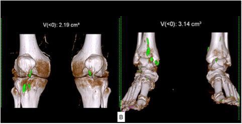
Other joints can also be involved. Monosodium urate crystal deposition can also occur in tendons and bursae.
(1)Well defined punched out erosion with sclerotic margins is seen involving head of 1st metatarsal (Figure A). These imaging findings are in favour of gout.
(2)Gout has an asymmetrical polyarticular distibution. The most commonly involved joint is the 1st metatarsophalangeal joint.
Picture This by Jankharia, Mumbai, Maharashtra 1MD, DNB (Radiology) 2MD, DMRD (Radiology)
(3)Dual-energy CT may be used to differentiate uric acid from calcium in musculoskeletal tissue, allowing gouty urate crystals to be distinguished from bone or dystrophic calcifications Postprocessing of dual-energy CT data yields colorcoded cross-sectional images (Fig B). DECT also provides sensitive and specific volumetric quantification of tophi which is important in monitoring treatment response.
M Chenniappan1
2 :
This is the ECG of 72 years old diabetic with tiredness; No previous ECGs were available.
1. Describe all ECG changes
2. Why is this clue?
3. What are practical implications?
ECG CHANGES :
The ECG shows sinus rhythm, slope ST segment in inferior leads (loss of concavity of ST segment) with reciprocal depression in L I and avL. The slope ST elevation is more prominent in LIII than L II (Right Coronary obstruction(RCA). In chest leads there is significant ST depression in all chest leads except in V1 where it is mild (discordant ST segment depression V1-V2). The ST depression in chest leads go beyond V3. (associated disease in left coronary artery system). In addition, there is 1 mm ST elevation in lead avR. (Probable LMCA Disease). The P wave in chest and inferior leads are bifid without significant terminal P negative force in V1 (Left Atrial enlargement is unlikely).
CLUE :
SUBTLE :
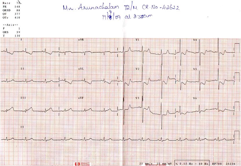
4. Probable LMCA occlusion
5. Triple Vessels Disease
6. Inter atrial conduction disturbance may indicate associated atrial infarction which may produce complications such as atrial arrhythmias, embolism and atrial rupture.
NOT SINGLE:
1. The slope ST in inferior leads as the earliest and hyperacute sign of Acute Total Occlusion of RCA (LIII>LII)
2. Discordant ST depression between V1 and V2 which is suggestive of RV infarct indicates proximal RCA occlusion.
3. ST depression beyond V3 indicates, associated LCA disease
4. Mild ST elevation in avR is suggestive of LMCA critical occlusion
5. ST depression in >5 leads are suggestive of Triple Vessels Disease (TVD)
6. Bifid P waves in the absence of typical deep terminal P negativity in V1 are suggestive of inter atrial conduction disturbance rather than Left Atrial Enlargement or Abnormality which can happen in atrial infarct as depolarization abnormality.
SINISTER:
1. Proximal RCA disease
2. RV infarct
3. Associated LCA involvement
1Adjunct Professor, Dr MGR Medical University, Tamilnadu; Senior consultant cardiologist, Tamilnadu; Ramakrishna Medical Centre, Apollo Speciality Hospital, Trichy
This is not single inferior wall infarction ; there are RV infarct, LCA Critical occlusion, LMCA disease , Triple Vessels Disease and Possible Atrial Infarction.
PRACTICAL IMPLICATION :
Because the of above said reasons, this patient should go for immediate CAG and suitable revascularisation rather than thrombolytic therapy if 5As (Affordability, Accessibility, Arrival time, Availability and About the institution) are met. Otherwise he should be thrombolysed immediately with Tenecteplase. Thrombolysis with Streptokinase is the last option.
Sukanti Bhattacharyya1
Forewords :
Lady Mary Wortley Montagu observed variolation in Constantinople and brought it to Britain in 1721. In Shetland Isles, Johnnie Notions had the reputation of not having lost a single patient with his self-devised inoculation. On May 14, 1796, Edward Jenner, the father of immunology, inoculated James Phipps with cow’s small pox blister-fluid ( as his 17th recorded case) , and to prove his hypothesis of induced immunity, he later challenged Phipps by injecting with variolous material successfully with no subsequent disease manifestations.
125 years after, on July 18, 1921, the first ever inoculation in human with a novel vaccine against tuberculosis was done through oral route on an orphan neonate in Charité Hospital, Paris by Benjamin Weill-Halle and Raymond Turpin in the philanthropic view to save the posthumous baby from potential risk of developing neonatal tuberculosis, as its mother died of pthisis soon after giving birth to a healthy baby.This vaccine was developed and already tested on animals by Calmette and Guérin. Initially it was named as include “Vaccin Bilié de Calmette et Guérin vaccine” and later on, as “Bacille de Calmette et Guérin vaccine” or BCG, in short. Thus, this 2021 is the first centenary of BCG vaccination in human.
Brief History :
Being challenged in 1895 by Theobald Smith regarding differences in phenotypes between human and bovine tuberculosis, Robert Koch revisited his previous thinking (1882) of identical natures of human and bovine tuberculosis bacilli. By 1901 Koch distinguished Mycobacterium bovis from Mycobacterium tuberculosis
This very finding and the success of vaccination in preventing smallpox inspired the immunologists to draw a parallel relationship between bovine tuberculosis and to frame a corollary that infection with bovine tuberculosis might also be protective against infection with human tuberculosis. But, their overzealous inoculation of unmodified M.bovis—as virulent as M. tuberculosis— in Italy in the late 19th century ended with disastrous results.
During such doldrums , Albert Calmette, a French physician and bacteriologist, and Camille Guérin, a veterinarian, were working at the Institut Pasteur de Lille (Lille, France) in 1908, ushered with a new idea in an effort to attenuate the bacilli. They tried subculturing virulent strains of the tuberculosis bacillus using different arbitrary culture media. They noted a glycerin- potato mixture grew bacilli well , but it was difficult to get homogenous suspension of bacilli. Upon intuitively adding ox bile into the medium, they astonishingly noted less clumping tendency and lesser virulence of the bacilli.
1MD, Associate Professor, Medical Physiology, IIMSAR and Dr B C Roy Hospital, Haldia, Purba Medinipur 721645
And this pre-empted them to undertake 239 subcultulturing over 13 years, even through the period of WW I .In 1919, they succeded to isolate avirulent bacilli, which were found to be unable to cause tuberculosis disease in research animals. Calmette and Guerin transferred to the Paris Pasteur Institute in 1919. The BCG vaccine was first used in humans in 1921 in Charité Hospital, Paris. It was a successful endeavour; but public acceptance of the vaccine was not apt, might be due to a disaster. In the summer of 1930 in Lübeck, 240 infants were vaccinated in the first 10 days of life; almost all developed tuberculosis and 72 infants died— due to improper storage of the BCG with virulent strains in the same incubator.
Dr R G Ferguson, working at the Fort Qu’Appelle Sanatorium in Saskatchewan, and few other forerunners developed the practice of vaccination against tuberculosis. In 1928, BCG was adopted by the Health Committee of the League of Nations (predecessor to the WHO)). But because of controversy and opposition, it took nearly two more decades to be widely used. In post WW II period ( from 1945 to 1948) relief organizations (International Tuberculosis Campaign or Joint Enterprises) vaccinated over eight million babies in eastern Europe and prevented the predicted post-war surge in TB.
Further Scopes :
BCG vaccine helps in preventing tubercular meningitis in infancy and childhood.
BCG also has some effectiveness against Buruli ulcer infection and other nontuberculous mycobacteria infections.
Additionally it is sometimes used as part of the treatment of non–muscle-invasive bladder cancer (NMIBC). BCG has been one of the most successful immunotherapies.
BCG has also been tried in colon cancer and colorectal cancer. Early inoculation with BCG vaccine, particularly in low income countries, tentatively exert a beneficial non-specific effect of on overall mortality, through reduction of sepsis and respiratory infections.
As of 2017, BCG vaccine is in the early stages of being studied in type 1 diabetes.
Use of the BCG vaccineis claimed to provide protection against COVID 19. However, epidemiologic observations in this respect are ambiguous. Spanish, French, German and Dutch research entities are preparing trials using genetically-modified BCG vaccine in this regard. BCG vaccine is in phase III trials in health care workers in Australia and Netherlands. BCG vaccine is being evaluated in Greece and Poland. Polish scientists are investigating on the immune training effect of BCG vaccine. The WHO has not yet recommended its use for prevention.
Luca S, Mihaescu T — History of BCG Vaccine, Maedica (Bucur) 2013; 8(1): 53-8. PMCID: PMC3749764
Rudrajit Paul1
Thalassemia and sickle cell disease are congenital haemolytic anemia syndromes with various systemic manifestations. While thalassemia major makes people transfusion dependent, sickle cell disease is associated with recurrent occlusive crises and other life threatening manifestations. For most patients, lifelong supportive therapy is the only means of management. Bone marrow transplant is able to cure these genetic diseases, but for most thalassemia patients in India, this is not an option due to1 cost and lack of suitable donors. However, new technology2 is now offering hope for cure of these genetic ailments.
On January 21, 2021, Frangoul et al from USA published a brief report in the NEJM where they have discussed a new therapeutic option for these patients. A brief overview of this method is described here:
CD34+ hematopoietic stem cells were isolated. Then, by electroporation, CRISPR-Cas9 gene editing kit was introduced inside these cells. This gene editing system targeted BCL11A. This is a transcription factor that silences ã-globulin gene expression. Enhancing the ã-globulin gene would lead to more HbF in the RBC and thus, decrease in transfusion requirement. The CRISPR gene editing system would silence the suppressor, which is BCL11A. Hence, fetal hemoglobin would increase. The stem cells, after CRISPR editing, were transfused into the body of patients after myeloablation. Thus, this was a bone marrow transplant procedure,
1MD, DNB, MRCP (UK), Consultant Physician, Kolkata
albeit with modified stem cells. At one year, it was seen that the patients had high levels of HbF with marked decrease in transfusion requirement and for sickle cell disease, abolition of further vasoocclusive crises.
The CRISPR technology is an area of intense research due to its ability to do gene editing with marked precision. In the study mentioned above also, there was no off-target editing. This is a remarkable achievement and the chance of inducing cancerous or dysmorphic mutations is almost nil. This makes CRISPR an ideal tool for clinical use. However, widespread clinical use is still far into the future.
Another method of gene editing is being tried in thalassemia. In September 2020, Gabr et al from Egypt published another study where they took stem cells of thalassemia patients and with CRISPR tool, tried to repair the mutation in the beta-globin gene itself. The edited cells were then cultured and it was found that the mutation had been successfully corrected. These repaired stem cells could then be transfused back into the patient like autologous bone marrow transplantation.
Clinicians of the future must be conversant with this new technology. In the future, when managing a patient of thalassemia, it may be worthwhile to discuss gene editing as a valid therapeutic option.
1Frangoul H — CRISPR-Cas9 Gene Editing for Sickle Cell Disease and β-Thalassemia. N Engl J Med 2021; 384: 252-60. Perspective
Anshuman Poddar, MBBS, MS (Gen. Surg.), Consultant, Dept. of Minimal Access and Bariatric Surgery, ILS Hospital, Kolkata)
1. The process of introduction of weakened pathogen into human body is called (a) Immunization (b) vaccination (c) attenuation (d) none of these
2. The first vaccine was developed by (a) Louis Pasteur(b) Edward Jenner (c) Carl Landsteiner(d) Joseph Miester
3. The process of weakening a pathogen is called (a) vaccination (b) attenuation (c) immunization (d) virulence reduction
4. A vaccine can be (a) an antigenic protein(b) weakened pathogen (c) live attenuated pathogen(d) all of these
5. Which of the following statement is true regarding vaccination
(a) vaccination is a method of active immunisation (b) vaccination is a method of passive immunisation (c) vaccination is a method of artificial passive immunisation (d) vaccination is a method of natural passive immunization
6. Passive immunisation include (a) introduction of antibodies directly (b) transfer of maternal antibodies across placenta (c) transfer of lymphocyte directly (d) all of these
7. Active immunity may be gained by (a) natural infection (b) vaccines (c) toxoids (d) all of these
8. Which of the following statement is not true about the Live attenuated vaccine?
(a)It is prepared using whole weakened living bacteria or virus
(b)It can generate a long-term immune response in an individual with a single dose of vaccine (c)Measles, MMR and oral polio vaccine are live attenuated vaccines
(d)It is stable at normal room temperature
9. Which of the following is not true about the important characteristics of the vaccine to be effective?
(a) It must be safe and easy to use with low side effects in humans
(b) It must provide immunity to at least 15% of the population (c) It must be available at low cost
(d) It must provide long-lasting immunity
10. Which of the following statement is false about influenza/flu vaccine?
(a) Flu vaccines may contain killed viruses or weakened viruses
(b) The influenza virus is a highly mutating virus, therefore annual vaccination is recommended
(c) The immune system gets activated 2-3 weeks after an individual is vaccinated
(d) None of the above
11. In December 2020 first vaccine for COVID-19 was approved in the Europe and in the USA, name the first biotechnology company which received the authorization to distribute the vaccine?
(a) Moderna (b) Pfizer-BioNtech (c)Astrazeneca (d)Novavax
12. Which of the following type of vaccine did the Oxford-Astra Zeneca and Serum Institute of India designed for COVID-19?
(a) mRNA vaccine(b) Subunit vaccine (c) Live attenuated vaccine(d) Vector borne vaccine
13. The two doses of Covishield should be taken (a) 2 days apart(b) 2 weeks apart (c) 4 weeks apart(d) 6 months apart
14.In which of the following, Herd Immunity cannot protect the individual?
(a)Tetanus(b)Diphtheria (c)Poliomyelitis (d)COVID-19
15.Which of the following Human papillomavirus subtypes are not covered by the Quadrivalent Anti-cervical cancer vaccine?
(a)Type 6(b)Type 7
(c)Type 11(d)Type 18
Objective : To review the literature for evidence of the efficacy of lincomycin in skin and soft tissue infections (SSIs) and in Upper respiratory tract infections (URTIs), and the current scope of lincomycin use in these infections.
Methodology : Computerized searches of the PubMed and Google scholar database were performed to identify primary and review articles about the therapeutic use of lincomycin between 2000 and 2020. The keywords used during the search were ‘lincomycin and skin infection’, ‘lincomycin and soft tissues infection’, lincomycin and urinary tract infection’, and ‘lincomycin review’.
Data analysis and conclusions : The efficacy of lincomycin in skin and soft tissue infections as well as in Upper Respiratory T ract Infections (URTIs)has been proven in many studies. Perhaps, local variations in antibiotic resistance of organisms lead to these variations. Most studies showed Staphylococcus aureus (S. aureus) accounting for 35-50% of infections.Lincomycin still seems to be an effective therapy in conditions like periodontitis, perioperative preventive and curative therapy, and certain skin conditions like folliculitis as well as topical application for acne. Lincomycin might still also be effective in cases of URTI not responding to the standard lines of treatment. It also seems to have an important role in Group A Beta-hemolytic Streptococcal Pharyngo-Tonsillitis.
Key words : Lincomycin, Soft Tissue Infections.
Lincomycin is obtained from the actinomyces Streptomyces lincolnensis. Chemically, it is a 6,8-dideoxy-6-aminooctose lincosamine1. It was isolated in 1962 and was the first lincosamide to be used in clinical practice. Lincomycin has an antibacterial effect on Gram-positivemicroorganisms (staphylococci, streptococci, pneumococci, diphtheria bacillus,and clostridia) and is usually reserved for serious bacterial infections like sepsis, osteomyelitis,septic endocarditis, pneumonia, pulmonary abscess, infected wounds, andpurulent meningitis, that areresistant to penicillin and other antibiotics2. Most Gram-positive cocci including staphylococci, pneumococci, and most streptococci [except S (faecalis)] are usually sensitive to lincomycin but Neisseriae and H influenzae are resistant. Other organisms that are sensitive to lincomycin include Mycoplasma hominis and M. pneumoniae (but not T strains) and Bacteroides spp3,5
Lincomycin acts by inhibiting protein synthesis at ribosomal binding sites. It is well absorbed after oral or intramuscular administration and is widely distributed in the body. It is also detected in cord blood and milk, although little gets into the normal cerebrospinal fluid. Due to its high concentration in the bone, it is often used in the management of acute staphylococcal osteomyelitis. Following oral dosing,it rapidly reaches peak levels in the serum and the minimum inhibitory concentration (MIC) of sensitive organisms is promptly exceeded; hence, a 4 to 6 hourly dosage regimen is recommended. Intramuscular administration of a single dose of 600 mg of lincomycin produces average peak serum concentrations of 11.6 mcg/mL at 60 minutes. The therapeutic concentration is maintained for 17 to 20 hours for most susceptible gram-positive organisms. A two-hour intravenous infusion of 600
1MD, FCP, PGDHEP, Director, Medical Affairs, Intellimed Healthcare Solutions, Mumbai 400070
2Pharm D, Medical Affairs Executive, Intellimed Healthcare Solutions, Mumbai 400070
Received on : 19/01/2021
Accepted on : 05/02/2021
[J Indian Med Assoc 2021; 119(2): 62-6]
mg of lincomycin achieves average peak serum concentrations of 15.9 mcg/mL and maintains therapeutic concentrations for 14 hours for most susceptible gram-positive organisms4,6
The excretion of lincomycin is mainly through the bile and faecal route. A small proportion of lincomycin (9% in 24 hours) is excreted by the kidneys; however, the serum concentrations of lincomycin can be very high in patients with renal failure 3. The biological half-life after intramuscular or intravenous administration is 5.4 ± 1.0 hours. In patients with hepatic impairment, serum halflife might be twice of that in patients with normal hepatic function6
This paper reviews the literature for evidence of the efficacy of lincomycin in skin and soft tissue infections (SSIs) and in Upper respiratory tract infections (URTIs), and the current scope of lincomycin use in these infections.
Methodology :
Computerized searches of the PubMed and Google scholar database were performed to identify primary and review articles about the therapeutic use of lincomycin between 2010 and 2020. Not many publications were found corresponding to this period. The search was then extended to include publications from 2000. Some articles published during the early years of its launch were also scanned to compare the change in bacterial sensitivity patterns over time. The keywords used during the search were ‘lincomycin and skin infection’, ‘lincomycin and soft tissues infection’, lincomycin and upper respiratory tract infections, and ‘lincomycin review’.The articles were supplemented by examining cited references.
Efficacy of lincomycin in Skin and
:
An early study in 1967 investigated the efficacy of lincomycin in the treatment of acute and chronic staphylococcalosteomyelitis and soft tissue infections 7 . It was a small cohort of 26 patients, comprising15 patients of osteomyelitis- 5 with the acute disease and 10 with thechronic form, and 11 with soft tissue infections. Most had not responded satisfactorily to various antibiotics,and many had
119, No 2, February 2021Journal
undergone repeated hospitalization and one or more surgeries. Following the administration of lincomycin,all patients of acute osteomyelitis recovered completely with no adverse effects. Of the 10 patients with chronic osteomyelitis, 7 recovered completely and the other three improved slightly. Of the 11 patients withsoft-tissue infections, 7 patients recovered completely, 1 improved, and 3 showed nobenefit. Two of these 3 patients were infected with organisms found to be insensitive tolincomycin.Favourable results in similar cases were reported by Kanee B Geddes et al in 1964 also reported that from a clinical point of view the drug appears to be particularly valuable in the treatment of staphylococcal osteomyelitis7. Another early study in 1974 reported thatlincomycin administered at the start of clean neurosurgical procedures reduced the infection rate from 5.1 to 2.3%8
However, over the decades there has been a significant change in antibiotic practice, due to the high rate of resistance of many microorganisms to most antibiotics. Hence, the results of early studies might not be applicable to the current clinical practice. The more recent studies have reported different outcomes with lincomycin in various skin and soft tissue infections.
A study published in 2014 evaluated the efficacy of Lincomycin Hydrocloride 500 mg capsules /irrigation solution when used as an adjunct to scaling and root planning (SRP) and/or pre and postsurgical debridement in periodontal diseases in 42 patients9. The patients were prescribed Lincomycin Hydrocloride 500mg capsules orally thrice a day for 5 days. All patients who were diagnosed for gingivitis, and those who underwent pre-surgical and post-surgical periodontal procedures achieved complete relief, while 96.85% patients diagnosed for periodontitis achieved complete relief.
A study in 2013 in China, explored the role of lincomycin combined with heparin sodium in the prevention central venous catheter infection in the ICU 10. A total of 172 patients who received central venous catheters were randomized into the trial and control groups with 86 cases in each group.The trial group received lincomycin and heparin sodium,and the control group received normal saline and heparin sodium.The incidence of infections in the trial group was 2.33% in 1-2 weeks after catheterization and 5.81% in 2-3 weeks after catheterization,and total incidence of infection was 9.30%.These were significantly lower than those in the control group (10.47%,15.12%,30.23% respectively), and there was statistical significance(P<0.05).The positive rate of blood culture in the trial group(12.50%)was significantly lower than that in the control group(53.85%); there was statistical significance (P0.05).
A retrospective study published in 2019 involved 60 patients with diabetic foot infections with or without osteomyelitis11. The patients were categorised as group 1mild infection and group 2-moderate infection. Both groups were treated using local wound debridement and the systemic administration of antibiotics. Group 1 patients were treated with either of the two regimens, A (amoxicillin/clavulanate + metronidazole) and B (clindamycin + metronidazole), for 1014 days. Group 2 patients were treated with either regimen A (ampicillin + cloxacillin + metronidazole) or B (lincomycin + metronidazole), for 6 weeks. In both groups, regimen B was
prescribed for patients who were either allergic to penicillin or already on penicillin without a response. Group 1 showed an 80% cure rate under regimen A and a 100% cure rate under regimen B.Group 2 had a 61.5% cure rate under regimen A and 11.53% improved, while regimen B patients had a 68.75% cure rate and 12.5% improved. The study reported that intravenous lincomycin and oral metronidazole show a higher cure rate among moderate diabetic foot infection patients with or without osteomyelitis.
A study among 40 randomly allocated patients, evaluated the effect ofintra abdominal lavage with an antibiotic solution of lincomycin and gentamycin in normal saline in decreasing the risk of postoperative infectionsafter surgeries for colorectal cancer12. Group 1 patients underwent intra abdominal lavage with normal saline, followed by a second lavage with a gentamicin–lincomycin solution, while group 2 underwent lavage with normal saline.There was a significant difference between the two groups in the incidence of postoperative wound sepsis. (5% in group1 and 45% in group2). In group1, the only isolated organism was Pseudomonas. In group2, three cases had E. coli and two cases each had Pseudomonas, Klebsiella, and Enterobacter infections (Fig 1).
A study of 30 patients with different skin infections was performed in India in 2017.Patients were prescribed Lincomycin 500 mg orally twice/thrice a day for 14 days depending on the severity of infection. A good response was seen in most types of infections Fig 213
In another Indian study about the efficacy of lincomycin against the strains of S. aureus isolated from various types of pyodermas in children, published in 2000, 75% strains of staphylococci were susceptible to lincomycin in vitro and 95% of the patients responded to 5-10 days treatment. Lincomycin was given to all these children in a daily dose of 30 mg/kg 5 - 10 days 14. Lincomycin appears to be still effective in children.
Lincomycin has also been used for the treatment of acne. In an early study in 1965, 14 patients with carbuncles, furuncles, folliculitis, cellulitis, laryngopharyngitis, lymphadenitis, pyonychias and dermatitis infectiosa eczematoides were treated with lincomycin. In every instance the sensitivity test with respect to staphylococci and/ or streptococci showed good inhibition with low MIC of lincomycin.
Group 1Group 2
P [n=20)(n(%)][n=20)(n(%)]
Wound sepsis
Negatives19 (95.0)11(55.0) 8.533*0.003*
Positives 1(5.0)9(45.0)
Intra-abdominal abscess
Negatives19 (95.0)18 (90.0)0.360 FEP=1.000
Positives 1(5.0)2(10.0)
Isolated organism
Pseudomonas 1(100.0)2(22.2)2.837 MCP=1.000
Klebsiella0 (0.0) 2(22.2)
Enterobacter0 (0.0)2(22.2) E coli0 (0.0)3(33.3)
*Signifant value P less than 0.05 FE, Fisher exact test; MC, MonteCarlo test
intra-abdominal abscess and isolated organism12
119, No 2, February 2021Journal
Lincomycin was very effective against coagulase-positive S aureus and against S.hemolyticus Type A15.Topical application of lincomycin has also been used in the treatment of acne. Amulticentric, randomized, double blind, placebo controlled, clinical trial was conducted in India in 2003 wherein lincomycin hydrochloride in 2% gel form was prescribed to 200 patients with grade II and grade III acne. About 70% cases showed a good to excellent response, which was significantly more as compared to 23% in the placebo group16
Dormanesh et al in 2005,attempted to study the antibiotic sensitivity of methicillin-resistant S aureus isolated from various types of hospital infections in children. Overall, 255 clinical samples from various types of infections including blood (n = 40), UTIs (n = 60), respiratory tract infections (n = 55), superficial and post-surgical wounds (n = 50) and burn infections (n = 50) were collected. Susceptibility of Methicillin-resistant S aureus (MRSA) isolates was tested. The most effective antibiotics were imipenem, lincomycin, vancomycin, cephalothin, cotrimoxazole and clindamycin Fig 318 Fig 3 Resistance of Methicillin Resistant Staphylococcus aureus Strains of Various Types of Clinical Infections in Pediatric Patients Against Commonly Used Antibiotics18
However, in another study of 1310 post-surgical wound swabs in 2011, only 50% of MRSA isolates were susceptible to lincomycin19. From the above evidences, it is obvious that the efficacy of lincomycin in skin and soft tissue infections varies between the studies. Perhaps, local variations in antibiotic resistance of organisms lead to these variations. Nevertheless, lincomycin still seems to be an effective therapy in conditions like periodontitis, perioperative preventive and curative therapy, and certain skin conditions like folliculitis. It is also indicated for patients who are intolerant to penicillin. Topical application of lincomycin for acne could be another regular indication.Its use in other deeper infections like osteomyelitis and diabetic foot might vary as many of the causative organisms might be highly resistant to several antibiotics. Hence, in such cases culture and sensitivity test is necessary before deciding antibiotic therapy. Upper Respiratory Tract Infection :
An early study in 1965, tested the sensitivity of different strains of streptococci and staphylococci to lincomycin. Of the 165 strains of streptococci tested, 164 were found to be sensitive to lincomycin.
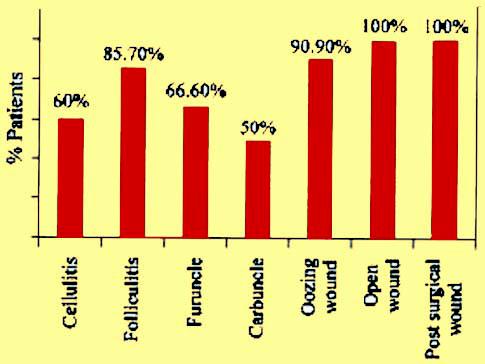
Fig 2 — Complete cure at the end of treatment13
Moreover, of the 3200 strains of staphylococci isolated from clinical material, only 40 were resistant to lincomycin. Nine of 14 patients with pneumonia, and 15 of 17 with acute exacerbations of bronchitis recovered completely with lincomycin therapy. Thirteen of the 14 patients of pneumonia had streptococcal infection. Notably, three patients showed partial resolution of the pneumonia with lincomycin but eventually resolved completely only when ampicillin was substituted for lincomycin after one week of treatment with lincomycin. Nevertheless, the authors reported that in the treatment of pneumonia and acute exacerbations of chronic bronchitis lincomycin appeared to be a satisfactory alternative to penicillin21
Later, Angeli et al. in 1997 compared the efficacy of lincomycin versus penicillin and clarithromycin in patients with acute pharyngitis/ tonsillitis caused by group A beta-hemolytic streptococci and in those
Type of Infections, %
AntimicrobialBlood (5) UTIs (18)RTIs(20) Wound BurnTotal (74) Agents Infections(18) Infections(18)
Ampicillin4(80) 15(83.33) 17(85) 16(88.88)17(94.44)69(93.24)
Centamycin-2(11.11)6(30) 8(44.44)8(44.44)24(32.43)
Lincomycin--1(5)2(11.11)2(11.11) 5(6.75)
Cephalothin-3(16.66)2(10)1(5.55)2(11.11)5(10.81)
Imipenem-1(5.55)-1(5.55)-2(2.70)
Tetracycline4(80)16(88.88) 18(90) 17(94..44) 18(100)73(98.64)
Vancomycin1(20) 1(5.55) 1(5) 1(5.55)1(5.55)5(6.75)
Ciprofloxacin1(20) 4(22.22)3(15)5(27.77)5(27.77)18(24.32)
Norfloxacin1(20) 5(27.77)5(25)6(33.33)7(38.88)24(32.43)
Cotrimoxazole1(20)2(11.11)2(10) 3(16.66)4(22.22)12(16.21)
Clindamycin1(20)2(11.11)3(15) 3(16.66)3(16.66)12(16.21)
Trimethoprimsulfa-methoxazole1(20) 3(16.66)6(30)7(38.88)9(50)26(35.13)
Penicillin4(80) 17(94.44) 18(90) 17(94.44)18(100) 74(100)
Oxacillin3(60) 15(83.33) 17(85) 16(88.88)18(100) 69(93.24)
Erythromycin2(40) 10(55.55) 12(60) 13(72.22)15(83.33)42(56.75)
Azithromycin2(40) 8(44.44)9(45)12(66.66)13(72.22)44(59.45)
Ceftriaxone1(20)2(11.11)3(15)5(27.77)5(27.77) 16(21.62)
Cefixime1(20)2(11.11)5(25) 7(38.88)7(38.88)22(29.72)
Fig 3 — Resistance of Methicillin Resistant Staphylococcus aureus Strains of Various Types of Clinical Infections in Pediatric Patients Against Commonly Used Antibiotics18
119, No 2, February 2021Journal
with a clinical history of recurrence, in an open-label, prospective, randomized, comparative, single-masked study22. The study was conducted across 8 centres and they found that all drugs had statistically similar clinical and bacteriologic efficacy as well as tolerability.
In 2012, a randomized study in India involving 41 patients of tonsillitis/sinusitis showed that the overall response rate of lincomycin hydrochloride was more than that of cefpodoximeproxetil. 67.89% of the patients in lincomycin group and 52.27% in cefpodoxime group achieved complete relief, in all the clinical symptoms24
Another study from India in 2018, studied the bacterial pathogens and their antibiotic sensitivity pattern in 100 patients with chronic otitis media. 48.84% of the Pseudomonas aeruginosa (P aeruginosa) isolates, 50% of S aureus isolates, 88.89% coagulasenegative staphylococci were sensitive to lincomycin Fig 425. This is an important finding considering that coagulase-negative staphylococci especially Staphylococcus epidermidis, are major nosocomial pathogens causing a variety of infections in humans20.
Interestingly, in 2013 a report of 2 cases of acute myopericarditis associated with concomitant Streptococcus pyogenes [group A Streptococcus (GAS)] pharyngotonsillitis was published. After investigations, although no definitive pathogenesis could be explained, toxin-mediated myocarditis seemed to be the most popular explanation for this condition. One of the 2 cases who did not respond to penicillin, recovered completely with lincomycin17
A long-term studytested clinical isolates of Streptococcus pyogenes (S. pyogenes) obtained from a Lexington, Kentucky hospital in 2004 and again in 2014,for their sensitivity to macrolide antibiotics. Interestingly, 22% of the isolates from 2004 were resistant tolincomycin, while the resistance decreased to 11% in 201426. A review was published in 2017 reported about the treatment challenges of Group A Beta-hemolytic Streptococcal Pharyngo-Tonsillitis (GABHS PT). It reported that lincomycin, clindamycin, and amoxicillin-clavulanate are more effective in relapsing GABHS PT27
From these results, theefficacy of lincomycin seems to be variable in different studies. However, lincomycin might still be effective in cases of URTIs not responding to the standard lines of treatment. It also seems to have an important role in GABHS PT. It must also be remembered though, that the results cannot be generalized, as the resistance patters can vary widely between different geographies
and different centres.
Interestingly, a recent study compared the effectiveness of lincomycin and azithromycin in the treatment of COVID-19 associated pneumonia in a cohort of 24 patients. Bronchoalveolar-lavage PCR results were compared after treatment. On the 6th day after starting treatment, negative bronchoalveolar PCR result was seen in 83.3% patients in the lincomycin group and in 33.3% patients in the azithromycin group (P < 0.05). In addition, other clinical outcomes like duration of hospitalization, temperature normalization, and radiological progression were also better in the lincomycin group28
Adverse Effects of Lincomycin :
Lincomycin has been noted to produce an unusually high incidence of diarrhoea when given orally32. While several authors have noted the presence of mild to moderate diarrhoea after the use of lincomycin, others have described more severe diarrheal symptoms that clinically simulate acute ulcerative colitis or pseudomembraneous colitis33 . However, no serious toxicity was reported during the clinical investigation of lincomycin in both UK and the United States (a total of approximately 2,500 patients)34 Similarly, none of the studies referenced above reported any severe adverse event. Some cases developed diarrhoea, which was controlled after reducing the dose of lincomycin.
Can Interchangeability of Lincosamides be Assumed in Clinical Practice ?
In 2010, the Australian therapeutic guidelines presented lincomycin and clindamycin as equivalent treatments for serious infections due to S. pyogenes, S Agalactiae and S Aureus 29. A paper published in 2014, mentioned that parenteral lincomycin use now exceeds parenteral clindamycin use in hospitals, including intensive care units in Australia. NinetyS. pyogenes, 45 S.agalactiae, and 100 S aureus isolates (50 methicillin susceptible and 50 methicillin resistant) were tested for MICs of clindamycin and lincomycin. All S pyogenes and S aureus isolates tested, had similar susceptibilities for clindamycin and lincomycin. Three of the S. agalactiae isolates had an erythromycinsusceptible, low-level clindamycin-resistant pattern, but were susceptible to lincomycin30
Summary :
It is important to have local hospital-based knowledge of the organisms causing various infections and their antibiotic sensitivity
Vol 119, No 2, February 2021Journal of the Indian Medical Association
patterns21. Lincomycin is still an effective therapy in conditions like periodontitis, perioperative preventive and curative therapy, and certain skin conditions like folliculitis. It is also effective in cases of URTIs not responding to the standard lines of treatment. Moreover, as seen from a recent review, lincomycin has an important role in GABHS PT infections and in cases of relapsing GABHS.
1Naveed S, Shah SN, Qamar F, Waheed N, Nazeer S — Simple UV spectrophotometric assay of Lincomycin. IJPRDD 2014; 1(2): 10-2.
2Vardanyan R, Hruby V — Synthesis of essential drugs. Elsevier; 2006 Mar 10.
3Ball AP, Gray JA, Murdoch JM — Lincomycin and Clindamycin. InAntibacterial Drugs Today 1978 (pp. 44-46). Springer, Dordrecht. Available at https://link.springer.com/chapter/10.1007/978-94-011-80047_10
4Greenwood D, Irving WL. Antimicrobial agents. InMedical Microbiology 2012 Jan 1 (pp. 54-68). Churchill Livingstone.
5Klein JO, Remington JS, editors — Infectious diseases of the fetus and newborn infant. WB Saunders; 7th Edition, 2011
6U.S. National Library Of Medicine. Drug Label Information LincocinFebruary 27, 2019.
7Hnatko SI — The treatment of acute and chronic staphylococcal osteomyelitis and soft tissue infections with lincomycin. Can Med Assoc J 1967 Sep 9; 97(11): 580-4. PMID: 6050890. Available at https:// www.ncbi.nlm.nih.gov/pmc/articles/PMC1923379/
8Savitz MH, Malis LI, Meyers BR — Prophylactic antibiotics in neurosurgery. Surg Neurol 1974; 2(2): 95-100. PMID: 4818644
9Katwe S — Dental Infections related to Gingivitis, Periodontitis and Pre/ Post surgical dental procedures in patients. J Indian Dent Assoc 2014; 8: p37-42. 6p
10LIAO YP, SONG YK — Effects of Lincomycin Combined with Heparin Sodium against Infection of Central Venous Catheter in ICU [J]. China Pharmacy 2013; 10.
11Aliakbar AH, Alsaadi MA, Barrak AA — Evaluation of the Surgical and Pharmacological Treatment of Diabetic Foot Infection: A Retrospective Study. Open Access Macedonian Journal of Medical Sciences 2019 May 15; 7(9): 1499-1504. Available at https://www.ncbi.nlm.nih.gov/ pmc/articles/PMC6542384/
12Elsisy AA, Hagag MG, Ewida MM — The effect of peritoneal lavage with a mixture of lincomycin–gentamicin on postoperative infection in cases of colorectal cancer surgery. Menoufia Medical Journal 2017; 30(2): 393-99
13Sarkar S — Clinical efficacy of Lincomycin in the Treatment of Skin Infiections: Results of a pilot study in Indian Adult Patients. Indian Medical Gazette Mar 2017.
14Rao RR, Padmavathi K, Ramani TV, Jyothi PA — Efficacy of Lincomycin against the strains of Staphylococcus aureus isolated from various types of pyodermas in children: in Vitro and in Vivo Study. Indian Journal of Dermatology, Venereology, and Leprology. 2000 Jul 1;66(4):185-87. Available at https://www.ijdvl.com/article.asp? issn=0378-6323;year=2000;volume=66;issue=4;spage=185;epage=187; aulast=Rao
15Kanee B — Lincomycin in dermatologic practice. Canadian Medical Association Journal 1965 Jul 31; 93(5): 220. Available at https:// www.ncbi.nlm.nih.gov/pmc/articles/PMC1928592/pdf/canmedaj011110033.pdf
16Sharma AD, Gupte PD, Sundaram M, Janaki VR, Rege VL, Bilimoria FE, et al — Topical lincomycin gel in acne vulgaris: A multicentric placebo controlled study. Indian Journal of Dermatology, Venereology, and Leprology. 2003 Jul 1; 69(4): 271.
17Chaudhuri A, Dooris M, Woods ML — Non-rheumatic streptococcal myocarditis–warm hands, warm heart. Journal of medical microbiology. 2013 Jan 1;62(1):169-72. Available at https:// www.microbiologyresearch.org/ content/journal/jmm/10.1099/ jmm.0.049569-0
18Dormanesh B, Siroosbakhat S, Darian EK, Afsharkhas L — Methicillinresistant Staphylococcus aureus isolated from various types of hospital infections in pediatrics: Panton-Valentine leukocidin, staphylococcal
chromosomal cassette mecSCCmecphenotypes and antibiotic resistance properties. Jundishapur J Microbiol. 2015 Nov;8(11). Available at https:/ /www.ncbi.nlm.nih.gov/pmc/articles/PMC4741056/
19Hussain S, Shams R, Ahmad K, Perveen R, Riaz B — Prevalence of methicillin resistant Staphylococcus aureus (MRSA) in surgical site infections in a tertiary care hospital. Journal Pathol 2005 Dec; 3(2): 815.
20Cremniter J, Slassi A, Quincampoix JC, Sivadon-Tardy V, Bauer T, Porcher R, et al — Decreased susceptibility to teicoplanin and vancomycin in coagulase-negative staphylococci isolated from orthopedic-deviceassociated infections. Journal of clinical microbiology 2010 Apr 1; 48(4): 1428-31.available at https://jcm.asm.org/content/jcm/48/4/1428.full.pdf
21Duncan IB, Jeans B — Lincomycin in hospital practice. Canadian Medical Association Journal. 1965 Sep 25;93(13):685. Available at https://www.ncbi.nlm.nih.gov/pmc/articles/PMC1928825/pdf/ canmedaj01119-0006.pdf
22Angeli G, Fukuda J, Gallegos GB, Ladue L, Miniti A, Suarez S, et al — Efficacy of lincomycin versus penicillin and clarithromycin in patients with acute pharyngitis/tonsillitis caused by group a beta-hemolytic streptococci and a clinical history of recurrence. Current therapeutic research 1997 Dec 1; 58(12): 917-29. Available at https:// www.sciencedirect.com/science/article/pii/S0011393X97800598
23Zhang J — Microbiological Analysis of Bacterial Population in Chronic Pharyngitis. Journal of Zhejiang University of Traditional Chinese Medicine 2009-03.
24Kothadiya A — A multicentric, open label, randomised, postmarketing efficacy study comparing multidose of lincomycin hydrochloride capsule 500 mg with multidosecefpodoximeproxetil tablet 200 mg in patients with tonsillitis, sinusitis. J Indian Med Assoc 2012 Aug 1; 110(8): 5803. Available at https://europepmc.org/article/med/23741829
25Kumar R, Singh G — Study of Bacterial Pathogens and Antibiotic Sensitivity Pattern of Ear Infections in Patients with Chronic Suppurative Otitis Media Attending a Tertiary Care Hospital in Panipat, India. J Med Sci and Health 2019; 5.
26Helton HL, Jackson RD, Watkins M — Macrolide Resistance Changes in Clinical Isolates of Streptococcus pyogenes in Kentucky. Available athttps://encompass.eku.edu/swps/2015/undergraduate/45/
27Brook I — Treatment challenges of group A beta-hemolytic streptococcal pharyngo-tonsillitis. Int Arch Otorhinolaryngol 2017 Sep; 21(3): 286-96. Available at https://www.scielo.br/pdf/iao/v21n3/1809-9777-iao-21-0300286.pdf
28Guvenmez O, Keskin H, Ay B, Birinci S, Kanca MF — The comparison of the effectiveness of lincocin® and azitro® in the treatment of covid-19associated pneumonia: A prospective study. Journal of Population Therapeutics and Clinical Pharmacology= Journal de la Therapeutique des Populations et de la Pharmacologie Clinique 2020 Jun 3;27(S Pt 1):e5-10. Available at https://europepmc.org/article/med/32543164
29Antibiotic Expert Group, Moulds RF — Therapeutic guidelines: antibiotic. Therapeutic Guidelines Limited; 2010.
30Porter MC, Henderson BA, Healy PE, Coombs GW, Ingram PR, McLellan D, et al — Can interchangeability of lincosamides be assumed in clinical practice? Comparative MICs of clindamycin and lincomycin for Streptococcus pyogenes, Streptococcus agalactiae and Staphylococcus aureus. Journal of Antimicrobial Chemotherapy 2014 Mar 1; 69(3): 8567. Available at https://pubmed.ncbi.nlm.nih.gov/24164721/
31Weldrick PJ, Iveson S, Hardman MJ, Paunov VN — Breathing new life into old antibiotics: Overcoming antibacterial resistance by antibioticloaded nanogel carriers with cationic surface functionality. Nanoscale 2019; 11(21): 10472-85. Available at https://pubmed.ncbi.nlm.nih.gov/ 31112150/
32Duncan IB, Jeans B — Lincomycin in hospital practice. Can Med Assoc J 1965 Sep 25; 93(13): 685-91. Available at https:// pubmed.ncbi.nlm.nih.gov/5828940/
33Le Frock JL, Klainer AS, Chen S, Gainer RB, Omar M, Anderson W — The spectrum of colitis associated with lincomycin and clindamycin therapy. J Infect Dis 1975 May; 131 Suppl: S108-15. available at https://pubmed.ncbi.nlm.nih.gov/1127264/
34Geddes AM, Sleet RA, Murdoch JM — Lincomycin Hydrochloride: Clinical and Laboratory Studies. Brit Med J 1964; 2: 670-72. Available at https://pubmed.ncbi.nlm.nih.gov/14171098/
S K Jindal1, J Lele2, A G Ghoshal3, S Nair4, S Kant5, A Parakh6, S K Banthia7, V D Nagda8, P Joshi9, T Masurkar10, SK Joshi11
Asthma is a clinical diagnosis; however, in cases where dilemma exists refer to an expert. In clinical practice for GPs, peak expiratory flow (PEF) can be a good indicator for asthma diagnosis if it showsreversibility on bronchodilator medication.
Asthma diagnosis in young children is mainly based on recurrent symptom patterns + assessment of family history + physical findings + differential diagnostic possibilities.
Asthma medications in inhaled form should be considered as compared to systemic formulations (oral/IV) to improve clinical outcomes in asthma.
Inhaled medicines work fast to improve the symptoms and outcomes. Inhaled route is safer as compared to oral or IV route.
ICS-containing controller treatment should be initiated immediately as needed (in mild asthma) or daily, to reduce the risk of serious exacerbations and to control the symptoms.
Lung function tests play a crucial role in evaluating asthma control and should be assessed at diagnosis, start of treatment, p ost 3-6 months of controller treatment and periodically thereafter.
[ J Indian Med Assoc 2021; 119(2): 67-70]
Key words : Asthma,stable asthma, exacerbations, spirometry, peak expiratory flow, bronchodilator, medication.
Background :
Despite an ever-increasing prevalence of asthma across all age groups, this condition remains poorly managed in India.1,2 Majority of Indian patients remain undiagnosed or wrongly diagnosed in general clinical practice and even those who get diagnosed, remain poorly or inadequately treated.Also, most of Indian patients with asthma continue to be on oral drugs rather than inhalation therapy.2
1MD, FCCP, FAMS, FNCCP, Emeritus Professor of Pulmonary Medicine, Postgraduate Institute of Medical Education and Research, Chandigarh 160012.
2MBBS,National Secretary, IMA HBI, IMA Building, J R Mhatre Marg, Juhu, Mumbai 400049.
3MD, DNB, FCCP, Ex-WHO Fellow ICS, FICP,Medical Director National Allergy Asthma Bronchitis Institutes, Kolkata 700017.
4MD, DNB, DTCD, Associate Professor Department of Pulmonary Medicine Medical College, Trivandrum 695011.
5MBBS, MD (Gold Medallist), MNAMS, FCCP (USA), FIAMS, FNCCP, FCAI, FIMSA, FIAB, FUPDA, FIACM, FICP, Professor and Head of The Department Respiratory Medicine, KGMU Lucknow 226003.
6MBBS, MD (Pediatric), DNB (Pediatric),Senior Consultant Pediatric Pulmonology, Allergy and Sleep Medicine, BLK Super Speciality Hospital, Delhi And Children’s Chest Clinic, Delhi 110005.
7MBBS, Faculty Consultant Banthia Hospital, Indore 452006.
8MBBS, General Physician Asthma Care Centre, Mumbai 400055.
9MBBS, Managing Board Member Sheth PT Surat General Hospital, Surat 395003.
10MBBS, Ex-President IMA Maharashtra State Branch, Mumbai 400049.
11MBBS, Research Team Member Prenatal Education Manashakti Research Centre, Lonavala 41040
Received on : 12/02/2021
Accepted on : 15/02/2021
To overcome these clinical challenges, the Indian Medical Association (IMA) took a lead in bringing about the transformation in the way asthma is managed in Indian scenario. Hence, a first step was taken to develop recommendations for assisting GPs to perform accurate diagnosis and appropriate management of asthma in India. This was achieved by having a group meetingbetween GPs having clinical experience in managing patients with asthma along withchest physicians and pediatricians. Important questions related to asthma diagnosis and its managementwere discussed and the final recommendation decisions were derived from the joint group discussion.
The executive summary of the recommendations are presented here for your reference.
For detailed version of the recommendations please go to https://www.ima-india.org/ima/
Introduction :
Asthma is a heterogeneous disease, usually characterized by chronic airway inflammation.3 According to WHO, India has the largest number of asthma deaths in the world, contributing to 22.3% of all global asthma deaths. Likewise, Indian patients with asthma have a high frequency of reported exacerbations (67%), resulting in substantial functional and emotional limitations.2
:
In most instances a detailed clinical history and vigilant physical examination should enable the doctor to achieve accurate diagnosis.4 Making an accurate diagnosis of asthma includes identification of both a characteristic pattern of respiratory symptoms (breathlessness, wheeze, cough and chest tightness) and variable
119, No 2, February 2021Journal
expiratory airflow limitation measured on peak expiratory flow meter or spirometry (Table: Please refer the detailed version of the guideline to know about the Diagnostic criteria for asthma in adults, adolescents, and children aged > 5 years). 5
During physical examination, the most common abnormal finding on chest is presence of rhonchi (bilateral and diffuse) predominantly heard during expiration (but it may be absent at presentation so normal findings on chest examination does not exclude asthma). Generally, investigations are essential only if the diagnosis of asthma is doubtful or other conditions are suspected to complicate asthma. Notably, this implies a referral to a secondary healthcare level where the symptoms and signs should be reassessed.4
Although, asthma is a clinical diagnosis in routine practice, peak flow rate and spirometry can be helpful in confirming the diagnosis of asthma. In particular, the bronchodilator reversibility testing can be an important diagnostic aid. For GPs, peak expiratory flow (PEF) done on PEF meter can be a good indicator for asthma diagnosis if it shows reversibility on bronchodilator medication. Bronchodilator reversibility is defined as anincrease PEF of >20% after bronchodilator administration with at least 60L/min absolute increment. If there is any ambiguity on diagnosis, refer to a specialist for spirometry and further evaluation. FEV1 from spirometry should be considered more reliable than PEF. However, if spirometry is not available, bronchodilator reversibility may be assessed with PEF meters.An algorithm is recommended for initial presentation for diagnosis of asthma in clinical practice (Figure: Please refer the detailed version of the guideline to view the Algorithm for initial presentation for diagnosis of asthma in clinical practice).5
Diagnosis of Asthma in Children Aged < 5 Years :
Making a confident diagnosis of asthma is challenging in children aged <5 years as common episodic respiratory symptoms such as wheezing and cough are present even in children without asthma, especially at the age of <2 years. Moreover, it is impossible to routinely assess airflow limitation or bronchodilator responsiveness in this age group.Based on the pattern of symptoms during and between viral respiratory infections, a probability-based approach, may prove to be useful for discussion with parents/carers (Figure: Please refer the detailed version of the guideline to know the Probability of asthma diagnosis in children aged <5 years). In children aged <5 years, diagnosis can often be based on clinical features, risk factors, therapeutic response to controller therapy, exclusion of alternate diagnoses and their characteristics suggestive of asthma.5
A positive family history of allergic disorders, or the presence of atopy or allergic sensitization should provide additional predictive support. For instance, early allergic sensitization increases the likelihood of a wheezing child to develop persistent asthma.5
For children aged <5 years, no specific tests can confirm the
diagnosis of asthma; but there are few useful adjuncts which are as follows:5
• Therapeutic trial : A therapeutic trial of asthma treatment (as needed short-acting beta 2-agonist and regular low dose inhaled corticosteroids) may be indicated to provide some guidance about the diagnosis of asthma. Daytime and night-time symptom control, frequency of wheezing episodes and exacerbations should be evaluated to determine the treatment response. However, a therapeutic trial may need to be repeated to confirm the diagnosis of asthma due to its variable nature in young children.
• Chest X-ray : A plain chest X-ray may help to exclude structural abnormalities (e.g. congenital lobar emphysema, vascular ring), if there is a doubt about the diagnosis of asthma in a wheezing or coughing child, chronic infections such as tuberculosis, an inhaled foreign body, or other diagnoses. Depending on the condition, other imaging investigations should be considered.
• Lung function testing: Spirometry has a limited role in diagnosis in this age group as most children aged <5 years are unable to perform reproducible expiratory manoeuvres.
The red flag signs for referral to a pediatrician of a child aged <5 years are mentioned in Table: Please refer the detailed version of the guideline to know the Red flag signs in a child <5 years 5 Management of Stable Asthma :
Preferred Route of Administration in Stable Asthma — Currently, inhalation therapy represents the first line of choice for delivery of drugs to treat asthma than oral medications.2,4 However, a recent Indian Asia Pacific – Asthma Insights and Management survey conducted in 9 urban cities have reported that only 36% and 50% Indian patients with asthma used controller and rescue inhalers with a majority still preferring the oral route of asthma medication. Due to preference given to the oral route, this study reported worst clinical outcomes in asthma despite the fact that entire spectrum of highly effective inhaled medications areavailable at affordable cost.2
Preferred Device for Managing Stable Asthma —
Inhalation devices used for the treatment of asthma are available as pressurized metered dose inhalers (pMDI), dry powder inhalers (DPI) or as nebulising solutions.4 There are both advantages and limitations for each inhaled device.6 Preferred device in children >6 years remains as MDI with spacer since it does not require a high inspiratory velocity, has the maximal drug deposition and lowest oropharyngeal deposition. Inhaled therapy constitutes the cornerstone of asthma treatment in children 5 years and younger.Inhaled medicines work fast to improve the symptoms and outcomes.A pressurized metered dose inhaler (pMDI) with a valved spacer (with or without a face mask, depending on the child’s age) is the preferred delivery system.5
The use of nebulizers should be discouraged except in occasional episodes or in emergency cases where there is need to give oxygen. 7 The controller treatment should never be initiated
Vol 119, No 2, February 2021Journal
with nebulization.5,7 pMDI with spacers are as effective as nebulizers for delivery of bronchodilators and are more advantageous in children as they have been shown to have less tachycardia.7, 8 Asthma Medications —
The drugs available for management of asthma can be divided into two broad categories—controller medications and reliever medications.5
Controller medications: Controller medications are essential to be taken regularly (irrespective of symptoms) and are primarily meant to prevent and control symptoms, reduce airway inflammation and/or decrease the risk of exacerbations. In mild asthma, controller treatment may be taken when symptoms occur and before exercise (eg, as-needed low dose ICS-formoterol).5
Examples of Controller Medications :5
• Inhaled corticosteroids (ICS):Beclometasone, budesonide, ciclesonide, fluticasone propionate, fluticasone furoate, mometasone, triamcinolone
• ICS and long-acting beta2-agonist bronchodilator combinations (ICS-LABA): Beclomethasone formoterol, budesonide formoterol, fluticasone furoate-vilanterol, fluticasone propionateformoterol, fluticasone propionate-salmeterol, and mometasoneformoterol
• Leukotriene modifiers : Montelukast
• Chromones : Sodium cromoglycate and nedocromil sodium Reliever Medications : Reliever medications (also known as rescue medications)are fast acting bronchodilators that are taken as and whenneeded to relieve the acute symptoms. They provide as-needed breakthrough symptomatic relief during worsening of asthma or exacerbations (eg, short-acting inhaled beta2-agonist bronchodilators [SABA]). They are also recommended for shortterm prevention of exercise-induced bronchoconstriction. Ideally, reducing the need for reliever treatment is considered to be an important goal in asthma management and a measure of the clinical success.5
Examples of relievermedications:5
• SABA:Salbutamol (albuterol), terbutaline
• Low-dose ICS-formoterol: Beclomethasone-formoterol or budesonide-formoterol
• Short-acting anticholinergics : Ipratropium bromide, oxitropium bromide
Add-on (controller) Therapies in Severe Asthma: They are ideally considered for persistent symptoms and/or exacerbations despite optimized treatment with high dose controller medications (e.g. a high dose ICS and a LABA). Add-on therapies can also be considered as a treatment of modifiable risk factors.5
Examples of add-on (controller)medications:5
• Long-acting anticholinergic: Tiotropium (in patients >6 years)
• Anti-Immunoglobulin E (Anti-IgE): Omalizumab (SC, in patients >6 years)
• Anti-Interleukin 5 (Anti-IL5) and anti-interleukin-5 receptor (Anti-IL5R) : Anti-IL5 mepolizumab (SC, in patients >12 years] or reslizumab (IV, in patients > 18 years), or anti-IL-5 receptor benralizumab [SC, in patients >12 years]
• Anti-interleukin-4 receptor (Anti-IL4R) : Dupilumab (SC, in patients >12years)
• Systemic corticosteroids : Prednisone, prednisolone, methylprednisolone, hydrocortisone
Starting treatment in stable asthma — ICS-containing treatment should be initiated after diagnosis. Managing stable asthma by starting with SABA-only treatment (without ICS) is not recommended especially in adults due to safety reasons. At all stages, it is recommended to give ICS with LABA bronchodilators (e.g. formoterol). Moreover, before starting initial controller treatment, it is crucial to record evidence for asthma diagnosis (if possible), document symptom control and risk factors and assess lung function. Always, the GP should train the patient to use the inhaler correctly, check their technique and finally schedule a patient follow-up visit. Likewise, after starting initial controller treatment it is vital to review response after 2–3 months, or according to clinical urgency.5
It is challenging to decide when a child aged >5 years should be initiated by controller treatment as many young children wheeze with viral infections. Nevertheless, the frequency and severity of wheezing episodes and the temporal pattern of symptoms (only with viral colds or also in response to other triggers) should be ideally considered. Any given controller treatment should be viewed as a treatment trial, with follow up scheduled after 2–3 months to review the response since the symptoms pattern tends to change over time in children.5
Please refer the detailed version of the guideline to know the Stepwise approach for adjusting asthma treatment in adults, adolescents, children aged 6–11 years and children >5 years
Asthma Assessment :
Severity assessmentin adults, adolescents, older children (>5 years) and younger children (<5 years) —
Asthma severity is currently evaluated retrospectively from the level of treatment needed to achieve symptom control and for reduction in exacerbations. It can be evaluated once the patient with asthma has been on controller treatment for several months. If appropriate, treatment step down has been considered to find the patient’s minimum effective level of treatment. Asthma severity may change over months or years and can be assessed when the patient has been on regular controller treatment for several months (Table: Please refer the detailed version of the guideline to know the Types of asthma based on its severity).5
Control assessment in adults, adolescents and children aged 6-11 Years —
For assessing asthma control in adults, adolescents and children (6-11 years), a consensus-based symptom control tool is
119, No 2, February 2021Journal
recommended (Table: Please refer the detailed version of the guideline to know the Assessment of asthma control in adults, adolescents and children 6-11 years ).5
Role of lung function tests in control assessment & Monitoring —
It is vital to assess lung function tests at diagnosis or start of treatment. Later on, the lung function tests should be performed after 3–6 months of controller treatment and periodically thereafter. Lung function is used for assessment ofpatient’s personal best FEV1. Generally, in adult patients with asthma, lung function should be recorded at least every 1-2 years. However, lung function tests should be recorded more frequently in higher risk patients (with exacerbations and with risk of lung function decline).5
In children, the lung function tests should be recorded more frequently based on asthma severity and clinical course. However, spirometry results cannot be reliably obtained until the age of >5 years.5
Once the diagnosis of asthma is confirmed, short-term PEF monitoring is used to assess treatment response, to evaluate triggers and to establish a baseline for management action plans. On the other hand, long-termPEF monitoring is only recommended in severe asthma,or in patients with impaired perception of airflow limitation.9
While assessing asthma symptom control in children >5 years, it is crucial to define satisfactory symptom control on information derived from family members and carers.5
Management of Non-Severe Exacerbations in Asthma (Adults, Adolescents, Children Aged 6-11 Years) :
Exacerbations of asthma are episodes characterized by a progressive increase in respiratory symptoms and progressive decrease in lung function, ie, they represent a change from the patient’s usual status that is sufficient to require a change in treatment. However, exacerbations which do not fulfil the criteria forsevere or life-threatening asthma are termed as ‘non-severe’ exacerbations. An algorithm is recommended for management of non-severe exacerbations in asthma (Figure: Please refer the detailed version of the guideline to know the Management algorithm of non-severe exacerbations in asthma).5,10
Article support and acknowledgement :
This support was provided by Insignia Learning Private Limited who received unconditional and unrestricted educational grant by Cipla Ltd.
The authors wish to thank Dr Tarun Jain* and Dr Swati Singh* for offering all their support in author meeting and manuscript/article preparation.
(*Employees of Insignia Learning Private Limited)
1 Salvi S, Apte K, Madas S, Barne M, Chhowala S, Sethi T, et al — Symptoms and medical conditions in 204 912 patients visiting primary health-care practitioners in India: a 1-day point prevalence study (the POSEIDON study). Lancet Glob Health 2015 Dec; 3(12): e776-84.
2 Salvi SS, Apte K, Dhar R, Shetty P, Faruqi RA, Thompson PJ, et al — Asthma Insights and Management in India: Lessons Learnt from the Asia Pacific – Asthma Insights and Management (AP-AIM) Study. J Assoc Physicians India 2015 Sep; 63(9): 36-43.
3 Reddel, Bateman E, Becker A, Boulet LP, Cruz AA, Drazen JM, et al — A summary of the new GINA strategy: A roadmap to asthma control. Eur Respir J 2015; 46: 579-82.
4 Jindal SK, Gupta D, Aggarwal AN, Agarwal R — World Health Organization; Government of India.Guidelines for management of asthma at primary and secondary levels of health care in India (2005). Indian J Chest Dis Allied Sci 2005 Oct-Dec; 47(4): 309-43.
5 Global Initiative for Asthma Management and Prevention, 2019. https:/ /ginasthma.org/wp-content/uploads/2019/04/gina-2019-main-pocketguide-wms.pdf (accessed Dec 17, 2019).
6 Erdem SB, Nacaroglu HT, UnsalKarkiner CS, Gunay I, Can D — Side effects of leukotriene receptor antagonists in asthmatic children. Iran J Pediatr 2015; 25(5): e3313.
7 Selecting the best inhaler device: Not always an easy task, 2010. http:/ /www.rtmagazine.com/2010/06/selecting-the-best-inhaler-device-notalways-an-easy-task/ (accessed Dec 17, 2019).
8 Reed CE — The natural history of asthma. J Allergy Clin Immunol. 2006 Sep; 118(3): 543-8.
9 Warning Signs of Heart Failure, 2019. https://www.heart.org/en/healthtopics/heart-failure/warning-signs-of-heart-failure (accessed Dec 17, 2019).
10 Agarwal R, Dhooria S, Aggarwal A, Maturu VN, Sehgal IS, Muthu V, et al — Guidelines for diagnosis and management of bronchial asthma: Joint ICS/NCCP (I) recommendations. Lung India 2015; 32(Suppl 1): S3-42.
Ifyouwanttosendyourqueriesandreceivethe responseonanysubjectfromJIMA,pleaseuse the E-mail or Mobile facility.
Website:https://onlinejima.com For Reception:Mobile : +919477493033
For Editorial:jima1930@rediffmail.com Mobile : +919477493027
For Circulation:jimacir@gmail.com Mobile : +919477493037
For Marketing:jimamkt@gmail.com Mobile : +919477493036
For Accounts: journalaccts@gmail.com Mobile : +919432211112
For Guideline:https://onlinejima.com

" Comprehensive Medical Toxicology " by V V Pillay, 3rd Edition, 2018, Published by Paras Medical Publisher, 5-1-475, First Floor, Putlibowli, Hyderabad 500095, Telangana, India, pp 1-1299, 22cm x 28 cm.
AN updated and improved version of 1st edition (2003) followed by 2nd edition (2008) is published as 3rd (latest) edition in 2018.
The book contains 1299 pages divided under 21 chapters covered in five sections namely : General Toxicology, Chemical Toxicology, Pharmaceutical Toxicology, Biotoxicology and Sociomedical Toxicology. There is a sixth
"Humanities in Medical Education" by Dr Tejinder Singh, Dr Rajiv Mahajan, Published by CBS Publishers & Distributors Pvt Ltd, CBS Plaza, 4819/XI, Prahlad St 24 Ansari Road, Darya Ganj, New Delhi, 110002, pp 1168, Kindle Edition Rs.247.80, Paperback Rs.259.60. OUR journey through medical education taught us that each learner is unique and learns at his/ her own pace. Similarly, each patient has unique medical and psychosocial need, which the doctor must be sensitive to. Unfortunately, medical treatment nowadays has become more technology based losing its human touch.
Competency based medical curriculum, which has been implemented since 2019 has emphasized this area, which was never an active component of medical curriculum. It has been rightly pointed out that science of
and final section of Appendices dealing with Poison Severity Score and Glasgow Coma Score.
Section I contains four chapters. First chapter of Introduction encompasses history of toxicology with introduction and idea of Poison Information center, Poison Control Center, Toxicity rating and Poison Severity Score. Second chapter deals with principles of diagnosis and management protocols of different poisons and overdose of substance in overall general but in detailed approach. Third chapter deals with medicolegal considerations in suspected poisoning cases. Fourth chapter is on analytical toxicology.
Section II contains six chapters dealing with namely Caustics, Inorganic Elements, Heavy Metals, Alcohols, Hydrocarbons & Pesticides and Toxic gases.
Section III covers Pharmaceutical Toxicology under seven chapters in details.
Section IV contains three chapters on Poisonous Plants, Food Poisoning and Venomous Bites & Stings.
Section V deals in details on the sociomedical aspects on Substances of Dependence & Abuse.
The whole book contains colorful pages of good quality depicting hundreds of relevant photographs and tables. The highlighted portions in each chapter discuss about several relevant famous or infamous case reports. The book contains updated information in every aspect.
Overall the book is an updated detailed literature on toxicology written in lucid language as a reference. Though the volume, weight and price of the book restricts its reach and applicability as a textbook as such, I am all in favor of referring this book to the departments of clinical medicine, clinical toxicology, emergency & critical care, forensic medicine including toxicology, pharmacology and Toxicology section of Forensic Science Laboratory. This book is very essential in running Poison Information center and Poison Control Center.
I wish every success of this great scientific literature.
Professor & Head, Prof (Dr) Biswajit Sukul Upgraded Department of Forensic & State Medicine Medical College, Kolkata
medicine is just not going to be enough to alleviate the physical suffering of a patient but to provide patient care by being compassionate and empathetic. For this purpose, AETCOM module has been prepared as a guide to facilitate institutions and faculty in implementing, a longitudinal program that will help students acquire necessary competence in the attitudinal, ethical and communication domains.
In the book, “Humanities in Medical Education,” two eminent medical teachers, Prof. Rajiv Mahajan and Prof. Tejinder Singh extensively covered all the relevant details of this newly introduced component. In the introductory part, they have actually covered major part of learning module of AETCOM for professional year 1. The communication aspect, which starts from 1st year and continues thereafter have been covered in the 2nd chapter of the book. It has also covered Professionalism, Attitudes, empathy
Vol 119, No 2, February 2021Journal


and altruism, all being in the learning modules of 2nd Professional year. The third and 4th chapter of the book deals with Teaching Learning methodologies and assessment. These two chapters will be particularly helpful for the faculties.
The book is written in simple and lucid language and it never drifted from its focus. The use of appropriate quotes has made the book more interesting. The authors were very much conscious that they are introducing something very new to their readers. However, addition of some real situations or real cases would make the book more interesting.
This book certainly ensures the stakeholders not to be overwhelmed by the change in curriculum, but to work hand in hand to get optimum benefits of CBME.
Department of Biochemistry
NRS Medical College Kolkata 700 014
(Answer : Mediquiz 02/2021)
119, No 2, February 2021Journal
[The Editor is not responsible for the views expressed by the correspondents]
SIR, — What is the scenario in the medical field during the last 100 years (1920 to 2020), let us examine :
(1)In Spanish Flu 1920 total 6 crore people died in a world of 200 crore but in 2020 total at best 1 crore people ill die in a world of 600 crores. This is definitely an achievement.
(2)During the last 100 years achievement in medical science is unimaginable, like –
•In 1921 – Insulin was discovered
•In 1942 – Sulfonamide was discovered .
•In 1945 – Penicillin was born
•In 1975 – Ultrasound was invented
•In 1980 – CT scan came in market
•In 1990 – MRI was invented.
•During last 100 Years many vaccine like Polio, Yellow Fever, Diphtheria, Chicken Pox flooded the market
•Many anti cancer drugs were discovered – now most cancer are not incurable.
We were enjoing our achievements & thought infectious disease is now a history of the past & totally forgot the old age saying "History Repeats Itself".
It was in 2019 winter in China , when a new virus strain Covid - 19 was discovered & soon we realized that we were living in Fool’s Paradise. We realized how insufficient was our infra- structure to combat Epidemic; especially the First World Countries.
Our health system has two wings : One vertical wing like Big hospitals with all modern facilities, other is preventive wing – ie, primary health care system where vaccination & preventive health care are delivered.
No doubt vertical development in USA is fantastic, Brain operation, Transplant surgery - State of the art is now in Zenith but Horizontal Preventive Care is neglected because there is no so called glamour in it & less money comes. On the contrary Cuba, a poor country with approx 1.5 crore people not only treated the victim of their country but also send medical help to itally & govt of itally have recognized it.
Now what should be our goal. More emphasis on horizontal care, more fund on preventive medicine. So long we are worshiping wrong God. Our God should be the persons who are working on preventive or community medicine. Health Care should not be left to the multinational pharma company.
Professor of General Medicine, Prof (Dr) S B Ganguly K P C Medical College, Kolkata
Life on the Celestial Sphere of the Earth Was brought to a Crashing Stop By Corona virus 2, or SARS-CoV-2. The Contagious Spread in the air Brought forth Confounding Sanctions And Constricting Social norms.
Many a Colleague was Struck, Especially among Cherished Seniors. For quite a few it was Curtains and Silence (death) Others Crawled and Scraped through the ICU, But have Continuing Shortness of breath. Charmed Sorts got just a Cough and Some fever But Carrier States went around as Callous Spreaders, Causing Statistically high numbers…!
Constitutional States (governments) under pressure Fueled by Correspondents Spewing fear, Corporates Seizing the opportunity Crisis Science kicked in Creating Serums (vaccines) on the double, With good Chance of Success, And a Creditable Safety profile.
Now we wait with Concerned Suspense, The arrival and Co-ordinated Sharing Of this Community Supportive vaccine.
Common Sense dictates that the Disease Calamity : Safety of Vaccine ratio Be understood, Confirmed and Surmised, by all. That, there is, Credible Security Against the Corona Syndromes.
The duration of Confirmed Shelter from Covid is not known So the Coping Strategies of CSM [Clean hands, Social distancing, Mask] Will Carefully Stretch for months more.
Yet, vaccination is individuals’ Choice Specific. And the non vaccinators will Continue Susceptibility Adding Considerably Significant Risk Of Cursed Suffering of the disease to themselves, Besides being the main Cause of Sustained viral Presence in all Cadres of Society.
This will delay return to CivilizedStyles of Living That were Considered Standards of the Pre Covid world. It will add to the Cash Shortages of millions, And deprive our young ones of Classes &Schooling To get their formal education and Cultural Skills Needed for the Confidence and Success in their lives.!
Senior Consultant Surgeon, Dr C S Rajan Bengaluru, Karnataka