LIST OF CONTRIBUTORS
Maria Roberta Cilio, MD, PhD
Professeure Ordinaire de Neurologie Pediatrique Université catholique de Louvain Epileptologie pédiatrique et néonatale
Cliniques universitaires Saint-Luc
Adjunct Professor of Neurology and Pediatrics University of California, San Francisco
Erika Claud, MD
Professor
Pediatrics and Medicine
The University of Chicago Chicago, Illinois
Alain C. Cuna, MD
Neonatologist
Children’s Mercy Kansas City Assistant Professor of Pediatrics
University of Missouri-Kansas City Kansas City, Missouri
Vincent Duron, MD
Assistant Professor, Surgical Director of Critical Care
Pediatric Surgery
Morgan Stanley Children’s Hospital/ New York-Presbyterian New York, New York
Lin Fangming, MD, PhD Director of Pediatric Nephrology Columbia University New York, New York
Kirsten Glaser, MD
University Children’s Hospital University of Wuerzburg Wuerzburg, Germany
Pamela Isabel Good, MD
Neonatal-Perinatal Medicine Fellow Department of Pediatrics
Morgan Stanley Children’s Hospital of New York—Presbyterian
Columbia University Medical Center New York, New York
Cathy Hammerman, MD
Director Newborn Nurseries
Neonatology
Shaare Zedek Medical Center
Professor Pediatrics
Hebrew University Faculty of Medicine
Jerusalem, Israel
William W. Hay, Jr., MD
Professor of Pediatrics (Neonatology)
Scientific Director, Perinatal Research Center
University of Colorado School of Medicine
Anschutz Medical Campus
Scientific Director, Perinatal Research Center Aurora, Colorado
Kendra Hendrickson, MS, RD, CNSC, CSP
Clinical Dietitian II
Neonatal Intensive Care Unit
University of Colorado Hospital
Department of Nutrition
Aurora, Colorado
Stuart Brian Hooper, BSc(Hons), PhD
The Ritchie Centre
Hudson Institute of Medical Research
The Department of Obstetrics and Gynecolory
Monash University
Melbourne, Australia
Thomas A. Hooven, MD
Assistant Professor Pediatrics
Columbia University
New York, New York
Elie G. Abu Jawdeh, MD
Neonatal-Perinatal Medicine
Kentucky Children’s Hospital
University of Kentucky College of Medicine Lexington, Kentucky
Erik A. Jensen, MD
Department of Pediatrics
Division of Neonatology
The Children’s Hospital of Philadelphia
The University of Pennsylvania School of Medicine
Philadelphia, Pennsylvania
Michael Kaplan, MB, ChB
Emeritus Director
Department of Neonatology
Shaare Zedek Medical Center
Professor of Pediatrics
Faculty of Medicine
Hebrew University
Jerusalem, Israel
Martin Keszler, MD
Professor of Pediatrics
Alpert Medical School of Brown University
Associate Director of NICU
Director of Respiratory Services
Women and Infants Hospital of Rhode Island
Providence, Rhode Island
Haresh Kirpalani, MB, MSc
Professor
Neonatology, Department of Pediatrics
The Children’s Hospital of Philadelphia Philadelphia, Pennsylvania
Emeritus Professor Clinical Epidemiology
McMaster University Hamilton, Canada
Ganga Krishnamurthy, MBBS
Assistant Professor of Pediatrics
Columbia University Medical Center Director, Neonatal Cardiac Care
Morgan Stanley Children’s Hospital of New York-Presbyterian
New York, New York
Satyan Lakshminrusimha, MBBS, MD, FAAP
Professor of Pediatrics
Chief, Division of Neonatology Director, Center for Developmental Biology of the Lung
University at Buffalo
The Women and Children’s Hospital of Buffalo Buffalo, New York
Abbot R. Laptook, MD
Professor of Pediatrics
Alpert Medical School of Brown University
Medical Director, Neonatal Intensive Care Unit
Staff Neonatologist
Women & Infants Hospital of Rhode Island
Providence, Rhode Island
Stéphanie Levasseur, MD, FRCPC
Assistant Professor of Pediatrics
Columbia University Medical Center
Morgan Stanley Children’s Hospital of New York-Presbyterian
New York, New York
Jack Lorenz, MD
Emeritus Professor of Pediatrics
Columbia University
College of Physicians & Surgeons
New York, New York
Shahab Noori, MD, MS, CBTI, RDCS
Fetal and Neonatal Institute
Division of Neonatology
Children’s Hospital Los Angeles
Department of Pediatrics
Keck School of Medicine
University of Southern California
Los Angeles, California
Camilia R. Martin MD, MS
Assistant Professor of Pediatrics
Harvard Medical School
Associate Director, NICU
Beth Israel Deaconess Medical Center
Boston, Massachusetts
Richard J. Martin, MD
Case Western Reserve University School of Medicine
Rainbow Babies& Children’s Hospital Cleveland, Ohio
Bobby Mathew, MBBS
Assistant Professor of Pediatrics
University of Buffalo
Attending Neonatologist, Associate Director
Neonatal Perinatal Medicine Fellowship Program
The Women & Children’s Hospital of Buffalo Buffalo, New York
Shahab Noori, MD
Associate Professor of Pediatrics
Keck School of Medicine of the University of Southern California, Attending Neonatologist
Children’s Hospital Los Angeles and the LAC USC Medical Center
Los Angeles, California
Brenda B. Poindexter, MD, MS
Professor of Pediatrics
Section of Neonatal-Perinatal Medicine
Indiana University School of Medicine
Riley Hospital for Children at IU Health Indianapolis, Indiana
Richard A. Polin, MD
William T. Speck Professor of Pediatrics College of Physicians and Surgeons
Columbia University
Director, Division of Neonatology
Morgan Stanley Children’s Hospital of New York—Presbyterian
New York, New York
Tara M. Randis, MD, MS
Assistant Professor
Department of Pediatrics and Microbiology
NYU School of Medicine
New York, New York
Veniamin Ratner, MD
Assistant Professor of Pediatrics
Columbia University Medical Center
Neonatologist
Morgan Stanley Children’s Hospital of New York-Presbyterian
New York, New York
Kimberly J. Reidy, MD
Assistant Professor Pediatrics/Nephrology
Children’s Hospital at Montefiore/Albert Einstein College of Medicine
Bronx, New York
Ana P. Duarte Ribeiro, MD
Case Western Reserve University School of Medicine
Rainbow Babies & Children’s Hospital Cleveland, Ohio
S. David Rubenstein, MD
Professor of Pediatrics
Columbia University Medical Center Director, Neonatal Intensive Care Unit
Morgan Stanley Children’s Hospital of New York-Presbyterian Director, Fellowship Training Program in Neonatal-Perinatal Medicine
New York Presbyterian Hospital, Columbia Campus
New York, New York
Ashley M. Reilly, PharmD
Clinical Pharmacy Specialist
Neonatal Intensive Care Unit/ Labor & Delivery
University of Colorado Hospital Department of Pharmacy
Aurora, Colorado
Calum T. Roberts Department of Paediatrics
Monash University
The Ritchie Centre
Hudson Institute of Medical Research
Monash Newborn
Monash Medical Centre
Melbourne, Australia
Tristan T. Sands, MD, PhD
Assistant Professor Neurology
Columbia University Medical Center
New York, New York
Istvan Seri, MD
Fetal and Neonatal Institute Division of Neonatology
Children’s Hospital Los Angeles Department of Pediatrics
Keck School of Medicine
University of Southern California Los Angeles, California
First Department of Pediatrics
Faculty of Medicine
Semmelweis University Budapest, Hungary
Michael Stark, BSc (Hons), MBChB, PhD
Associate Professor
Department of Neonatal Medicine
Women’s and Children’s Hospital
The Robinson Research Institute University of Adelaide Adelaide, Australia
Steven Stylianos, MD
Rudolph Schullinger Professor of Pediatric Surgery
Department of Surgery
Columbia University School of Physicians & Surgeons
Surgeon-in-Chief
Morgan Stanley Children’s Hospital
New York, New York
Arjan B. te Pas, MD, PhD Division of Neonatology
Department of Pediatrics
Leiden University Medical Center Leiden, The Netherlands
Payam Vali, MD
Assistant Professor of Clinical Pediatrics
University of California Davis Sacramento, California
Clyde J. Wright, MD
Section of Neonatology
Department of Pediatrics
University of Colorado School of Medicine and Children’s Hospital Colorado Aurora, Colorado
Tai-Wei Wu, MD
Fetal and Neonatal Institute
Division of Neonatology
Children’s Hospital Los Angeles Department of Pediatrics
Keck School of Medicine
University of Southern California Los Angeles, California
Ariela Zenilman, MD
Columbia University Medical Center New York, New York
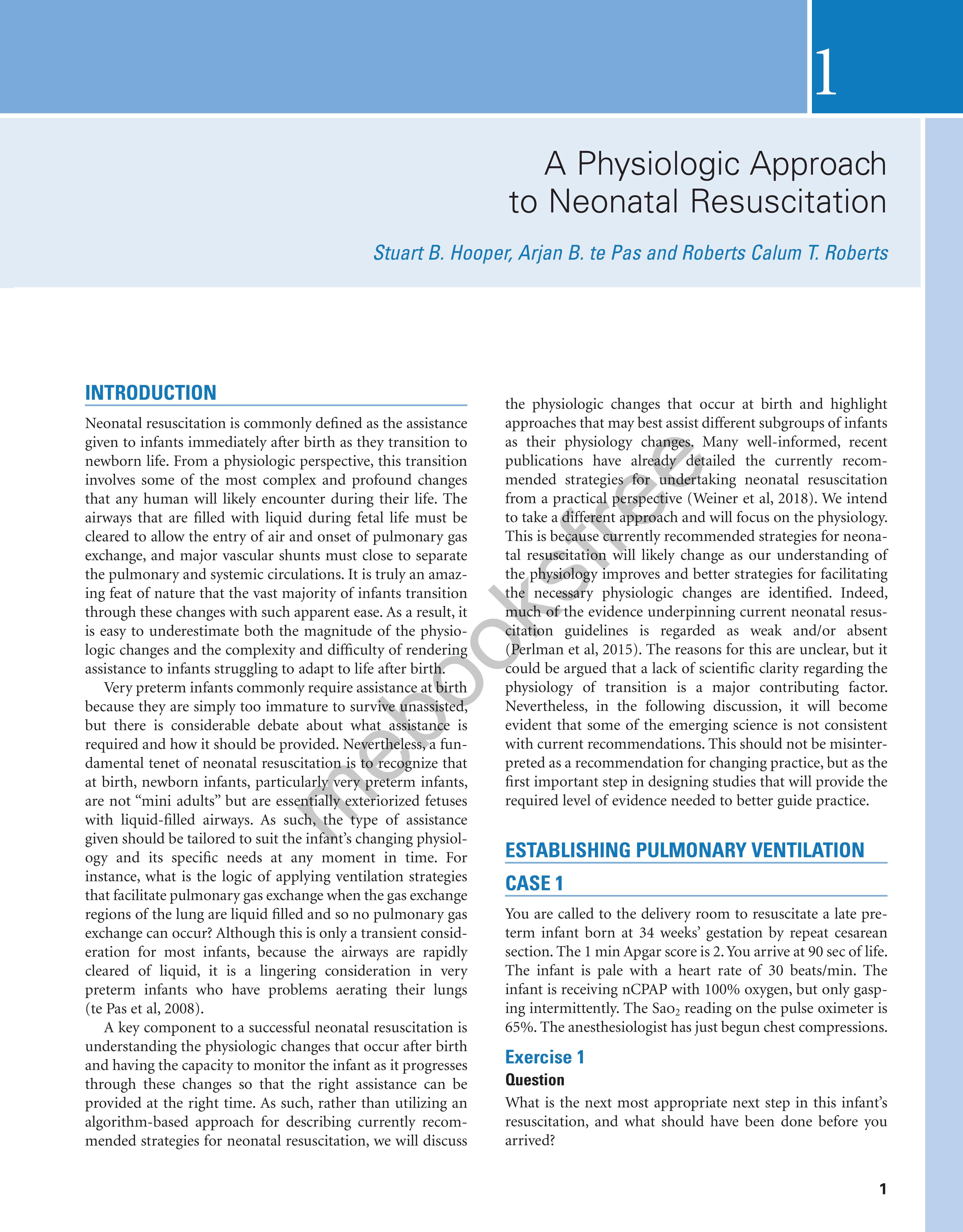
A Physiologic Approach to Neonatal Resuscitation
Stuart B. Hooper, Arjan B. te Pas and Roberts Ca/um T. Roberts
INTRODUCTION
Neonatal resuscitation is commonly defined as the assistance given to infants immediately after birth as they transition to newborn life. From a physiologic perspective, this transition involves some of the most complex and profound changes that any human will likely encounter during their life. The airways that are filled with liquid during fetal life must be cleared to allow the entry of air and onset of pulmonary gas exchange, and major vascular shunts must close to separate the pulmonary and systemic circulations. It is truly an amazing feat of nature that the vast majority of infants transition through these changes with such apparent ease. As a result, it is easy to underestimate both the magnitude of the physiologic changes and the complexity and difficulty of rendering assistance to infants struggling to adapt to life after birth.
Very preterm infants commonly require assistance at birth because they are simply too immature to survive unassisted, but there is considerable debate about what assistance is required and how it should be provided. Nevertheless, a fundamental tenet of neonatal resuscitation is to recognize that at birth, newborn infants, particularly ery Rreterm infants, are not "mini adults" but are essen t ially exteriorized fetuses with liquid-filled airways. As such, the type of assistance given should be tailored to suit the infant's changing physiology and its specific needs at any moment in time. For instance, what is the logic of applying ventilation strategies that facilitate pulmonary gas exchange when the gas exchange regions of the lung are liquid filled and so no pulmonary gas exchange can occur? Although this is only a transient consideration for most infants, because the airways are rapidly cleared of liquid, it is a lingering consideration in very preterm infants who have problems aerating their lungs (te Pas et al, 2008).
A key component to a successful neonatal resuscitation is understanding the physiologic changes that occur after birth and having the capacity to monitor the infant as it progresses through these changes so that the right assistance can be provided at the right time. As such, rather than utilizing an algorithm-based approach for describing currently recommended strategies for neonatal resuscitation, we will discuss
the physiologic changes that occur at birth and highlight approaches that may best assist different subgroups of infants as their physiology changes. Many well-informed, recent publications have already detailed the currently recommended strategies for un_g_er aking neonatal resuscitation from a practical Rerspective (Weiner et al, 2018). We intend to take a d ifferent approach and will focus on the physiology. This is because currently recommended strategies for neonatal resuscitation will likely change as our understanding of the p t.siology improves and better strategies for facilitating the necess ry physiologic changes are identified. Indeed, much o f, the evidence underpinning current neonatal resuscitati 0 n guidelines is regarded as weak and/or absent SPerlman et al, 2015). The reasons for this are unclear, but it could be argued that a lack of scientific clarity regarding the physiology of transition is a major contributing factor. Nevertheless, in the following discussion, it will become evident that some of the emerging science is not consistent with current recommendations. This should not be misinterpreted as a recommendation for changing practice, but as the first important step in designing studies that will provide the required level of evidence needed to better guide practice.
ESTABLISHING PULMONARY VENTILATION
CASE1
You are called to the delivery room to resuscitate a late preterm infant born at 34 weeks' gestation by repeat cesarean section. The 1 min Apgar score is 2. You arrive at 90 sec of life. The infant is pale with a heart rate of 30 beats/min. The infant is receiving nCPAP with 100% oxygen, but only gasping intermittently. The Sao 2 reading on the pulse oximeter is 65%. The anesthesiologist has just begun chest compressions.
Exercise 1
Question
What is the next most appropriate next step in this infant's resuscitation, and what should have been done before you arrived?
Answer
Positive pressure ventilation should have been started immediately.
From a teleologic perspective, it is logical that the physiologic changes required for survival after birth are triggered by the one event that cannot occur in utero, lung aeration. Aerating the lung and establishing pulmonary ventilation triggers the physiologic changes that underpin the transition to newborn life (Hooper et al, 2015a). However, it is far too simplistic to assume that the primary benefit of “establishing pulmonary ventilation” is reestablishing oxygen and carbon dioxide exchange lost following umbilical cord clamping. Lung aeration not only triggers the switch to pulmonary gas exchange but also triggers a very large reduction in pulmonary vascular resistance (PVR), which initiates a series of cardiovascular changes that are also essential for survival after birth (see later). Positive pressure ventilation also enhances reabsorption of lung fluid.
CASE 1 CONTINUED
With initiation of positive pressure ventilation, the heart rate increases to 120/min and the saturation increases to 85% by 7 min of life. The infant is breathing regularly at 120 breaths/ min. Auscultation reveals fine rales and wet sounding rhonchi. You suspect the infant has a “wet lung syndrome.”
Exercise 2
Question
When is lung liquid reabsorbed? How did the mode of delivery influence the resorption of lung liquid?
Answer
Resorption of lung liquid begins antenatally and continues during labor and delivery. However, most lung liquid is reabsorbed postnatally when spontaneous or assisted ventilations begin. Infants delivered by cesarean section do not undergo the postural changes of vaginally delivered infants; those changes help to expel liquid from the lungs.
AIRWAY LIQUID CLEARANCE BEFORE BIRTH AND DURING LABOR
Although there is some evidence to suggest that airway liquid clearance begins late in gestation before labor onset (Jain and Eaton, 2006), this is not a consistent finding, and the role of experimental artefacts is unclear with regard to the original observations (Harding and Hooper, 1996). Nevertheless, considering the capacity of the lung to clear airway liquid during labor and after birth (see later), whether small amounts of liquid are cleared before labor appear inconsequential. However, it is clear that airway liquid clearance can begin during labor and vaginal delivery (Olver et al, 2004). The release of adrenaline in response to the stress of labor activates Na1 channels located on the luminal surface of airway epithelial cells, which promotes Na1 reabsorption from the airways
into lung tissue (Olver et al, 1986). This reverses the osmotic gradient for liquid movement across the airway epithelium, leading to liquid reabsorption, rather than secretion as occurs in utero. However, Na1 reabsorption requires high levels of adrenaline, is relatively slow, only arises late in gestation, and so is not active in very preterm infants (Hooper et al, 2016). Similarly, as cesarean section delivery in the absence of labor avoids the stress of labor, this mechanism is unlikely to be activated in infants delivered by cesarean section without labor (Jain and Eaton, 2006).
Partial airway liquid clearance can also occur during labor as a result of induced postural changes before and during delivery of the head (te Pas et al, 2008). The fetus is forced into an exaggerated “fetal position” with the enhanced dorso–ventral flexion causing an increase in abdominal pressure and rostral displacement of the diaphragm (Harding et al, 1990). This increases intrathoracic pressures and forces liquid to leave the lungs via the trachea (Hooper and Harding, 1995; Harding and Hooper, 1996). As the fetal respiratory system is highly compliant, only small increases in intrathoracic pressure are needed for large reductions in airway liquid volumes (Hooper and Harding, 1995; Harding and Hooper, 1996). Although this mechanism is applicable to infants born vaginally, as per Na1 reabsorption, it is not readily applicable to infants born by cesarean section, particularly in the absence of labor.
Airway Liquid Clearance After Birth
Lung aeration has significant implications for respiratory function in the newborn period, and to better understand these consequences, the process of lung aeration can be divided into a series of phases that give rise to separate challenges (Hooper et al, 2016).
1. The first phase commences at birth with liquid-filled airways, and so the primary challenge is to clear the airways of liquid, which occurs across the distal airway wall.
2. Airway liquid is cleared from the airways into the surrounding lung tissue at a much greater rate (over minutes) than it is cleared from the tissue (over hours). As such, airway liquid accumulates within lung tissue for the first few hours after birth, forming “perivascular fluid cuffs,” expanding the chest wall and increasing interstitial tissue pressures, essentially making the lung edematous.
3. Airway liquid is gradually cleared from lung tissue via the circulation and lymphatics, after which lung function and mechanics stabilize.
Exercise 3
Question
What is the importance of spontaneous breathing (or positive pressure ventilation) on promoting the clearance of lung water?
Answer
To clear lung liquid from the airways and alveoli, positive pressure ventilation (either spontaneous or assisted) must begin. Ventilation moves the liquid through the airways to
the distal respiratory units, where it is absorbed into the lung interstitium and then into lymphatics.
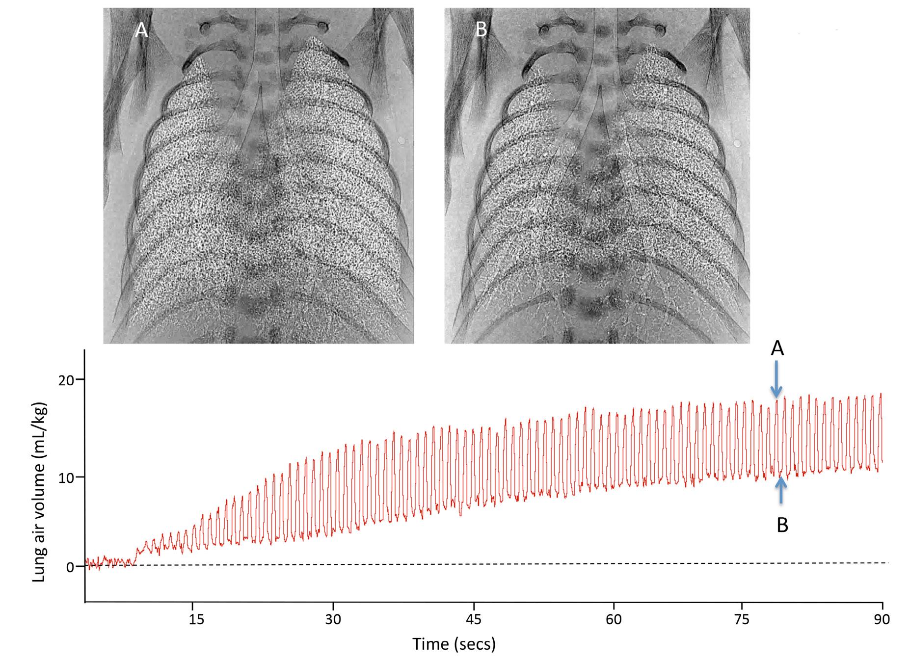
As noted earlier, the majority of liquid remaining in the airway is cleared across the distal airway wall. For this to occur, the liquid must move distally through the airways before leaving the airways and entering the surrounding distal lung tissue (Hooper et al, 2016). This process has been observed in newborn rabbits using phase contrast x-ray imaging, showing that the air/liquid interface moves distally during each inspiration (Hooper et al, 2007; Siew et al, 2009b) (Fig. 1.1). As no further distal movement occurred between breaths, lung aeration and the creation of a functional residual capacity (FRC) occurs in a stepwise fashion, increasing with each successive inspiration (Fig. 1.1). This led to the recognition that hydrostatic pressure gradients (between airways and lung tissue) generated by inspiration are largely responsible for airway liquid clearance after birth (Hooper et al, 2007; Siew et al, 2009b). Importantly, this mechanism provides a rational explanation for why very preterm infants who have little or no capacity to reabsorb Na1 are still able to clear their airways of liquid. As the air/liquid interface can also move proximally between breaths, causing a reduction in FRC, it is possible that liquid can reenter the airways between breaths, necessitating its reclearance with the next inspiration (Hooper et al, 2007; Siew et al, 2009b).
Exercise 4 Question
During the transition to postnatal life, what are the factors that govern whether airway liquid clearance is fast or slow?
Answer
Variables that regulate the rate of resorption of liquid include:
A. The surface area of the lung
B. Airway resistance (liquid has a higher resistance than air)
C. Resistance to moving the liquid across the walls of the distal airways
D. Tightness of the epithelial barrier
The initial resistance to air entering the lungs at birth is governed by both the resistance to moving liquid through the airways and by the resistance to moving this liquid across the distal airway wall. As water has a much higher viscosity than air, the resistance to moving air into the lungs is much greater when the airways are liquid filled compared with a few moments later when they are air filled (te Pas et al, 2009a, 2016). Consequently, airway resistance decreases markedly during the initial phase of lung aeration, as progressively more of the airways aerate and the reduction follows an exponential function that is difficult to predict (te Pas et al, 2009a, 2016).
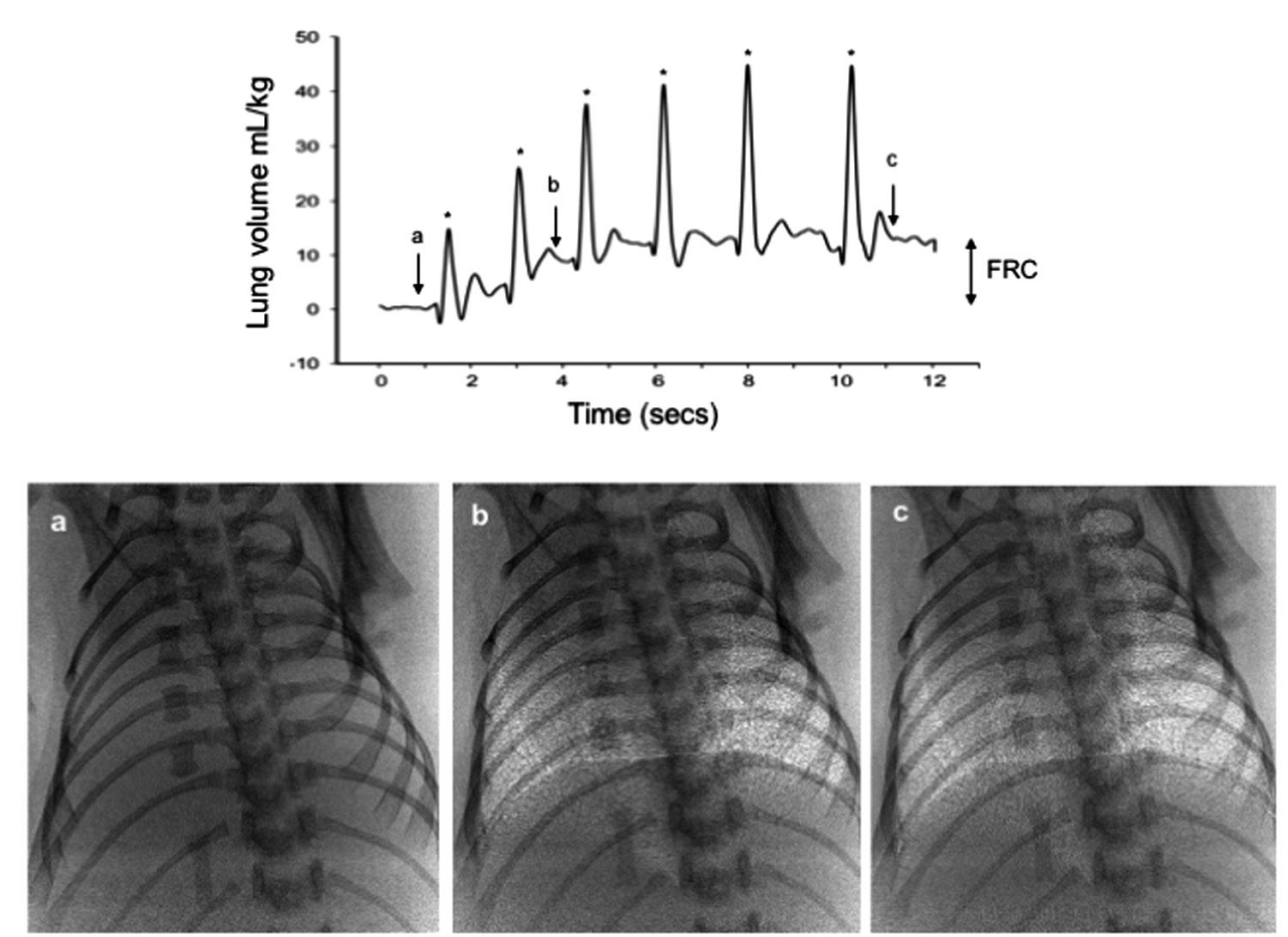
Fig. 1.1 Simultaneous plethysmograph recording and phase contrast x-ray images of a spontaneously breathing newborn rabbit during lung aeration. Upper panel: Plethysmograph recording showing 6 spontaneous breaths over a 10 sec period along with the gradual recruitment in FRC that occurs with each breath. Spontaneous breaths are the large increases in lung volume (indicated by an *) that decrease to a gradually increasing baseline (functional residual capacity: FRC). The reduction in lung volume after each breath is an artefact from the plethysmograph measurement. Bottom panel: Phase contrast x-ray images of the newborn rabbit’s chest, acquired at the time points indicated on the plethysmograph recording (indicated by an arrow and the corresponding letter for each image). Little to no aeration is present in image A, whereas a significant amount of aeration is present in image C, which was acquired approximately 10 seconds later.
On the other hand, little is known about the contribution that the resistance to liquid movement across the distal airway wall makes to the overall resistance to airway liquid clearance. Based on the volume of liquid that can be cleared during one inspiration (up to 3 mL/kg), and knowing the duration of inspiration (100–200 mSec), the liquid flux across the pulmonary epithelium can be as high as 15 to 30 mL/kg/sec or 0.9 to 1.8 L/kg/min. Although transient, this is surprisingly high for liquid movement across the relatively tight pulmonary epithelium (Egan et al, 1975). A large surface area is one obvious factor that allows the lung to clear liquid at this rate, but the “tightness” of the epithelial barrier likely resists water transfer.
The immature lungs of preterm infants have airways that are smaller in diameter and have few if any alveoli. As reducing the radius of a tube increases its resistance by the 4th power and as the absence of alveoli markedly reduces the lung’s surface area, the resistance to airway liquid clearance is higher in preterm infants than in term infants (te Pas et al, 2016). As a result, either the process of lung aeration will be much slower, or preterm infants will require larger inspiratory efforts or higher inflation pressures to overcome this higher resistance. This concept is at odds with current resuscitation guidelines that suggest using lower inflation pressures during the initiation of lung aeration in very preterm infants compared with term infants (Perlman et al, 2015). This recommendation is based on an extrapolation from studies in aerated lungs suggesting that higher pressures cause lung injury. However, considering it is the volume change and not the pressure per se that causes lung injury (Jobe et al, 2008) and that a liquid-filled lung is orders of magnitude less compliant than an air-filled lung (te Pas et al, 2009a), this recommendation may be flawed and requires further investigation.
Previous studies have demonstrated that the fetal pulmonary epithelium is relatively tight, which restricts the entry of even relatively small molecules into lung liquid during development (Egan et al, 1975). However, at birth these pore sizes markedly increase, which likely reduces the resistance to liquid movement across the epithelium in term newborns (Egan et al, 1975). However, it is unknown whether this occurs in preterm infants or whether it occurs to a greater degree due to the immaturity of the epithelium. Although this could reduce the resistance to liquid clearance, it may also contribute to the entry of plasma proteins into the lumen, which will interfere with surfactant function.
Exercise 5
Question
In the delivery room, how will I know when this infant’s lungs are optimally aerated?
Answer
Heart rate, oxygen saturation and expired CO2
Heart rate and peripheral oxygen saturation levels, measured using a pulse oximeter and/or ECG leads, are commonly used to assess when neonatal resuscitation has
been “successful” (Dawson et al, 2010a, 2010b). The idea that an increasing heart rate is a sign of lung aeration is based on the concept that a low heart rate indicates a vagal-induced bradycardia in response to perinatal asphyxia (Dawes, 1968). As such, an increasing heart rate is assumed to reflect improved oxygenation following the onset of pulmonary gas exchange. However, it is now clear that an increase in heart rate can also occur after birth in the absence of an increase in oxygenation (Lang et al, 2015). In this instance, the increase in heart rate is secondary to an increase in PBF (in response to lung aeration), which increases venous return and left ventricular preload (Lang et al, 2015). Nevertheless, whether the increase in heart rate results from increased oxygenation or an increase in PBF, both only occur as a result of lung aeration.
An alternate indicator for lung aeration is the use of expired CO 2, which is closely related to the degree of lung aeration (Hooper et al, 2013). Indeed, it is such a sensitive indicator that it can detect breath-by-breath changes in lung aeration in parallel with the changing tidal volumes and increases much more quickly in response to lung aeration than an increase in both heart rate and oxygenation in infants (Hooper et al, 2013; Blank et al, 2014; Schmolzer et al, 2015). The close relationship between end-tidal expired CO 2 levels and tidal volumes is because CO 2 exchange is surface-area limited during lung aeration. As CO 2 has a high solubility, its diffusion across the pulmonary epithelium is very efficient and is not normally surface-area limited. As such, end-tidal CO 2 levels are commonly used to estimate pulmonary arterial blood P co 2 levels and can be used to calculate cardiac output (Trillo et al, 1994). However, when the lung is not fully aerated, the surface area available for gas exchange at end inspiration is dependent on the size of the tidal volume (Hooper et al, 2013). When tidal volumes are larger, the surface area for gas exchange increases, which increases the efficiency of CO 2 exchange.
Although CO2 monitoring in the delivery room is currently not routine, in combination with tidal-volume monitoring, it provides a reliable method for assessing the effectiveness of pulmonary ventilation immediately after birth. Indeed, considering that the dead space of the lower airways is 2 to 3mL/kg and that the pharynx is expandable (Crawshaw et al, 2017), it is possible to achieve significant tidal volumes (3–4 mL/kg) without gas entering the gas exchange regions of the lung. As such, the baby would appear to be ventilated, but oxygenation levels and heart rate would likely remain low. However, the absence of any expired CO2 would indicate that the gas exchange regions are not being ventilated. Some of the new respiratory function monitors include the ability to measure expired CO2 levels, although increasing dead-space volume is an issue, and are most effectively used in combination with tidal-volume monitoring. Alternatively, a colorimetric CO2 indicator, which changes color in response to expired CO2, can be used to indicate when gas exchange has commenced (Blank et al, 2014).
Exercise 6 Question
During resuscitation, what alternate resuscitation strategies might be used to improve uniform lung aeration and better ventilation in this infant?
Answer
Increase in positive end expiratory pressure (PEEP) or sustained lung inflation
Recognition that airway liquid clearance after birth results from the generation of hydrostatic pressure gradients (between airways and lung tissue) has provided opportunities for developing strategies that facilitate this process in very preterm infants. Indeed, in a simplistic sense, all that is required is to apply a gas pressure to the airways to overcome the high resistance of moving liquid through the airways and across the distal airway wall. This rationale is consistent with the current recommendations for using either intermittent positive pressure ventilation (iPPV) or continuous positive airway pressure (CPAP) in combination with the infant’s spontaneous breathing to assist preterm infants initiating pulmonary gas exchange after birth (Perlman et al, 2015; Weiner et al, 2018).
However, the big question is how much pressure should be applied? Indeed, if the applied pressure is too low, it will be insufficient to overcome the resistance required to move the liquid distally through the airways. If it is too high, then there is a risk of causing overinflation and lung injury in lung regions that have already aerated (Jobe et al, 2008). To add to the complexity, as the airway resistance dramatically decreases (by 100 fold) with airway liquid clearance, the pressures required to move the liquid at any given flow rate will also greatly reduce (te Pas et al, 2016). Considering the huge variability expected between individual infants at birth, particularly with the amount of liquid present in the airways and the level of inspiratory effort each will apply, stipulating a single set inflation pressure or CPAP level to assist preterm infants to aerate their lungs at birth ignores this complexity. Clearly the pressure required will be different in different infants and will change as the lung aerates. Although we now have a grasp of the complexities involved in facilitating lung aeration in very preterm infants, the challenge is to apply this knowledge in a useful and practical manner (Jobe, 2011).
During lung aeration, ideally the respiratory support applied should change in accordance with the change in resistance caused by airway liquid clearance. High airway pressures could be applied initially when the resistance is high, which decrease as the airway resistance decreases to avoid overinflation and lung injury. However, to decrease the applied pressure in synchrony with the decreasing resistance requires complex feedback information regarding the changing airway resistance. Although modern ventilators can measure airway resistance on a breath-by-breath basis, they are rarely used in the delivery room even if the infant is intubated. Instead, low-technology devices such as resuscitation bags or t-piece devices are more commonly used, mostly in
combination with noninvasive ventilation (Schmolzer et al, 2010; Schilleman et al, 2013). These provide little or no opportunity to measure airway resistance and provide little information on how to modify the required ventilation parameters as lung mechanics change unless it is combined with a respiratory function monitor. It seems counterintuitive that sophisticated ventilators and monitoring equipment are commonly used in the NICU once the lung has aerated and respiratory mechanics have stabilized, but they are not routinely used in the delivery room when respiratory mechanics are rapidly changing and respiratory function is very difficult to manage in a safe and effective way.
SUSTAINED INFLATION DURING RESUSCITATION
The movement of air into the lung at birth is primarily determined by airway resistance and the applied pressure gradient, as defined by F 5 DP/R; where F is flow, DP is the pressure gradient and R is airway resistance, which includes the resistance to moving liquid across the distal airway wall. As flow equals volume (V) divided by time (T), the factors determining the movement of air (inflation volume) into the lung can be defined as V 5 (DP 3 T)/R. As such, the main controllable factors determining inflation volume are the applied pressure gradient (DP) and time (T) over which the pressure is applied (inflation time). Although increasing the inflation pressure can overcome the high initial resistance, as the resistance decreases with lung aeration, there is a high risk of overinflating and injuring the lung if the pressure is not simultaneously reduced. Alternatively, increasing the inflation time using a slower, sustained inflation (SI) allows lower inflation pressures to be used. Although the initial flow of gas into the lung is slow, it rapidly increases as more of the airways aerate and the resistance decreases (te Pas et al, 2016). Theoretically, this approach has multiple advantages, as during a sustained inflation the lung’s end inflation volume is self-limiting and determined by the inflation pressure, which can be much lower than the pressure required to initially aerate the lung with a shorter inspiratory time.
As different lung regions aerate at different rates, a SI allows more lung regions to aerate during a single inflation (te Pas et al, 2009b, 2009a). This has important implications for lung injury, because during the subsequent inflation, air will rapidly flow into and expand aerated lung regions first due to the much lower airway resistance. Therefore if the inflation time is short (as occurs with iPPV), the entire tidal volume will only enter aerated regions, potentially causing overexpansion and injury in those regions with little further lung aeration (Siew et al, 2009a, 2011). Furthermore, as gas exchange only occurs when the distal gas exchange regions aerate, there is no reason to terminate the inflation to allow exhalation when these regions are liquid filled. These explanations underpin the rationale for providing a SI for the first inflation after birth, but the benefits described in animal studies have not been replicated in humans (van Vonderen et al, 2014a; Lista et al, 2015). Although the reasons are
unclear, in animal studies the SI was applied with an endotracheal tube, whereas in all human studies, the SI was applied noninvasively, usually with a face mask (including the SAIL trial). This is a major point of difference (see later), and studies that are restricted to comparing a SI with conventional ventilation in intubated infants may possibly reveal results that are as clear cut as the animal studies.
Exercise 7
Question
What are the adverse effects of higher levels of PEEP or sustained inflation during resuscitation?
Answer
High levels of PEEP or a prolonged sustained inflation can decrease venous return and reduce cardiac output.
Recent studies have suggested that a stepwise PEEP recruitment maneuver (up to 20 cmH2O) that extends over 2 to 3 minutes can achieve better postmaneuver lung mechanics than an SI (McCall et al, 2016). This suggestion is consistent with the concept that lung aeration is a function of applying an elevated pressure over an extended period, and the results show significant improvements in lung mechanics (Tingay et al, 2016, 2017). However, applying this maneuver ignores the cardiovascular consequences of applying high elevated airway pressures that increase intrathoracic pressures for an extended period. Simple physics dictates that as soon as intrathoracic pressures exceeds central venous pressure, then all venous return to the heart will cease and, as such, cardiac output must decrease (see later). Furthermore, in the aerated lung, high PEEP levels reduce PBF, and this effect on PBF is not completely reversed following the reduction in PEEP (Polglase et al, 2005). Although this adverse effect of increased intrathoracic pressure on PBF is applicable to both a sustained inflation and PEEP recruitment maneuver, a sustained inflation does not influence the time related increase in PBF, perhaps because sustained inflation is only 10 to 30 seconds long (Sobotka et al, 2011). However, the PEEP recruitment maneuver can take 2 to 3 minutes (Tingay et al, 2016, 2017), and it is unclear how it influences the increase in PBF at birth. There is also a need to be cautious of any rebound in cardiac output, as per a Valsalva maneuver that may occur post maneuver.
Whether a sustained inflation or a PEEP recruitment strategy is the most effective approach for aerating the lung remains unclear, and more studies are required. In particular, there is much debate about what is the most appropriate starting pressure and duration of the sustained inflation. However, evidence from animal studies indicate that these are not the correct starting points (McCall et al, 2016), particularly as a “one-size-fits-all” approach is unlikely to be successful in different infants (te Pas et al, 2009a, 2016). Targeting a set inflation volume instead of a fixed inflation time and using a ramped pressure increase, which is then held constant once gas starts to move into the lungs, may be more appropriate (Polglase et al, 2014; McCall et al, 2016; te Pas et al, 2016). Measurement of CO2 levels in the expired air
can then indicate whether a second inflation is needed. This approach is easy to implement in animals, but its application in humans will depend on the use of sophisticated approaches to monitor newborns immediately after birth (McCall et al, 2016).
CASE 1 CONTINUED
At 60 min of life the infant’s respiratory rate is 120/min. There is intermittent grunting. The inspiratory oxygen concentration is 100% to achieve a saturation of 90%.
Exercise 8
Question
Why is this infant tachypneic?
Answer
The increased water in the interstitium of the lung has reduced the infant’s lung compliance and reduced its inspiratory reserve volume by expanding the chest wall and flattening the diaphragm.
At birth, the liquid residing in the airways following the first breath is rapidly cleared into lung tissue, forming perivascular fluid cuffs (Bland et al, 1980). As liquid clearance from the tissue takes hours, the volume of liquid residing in the airways at birth must be accommodated within the lung’s interstitial tissue during this time (Bland et al, 1980; Miserocchi et al, 1994). This has consequences for respiratory function in the newborn period (Berger et al, 1996), including an increase in interstitial tissue pressure (Miserocchi et al, 1994). This in turn increases the potential for liquid to reenter the airways during expiration and expansion of the chest wall (Hooper et al, 2007; McGillick et al, 2017). Indeed, although the liquid has cleared from the airways, it remains within the thorax, forcing the chest wall to expand to accommodate both the liquid and the air that creates the newly formed FRC (Hooper et al, 2007; McGillick et al, 2017).
Exercise 9
Question
Assuming this infant’s lung water is increased, how can respiratory function be improved?
Answer
Increasing the positive end expiratory pressure
It is both fortuitous and necessary that the newborn’s chest wall is very compliant so that it can easily expand without further increasing interstitial tissue pressures and opposing FRC formation. This explains the importance of applying an end expiratory pressure on the airways during the immediate newborn period (Siew et al, 2009a). The positive airway pressure not only prevents the lung from collapsing but also opposes the elevated interstitial pressure and prevents liquid reentering the airways during expiration (Siew et al, 2009a). Phase contrast x-ray imaging in ventilated very preterm rabbits (Figs. 1.2 and 1.3) has clearly demonstrated that a FRC will not develop, largely due to liquid
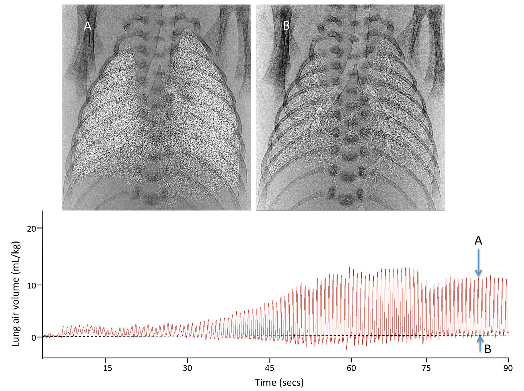
Fig. 1.2 Phase contrast x-ray images and a plethysmograph recording of a preterm newborn rabbit immediately after birth ventilated from birth in the absence of a positive end expiratory pressure (PEEP). In the absence of PEEP, preterm rabbits failed to develop a functional residual capacity (FRC) resulting in liquid reentry or airway collapse at end expiration. Phase contrast x-ray images (A and B) were recorded at each time point on the plethysmograph trace. Image A was acquired at end inspiration, whereas image B was acquired at FRC.
reentry into the airways, in the absence of an end expiratory pressure (Siew et al, 2009a).
Exercise 10
Question
Does this infant have transient tachypnea of the newborn? (TTN)
Answer
The infant’s clinical signs are consistent with TTN. As the volume of airway liquid differs greatly between individuals at birth (Harding and Hooper, 1996), the volume of liquid entering lung tissue will also vary, causing large variations in lung tissue and chest wall mechanics. For instance, accommodating larger liquid volumes in lung tissue increases chest wall expansion (including flattening of the diaphragm), reduces FRC, increases lung tissue stiffness (McGillick et al, 2017) and likely further increases pulmonary interstitial tissue pressures and pressure within the fluid cuffs surrounding pulmonary blood vessels (Bland et al, 1980; Miserocchi et al, 1994). These consequences may explain the pathology
associated with transient tachypnea of the newborn (TTN). Indeed, the tachypnea maybe caused by expansion of the chest wall and flattening of the diaphragm, which reduces the infant’s inspiratory reserve volume. This limits the infant’s capacity to increase tidal volume, making an increase in respiratory rate the primary option for increasing minute ventilation and CO2 clearance. Because of the increased respiratory rate, many of these infants develop a respiratory alkalosis. Furthermore, the associated reduction in FRC commonly induces expiratory braking and grunting, which is another characteristic of infants with TTN.
Exercise 11
Question
How long will the tachypnea persist?
Answer
The tachypnea can resolve in a day or two or last as long as 5 to 7 days as liquid is reabsorbed.
Although TTN is usually transient and self-resolving, some infants exhibit continuing respiratory morbidity for up
Fig. 1.3 Phase contrast x-ray images and a plethysmograph recording of a preterm newborn rabbit immediately after birth ventilated from birth with a positive end expiratory pressure (PEEP) of 5 cmH2O. With this level of PEEP, preterm rabbits gradually develop a significant functional residual capacity (FRC) with most distal airways (not all, see basal lung regions) remaining aerated at FRC. Phase contrast x-ray images (A and B) were recorded at each time point on the plethysmograph trace. Image A was acquired at end inspiration, whereas image B was acquired at FRC.
to 72 hours, and some develop persistent pulmonary hypertension (Jain and Dudell, 2006; Ramachandrappa and Jain, 2008). Thus the morbidity associated with TTN may not be unidimensional, as the consequences of too much liquid in lung tissue should resolve as the liquid is cleared. It is possible that some infants experience continuing morbidity due to lung injury associated with bidirectional liquid movement across the epithelium. In newborn rabbits, liquid can reenter the airways at FRC due to the elevated pressures in lung tissue, which is then recleared during the subsequent inspiration/inflation (Hooper et al, 2007; Siew et al, 2009b).
Liquid entry rates into lung tissue during inspiration are very high, and although liquid movement back into the airways is considerably lower, 1 to 2 mL/kg can reenter the airways over a 1 to 2 sec expiratory time in newborn rabbits (Hooper et al, 2007; Siew et al, 2009b). As the liquid is recleared during the next inspiration, bidirectional liquid movement across the pulmonary epithelium is likely a normal feature of lung aeration (Hooper et al, 2007; Siew et al, 2009b). However, unlike the kidney, which has a fenestrated epithelium, the pulmonary epithelium is not designed for sustained high liquid flux rates. It is possible that if lung tissue liquid volumes are elevated, the propensity for airway
liquid reentry increases, leading to increased bidirectional liquid movement across the airway wall, resulting in lung injury. Thus although the respiratory morbidity associated with TTN may initially be caused by too much liquid within lung tissue, lung injury may be responsible for the continuing morbidity in some TTN infants. Nevertheless, this explanation provides an understanding for why CPAP, which opposes airway liquid reentry, can be used to treat TTN.
CASE 1 CONTINUED
The infant continues to receive nCPAP with 100% oxygen. Because of intermittent episodes of bradycardia and hypoxemia, he required positive pressure ventilation using nasal prongs (nIPPV). However, the Sao2 only rises to 90%. You consider intubating this infant but worry about injuring the lung with positive pressure ventilation.
Exercise 12
Question
Why was the noninvasive positive pressure ventilation (nIPPV) ineffective?
Answer
Noninvasive positive pressure ventilation is often ineffective because the vocal cords adduct during apnea and prevent the positive pressure from being transmitted to the lower airway. In addition, the dead-space volume of the nasopharynx may be considerable and mean that higher pressures and tidal volumes are needed when using noninvasive ventilation.
Until recently, intubation and mechanical ventilation were the most common form of respiratory support for very preterm infants. However, as it is invasive and associated with a higher risk of lung injury, noninvasive respiratory support has become the preferred method of respiratory support (Finer et al, 2010). But this change has occurred without understanding how noninvasive respiratory support interacts with the infant’s physiology at birth. In particular, the physiologic significance of an endotracheal tube, which bypasses the infant’s upper airway, has been largely overlooked. As a result, it is widely assumed that iPPV is equally effective when applied noninvasively as when applied via an endotracheal tube, which ignores the role of the larynx in regulating gas flow into and out of the lung.
When applied via an endotracheal tube, iPPV bypasses the larynx and has direct access to the sublaryngeal airways, unless they are blocked with mucous or foreign matter (e.g., meconium). However, when applied noninvasively (usually via a mask), airflow must pass through the upper airways and larynx before entering the trachea and lower airways. As the larynx can seal the airways even against very high pressures (.100 mm Hg), a patent airway is heavily dependent on whether the larynx is open during noninvasive ventilation.
In adults, the larynx is mostly open except during swallowing, some postural movements, and abdominal evacuation, etc. However, regulation of the larynx in the fetus and newborn is very different from in the adult (Harding et al, 1986; Praud et al, 1992). Before birth, airway liquid is secreted across the pulmonary epithelium, and while it flows out of the lungs via the trachea, the fetal larynx acts to restrict the rate of efflux to maintain the lungs in a hyperexpanded state (FRC of 35–40 mL/kg vs. 20–25 mL/kg after birth) (Hooper and Harding, 1995; Harding and Hooper, 1996). This is because lung expansion provides the primary stimulus for lung growth (Hooper and Harding, 1995; Harding and Hooper, 1996). During apnea, the larynx adducts to restrict liquid loss from the airways, causing it to accumulate within the airways and expand the lung. During fetal breathing movements (FBM), the larynx opens and liquid leaves the lungs at an increased rate, despite contractions of the diaphragm (Hooper and Harding, 1995; Harding and Hooper, 1996). Thus the larynx plays an important role in maintaining fetal lung expansion by adducting during apnea and preventing airway liquid loss. As this fetal pattern of laryngeal activity persists into newborn life (Crawshaw et al, 2017) it has major implications for the effective application of noninvasive ventilation in the newborn. It also reinforces the concept that infants, particularly very preterm infants, are not “mini adults” but are essentially exteriorized fetuses at
birth and, therefore, automatically extrapolating treatment strategies from adult medicine may not be helpful.
In the context of neonatal resuscitation, the consequence of having a fetal pattern of laryngeal activity in the newborn is that apneic infants will mostly have a closed larynx. This has recently been demonstrated using phase contrast x-ray imaging (Crawshaw et al, 2017), whereby the patency of the larynx was found to depending on whether the newborn was apneic (closed larynx) or in a stable breathing pattern (open larynx). As a closed larynx prevents iPPV from ventilating the lung, no matter how much pressure is applied (Crawshaw et al, 2017), iPPV will be ineffective in apneic infants until they become so hypoxic and bradycardic that the larynx relaxes. Laryngeal closure is most likely seen as “airway obstructions” and explains why noninvasive ventilation in the delivery room has a high failure rate, requiring intubation (Schmolzer et al, 2010; Schilleman et al, 2013). However, if the newborn is breathing, the larynx will open, allowing iPPV to assist spontaneous breaths to aerate the lung (Crawshaw et al, 2017). Thus in contrast to ventilation via an endotracheal tube, the success of noninvasive ventilation is dependent on the presence of spontaneous breathing, when the larynx must open.
During noninvasive ventilation, particularly with a face mask, the dead-space volume may be significantly larger than in intubated infants. This is because of the contribution of the nasopharynx to the overall dead-space volume, which increases in relative size as the infant gets smaller (Nieves et al, 2018). As such, a 2 kg infant is estimated to have a nasopharynx dead-space volume of 2 mL/kg (Nieves et al, 2018) and in addition, the pharynx has been shown to expand further in response to an inflation pressure during iPPV (Crawshaw et al, 2017). This raises a number of questions about targeting the same tidal volumes during iPPV in intubated and mask-ventilated infants. Indeed, this is consistent with the finding that tidal volumes were significantly reduced following intubation, despite receiving the same inflation pressure, in infants previously receiving iPPV via a facemask (van Vonderen et al, 2014b).
Exercise 13
Question
If an infant is apneic at birth, should noninvasive ventilation be tried in the delivery room?
Answer
Noninvasive ventilation (NiPPV) might be acceptable once the apnea has resolved. Although NiPPV might not ventilate the infant more effectively (because of vocal cord adduction), it can provide a higher mean airway pressure.
From a physiologic perspective, an infant who is apneic at birth, particularly if it has good tone, will most likely have an adducted larynx (see earlier) that prevents noninvasively applied iPPV from ventilating the lung (Crawshaw et al, 2017). Although this scientific knowledge has come from animal studies, physiologic recordings from preterm infants during sustained inflation studies indicate that this also
occurs in humans (van Vonderen et al, 2014a; Lista et al, 2015). If the infant doesn’t commence breathing, it will progressively become more hypoxic and will eventually lose tone and become bradycardic. Although all of these facts have been well documented scientifically, it is unclear at what point reflexes, such as those controlling laryngeal adduction, are lost as the asphyxia worsens. Nevertheless, it appears that at some point the larynx relaxes, despite the infant being apneic, which allows positive pressure ventilation to be delivered noninvasively (Schmolzer et al, 2010; Schilleman et al, 2013). Although noninvasive iPPV may eventually be successful, clearly the respiratory support is not optimal if the infant first becomes hypoxic and bradycardic before ventilation is successful. This raises the question as to whether current recommendations incorrectly assume that, when using noninvasive respiratory support, iPPV is a reliable “backup” for apneic infants. Instead, as closure of the larynx may render iPPV ineffective, perhaps the focus should shift toward stimulating breathing and avoiding known causes of apnea, such as hypoxia.
Exercise 14
Question
In an apneic infant, what other strategies might be helpful in the delivery room?
Answer
Physical stimulation and caffeine
As the larynx is mostly open during breathing in newborns, except when making expiratory braking maneuvers (e.g., grunting) (Crawshaw et al, 2017), it seems logical that stimulating breathing should be the primary focus of noninvasive respiratory support in the delivery room. This can be achieved by applying physical stimulation and avoiding hypoxia (as it inhibits breathing) and by giving respiratory stimulants such as caffeine (Dekker et al, 2017a, 2018). Recent studies have attempted to assess how—and how much— physical stimulation should be applied in the delivery room (Dekker et al, 2017b; Gaertner et al, 2018). In one study, standardizing and increasing the application of a physical stimulus was found to increase oxygenation in preterm infants, despite the use of a significantly lower Fio2 and an unexpected increase in physical stimulation within the control group (Dekker et al, 2018). This was thought to result from an increase in minute ventilation (tidal-volume 3 respiration rate), which wasn’t quite significant. Similarly, it has been shown that the administration of caffeine into the umbilical vein (using a butterfly needle) within the first few minutes after delivery can significantly increase breathing efforts in preterm infants (Dekker et al, 2017a). Thus rather than waiting for the infant to reach the NICU, it is both feasible and potentially beneficial to administer caffeine as soon as possible after birth.
Exercise 15
Question
How should the inspiratory oxygen concentration be regulated when noninvasive ventilation is used?
Answer
ILCOR recommends starting resuscitation with 21% to 30% oxygen in preterm infants. Theoretically, higher inspiratory oxygen concentrations used immediately after birth may stimulate respirations, but oxygen requirements decrease exponentially thereafter. Therefore, if an increased concentration of oxygen is used, it should be rapidly weaned. Infants who remain bradycardic despite use of 30% oxygen should receive 100% oxygen and positive pressure ventilation until the bradycardia is resolved.
The role of oxygen in stimulating/sustaining spontaneous breathing efforts in preterm infants at birth has been overshadowed by the debate on starting Fio2 levels and the risk of hyperoxia-induced organ injury. This risk has been very well documented and prompted a change in the 2010 international guidelines, recommending that respiratory support for preterm infants should change from starting in high Fio2 levels to low levels (30%) or air (Perlman et al, 2010). However, this change also coincided with a switch in the preferred respiratory support for preterm infants, which switched from invasive to noninvasive ventilation (Morley and Davis, 2008; Morley et al., 2008) without recognizing that a change to lower initial Fio2 levels may affect spontaneous breathing. Presumably this was not considered to be important because it was assumed that iPPV is equally effective when applied noninvasively as it is when applied via an endotracheal tube.
A retrospective study in preterm infants has shown that increasing Fio2 levels can significantly increase respiratory efforts; however, oxygen requirements usually decrease exponentially thereafter (van Vonderen et al, 2013). It was surmised that the initial high Fio2 was required to provide a large air/blood Po2 gradient to increase oxygenation and stimulate breathing when the surface area for gas exchange was initially low. However, the resulting increase in respiratory effort increased lung aeration and exponentially increased the capacity for gas exchange due to an exponential increase in surface area (van Vonderen et al, 2013). Thus although high Fio2 levels may be required initially to stimulate and/or support breathing at birth, the requirement likely decreases exponentially as the lung aerates. Nevertheless, in view of the requirement for oxygen to stimulate and support breathing at birth in infants receiving noninvasive respiratory support, the debate over starting Fio2 levels seems to be esoteric and not particularly useful. Clearly, this will depend on the infant’s oxygenation level at delivery, the level of respiratory effort, the level of stimulation (either physical or chemical) and the degree of lung aeration at any one moment in time during a process that changes exponentially. This is an extraordinarily complex question and it is highly unlikely to have one correct answer, as the need will vary considerably between infants. This brings into question the rationale of current trials trying to find a starting Fio2 that can be applied to all infants. Instead, the complexity emphasizes the need to be able to rapidly titrate Fio2 as required (to avoid both hypoxia and hyperoxia) and to measure the infant’s oxygenation to guide titration, with the understanding
that the capacity for oxygen exchange can increase exponentially as the lung aerates (van Vonderen et al, 2013).
FACILITATING THE CARDIOVASCULAR TRANSITION AT BIRTH
Exercise 16
Question
How does lung aeration affect the cardiovascular transition at birth?
Answer
Lung aeration initiates the cardiovascular transition at birth by increasing pulmonary blood flow (PBF) and increasing venous return to the left ventricle.
As indicated earlier, aerating the lung at birth is not just about transferring the site of respiratory gas exchange to the lung. Lung aeration also plays a vital role in initiating the cardiovascular transition at birth by stimulating a large
decrease in PVR (Hooper et al, 2015c). This is responsible for increasing PBF (10–30 fold) and allowing the pulmonary circulation to accept 100% of right ventricular output while also allowing pulmonary arterial pressures to substantially decrease (Hooper et al, 2015c). The increase in PBF is not just important for enhancing pulmonary gas exchange but is also vital for taking over the role of providing preload for the left ventricle and thereby sustaining cardiac output (Fig. 1.4). The decrease in PVR along with the increase in afterload caused by removal of the placental circulation may also contribute to closure of the ductus arteriosus (DA) and separation of the two circulations (Hooper et al, 2015c).
Exercise 17
Question
Why is the increase in PBF so important after birth?
Answer
The increase in PBF allows the lungs to serve as the organ of gas exchange and helps to maintain cardiac output by
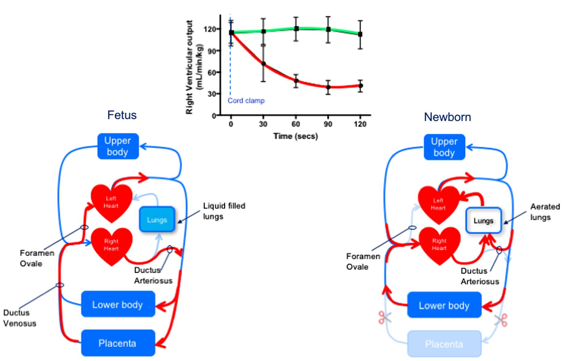
Fig. 1.4 Diagrammatic representation of the fetal and newborn circulation, also showing the changes in right ventricular output if the cord is clamped before (red) or after (green) ventilation onset. In the fetus, pulmonary vascular resistance (PVR) is high, and so the majority of right ventricular output flows through the ductus arteriosus (DA) with only a small amount flowing through the lungs. As a result, pulmonary blood flow (PBF) is low, and so much of the preload supplying the left ventricle is derived from the placenta, with umbilical venous return flowing via the ductus venosus and foramen ovale to directly enter the left side of the heart. After birth, when the umbilical cord is clamped, the supply of umbilical venous return to the left ventricle is lost, and so cardiac output decreases until the lungs aerate and PBF increases to restore preload for the left ventricle. However, if the lungs aerate and PBF increases before the umbilical cord is clamped, the supply of preload for the left ventricle can immediately switch from umbilical to pulmonary venous return following cord clamping. The reduction in PVR following lung aeration causes blood to flow from the systemic into the pulmonary circulation (left to right) through the DA, which greatly contributes to the increase in PBF. (Right ventricular output values are replotted data from Bhatt et al, 2013.)
taking over the role of providing venous return to the left ventricle.
Fetal Circulation: the Starting Point
To understand the extent of the cardiovascular changes at birth, it is important to first understand the starting point for the transition, which is the structure and function of the fetal circulation (Fig. 1.4). Before birth, the lungs are not involved in gas exchange, and the main role of PBF is to provide oxygen and nutrients for developing lung tissue. In contrast, after birth, PBF is vital for the efficient exchange of respiratory gases and supplies 100% of venous return to the left ventricle. In the fetus, instead of flowing through the lungs, much (up to 90%) of right ventricular output passes through the DA and directly enters the descending aorta (Fig. 1.4). Although it is often perceived that PVR in the fetus is fixed at a high level, this is not correct (Polglase et al, 2004). During development, PVR gradually decreases as the pulmonary vascular network grows and develops (Rudolph, 1979). This leads to a large increase in the cross-sectional area of the pulmonary vascular bed, particularly with the formation and growth of small vessels and alveolar capillaries. As such, the capacity of premature infants to dilate their pulmonary vascular bed and reduce PVR must be limited, because the vascular bed has yet to develop a large cross-sectional area. In the fetus, PBF can also vary markedly depending on fetal activity (Polglase et al, 2004). It can increase during periods of FBM, increasing 8- to 10-fold during periods of accentuated FBMs (Polglase et al, 2004). This is likely due to the reduction in thoracic pressure and an expansion-induced increase in alveolar capillary caliber (as occurs in adults), because the increase in PBF is closely associated with each individual breath (Polglase et al, 2004).
As PBF is low during fetal life, pulmonary venous return is unable to provide the left ventricle with sufficient venous return (preload) to sustain its output (Rudolph, 1979). Instead, the left ventricle obtains the majority of its preload from umbilical venous return, which flows via the ductus venosus (DV) and foramen ovale (Rudolph, 1979) (Fig. 1.4). As a result, the left ventricle receives oxygenated blood directly from the placenta (site of gas exchange), which accounts for the higher oxygenation levels in preductal arteries (arteries that branch off the aorta upstream of the DA/aorta junction) (Rudolph, 1979); this is analogous to the adult, as the left ventricle also receives the oxygenated blood, but in this case from the lungs.
Exercise 18
Question
What is the relationship between PBF and flow through the ductus arteriosus in fetal life?
Answer
Blood flow through the ductus arteriosus at birth can be right to left, bidirectional, or left to right depending on how quickly PVR diminishes. Ultimately most preterm infants exhibit left-to-right blood flow through the ductus arteriosus before it closes.
The relationship between PBF and flow through the DA is highly dynamic and simply determined by the pressure gradient between the pulmonary and systemic circulations (Hooper, 1998). In turn, these are controlled by downstream resistances in the two circulations. When PVR is high, the majority of right ventricular output flows across the DA into the systemic circulation, whereas PBF in the left and right pulmonary arteries is bidirectional (Figs. 1.4 and 1.5). During systole, PBF is forward (antegrade) in direction, entering the lungs, but during most of diastole, blood flows retrogradely, away from the lungs, leaving the pulmonary circulation and entering the systemic circulation through the DA (Crossley et al, 2009) (indicated by negative PBF in Fig. 1.5). Thus blood flows through the DA continuously, from the pulmonary and into the systemic circulation (rightto-left), throughout the cardiac cycle (Fig. 1.5); in contrast, flow in the main pulmonary trunk (upstream of the DA and left and right pulmonary arteries) is reduced to zero during diastole (Rudolph, 1979).
During FBM, PVR decreases and mean PBF increases, which is almost entirely due to a reduction in retrograde flow when diastole coincides with the reduction in intrathoracic pressure during each breath (Polglase et al, 2004). As a result, right-to-left shunting through the DA decreases and the contribution of right ventricular output to flow in the systemic circulation is reduced. This competitive relationship between flow in the pulmonary and systemic circulations persists after birth (see later), for as long as the DA remains open (Bhatt et al, 2013; Blank et al, 2017).
Transitioning the Circulation From a Fetal Into a Newborn Circulatory Pattern
At birth, lung aeration stimulates a 10- to 30-fold increase in PBF, and although the precise mechanisms are still unclear, numerous factors are involved that act in combination or in a hierarchical manner (Hooper et al, 2015c). As in adults, oxygen is a potent stimulus for pulmonary vasodilation in the fetus, which is thought to be mediated by NO release. Other factors include the release of vasodilators and an increase in lung recoil caused by the formation of surface tension (Gao and Raj, 2010). More recently, x-ray imaging has been used to examine the spatial relationship between ventilation and perfusion within the lung during lung aeration (Lang et al, 2014). Partial lung aeration caused a global increase in PBF (Fig. 1.6), increasing similarly in both aerated and unaerated lung regions, irrespective of whether the ventilation gas was air, 100% O2, or 100% N2 (Lang et al, 2015). Clearly, an increase in oxygenation is not responsible, although ventilation with 100% O2 enhanced the PBF increase in aerated lung regions (Lang et al, 2015). Subsequent studies revealed that vagal denervation could block the global response to lung aeration, suggesting that a neural reflex was involved that may be triggered by J-receptors activated by the movement of liquid into lung interstitial tissue during lung aeration (Lang et al, 2017).
Irrespective of the mechanism, these studies demonstrate the potential for a ventilation–perfusion mismatch in the
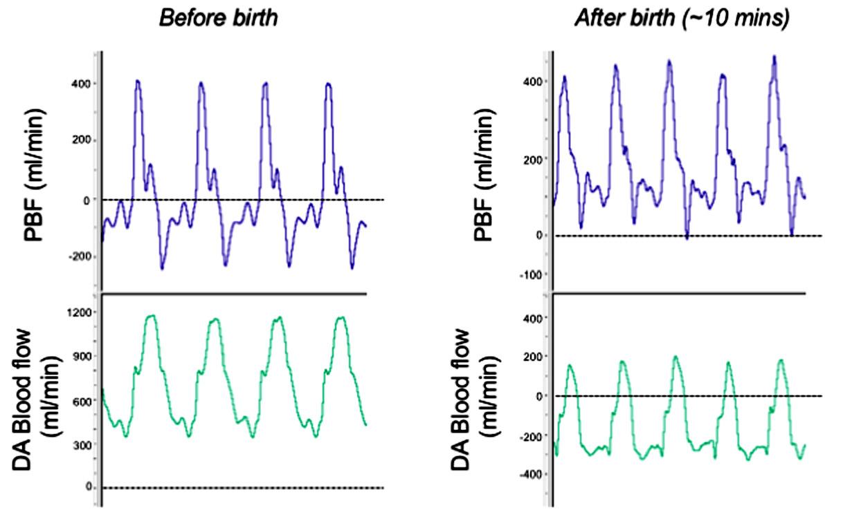
Fig. 1.5 Blood flow waveforms in the left pulmonary artery and in the ductus arteriosus (DA) before and immediately after birth. Before birth, pulmonary blood flow (PBF) flows toward the lungs (positive flow) only briefly during systole and then during late systole and throughout most of diastole, PBF is mostly retrograde (negative value), flowing away from the lungs and passing through the DA. This retrograde PBF accounts for the high levels of diastolic flow in the DA during fetal life. After birth, the decrease in pulmonary vascular resistance facilitates antegrade flow in the pulmonary arteries throughout the cardiac cycle, with relatively high flows occurring even during diastole. These diastolic flows are due to the left-to-right shunting (indicated by negative flows) through the DA, contributing to flow during this time. Although net blood flow across the DA is predominantly left to right, the flow waveform demonstrates distinct bidirectional characteristics due to the changing pressure gradient across the DA associated with the cardiac cycle.
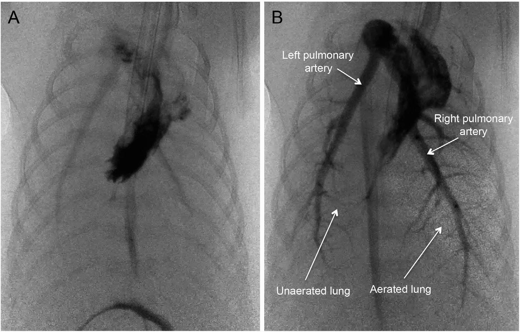
Fig. 1.6 Simultaneous phase contrast and angiographic x-ray images of a near term rabbit kitten before lung aeration (A) and following partial aeration of the right lung (B). Before lung aeration, blood flow in the left and right pulmonary arteries is low. In B, aerated lung regions can be seen as “speckle” in the image and are due to x-ray refraction at the air/water interface. Although only part of the right lung is aerated, blood flow is increased in both the left and right pulmonary arteries. Aerated and unaerated lung regions are indicated by arrows
lung at birth and raises the question as to whether this mismatch is problematic or advantageous. Indeed, it is possible this spatial “disconnect” (Fig. 1.6) may be advantageous, as the decrease in PVR and increase in PBF is more vital for the infant’s survival than complete lung aeration. This is because the increase in PBF is vital for maintaining left ventricular preload and cardiac output after birth (Bhatt et al, 2013), whereas only partial lung aeration is needed to achieve sufficient gas exchange for survival.
Both right and left ventricles contribute to the increase in PBF (Fig. 1.4) after lung aeration (Crossley et al, 2009) because the decrease in PVR allows the lung to accept 100% of right ventricular output and at the same time causes pulmonary arterial pressures to decrease. This reverses the pressure gradient between the pulmonary and systemic circulations, causing blood flow through the DA to reverse (compared with the fetal state), mostly flowing left to right (Figs. 1.4 and 1.5) (Crossley et al, 2009). As such, PBF into the lung occurs continuously throughout the cardiac cycle, with left-to-right DA flow maintaining elevated PBF during diastole (Figs. 1.4 and 1.5) and contributing up to 50% of total PBF (Crossley et al, 2009). This redirection of blood flow, originating from both left and right ventricles through the lungs, “steals” blood flow from the lower body and placenta (Blank et al, 2017), if the cord is still intact (see later) (Fig. 1.4). During this time, while most of the DA blood flow is left to right, instantaneous flow is bidirectional; right to left initially during systole and then left to right during late systole and throughout diastole (Fig. 1.5). This is thought to be because the pressure wave emanating from the right ventricle reaches the pulmonary artery/DA junction before the pressure wave coming from the left ventricle reaches the DA/aorta junction (Hooper et al, 2015c). As a result, flow is initially right to left and then rapidly changes to left to right as the pressure gradients change. It is currently unclear whether the resulting turbulence contributes to DA closure.
Exercise 19
Question
What are the physiologic advantages of delayed cord clamping?
Answer
Infants who are delivered after delayed cord clamping exhibit greater hemodynamic stability and have greater blood volumes and decreased need for RBC transfusion. In preterm infants, there are lower incidences of intraventricular hemorrhage and necrotizing enterocolitis.
Delayed Umbilical Cord Clamping (DCC) and Placental Transfusion
Delayed umbilical cord clamping (DCC) after birth is not a new concept, as it dates back to Aristotle and has been revisited by many commentators over the centuries, including Erasmus Darwin (Charles Darwin’s grandfather) in 1801 (Darwin, 1801). However, immediate cord clamping became standard practice following implementation of the active
(vs. expectant) management of the third stage of labor, which is aimed at reducing the risk of postpartum hemorrhage (PPH) (Begley et al, 2011). The three components of this approach were early administration of a potent uterotonic (e.g. oxytocin), immediate cord clamping, and gentle traction on the cord to reduce the length of third stage.
Although this active management strategy significantly reduces the risk of PPH, it also significantly reduces birth weights because of a lower blood volume (Begley et al, 2011). This indicates that although it has very clear benefits for the mother, it may have adverse implications for the infant that were not broadly considered upon implementation. As oxytocin administration at the end of third stage of labor is equally as effective at reducing the risk of PPH (Soltani et al, 2010), the need for immediate cord clamping to reduce the risk of PPH has become obsolete. This raises the question about the need for immediate versus delayed cord clamping, as the timing within third stage does not have an impact on the mother’s risk of PPH.
For many years the debate around the timing of cord clamping has focused on the concept of “placental transfusion,” whereby DCC advocates claim that after birth a volume of blood moves from the placenta into the infant, giving the infant a “blood transfusion” (McDonald et al, 2013); this explains the higher birth weights in infants delivered with “expectant” versus “active” management of third stage of labor (Begley et al, 2011). The concept of placental transfusion is largely based on studies that used radiolabelled (125I) albumin to measure blood volumes in infants who received cord clamping at different times after birth (Yao et al, 1969). Numerous studies have reported increased birth weight changes, hematocrits, hemoglobin levels, and iron stores and reduced need for transfusions in infants receiving delayed cord clamping (McDonald et al, 2013).
Exercise 20
Question
What is physiologic based cord clamping (PBCC)?
Answer
With physiologic based cord clamping or baby-directed umbilical cord clamping, the timing of cord clamping is based on the infant’s physiology rather than on a set period after birth. It supports preload (and cardiac output) at a time when umbilical venous return is ending. Clinically it means delaying clamping of the umbilical cord until lung aeration has been established.
The Physiology of Umbilical Cord Clamping at Birth
As highlighted earlier, cord clamping at birth removes umbilical venous return as a source of preload for the left ventricle, making it dependent on PBF and any residual flow through the foramen ovale for preload (Hooper et al, 2015c) (Fig. 1.4). In addition, arterial pressure (afterload) is greatly increased by cord clamping due to the loss of the low-resistance placental circulation (Bhatt et al, 2013), which
during fetal life receives a large proportion (30%–50% depending on GA) of cardiac output (Rudolph, 1985). As a result, the combined loss of preload and the increase in afterload cause a large reduction in cardiac output, which remains reduced until the lungs aerate, PBF increases and the supply of preload is restored (Bhatt et al, 2013) (Fig. 1.4). On the other hand, if the lung aerates and PBF increases before the umbilical cord is clamped, then the elevated PBF can immediately take over the role of providing preload for the left ventricle when umbilical venous return is lost (Bhatt et al, 2013) (Fig. 1.4). As a result, cardiac output is sustained throughout the transition. This has been termed physiologic based cord clamping (PBCC) (Kluckow and Hooper, 2015) or baby-directed umbilical cord clamping (Blank et al, 2018), whereby the timing of cord clamping is based on the infant’s physiology rather than on a set period after birth. In addition, if PVR is reduced before the umbilical cord is clamped, the increase in arterial pressure (afterload) caused by cord clamping is reduced because the pulmonary circulation is able to serve as an alternate low resistance pathway for blood to flow (Bhatt et al, 2013). In view of these findings, many now believe that ideally the timing of umbilical cord clamping should be delayed until after the lungs have aerated and PBF has increased (Knol et al, 2018). Two recent feasibility studies have provided the first evidence indicating the potential benefits of resuscitating infants on the cord (Blank et al, 2018; Brouwer et al, 2018).
Physiologic Based Umbilical Cord Clamping
Whatever the explanation for “placental transfusion,” it is unfortunate that this concept has been the only focus of debate over DCC. Indeed, such a unidimensional focus ignores the known benefits of DCC that have a logical scientific explanation (e.g., maintenance of cardiac output during transition), leading to (1) a series of poorly designed clinical studies, (2) an unproductive circular debate between the risk of hypovolemia/anemia and hyperbilirubinemia (Weeks and Bewley, 2018) and (3) the belief that time is the major benefactor of delayed cord clamping, irrespective of the infant’s physiologic state and whether it needs resuscitation. It also limits thinking on the way in which this very simple procedure, which has no cost, can be most effectively applied and in which infants it will have most benefit. For instance, a Tanzanian study has shown that for every 10 sec delay (up to 2 min) between breathing onset and umbilical cord clamping, there is a 20% reduction in mortality and/or admission into intensive care (Ersdal et al, 2014).
Recognition that immediate cord clamping may have an adverse impact on cardiovascular function in the immediate newborn period first arose with the formation of the Dawson nomograms (Dawson et al, 2010a, 2010b). The aim of developing these nomograms was to describe the normal heart rate and oxygenation ranges for healthy term (and preterm) newborn infants to provide target ranges immediately after birth for infants requiring resuscitation. They showed that 50% of normal healthy term infants had a heart rate under 100 bpm at 1 min after birth (Dawson et al, 2010b). This was
surprising, as the infants were thought to be well oxygenated and healthy, and so a chemoreceptor mediated vagal bradycardia was considered unlikely. Instead, it was suggested that a reduction in venous return was responsible (Dawson et al, 2010b), which prompted the study demonstrating that cord clamping before ventilation onset causes a marked reduction in cardiac output (Bhatt et al, 2013). More recently, two studies have examined the feasibility of resuscitating infants while still “on the cord,” and both have found a much higher heart rate than would be expected from the Dawson nomograms (Blank et al, 2018; Brouwer et al, 2018). This suggests that (1) immediate cord clamping has a much wider adverse impact (lower than desired heart rate and cardiac output) than expected and (2) new nomograms will be required for infants receiving physiologic based cord clamping.
Although current guidelines recommend that DCC should be applied to healthy term infants not requiring resuscitation (Perlman et al, 2015), these infants are likely to benefit least. Based on the available scientific evidence, infants who are likely to receive the greatest benefit of PBCC are those who have the longest delay between delivery and the onset of pulmonary ventilation (Hooper et al, 2015b). As the increase in PBF will also be delayed and/or reduced in magnitude, the switch from umbilical to pulmonary venous return for providing left ventricular preload will also be reduced or delayed. These include very preterm infants and infants with lung hypoplasia who have difficulty in aerating their lungs and have a poorly developed pulmonary vascular bed at birth. Other infants likely to benefit include apneic infants with poor tone at birth as a result of intrauterine hypoxia (see later).
Currently there is still no robust scientific explanation for the net movement of blood from the placenta into the infant during DCC after birth. Nevertheless, it is important to understand the mechanisms involved, because if the causes are unknown, it is difficult to identify the factors that impede or prevent it. Suggested mechanisms include uterine contractions, gravity, an increase in pulmonary blood volume with the increase in PBF, and a decrease in intrathoracic pressure caused by inspiration, but none of these explanations have been substantiated scientifically (Hooper and Kluckow, 2018).
The absence of an explanation for placental transfusion is not proof that it does not occur, but it may be that our understanding of the underlying circumstances is inaccurate. Indeed, the debate concerning placental transfusion has entirely focused on blood volume accumulation within the newborn after birth without any thought as to what happens to fetal blood volume before birth, during labor. This is a major oversight, as the common assumption is that fetal blood volume remains constant before birth and then suddenly increases after birth due to placental transfusion. Alternatively, it is possible that the fetus loses blood volume into the placenta during labor, perhaps due to increased dorso–ventral flexion imposed by uterine contractions (Harding et al, 1990). As such, an apparent “placental transfusion” after birth may result from restoration of this blood
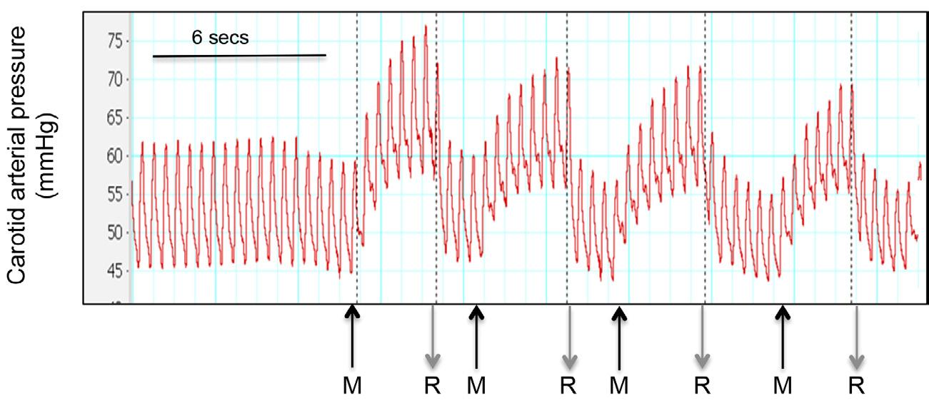
Fig. 1.7 Changes in carotid arterial pressure in response to four consecutive umbilical cord “milkings.” Each cord milk is indicated “M,” whereas the cord release that occurred at the end of each milk is indicated by “R.” Note the large increases in arterial pressure that occurred with each milk.
volume back into the infant as the two circulations come back into balance. This would explain why placental transfusion is less evident following cesarean section deliveries and provides a much more compelling argument for DCC. That is, rather than the infant acquiring blood volume and red blood cells that it never had during fetal life, which increases the risk of hyperbilirubinemia, maybe it is simply reclaiming the blood volume and red blood cells it temporarily lost during delivery.
Exercise 21
Question
Does umbilical cord milking (UCM) provide the same benefits as DCC?
Answer
Depending on the circumstances, umbilical cord milking and delayed cord clamping can both transfer a volume of blood to the newborn infant. The potential for transfer is more rapid with UCM, but it also causes large changes in arterial pressure and flow that are potentially injurious. This may explain the increased likelihood of intraventricular hemorrhage in infants under 28 weeks’ gestation. As studies now show that resuscitation can occur while infants are still attached to the cord, it is debatable whether the urgent need for resuscitation is a compelling argument for replacing DCC with UCM.
UCM involves squeezing the cord between thumb and finger and then sliding them along the cord, forcing blood to move from the cord into the infant. Depending on the protocol, this can be done before or after cord clamping and either once or multiple times. UCM has been proposed as an alternative to DCC, providing the infant with a blood transfusion in a shorter period (Katheria et al, 2018). This is thought to be desirable, allowing the infant to gain a blood transfusion while still allowing rapid transfer to a resuscitation table if the infant requires some form of intervention (Katheria et al, 2014, 2017). Although clinical evidence suggests that UCM provides benefits over immediate cord clamping, a recent
scientific study has raised several concerns (Blank et al, 2017). When the cord remains unclamped and is milked several times, whether a net volume of blood is transferred into the newborn depends on the milking protocol (Blank et al, 2017). As milking the cord necessarily causes cord occlusion, each milk of the cord produces arterial pressure and cerebral blood flow changes that are identical to immediate cord clamping (Fig. 1.7), effectively replicating the adverse effects multiple times (Blank et al, 2017). Other concerns that are yet to be answered relate to forcing blood under pressure into the low-pressure venous circulation and the release of cellular debris and cytokines into the newborn circulation if milking occurs more than once.
The underlying assumption of UCM is that the primary benefit of DCC is placental transfusion (Katheria et al, 2017), which is clearly wrong. Like immediate cord clamping, if the milking occurs before the lungs have aerated, venous return and cardiac output must be reduced as the volume entering the newborn with each milk is only a small fraction of umbilical venous flow. The argument for UCM versus DCC centers around time and the need to initiate resuscitation as soon as possible (Katheria et al, 2017). However, the perception that UCM is a time-condensed equivalent of DCC reduces the focus on the more important issue of developing perinatal (as opposed to neonatal) resuscitation teams that can overcome the logistics of resuscitating infants on the cord, which is likely to be the most efficacious approach.
EXERCISE 22
Question
How does DCC influence the infant’s body temperature during resuscitation with an intact cord?
Answer
Although still anecdotal, scientific studies suggest that the requirement for external heat input to maintain newborn body temperature with the cord intact is less than that required following cord clamping.
The management of body temperature in newborn infants in the delivery room is important, particularly in preterm infants. Depending on whether the infant is full term or preterm, this is usually achieved by drying the infant, replacing wet towels with dry ones, use of a radiant heat warmer, and placing a hat on the infant’s head to reduce heat loss (Weiner et al, 2018). In addition, preterm infants are commonly placed within a plastic wrap to further reduce heat loss and maintain body temperature. Although it is unclear whether body temperature is more or less of a problem during DCC, logistically, it is likely that all of the procedures that have been adopted can also be applied in infants during delayed cord clamping. Indeed, “resuscitation tables” that have or are being developed for this purpose include heated mats and/or an overhead radiant warmer to maintain the infant’s temperature (Brouwer et al, 2018). Similarly, studies not employing the use of a table are simply resuscitating the infant on a heat pad on the mother’s legs (Blank et al, 2018).
However, it is possible that the infant’s body temperature control will be more stable and less prone to rapid reductions if it is resuscitated with an intact cord, because the placenta is still within the mother and should heat the infant’s blood as it perfuses the placenta. Although still anecdotal, scientific studies suggest that the requirement for external heat input to maintain newborn body temperature with the cord intact is less than that required following cord clamping. On the other hand, considering the contribution of warmed placental blood to maintaining the infant’s body temperature, applying the same heat loss/heat input procedures as are applied in infants with a cord that has been ligated may increase the risk of hyperthermia in infants resuscitated with an intact cord (i.e., with delayed cord clamping).
MANAGING THE ASPHYXIATED INFANT
From a medicolegal perspective, “birth asphyxia” usually refers to the severest form of asphyxia, but the physiologic reality is that perinatal asphyxia is a continuum, ranging from the prolonged and very severe to transient and milder forms. Irrespective of the severity, the initial physiologic response to perinatal asphyxia is identical and well described (Dawes, 1968). Initially, it involves a chemoreceptor mediated, vagal-induced, bradycardia, apnea, a mild hypertension, and an inhibition of body movements, along with a marked redistribution of cardiac output to increase blood flow and maintain oxygen supply to the heart and brain (Reuss and Rudolph, 1980). As the asphyxia is prolonged, the response diverges depending on the severity of the reduction in oxygenation. With less severe reductions, the increase in blood flow to the heart and brain maintains oxygen delivery to these vital organs to sustain their function, with the increase in blood flow being very closely matched with the decrease in blood oxygen content (organ O2 delivery 5 blood O2 content 3 organ blood flow) (Reuss and Rudolph, 1980). This adaptive response is so effective that the fetus can survive for days,
weeks, and even months, with the later resulting in the “brain sparing” associated asymmetric fetal growth restriction. In addition, an inhibition of fetal breathing and body movements contribute to the maintenance of oxygen delivery to the heart and brain through a reduction in total body oxygen consumption (Rurak and Gruber, 1983).
CASE 2
A term infant is born to a woman with an acute placental abruption via a spontaneous vaginal delivery. Following delivery, the obstetrician palpates the umbilical cord and detects a heart rate of 20 beats/min.
Exercise 23
Question
What are the physiologic advantages of resuscitating an infant with severe asphyxia with an intact cord?
Answer
When cord clamping occurs immediately after birth in an asphyxiated infant, it reduces cardiac output (as the low resistance placental circuit is removed) and interferes with the neonate’s ability to adapt to asphyxia and protect the brain and myocardium. However, ventilating the lungs of a depressed infant before the cord is clamped is technically complex, requires special equipment, and is still unproven. In most developed countries with good obstetric management, the most common form of perinatal asphyxia is the less severe, mildly asphyxiated infant who is apneic and bradycardic, with little or no tone at birth and low initial Apgar scores. These infants are unable to initiate breathing at birth (due to a hypoxic inhibition of breathing), and current guidelines recommend immediate umbilical cord clamping and transfer to a stable platform for resuscitation (Perlman et al, 2015). However, it is questionable (and controversial) as to whether this is the correct approach. As indicated earlier, the physiologic response to protect the brain from hypoxia is to increase cerebral blood flow through a redistribution of cardiac output (Reuss and Rudolph, 1980). In the absence of an increase in PBF, cord clamping reduces cardiac output (Bhatt et al, 2013), which undermines the newborn infant’s capacity to adapt to the asphyxia and maintain oxygen delivery to the heart and brain, increasing the risk of hypoxic/ ischemic brain injury. In essence, it deprives the infant from using the primary defense mechanism that it has against hypoxia, which is a redistribution of cardiac output.
On the other hand, ventilating the lungs before the cord is clamped is complex but has theoretical advantages. It avoids the delay in resuscitation onset caused by the time required for cord clamping and transferring the infant to a resuscitation table and avoids the loss in cardiac output caused by cord clamping before PBF has increased. It also allows for a recovery in the infant’s oxygenation with a relatively stable cardiac output that is physiologically determined rather than being artificially limited by cord clamping.